Article

Characteristics of Initial Attachment and Biofilm Formation of Pseudomonas aeruginosa on Microplastic Surfaces

Department of Environmental Engineering, Seoul National University of Science & Technology, Seoul 01811, Korea
*
Author to whom correspondence should be addressed.
These authors contributed equally to this work.
Appl. Sci. 2022, 12(10), 5245; https://doi.org/10.3390/app12105245
Submission received: 25 April 2022 / Revised: 18 May 2022 / Accepted: 19 May 2022 / Published: 22 May 2022

Abstract

:
The toxic effect of microplastics on living organisms is emerging as a serious environmental issue nowadays. The biofilm formed on their surface by microorganisms can further increase the toxicity, but the mechanism of biofilm formation on microplastics is not yet fully understood because of the complexities of other factors. This study aimed to identify the factors with an important influence on biofilm formation on microplastic surfaces. The microtiter plate assay was used to evaluate the biofilms formed by Pseudomonas aeruginosa PAO1, a model microorganism, on four types of microplastics, including polyethylene, polystyrene, polypropylene, and polytetrafluoroethylene. The density of microplastics was found to be a key factor in determining the amount of biofilm formation because the density relative to water has a decisive effect on the behavior of microplastics. Biofilm formation on plastics with densities similar to that of water showed remarkable differences based on surface characteristics, whereas biofilm formation on plastics with a higher density was significantly influenced by particle movement in the experimental environment. Furthermore, biofilm formation was inhibited by adding a quorum quenching enzyme, suggesting that QS is critical in biofilm formation on microplastics. This study provides useful information on biofilm formation on microplastic surfaces.

1. Introduction

Since the 1950s, plastic production has steadily increased owing to its advantages of being inexpensive, light, strong, durable, and corrosion-resistant. Plastic production increased from 1.7 million tons in 1950 to 359 million tons in 2018, more than 200 times the volume produced 68 years ago [1,2,3]. Owing to the rapid increase in the usage of plastics globally, a significant amount of nonrecycled, landfilled and reused plastics has been released into the environment and has accumulated.
As plastics, which are hardly biodegradable, accumulate in the environment, a new environmental issue that has emerged is microplastic pollution. Microplastics are plastics that have been shattered by various weathering processes or fabricated into small sizes for specific purposes with diameters of 5 mm or less. Microplastics found in terrestrial, aquatic, and atmospheric environments have various polymer types, shapes, and sizes [4,5,6]. Although microplastics do not have short-term lethal effects on living organisms, long-term exposure can induce chronic toxicity through various mechanisms [7,8]. Toxic chemicals used in the production of plastics can induce toxicity, and the small and pointy structure of microplastics can damage organisms and cause inflammation. Ingestion of tiny microplastics has been shown to cause malnutrition and alterations in reproductive function in some organisms [9,10]. Additionally, persistent organic pollutants adsorbed on plastics can reach concentrations up to a million times higher than ambient concentrations, and these compounds can be further desorbed inside the organism, exacerbating the bioaccumulation of the persistent organic pollutants [11,12].
Biofilms are microbial colonies that attach to and grow on biotic and abiotic surfaces [13]. Microplastics provide a new artificial adhesive substrate for biofilm formation [14,15]. The formation and heteroaggregation of biofilms have been reported to affect not only the physical properties of microplastics [16] but also the chemical properties owing to the presence of bacteria in the biofilm [17,18]. Such modified microplastics have been found to enhance the carrier effect on heavy metals while also increasing the associated toxicity [19,20,21]. Furthermore, when harmful microorganisms form a biofilm on microplastics, antibiotic resistance increases, increasing the toxic potential in the aquatic ecosystem [22]. In the wastewater treatment process, biofilm formation on the microplastic surface affects the microplastic removal efficiency of the sedimentation tank through the change in density [23]. In the membrane water treatment process, microplastics act as a substrate for microorganisms to adhere and grow, causing increased membrane biofouling [24].
Biofilm formation on the surface of microplastics can be affected by environmental conditions, as well as the properties of plastics and biological interactions between microorganisms, but specific research results to explain the mechanism are still lacking [25]. Therefore, this study aims to analyze the difference in the biofilm formation of microorganisms based on various types of microplastics. Using the Pseudomonas aeruginosa PAO1 strain as a model microorganism, biofilm formation on the surfaces of four types of microplastics including polyethylene (PE), polypropylene (PP), polytetrafluoroethylene (PTFE), and polystyrene (PS) were analyzed through batch experiments with microplates. The difference in biofilm formation rate based on the material and surface characteristics of microplastics was investigated, as well as the effect of the interaction between microorganisms and plastics on initial attachment and biofilm development. Finally, the effect of quorum sensing (QS) (a cell–cell communication mechanism) on biofilm formation on PE microplastic surfaces was analyzed.

2. Materials and Methods

2.1. Bacterial Strains and Growth Conditions

Pseudomonas aeruginosa PAO1, an aerobic Gram-negative bacterium, was selected as a model microorganism for biofilm formation. PAO1 has been reported to regulate biofilm formation using self-generated QS signals [26]. Escherichia coli TOP10-AiiO, which had previously been confirmed to degrade N-acylhomoserine lactone (AHL), a QS signal molecule [27], was used as the quorum quenching (QQ) bacterium, and E. coli TOP10-Empty transformed with the empty pTrcHis2 plasmid was used as the negative control. Agrobacterium tumefaciens A136 was used as a reporter strain for detecting AHL [28]. All strains used in this research were cultured using the Luria–Bertani medium (LB) (BD-Difco, Sparks, MD, USA).

2.2. Analysis of Microplastic Surface Properties

The microplastics used in this experiment were spherical beads made of PE, PP, PS, and PTFE (Cospheric, Santa Barbara, CA, USA). The density and diameter of each microplastic bead are listed in Table 1.
The contact angle and surface roughness of the microplastics used in the experiment were analyzed to determine their hydrophobicity and surface properties. The contact angle was measured by sessile drop using the KSV CAM-200 (KSV Instruments, Helsinki, Finland). The angle between the microplastic surface and the water–air interface was measured in about 5 s after 2 μL of deionized water was dropped on the microplastic surface. All contract angle measurements were repeated five times in each of the microplastics. An atomic force microscope (XE-100, PSIA Corp., Sungnam, Korea) was used to measure the surface roughness of the microplastics, which can significantly affect the adhesion of microorganisms. The microplastic beads were attached to a microscope slide plate, and an area of 20 × 20 μm2 of the upper surface per sample was analyzed.

2.3. Biofilm Formation Assay

Using a loop, a single colony of PAO1 was inoculated into 5 mL LB broth and incubated overnight at 200 rpm and 37 °C. The bacterial culture was diluted to OD600 0.03 using sterilized M9 minimal medium (containing 9 mM NaCl, 22 mM, KH2PO4, 48 mM Na2HPO4, 19 mM NH4Cl, 2 mM MgSO4, 100 μM CaCl2, and 0.4% glucose, with pH = 7.0), and an aliquot of 1 mL was inoculated into each well of a surface-treated 24-well plate (#30024, SPL, Pocheon-si, Korea). After adding 10 plastic beads to each well, the microplate was sealed with parafilm and incubated at 37 °C with shaking (180 rpm) for 6 h. The microplastics were sterilized with 99% ethanol before the biofilm experiment.

2.4. Biofilm Quantification

The biofilms formed on the surfaces of the 24-well plate and the microplastics were quantified by crystal violet (CV) staining. After biofilm cultivation, the microplastics were transferred to a new 24-well plate, and the culture solution was removed. Both the biofilms formed on the surface of the well plate and those formed on the microplastics were washed with 1 mL of phosphate-buffered saline (PBS) buffer (Sigma–Aldrich, St. Louis, MO, USA). Thereafter, the biofilms were dried for 30 min and stained with 1 mL of 0.1% CV (Sigma–Aldrich, USA) solution. After 20 min of incubation at room temperature, the CV solution was removed and washed twice with 1 mL PBS buffer. In each well, 1 mL of high-purity ethanol (~99.9%, Duksan, Ansan-si, Korea) was added to dissolve the CV stains in the biofilm. The amount of biofilm was quantified by absorbance analysis at a wavelength of 550 nm using a microtitre plate reader (Gen 5, Biotek, Winooski, VT, USA). To quantify the biofilm formed on the microplastic surface, the microplastics were removed from the solution before the absorbance measurement.

2.5. Effect of QS on Biofilm Formation on the Microplastic Surface

To investigate the role of bacterial signaling by QS in the biofilm formed on the surface of the microplastics, AHL signaling molecules were added to the biofilm experiment described above. Only PE, which forms the most biofilm among the four types of plastics, was used in this experiment. N-butanoyl-L-homoserine lactone (C4-HSL) and N-(3-oxododecanoyl)-L-homoserine lactone (3-oxo-C12-HSL), which are produced and utilized by PAO1, were dissolved in dimethyl sulfoxide (DMSO, Sigma–Aldrich, USA) to generate AHL cocktails. The AHL cocktails were injected into a 24-well plate containing PAO1 (OD600 of 0.03) and PE microplastic beads (10 each) at final concentrations of 5, 10, and 20 μM. The control was injected with DMSO in a volume corresponding to the injected AHL cocktails. After sealing with parafilm and incubating at 180 rpm and 37 °C for 6 h, the biofilm was quantified with the method described in Section 2.4.

2.6. Effect of QQ on Microplastic Surface Biofilm Formation

The E. coli strains were cultured overnight at 37 °C with 180 rpm shaking, and the cultured solution was divided into 50 mL centrifuge tubes and centrifuged (4500 rpm, 24 min, 4 °C). The supernatant was discarded, and the pellet was resuspended in 50 mM Tris buffer (Trizma hydrochloride solution, Sigma–Aldrich) and adjusted at OD600 of 10. Cell lysis was performed on the resuspended culture using a sonicator (JY92-IIDN Ultrasonic Homogenizer, Ningbo Scientz Biotechnology Co., Ltd., Ningbo, China). After repeating the 40 s sonication and 30 s rest 25 times, the mixture was divided into 10 tubes (1 mL) and centrifuged (12,000 rpm, 20 min, 4 °C). Subsequently, the supernatant was filtered with a 0.2 μm PES filter. The extracted QQ enzyme solution was stored at 4 °C until it was utilized.
The enzyme extracted by the method described above was tested for activity using C8-HSL, a type of AHL. About 100 μL extracted QQ enzyme, 400 μL tris buffer, and 500 μL C8-HSL (400 μM) were mixed. The reaction was carried out at 30 °C for 3 min and 15 min, respectively, without stirring after gently shaking the tube into which the sample was placed. The supernatant was subsequently collected, and the enzymatic reaction was stopped with heat treatment (95 °C, 2 min) and centrifuged (13,500 rpm, 3 min, 4 °C). After that, 200 μL of the supernatant was carefully collected. AHL was quantified using A. tumefaciens A136, the reporter strain, and β-galactosidase produced by A136 reacting with AHL was converted to a luminescence signal using the Beta–Glo system (Promega, Madison, WI, USA). Measurements were conducted using microplates.
After confirming QQ activity, QQ enzyme extracts were injected into the PAO1-incubated microplate. After 100 μL of QQ enzyme extract and 900 μL PAO1 were combined in a 24-well plate, five PE plastic beads were added to each well. After injection, the 24-well plates were shaken at 180 rpm 2, 4, 6, and 8 h at 37 °C. In the cultured 24-well plate, the biofilm was quantified by the method described in Section 2.4.

2.7. Statistical Analysis

All statistical analyses were performed using Origin 2022 (Originlab, Northampton, MA, USA). One-way analysis of variance (ANOVA) with the Tukey multiple comparison test and two-way ANOVA with the Bonferroni multiple comparison tests were applied throughout the analysis.

3. Results and Discussion

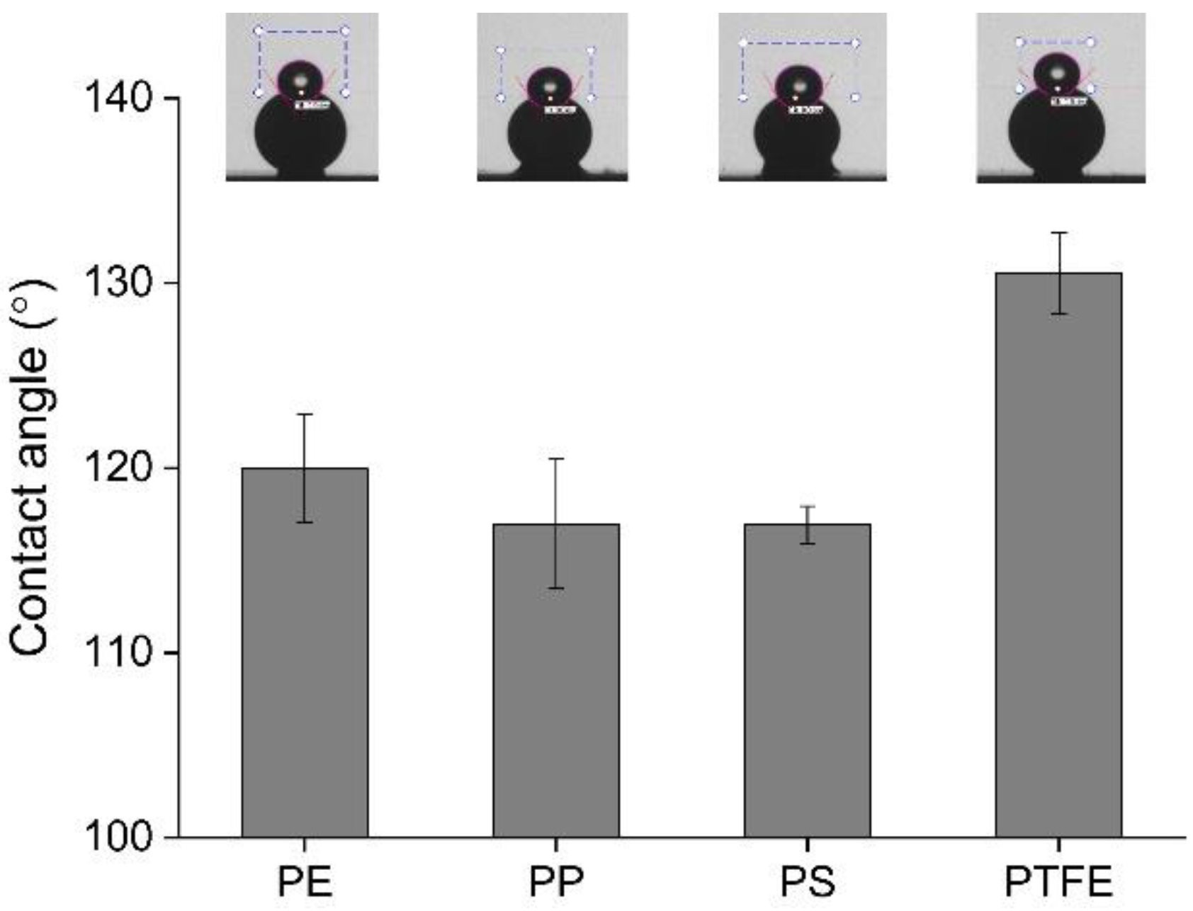
3.1. Surface Characterization of Microplastics

Considering that the larger the contact angle, the higher the hydrophobicity, PTFE had the highest hydrophobicity at 130°, and PS and PP had the lowest hydrophobicity at 117° (Figure 1). PE had a contact angle of 120°, which was slightly higher than PP and PS. The attachment properties of microorganisms with regard to the microplastic surface can be explained by the interfacial energy between the microplastic and water. Since a microplastic surface with high hydrophobicity has high interfacial energy with water, it can be lowered by attaching microorganisms with lower hydrophobicity [29,30]. Therefore, microorganisms are expected to attach better to surfaces following the hydrophobicity order: PTFE > PE > PP = PS.
The contact angles are not only determined by hydrophobicity due to the chemical properties of the materials but also by surface roughness. The surface roughness is explained by the Wenzel equation, which states that the contact angle of a hydrophobic surface increases as the surface roughness increases [31]. Therefore, the roughness of the surface may have affected the contact angle of each microplastic bead differently. The surface roughness (Rq) of PS and PP was the highest at 119.28 and 116.42 nm, respectively, and the surface roughness of PE and PTFE was much lower at 55.09 and 71.24 nm, respectively (Table 2). However, since the microplastic with the highest contact angle was identified as PTFE (Figure 1), the difference in contact angle was thought to be due to differences in the chemical properties of the material rather than surface roughness effects.

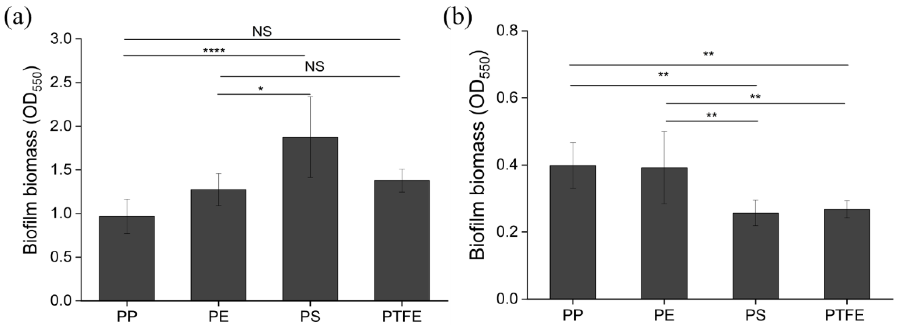
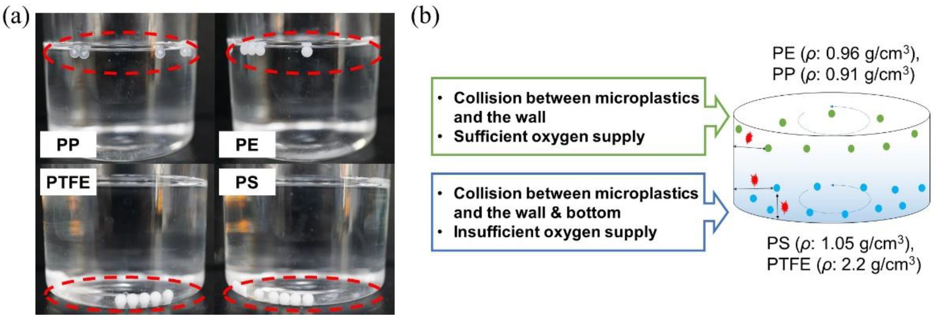
3.2. Biofilm Formation on Microplastic Surfaces

P. aeruginosa PAO1 was incubated in a 24-well plate with each microplastic under shaking conditions, and the amount of biofilm formation on the surface of the well plate and the microplastic was measured. When comparing the amount of biofilm formed on the surface of the well plate, the wells injected with PS and PTFE (OD550: 1.38~1.88) produced more than the wells injected with PP and PE (OD550: 0.97~1.27) (Figure 2a). Conversely, the biofilms on the microplastic surfaces were observed to form more PP and PE (OD550: 0.39~0.40) than on PS and PTFE (OD550: 0.26~0.28) (Figure 2b). As stated in Section 3.1, the higher the hydrophobicity of the plastic, the better the adhesion of microorganisms and biofilm formation. However, in this study, although PTFE had the highest hydrophobicity, it had the least amount of biofilm formed. Also, in the case of other plastics, there was no significant correlation between the amount of biofilm formation and the degree of hydrophobicity of the plastics. This experiment revealed that biofilm formation may be influenced by factors other than the hydrophobicity of microplastics.
The reduced biofilm formation on the PS and PTFE surfaces could be due to the relatively high densities of the two plastics. Since PS and PTFE have a higher density than water (Table 1), they sink to the bottom of the well plate (Figure 3a). During the incubation period, the microplastic moves and collides with the surface of the well plate. During this process, PS and PTFE collide with the bottom and wall surface of the well. However, PP and PE, which have similar densities to water and float on the culture medium (Figure 3a), collide only with the wall surface, as shown in Figure 3b. As a result of the frequency of impacts, biofilms on PS and PTFE surfaces appear to be more easily peeled off. Furthermore, oxygen concentration, which is an important factor in biofilm formation and dispersion, may also have contributed to the reduced biofilm formation on the microplastic surfaces. Although the shaking of the well plate provides oxygen to the inside of the culture medium, oxygen concentration inside the well plate is rapidly consumed by PAO1 development. Hence, oxygen concentration at the bottom surface may be lower than the water surface in contact with oxygen [32]. Therefore, in the case of microplastics moving in a low oxygen region, such as PS and PTFE, it is assumed that biofilm does not grow effectively or is easily dispersed [33]. The biofilm formed on the surface of the PS and PTFE microplastics was low because of the influence of movement characteristics rather than surface characteristics.

3.3. Effect of Microplastics at Different Biofilm Developmental Stages

The time-dependent biofilm formation of PAO1 was observed in an empty 24-well plate before testing the growth of the PAO1 biofilm formed on the microplastic surface. The amount of biofilm was not large at 1 or 2 h, but it significantly increased after 4 h (Figure 4a). These observations indicated that the initial attachment and aggregation phase proceeded for about 2 h, followed by the biofilm formation and growth phase between 2 h and 6 h.
The time-dependent biofilm formation of PAO1 was also assessed by performing biofilm tests in the presence of PP, PE, PS, and PTFE (Figure 4). To compare biofilm formation on two substrates (i.e., the well surface and microplastic surface) with different surface areas, the number of biofilms formed per surface area was presented. During the first 1–2 h (initial attachment and aggregation phase), more microorganisms were attached to the microplastic surface (4.30–8.92 mm−2) than to the well plate (2.15–3.49 mm−2). The 24-well plate used in this study was made of PS, and its surface hydrophilicity was increased by changing electrostatic characteristics through surface treatment, making hydrophobic microorganisms less able to attach to it. PE (8.36–8.92 mm−2) had a larger amount of microorganisms attached to its surface than PP (5.37–5.77 mm−2) (Figure 4b), which could be due to the higher hydrophobicity of PE. However, among the four microplastics, PTFE, which had the highest hydrophobicity, allowed the lowest attachment of microorganisms on its surface. This is presumably due to the maximum density of the PTFE, which caused it to sink to the bottom and frequently collide with the surface of the well plate, as explained in Section 3.2.
Unlike the initial attachment and aggregation phase (1–2 h), the biofilm on the well surface increased exponentially during the biofilm formation and growth phase (2–6 h), whereas it did not significantly increase on the microplastic surface (Figure 4). This could be explained by the collision between the microplastics and the well plate. The entire surface area of microplastics was exposed to the collisions, whereas only a specific part of the well plate, the upper or lower part, was exposed to the collisions with microplastics. This is presumed to be the cause of the failure of the biofilm to grow exponentially on the microplastic surface as on the well plate surface. A previous study that investigated bacteria–plastic interactions with various types of flat plastic surfaces reported that the surface properties of plastics, especially surface hardness, play an important role in the adhesion of bacteria [34]. However, as in the current study, when the plastic particles move, it can be seen that the behavior in water according to the density of plastic particles has a decisive effect on biofilm formation.

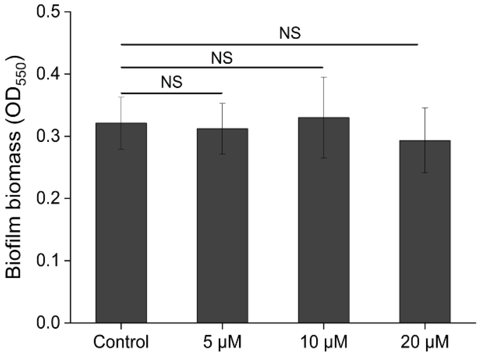
3.4. Effect of QS Signaling on Biofilm Formation on Microplastic Surfaces

3.4.1. Injection of Exogenous QS Signaling Molecules

AHL, a QS signal molecule, is known to play an important role in PAO1 biofilm formation. Therefore, to investigate the effect of QS on microplastic surface biofilm formation, an additional experiment was conducted by injecting various concentrations of exogenous AHLs into the PAO1 culture during biofilm formation. As shown in Figure 5, when AHL (the cocktail of C4-HSL and 3-oxo-C12-HSL) was injected at final concentrations of 5, 10, and 20 μM, respectively, no difference was detected in the amount of biofilm when compared to the control group (w/o AHL injection). It was reported that the deletion of genes that synthesize C4-HSL and 3-oxo-C12-HSL in the PAO1 resulted in a reduction in biofilm formation [35]. However, no research has been done to see if the addition of exogenous AHLs to PAO1 promotes biofilm formation. As shown in this experiment, the biofilm formation of PAO1 was not significantly affected by exogenous AHLs, which were additionally injected at high concentrations.

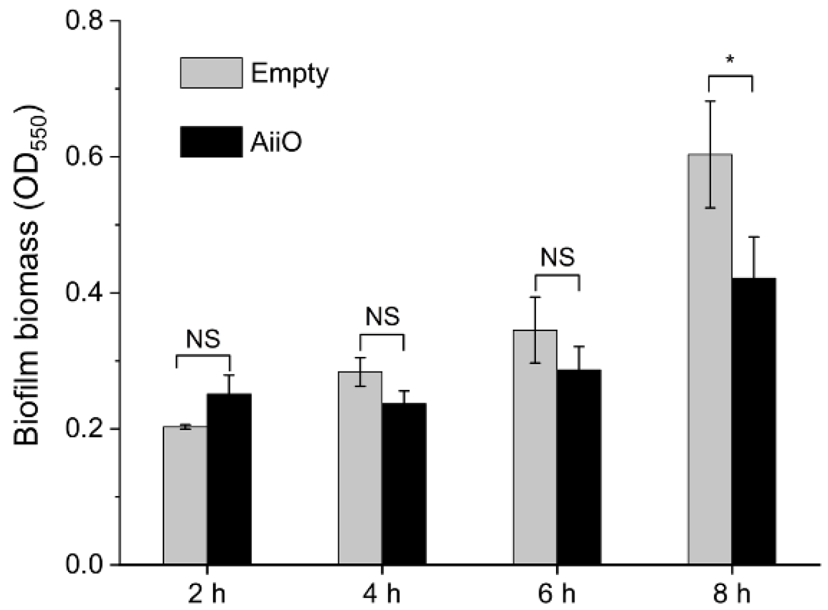
3.4.2. Injection of QQ Enzyme

After confirming that exogenous AHL did not affect PAO1 biofilm formation, the effect of AHL removal on biofilm formation on the microplastic surface was investigated. The removal of AHL was achieved by adding a QQ enzyme extracted from E. coli AiiO.
As shown in Figure 6, the overall biofilm formation was reduced by the addition of the AiiO enzyme extract (p = 0.0138). There was no significant difference in the number of biofilms formed on the PE microplastic surface in the control group (Empty) and the QQ treatment group (AiiO) in the 2–6 h period. However, the QQ treatment reduced biofilm formation by 30.1% (p = 0.019) after 8 h. This is consistent with previous reports, which found that the effect of AHL on the biofilm formation of microorganisms is greater at the biofilm growth stage than at the initial attachment stage [36]. Therefore, the biofilms formed on the moving microplastic surface showed slightly different tendencies from those formed on the fixed surface. However, it was confirmed that QS remains an important mechanism for biofilm formation on microplastic surfaces. This suggests that the QS mechanism must be considered in future studies on biofilm formation on microplastic surfaces.

4. Conclusions

In this study, biofilm formation on microplastic surfaces over time was analyzed to observe the difference in the biofilm formed depending on the type of microplastic. Biofilms did not grow well on the surfaces of PS and PTFE microplastics, which are denser than water, because of the detachment of biofilm due to frequent collisions with the bottom or restricted biofilm growth induced by low oxygen concentrations. Biofilms formed better on PE and PP microplastic surfaces, which have similar densities to water, and PE, which exhibited a relatively high hydrophobicity, was observed to have the best bacterial attachment at the initial stage of biofilm formation. Furthermore, the importance of the QS mechanism in the biofilm formation on the microplastic surfaces was validated by observing that biofilm formation was inhibited by QQ enzyme injection. The results of this study will help in understanding the interaction between microplastics and microorganisms and provide advanced insight into the behavior of microplastics in water systems and their environmental impact.

Author Contributions

Conceptualization, P.T.A., J.-H.K. and H.-S.O.; methodology, P.T.A., J.-H.K. and H.-S.O.; investigation, P.T.A. and J.-H.K.; formal analysis, P.T.A.; writing—original draft preparation, P.T.A. and J.-H.K.; writing—review and editing, J.-H.K. and H.-S.O.; supervision, funding acquisition, and project administration, H.-S.O. All authors have read and agreed to the published version of the manuscript.

Funding

This study was supported by a research program funded by SeoulTech (Seoul National University of Science and Technology) (2019-0493).

Institutional Review Board Statement

Not applicable.

Informed Consent Statement

Not applicable.

Conflicts of Interest

The authors declare no conflict of interest.

References

  1. Geyer, R.; Jambeck, J.R.; Law, K.L. Production, use, and fate of all plastics ever made. Sci. Adv. 2017, 3, e1700782. [Google Scholar] [CrossRef] [PubMed] [Green Version]
  2. Horton, A.A.; Dixon, S.J. Microplastics: An introduction to environmental transport processes. Wiley Interdiscip. Rev. Water 2018, 5, e1268. [Google Scholar] [CrossRef] [Green Version]
  3. Haram, L.E.; Carlton, J.T.; Ruiz, G.M.; Maximenko, N.A. A plasticene lexicon. Mar. Pollut. Bull. 2020, 150, 110714. [Google Scholar] [CrossRef] [PubMed]
  4. Amaral-Zettler, L.A.; Zettler, E.R.; Slikas, B.; Boyd, G.D.; Melvin, D.W.; Morrall, C.E.; Proskurowski, G.; Mincer, T.J. The biogeography of the Plastisphere: Implications for policy. Front. Ecol. Environ. 2015, 13, 541–546. [Google Scholar] [CrossRef]
  5. Shabbir, S.; Faheem, M.; Ali, N.; Kerr, P.G.; Wang, L.-F.; Kuppusamy, S.; Li, Y. Periphytic biofilm: An innovative approach for biodegradation of microplastics. Sci. Total Environ. 2020, 717, 137064. [Google Scholar] [CrossRef]
  6. Rosato, A.; Barone, M.; Negroni, A.; Brigidi, P.; Fava, F.; Xu, P.; Candela, M.; Zanaroli, G. Microbial colonization of different microplastic types and biotransformation of sorbed PCBs by a marine anaerobic bacterial community. Sci. Total Environ. 2020, 705, 135790. [Google Scholar] [CrossRef]
  7. Li, J.; Liu, H.; Chen, J.P. Microplastics in freshwater systems: A review on occurrence, environmental effects, and methods for microplastics detection. Water Res. 2018, 137, 362–374. [Google Scholar] [CrossRef]
  8. Sun, J.; Dai, X.; Wang, Q.; van Loosdrecht, M.C.; Ni, B.-J. Microplastics in wastewater treatment plants: Detection, occurrence and removal. Water Res. 2019, 152, 21–37. [Google Scholar] [CrossRef]
  9. Bhattacharya, P.; Lin, S.; Turner, J.P.; Ke, P.C. Physical adsorption of charged plastic nanoparticles affects algal photosynthesis. J. Phys. Chem. C 2010, 114, 16556–16561. [Google Scholar] [CrossRef]
  10. Besseling, E.; Wang, B.; Lürling, M.; Koelmans, A.A. Nanoplastic affects growth of S. obliquus and reproduction of D. magna. Environ. Sci. Technol. 2014, 48, 12336–12343. [Google Scholar] [CrossRef]
  11. Rios, L.M.; Jones, P.R.; Moore, C.; Narayan, U.V. Quantitation of persistent organic pollutants adsorbed on plastic debris from the Northern Pacific Gyre’s “eastern garbage patch”. J. Environ. Monit. 2010, 12, 2226–2236. [Google Scholar] [CrossRef] [PubMed]
  12. Browne, M.A.; Niven, S.J.; Galloway, T.S.; Rowland, S.J.; Thompson, R.C. Microplastic moves pollutants and additives to worms, reducing functions linked to health and biodiversity. Curr. Biol. 2013, 23, 2388–2392. [Google Scholar] [CrossRef] [PubMed] [Green Version]
  13. Hall, C.W.; Mah, T.-F. Molecular mechanisms of biofilm-based antibiotic resistance and tolerance in pathogenic bacteria. FEMS Microbiol. Rev. 2017, 41, 276–301. [Google Scholar] [CrossRef]
  14. Zettler, E.R.; Mincer, T.J.; Amaral-Zettler, L.A. Life in the “plastisphere”: Microbial communities on plastic marine debris. Environ. Sci. Technol. 2013, 47, 7137–7146. [Google Scholar] [CrossRef] [PubMed]
  15. Reisser, J.; Shaw, J.; Hallegraeff, G.; Proietti, M.; Barnes, D.K.; Thums, M.; Wilcox, C.; Hardesty, B.D.; Pattiaratchi, C. Millimeter-sized marine plastics: A new pelagic habitat for microorganisms and invertebrates. PLoS ONE 2014, 9, e100289. [Google Scholar] [CrossRef] [PubMed] [Green Version]
  16. Rummel, C.D.; Jahnke, A.; Gorokhova, E.; Kühnel, D.; Schmitt-Jansen, M. Impacts of biofilm formation on the fate and potential effects of microplastic in the aquatic environment. Environ. Sci. Technol. Lett. 2017, 4, 258–267. [Google Scholar] [CrossRef] [Green Version]
  17. Yuan, J.; Ma, J.; Sun, Y.; Zhou, T.; Zhao, Y.; Yu, F. Microbial degradation and other environmental aspects of microplastics/plastics. Sci. Total Environ. 2020, 715, 136968. [Google Scholar] [CrossRef]
  18. Cappitelli, F.; Sorlini, C. Microorganisms attack synthetic polymers in items representing our cultural heritage. Appl. Environ. Microbiol. 2008, 74, 564–569. [Google Scholar] [CrossRef] [Green Version]
  19. Ju, W.; An, J.; Jho, E. Adsorption characteristics of Cd and Pb on microplastic films generated in agricultural environment. J. Korean Soc. Environ. Eng. 2021, 43, 32–42. [Google Scholar] [CrossRef]
  20. Barboza, L.G.A.; Vieira, L.R.; Branco, V.; Figueiredo, N.; Carvalho, F.; Carvalho, C.; Guilhermino, L. Microplastics cause neurotoxicity, oxidative damage and energy-related changes and interact with the bioaccumulation of mercury in the European seabass, Dicentrarchus labrax (Linnaeus, 1758). Aquat. Toxicol. 2018, 195, 49–57. [Google Scholar] [CrossRef]
  21. Urbanek, A.K.; Rymowicz, W.; Mirończuk, A.M. Degradation of plastics and plastic-degrading bacteria in cold marine habitats. Appl. Microbiol. Biotechnol. 2018, 102, 7669–7678. [Google Scholar] [CrossRef] [PubMed] [Green Version]
  22. Kumar, A.; Alam, A.; Rani, M.; Ehtesham, N.Z.; Hasnain, S.E. Biofilms: Survival and defense strategy for pathogens. Int. J. Med. Microbiol. 2017, 307, 481–489. [Google Scholar] [CrossRef]
  23. Zhang, Y.; Diehl, A.; Lewandowski, A.; Gopalakrishnan, K.; Baker, T. Removal efficiency of micro-and nanoplastics (180 nm–125 μm) during drinking water treatment. Sci. Total Environ. 2020, 720, 137383. [Google Scholar] [CrossRef] [PubMed]
  24. Xiong, X.; Siddique, M.S.; Graham, N.J.; Yu, W. Towards microplastics contribution for membrane biofouling and disinfection by-products precursors: The effect on microbes. J. Hazard. Mater. 2022, 426, 127797. [Google Scholar] [CrossRef] [PubMed]
  25. Arias-Andres, M.; Rojas-Jimenez, K.; Grossart, H.-P. Collateral effects of microplastic pollution on aquatic microorganisms: An ecological perspective. TrAC Trends Anal. Chem. 2019, 112, 234–240. [Google Scholar] [CrossRef]
  26. Evans, L.R.; Linker, A. Production and characterization of the slime polysaccharide of Pseudomonas aeruginosa. J. Bacteriol. 1973, 116, 915–924. [Google Scholar] [CrossRef] [Green Version]
  27. Oh, H.-S.; Tan, C.H.; Low, J.H.; Rzechowicz, M.; Siddiqui, M.F.; Winters, H.; Kjelleberg, S.; Fane, A.G.; Rice, S.A. Quorum quenching bacteria can be used to inhibit the biofouling of reverse osmosis membranes. Water Res. 2017, 112, 29–37. [Google Scholar] [CrossRef]
  28. McLean, R.J.; Whiteley, M.; Stickler, D.J.; Fuqua, W.C. Evidence of autoinducer activity in naturally occurring biofilms. FEMS Microbiol. Lett. 1997, 154, 259–263. [Google Scholar] [CrossRef]
  29. Lee, J.; Chae, H.-R.; Won, Y.J.; Lee, K.; Lee, C.-H.; Lee, H.H.; Kim, I.-C.; Lee, J.-m. Graphene oxide nanoplatelets composite membrane with hydrophilic and antifouling properties for wastewater treatment. J. Membr. Sci. 2013, 448, 223–230. [Google Scholar] [CrossRef]
  30. Krishnan, S.; Weinman, C.J.; Ober, C.K. Advances in polymers for anti-biofouling surfaces. J. Mater. Chem. 2008, 18, 3405–3413. [Google Scholar] [CrossRef]
  31. Jin, H.; Tian, L.; Bing, W.; Zhao, J.; Ren, L. Bioinspired marine antifouling coatings: Status, prospects, and future. Prog. Mater. Sci. 2022, 124, 100889. [Google Scholar] [CrossRef]
  32. Meier, K.; Klöckner, W.; Bonhage, B.; Antonov, E.; Regestein, L.; Büchs, J. Correlation for the maximum oxygen transfer capacity in shake flasks for a wide range of operating conditions and for different culture media. Biochem. Eng. J. 2016, 109, 228–235. [Google Scholar] [CrossRef]
  33. McDougald, D.; Rice, S.A.; Barraud, N.; Steinberg, P.D.; Kjelleberg, S. Should we stay or should we go: Mechanisms and ecological consequences for biofilm dispersal. Nat. Rev. Microbiol. 2012, 10, 39–50. [Google Scholar] [CrossRef] [PubMed]
  34. Cai, L.; Wu, D.; Xia, J.; Shi, H.; Kim, H. Influence of physicochemical surface properties on the adhesion of bacteria onto four types of plastics. Sci. Total Environ. 2019, 671, 1101–1107. [Google Scholar] [CrossRef]
  35. Davies, D.G.; Parsek, M.R.; Pearson, J.P.; Iglewski, B.H.; Costerton, J.W.; Greenberg, E.P. The involvement of cell-to-cell signals in the development of a bacterial biofilm. Science 1998, 280, 295–298. [Google Scholar] [CrossRef] [Green Version]
  36. Anburajan, P.; Kim, Y.; Rice, S.A.; Oh, H.-S. Bacterial signaling and signal responses as key factors in water and wastewater treatment. J. Water Process Eng. 2021, 44, 102434. [Google Scholar] [CrossRef]
Figure 1. Contact angle measurement of microplastics.
Applsci 12 05245 g001
Figure 2. (a) Biofilm formation on the well surface. Error bars were defined as standard deviations (n = 8, biological replicates). One-way ANOVA was performed and the Tukey post hoc test was conducted to compare the number of biofilms where significant differences are indicated as follows: NS, not significant p > 0.05, * p < 0.05, and **** p < 0.0001. (b) Biofilm formation on the microplastic surface (n = 8, biological replicates). ** p < 0.01.
Applsci 12 05245 g002
Figure 3. (a) Images of microplastics in water. (b) The possible mechanism of biofilm detachment from the microplastic surface.
Applsci 12 05245 g003
Figure 4. (a) Biofilm formation on the well surface. (b) Biofilm formation on the microplastic surface. Error bars were defined as standard deviations (n = 6, biological replicates).
Applsci 12 05245 g004
Figure 5. Effect of AHL on the biofilm formation on PE microplastic surface. Error bars were defined as standard deviations (n = 12, biological replicates). One-way ANOVA was performed and the Tukey post hoc test was conducted to compare the AHL treatment to the negative control, where significant differences are indicated as follows: NS, not significant p > 0.05.
Applsci 12 05245 g005
Figure 6. Effect of QQ enzyme on the biofilm formation on the PE microplastic surface. Error bars were defined as standard deviations (n = 3, biological replicates). A two-way ANOVA was performed and the Bonferroni post hoc test was conducted to compare the QQ treatment (AiiO) to the negative control (Empty), where significant differences are indicated as follows: NS, not significant p > 0.05 and * p < 0.05.
Applsci 12 05245 g006
Table 1. The density and diameter of the microplastic beads used in the experiment.
PEPPPSPTFE
Diameter (mm)3.23.22.963.2
Density (g/cm3)0.960.911.052.2
Table 2. Analysis of surface roughness of microplastic beads by AFM.
PEPPPSPTFE
Rq * (nm)116.4255.09119.2871.24
* Rq is one of the most commonly reported measures of surface roughness from AFM images.
Publisher’s Note: MDPI stays neutral with regard to jurisdictional claims in published maps and institutional affiliations.

