Article

Synthesis of Gold-PVP Nanostructured Composites by Microplasma: A Test to Study Their Inhibiting Tendency of Avian Influenza Virus Activity

1
Department of Physics, University of Engineering and Technology, Lahore 54890, Pakistan
2
Institute of Microbiology, University of Veterinary and Animal Sciences, Lahore 54000, Pakistan
3
Zoology Department, College of Science, King Saud University, Riyadh 11451, Saudi Arabia
4
School of Pharmacy and Biotechnology, Coleraine Campus, Ulster University, Cromore Rd., Coleraine BT52 1SA, UK
*
Author to whom correspondence should be addressed.
Appl. Sci. 2022, 12(11), 5352; https://doi.org/10.3390/app12115352
Submission received: 17 April 2022 / Revised: 19 May 2022 / Accepted: 20 May 2022 / Published: 25 May 2022
(This article belongs to the Section Nanotechnology and Applied Nanosciences)

Abstract

:
Gold–polymer nanostructured composites have a great potential in the biomedical and advanced materials field as an antimicrobial agent against various pathogens, especially viruses. In the present work, gold and gold-PVP colloids have been prepared by the electrochemical reduction of hydrogen tetrachlorauric acid (HAuCl4·3H2O) precursor. The atmospheric pressure microplasma technique was used as a reducing agent, while D-Fructose was used as a stabilizing agent in the synthesis process. X-ray Diffraction (XRD) confirmed the crystalline behavior of both gold nanostructured particles and gold-PVP nanocomposites. The morphology of the nanoparticles was examined by SEM. The absorption characteristic peaks at 541 nm and 542 nm in UV-Vis spectra confirmed the surface plasmon resonance in gold nanostructured particles and gold-PVP nanostructured composites, respectively. Dynamic light scattering studies with percentage intensity distribution revealed particle size distribution ranges from 8–288 nm for gold colloids and 15–297 nm for gold-PVP colloids. Gold-PVP nanostructured composites have shown an improvement in the antiviral activity against the H9N2 virus as compared to bare gold nanostructured particles.

1. Introduction

Influenza viruses, causing serious illness, deaths, and fear, have globally devastated the human and poultry population [1]. Avian Influenza viruses belonging to the orthomyxoviridae family are generally categorized by two surface antigens, Hemagglutinin (HA) and neuraminidase (NA), which bridge the biological system to infect birds, humans, and other mammalians [2]. The Avian Influenza Virus H9N2, initially isolated from china poultry forms in 1994, has been shown to infect humans by crossing species barriers [3]. The concerns about its pandemic potential have been compelling researchers to recognize and sort alternative anti-influenza agents with a different mechanism of action [4]. However, the shifting and drifting of the various influenza virus strains, their emerging drug resistance, and the limitations of antiviral drugs have posed a serious threat against the survival of humans and animals [5]. Different kinds of influenza have different abilities to resist antiviral medicines, but mainly two approaches, injectable vaccines and antiviral agents, are used to control it [1].
In the biomedical field, metal nanoparticles have been studied enormously to develop vaccines and antiviral drugs against influenza viruses. Xiang et al. comprehensively studied the antiviral activity of silver nanoparticles (AgNPs) on the H3N2 virus, where AgNPs selectively destroyed the virus structure [6]. In another previous investigation, AgNPs showed an efficient inhibitory activity on H1N1 influenza A virus in the hemagglutination of a chicken RBCs Assay Inhibition test by using an embryonated chicken egg as a cell culture [7]. Gold nanoparticles (AuNPs) have ideal opportunities to have bio conjunctions, particularly with influenza viruses, and can interact directly with the cell surface and block viral attachments [8]. More recently, J. Kim and co-workers reported a strong antiviral activity of porous AuNPs on H1N1 and H9N2 influenza virus strains in an HA protocol using MDCK cells as a culture medium [9]. In one of the previous cases of research, gold nanoparticles of a size of 14 nm were found to be more effective in inactivating hemagglutination than that of a 2 nm size [10]. It is seen in previous research that metallic NPs have been extensively explored as antimicrobial agents, but at the same time, there have been stability, biocompatibility, non-degradability, and toxicity issues [11,12]. These issues have limited their applications in the medical field [13]. Thus, the researchers have revisited their strategies in exploring noble metal nanostructures in the medical field [14].
The behavior of a mixture of nanoparticles (NPs) and polymers, referred to as nanocomposites (NCs), has piqued the scientific community’s curiosity. A nanocomposite is a mixture of a metal–polymer material with at least one dimension in a nanoscale regime of 1–100 nm [15]. PVP and PEG are WHO-endorsed synthetic polymers, known to be well tolerated by the human body [16]. Their association with noble metal NPs makes most admirable hybrid nanostructures. These nanocomposites can mitigate the effects of microbial infections by enhancing stability, boosting biological activity, and lowering toxicity [17]. Recently an enhanced antiviral activity of ZnO-PEG NPs as compared to bare ZnO NPs has been reported [18]. In another research activity, small silver NPs in chitosan showed a stronger capability for inhibiting the H1N1 virus [19].
For the synthesis of metallic NPs/NCs, different approaches have been adopted, including physical [20], Chemical [21], and biological routes [22]. Among these techniques, atmospheric pressure microplasma has been the most admired bottom-up approach to nanoparticle synthesis [23]. In this process, electrons with other plasma species are produced in an argon/helium gas-assisted DC microplasma discharge at the surface of the metal precursor solution. The reduction of metal cations occurs due to interaction with microplasma-generated energetic electrons. Metal nanoparticles are formed via nucleation and growth through Van der Waals forces [24,25]. The details of the synthesis mechanism are described in the experimental section. The main advantage of this synthesis route is that the size and morphology of nanoparticles are tuned by controlling the experimental parameters, such as microplasma discharge time, gas flow rate, and the concentration of precursor salt [26,27]. This makes microplasma an ideal technique to fabricate biocompatible nanoparticles with promising biomedical applications [28].
These studies aimed to take advantage of microplasma-assisted fabrication of gold nanocomposites. Povidone (PVP), with its established capping and shape-regulating character, can conjugate with AuNPs to develop Au-PVP nanostructured composites [29]. The Au-PVP nanocomposites with tunable physicochemical properties are expected to be equipped with biocompatible and biodegradable characteristics mandatory for their use as antiviral agents [30]. To the best of our knowledge, there is no evidence of such investigations with a gold-PVP hybrid nanostructure synthesized by microplasma on the H9N2 influenza virus strain.

2. Materials and Method

Analytical-grade Hydrogen tetrachloroaurate(III) trihydrate HAuCl4·3H2O (MERCK, D-6100 Darmstadt OR G 7/8025/001 HK1, 393.83 gm/mole), as a metal precursor; D-Fructose (MOL WT 180.16 gm/mole BATCH NO; 4088-2629 AVONCHEM UK), as a stabilizer; Polyvinylpyrrolidone (PVP) (MW 40,000; 111.14 g/mole, Sigma-Aldrich), as capping agent; and distilled water, as a solvent, were used. A 100-mL precursor of 1.0 mM concentration Hydrogen tetrachloroaurate(III) trihydrate HAuCl4·3H2O and 5 mM D-Fructose was prepared as follows. A total of 4 mL taken from a 25 mM stock solution of 25 mM Hydrogen tetrachloroaurate(III) trihydrate (HAuCl4·3H2O) was mixed in 96 mL of distilled water. Further, 0.089 gm of D-Fructose was added to a 100 mL prepared solution. The whole solution was stirred magnetically for 20 min to ensure the homogeneity of the required precursor.

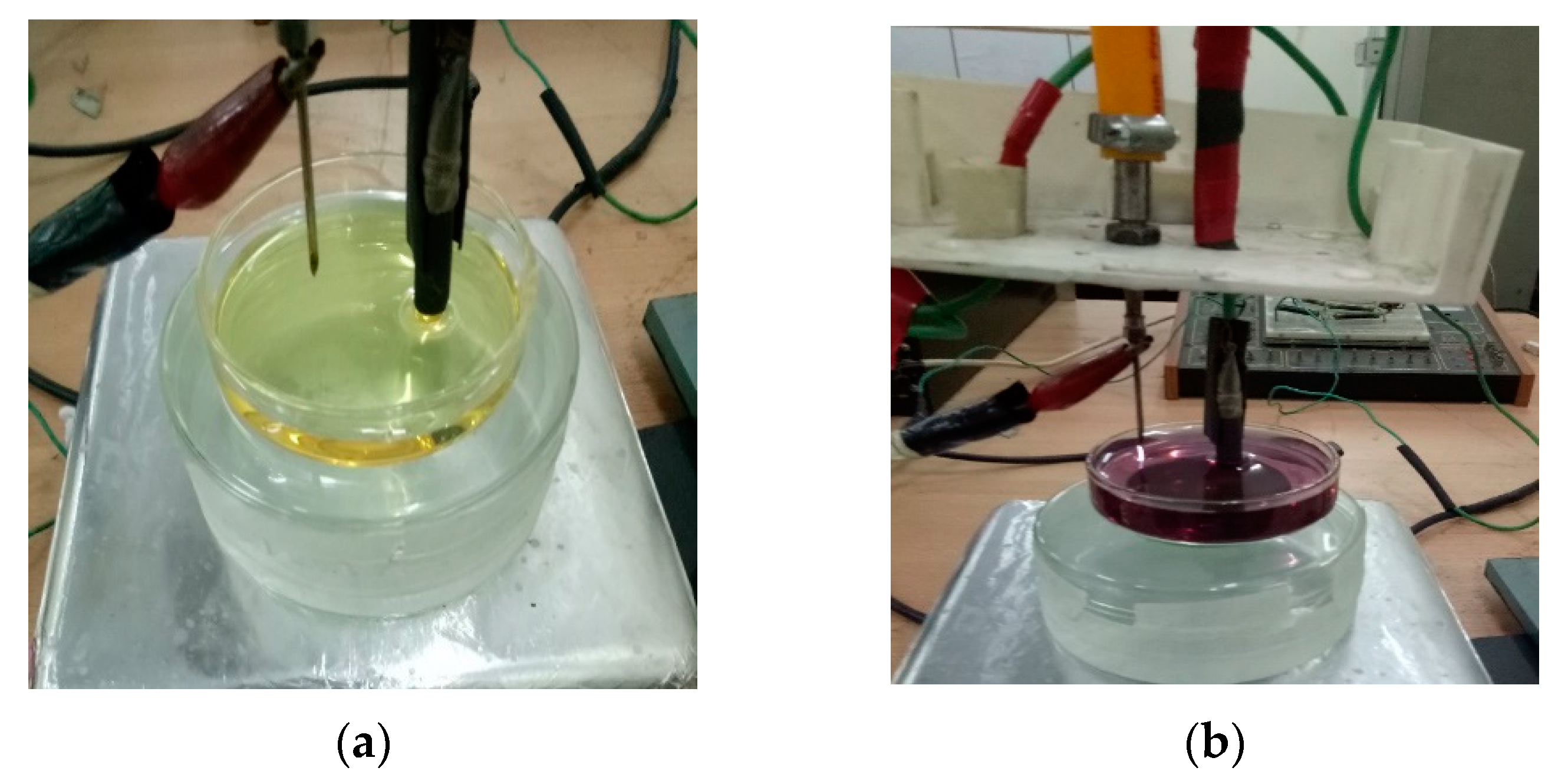
2.1. Synthesis of Gold Nanostructured Particles and Gold-PVP Nanostructured Composites

The microplasma was sustained by a high dc voltage (up to 5–6 kV) applied at the carbon rod, while the stainless steel capillary (through which argon gas flows at a constant rate) was grounded through a ballast resistor of 2.5 MΩ. The plasma current was maintained at 2–2.5 mA (power supply output of 30 KV, 66 mA Glassman high voltage, INC).
A reaction cell of volume 100 mL containing hydrogen tetrachloroaurate(III) trihydrate HAuCl4·3H2O was used as a precursor. The precursor solution was exposed to atmospheric pressure microplasma plasma for 20 min (named Au-20). The microplasma discharge provides a cascade of electrons and other plasma species which interact with precursor solutions to form gold NPs. The change of color of the solution from golden (Figure 1a) to purplish (Figure 1b) after 2–3 min of plasma exposure time was the first visual indication of the formation of gold nanostructured particles. To prepare the second sample of Au-PVP nanostructured composites (named Au-PVP-20), an amount of 0.028 gm of Polyvinylpyrrolidone PVP, (Molecular Weight = 40,000; 111.14 g/mole) was added and magnetically stirred for 20 min. Further, the color of Au-PVP-20 remained the same as that of Au-20.

2.2. Investigations of Antiviral Activity

To test the antiviral activities of the synthesized nanostructured particles, a standard three-step procedure was followed. These steps included inoculation, harvesting of the chorioallantoic fluid, and the Hemagglutination Assay protocol. In this procedure, a volume of 100 μL each of synthesized gold and gold-PVP colloids was separately mixed with an equal volume of confirmed Avian Influenza Virus (AIV) (H9N2) of 106 EID50 in a microfuge tube. The AIV (H9N2) of 106 EID50 was procured from Influenza lab, Institute of Microbiology, the University of Veterinary and Animal Sciences, Lahore.
The mixture of nanostructured gold particles (Au-20) and gold-PVP nanostructured composites (Au-PVP-20) and the virus was separately incubated for 60 min at room temperature to interact. Inoculation is proceeded by injecting the above samples into the allantoic cavity of 9–11 days’ serum pathogen-free fertile chicken eggs with a 22 G needle attached to a 1 mL syringe as shown in Figure 2a. The hole was sealed with wax. After 48 hrs. of incubation at 37 °C, the eggs were chilled for at least 4 h at 4 °C to let the virus propagate. Chilled eggs with air sacs facing up into a holder were transferred to the biosafety lab. The eggshell above the air sac was removed carefully with sterilized scissors without damaging the chorioallantoic membrane, as shown in Figure 2b. With sterile forceps, the chorioallantoic membrane was removed, and allantoic fluid was collected as much as possible with a small spatula or 1 mL Eppendorf pipette.
The chorioallantoic fluid (CAF) harvested from the Embryonated chicken eggs as a virus stock was the major part of the laboratory-based study of influenza A virus titration in the hemagglutination Assay protocol procedure referenced by Alexander and Chettle (1977) is described below.

Hemagglutination Test (HA)

In a typical Hemagglutination assay (HA) protocol, an influenza virus titer can be evaluated by utilizing serial dilutions of a virus on a 96-well plate (either V- or round-bottom) and introducing a fixed amount of red blood cells, for example, 1% Chicken RBCs.
Using a multi-channel micropipette, 50 μL of normal saline (NS) was dispensed into all of the wells of the micro-titration plate. A volume of 50 μL of harvested CAF was dispensed in the first well. Two-fold serial dilutions were made from 1:2 to 1:2048. It was accomplished by putting 50 μL into the next well and so on until the 11th well was reached. A total of 50 µL of 1% RBC suspension was dispensed into all of the wells. The 12th well, which contained only 1% RBC suspension and normal saline, was preserved as a negative control. The micro-titration plate was then incubated for 20–30 min at 37 °C. The RBCs in the 12th well had settled down to form a visible bead at the bottom of the well. To assure quality findings, the HA test was repeated three times. HA activity negative means that the virus has been disabled or have become neutral with activity of nanoparticles. Consequently, RBCs have settled down to the bottom of the well in the form of a bead. HA activity positive confirms the presence of a virus that adheres to the surface of RBCs causing agglutination. Consequently, preventing RBCs from settling down and a matt reddish suspension is seen. The HA activity was recorded as shown in Figure 3. The Figure 3a shows HA activity of Au-20 and dilution, upto which their tendency of inhibiting influenza virus activity of agglutination is observed. Whereas in Figure 3b, the dilution of Au-PVP-20 upto which their ability of inhibiting virus activity of agglutination is displayed. The virus has a tendency to adhere to the surface of red blood cells, causing agglutination and hence preventing them to settle at the bottom. This is called HA activity positive.

3. Results and Discussions

3.1. X-rays Diffraction (XRD) Analyses

The crystallinity and phases of gold nanostructured in samples Au-20 and Au-PVP-20 were probed with Bruker D8 DISCOVER Advance in Bragg–Brentano mode with Cu Kα radiation (40 KV, 1.54 A, and 40 mA) over 2Ɵ angle ranging from 10° and 70°. The samples for XRD analysis were prepared by drop-casting colloids separately on glass strips followed by drying at room temperature. The XRD data were compared with the JCPDS data card 04-0784 for gold, as depicted by graph (a) in Figure 4. The patterns of Au-20 and Au-PVP-20 appeared to be identical, as shown by graphs (b) and (c), respectively, in Figure 4. The Bragg’s reflections peaks of both samples appeared at 38.24°, 44.3°, and 64.7°, corresponding to (111), (200), and (220) planes, respectively, indicating the FCC lattice.
The maximum intensity peak at 38.24° for both samples indicated the preferential growth in the (111) direction, which is in agreement with previously reported results [30]. However, a decrease in (111) peak intensity and a slight increase in the intensity of (200) and (220) directions in the case of Au-PVP-20 was observed [30]. This increase in intensity might have been due to increased stacking in the (200) and (220) planes because of the reorientation of the (111) planes in the dielectric polymeric medium of a relatively higher refractive index after PVP capping [31]. This change in intensities is also attributable to the changes in the shape of the nanostructures, as evidenced by the SEM micrographs in Figure 5. Here PVP has demonstrated a shape-directing character of AuNPs metal cores when it adsorbed to gold colloids [32].

3.2. SEM Analysis

The morphology of Au-20 and Au-PVP-20 samples were investigated by a ZEIS Sigma 500 Field Emission Electron Microscope (FE-SEM). Dried drop-casted samples of Au-20 and Au-PVP-20 colloids were prepared on glass slides. Without PVP, the sizes of the majority of nanostructured particles were not uniform and were of irregular shapes, as shown in Figure 5a. However, a small number of AuNPs had a uniform size and shape. With PVP capping, the morphology of the particles changed as we expected from the PVP association. A strong shape-regulating behavior of PVP with AuNPs was observed, as shown in Figure 5c. This is in agreement with the previous investigations [31]. Now, the majority of the nanostructured particles seemed spherical with a controlled size range, whereas a minority of the particles also seemed to have an irregular shape. In Figure 5a,c, the inset histogram shows the size distribution of Au-20 and Au-PVP-20, respectively, extracted from SEM micrographs.
The Energy Dispersive X-ray (EDS) spectroscopy of both samples was executed for the elemental analysis of Au-20 and Au-PVP-20. The spectrum in Figure 5b confirms metallic gold, masked by C and O peaks present in D-Fructose. Cl is the constituent of the gold precursor (HAuCl4.3H2O), while the Si peak is due to the substrate. Argon is present because of being a base gas to produce plasma. Figure 5d is the EDX spectrum of Au-PVP-20, where the same elemental peaks of Au-20 appear with an extra N peak due to PVP capping. The N peak confirms the PVP capping [30]. The inset histogram shows the size distribution of nanostructured gold particles and nanostructured gold-PVP composites extracted from SEM micrographs. It depicts that in both the cases, the size ranged from 1 nm to 270 nm, with the maximum number particles in the size range of 30 nm to 60 nm.

3.3. Dynamic Light Scattering (DLS) Analysis

The particle size distribution of Au-20 and Au-PVP-20 colloids was analyzed by Dynamic Light Scattering Techniques using a standard He–Ne laser (632.8 nm) as a light source. BI-200SM, Brookhaven Instrument Corp., was used for this purpose. The Particle Size Distribution (PSD) profiles of Au-20 and Au-PVP-20 are shown in Figure 6. In the case of Au-20 (Figure 6a), there were two size distributions. The first distribution lay in the range from 8 nm to 27 nm, with a peak at 19 nm, while a majority of the nanostructured particles lay in the second distribution, which ranged from 74 nm to 288 nm, with a peak at 121 nm. In Au-PVP-20 (Figure 6b), again, there were two size distributions. The first distribution lay in the range from 15 nm to 19 nm, with a peak at 19 nm, while a majority of the nanostructured particles lay in the second distribution, which ranged from 98 nm to 297 nm, with a peak at 153 nm.
With PVP capping, the hydrodynamic size distribution was 15 nm to 297 nm. Conversely, without PVP capping, the hydrodynamic size distribution was 8 nm to 288 nm. These size distributions lie within the range of the size distribution determined by Yang and co-workers for Au-PVP nanocrystals, which range from 100–300 nm [33,34].
It is to be noted that in DLS measurements, the size distribution of the AuNPs in composites is not very different from fructose-stabilized AuNPs. This justifies the co-stabilizing character of PVP in controlling the size distribution.

3.4. UV-VIS Spectroscopic Analysis

In the present investigations, the UV-VIS spectroscopic studies of Au-20 and Au-PVP-20 were accomplished by using Agilent Technologies Cary 60 ranging from 200–800 nm, with a resolution of 1 nm. The absorbance spectrum of Au-20 exhibited a maximum peak at the wavelength of 541 nm, whereas Au-PVP-20 exhibited a maximum peak at 542 nm, as shown in Figure 7, which lies within the range previously reported (520 nm–580 nm) for gold nanostructured particles with a size range of 10 nm–100 nm [28,35]. There was no reasonable shift in the maximum absorption in Au-PVP-20 as compared to Au-20. This is because the immediate mixing of PVP with gold nanoparticles gives no sufficient time for Au-20 nanostructured particles to agglomerate or to change the effective cores of nanostructured particles [36]. PVP itself is best known for its stability, which has prevented the Au-20 nanostructured particles to agglomerate [37]. Particularly, previous investigations suggest that the maximum absorption peak is red-shifted with an increase in the size of the nanostructured particles and blue-shifted with the decrease in nanostructured particles’ size [28,37].
However, in our case, the DLS analysis (Figure 6) revealed that the sizes of the metallic cores of the nanostructured particles in the distribution did not significantly change with PVP capping. Eventually, the surface plasmon resonance response of conduction electrons in both Au-20 and Au-PVP-20 remains almost identical [38].
By using the UV-Vis absorption spectra of Au-20 and Au-PVP-20, the optical bandgap was determined by exploiting the Tauc equation, which is a relation between the absorption coefficient α, and Eg is the bandgap energy Eg. [39,40].
(αhʋ)2 = k(hʋ − Eg)
where hʋ is the energy of the incident photon, k is a constant, and the Tauc plot is a graph of hʋ (abscissa) Versus (αhʋ)2 (ordinate). The extrapolation of the tangent in the Tauc plot to (αhʋ)2 = 0 gives the value of the direct bandgap energy.
The optical bandgap values extracted from the Tauc plot (Figure 8) were found to be the same both for Au-20 and Au-PVP-20, i.e., 2.58 eV, which justifies the identical absorption response. However, with the PVP linkage, the Intensity of absorption increased reasonably. This difference in the intensity of absorption is usually associated with the nanoparticles’ concentration affected by the mild acidic environment of PVP (3 > pH < 4.8) [41].

3.5. Antiviral Activity

To examine the inhibitory properties of gold nanostructured particles and PVP-gold nanostructured composites in the pre-entry stages of the influenza virus life cycle, we tested the tendency of these nanostructured particles to inhibit hemagglutination by the method referenced by Alexander and Chettle [42]. In a typical HA test, the comparison of the Influenza H9N2 virus’ tendency to agglutinate chicken Red Blood Cells (RBCs) and the gold nano-structured particles’ property of inhibiting virus agglutination was studied. Our findings based on the observations on the HA protocol evidenced that gold nano-structured particles synthesized by the atmospheric pressure microplasma technique showed an inhibition of the H9N2 influenza virus activity of agglutinating Chicken RBCs, as has already been shown in Figure 3 in Section 2.2 (HA).
In our present investigations, the concentration of nanostructured gold particles, which attenuated the infectivity of the H9N2 Influenza virus strain in the HA protocol, was evaluated to be 1.96 μg/mL by using the relation [43].
W e i g h t = M o l a r i t y × M o l e c u l a r   W e i g h t × V o l u m e
Recently, porous gold nanoparticles synthesized by the surfactant-free emulsion method have been investigated against the H1N1, H3N2, and H9N2 influenza virus strains in the HA protocol using MDCK cells as a culture medium [9]. In their findings, the concentration of porous gold cabable to disable these multiple avian influenza viruses was 0.2 mg/mL. More recently, gold NPs synthesized by the chemical method using citrate as a reducing agent demonstrated a convincing inhibitory effect against the H1N1 virus in MDCK cells with medium-size gold nanoparticles [44].
Similar kinds of investigations were carried out by a previous research group with silver nanoparticles synthesized by biological roots. AgNPs with a concentration range of 12.5 μg/mL to 100 μg/mL exhibited a potential tendency of inhibiting H1N1 influenza A virus activity [6].
To study the inhibitory role of gold particles in the hybrid nanostructure, we investigated the antiviral activity of gold-PVP nanostructured composites. Quite interestingly, gold nanostructured in the composite form disabled the Avian virus activity of agglutination with more dilution, and hence less concentration, as compared to bare gold nanoparticles, as evidenced in Figure 3. We separately investigated the antiviral activities of Povidone (PVP) with the same concentration mixed with distilled water, where no antiviral activity was observed against AIV/H9N2 influenza viruses. This confirmed that the antiviral activity was only due to the presence of AuNPs in composites. Thus, we argue that the PVP attachment to the gold nanostructured particles boosted their antiviral activities. This strong inhibitory role of AuNPs in a hybrid nanostructure against the AIV/H9N2 influenza virus is due to the supporting role of PVP in Au-PVP nanostructured composites. PVP, due to its shape-directing, regulating, and controlling character [30,32], as evidenced by our morphological studies in the FESEM analysis, boosted the biocompatibility and, hence, improved the antiviral tendencies.
The supporting antiviral behavior of other polymers, such as polyethylene glycol (PEG) and chitosan, with other metal nanostructured particles has also been investigated by previous research groups. Recently, ZnO NPs and ZnO-PEG NPs synthesized by the mechanical ball milling method [45] have been investigated to test their antiviral activity against the H1N1 influenza virus in the MDCK cell as a culture medium. PEGylated ZnO NPs showed an enhanced antiviral activity compared to bare ZnO NPs [18]. In another research activity, small silver NPs in chitosan were found to possess a greater capability of inhibiting the H1N1 virus [19]. The size distribution of colloids, as determined in our DLS studies, was 8–288 nm for nanostructured gold particles and 15–297 nm for gold-PVP nanostructured particles. These size distributions lie within the range of the sizes of the Orthomyxoviruses family to which H9N2 belongs [46]. These compatibilities in the size distributions of nanostructured gold particles, particularly in composite form, and the possible size of influenza viruses have provided an ideal platform to demonstrate biological interactions [8]. The supporting role of PVP in enhancing the inhibiting ability of nanostructured gold composites is also reflected in UV-Vis spectroscopic studies. The increase in the intensity peak with PVP association revealed the provision of the possible character of PVP as co-reducing agent [47] in addition to microplasma, resulting in the fabrication of more colloids and, hence, more interactions with the biological system and, hence, more antiviral activities [48].
In light of our present investigations, it was evidenced that gold nanostructured particles and nanostructured gold composites fabricated by an atmospheric pressure microplasma have been proven to exhibit a strong tendency of inhibiting the avian influenza H9N2 virus strain. These gold nanostructured particles are expected to equally fight multiple influenza virus strains with the same potential. This argument is well supported by the previous investigations with porous gold nanoparticles [9] and the effective inhibition of multiple influenza virus strains with anionic gold [49].
The mechanisms of antiviral activity of metallic nanostructures and their composites fabricated by any kind of physical [20], chemical [21], and biological routes [22] against multiple influenza virus strains may be hypothesized within the same pattern of action. These nanostructures adsorb to the virions spikes (viral envelope glycoproteins) by Van der Waals interactions [50]. This adsorption may disrupt the virus attachment to the cellular receptors, thereby inhibiting virus penetration to the host cell [51]. In light of already reported studies and our present investigations, attention must be paid to the further exploration of metal NPs in a nanocomposite form as antiviral agents that could be better alternatives to currently available antiviral drugs.

4. Conclusions

Fructose-stabilized gold collides were synthesized by employing an atmospheric pressure microplasma technique by electrochemical reduction of HAuCl4.3H2O. Au NPs having an FCC structure with 111, 200, and 220 phases were synthesized. Au nanoparticles were then mixed in PVP to form gold-PVP nanocomposites. PVP adsorption on Au collides’ resulted in a reduced growth rate of 111 direction, whereas 200 and 220 growths were evidenced to be increased. There was a slight change in the size distribution of PVP-associated Au NPs (15–297 nm) as compared to Au NPs (8–288 nm). A noticeable change in the morphology of the NPs after PVP association was evident in the shape-directing behavior of PVP. There was no reasonable shift in the maximum absorption peaks of PVP-associated NPs, with respect to Au NPs depicting the stabilizing character of D-Fructose and PVP, preventing the particles from agglomerating. In the Hemagglutination assay protocol, gold nanoparticles showed strong antiviral tendencies in combating the H9N2 Avian Influenza virus. Gold nanoparticles with PVP in a nanocomposite form significantly improved their inhibitory tendency. Our studies have set a direction that gold-polymer nanocomposites could be promising and novel antiviral drugs against the H9N2 influenza virus infection. In the future, the corroboration of microplasma techniques in the synthesis of metallic nanocomposites and their antiviral tendencies will open up new perspectives in managing other influenza virus infections and even currently prevailing COVID-19 pandemics.

Author Contributions

Conceptualization, M.Z. and M.S.R.; methodology, M.Z.; software, H.G.; validation, M.Z., A.K. and M.S.R.; formal analysis, S.Y.A.; investigation, M.Z. and T.Y.; resources, T.Y. and M.S.R.; data curation, M.Z. and T.Y.; writing—original draft preparation, M.Z.; writing—review and editing, M.Z., A.K and M.S.R.; visualization, M.Z.; supervision, M.S.R. and T.Y.; project administration, M.S.R.; funding acquisition, S.Y.A. and H.G. All authors have read and agreed to the published version of the manuscript.

Funding

This research received no external funding.

Institutional Review Board Statement

It is hereby stated that all the experimental manipulations concerning the project entitled “Synthesis of Gold-PVP Nanocomposites by Microplasma: A Test to Study Their Inhibiting Tendency of Avian Influenza Virus Activity” were undertaken in compliance with the Institutional guidelines of the Ethical Review Committee, Institute of Microbiology, University of Veterinary and Animal Sciences, Lahore. The documentation of compliance with ethical standards from the concerned authorities may be provided if asked during peer review or upon acceptance of the manuscript.

Informed Consent Statement

No human and animals were involved.

Data Availability Statement

It is stated that all necessary data supporting this research article have been included; however during the editorial process, if any kind of data information are needed, their availability will be made accordingly.

Acknowledgments

This project was supported by the Researchers Supporting Project number (RSP-2021/35), King Saud University, Riyadh, Saudi Arabia.

Conflicts of Interest

The authors declare no conflict of interest.

References

  1. Wieczorek, K.; Szutkowska, B.; Kierzek, E. Anti-influenza strategies based on nanoparticle applications. Pathogens 2020, 9, 1020. [Google Scholar] [CrossRef] [PubMed]
  2. Brown, I.H. Summary of avian influenza activity in Europe, Asia, and Africa, 2006–2009. Avian Dis. 2010, 54, 187–193. [Google Scholar] [CrossRef] [PubMed]
  3. Peacock, T.T.P.; James, J.; Sealy, J.E.; Iqbal, M. A global perspective on H9N2 avian influenza virus. Viruses 2019, 11, 620. [Google Scholar] [CrossRef] [Green Version]
  4. Gurunathan, S.; Qasim, M.; Choi, Y.; Do, J.T.; Park, C.; Hong, K.; Kim, J.-H.; Song, H. Antiviral potential of nanoparticles—Can nanoparticles fight against coronaviruses? Nanomaterials 2020, 10, 1645. [Google Scholar] [CrossRef] [PubMed]
  5. Ding, Y.; Dou, J.; Teng, Z.; Yu, J.; Wang, T.; Lu, N.; Wang, H.; Zhou, C. Antiviral activity of baicalin against influenza A (H1N1/H3N2) virus in cell culture and in mice and its inhibition of neuraminidase. Arch. Virol. 2014, 159, 3269–3278. [Google Scholar] [CrossRef] [PubMed]
  6. Xiang, D.; Zheng, Y.; Duan, W.; Li, X.; Yin, J.; Shigdar, S.; O’Connor, M.L.; Marappan, M.; Zhao, X.; Miao, Y. Inhibition of A/Human/Hubei/3/2005 (H3N2) influenza virus infection by silver nanoparticles in vitro and in vivo. Int. J. Nanomed. 2013, 8, 4103. [Google Scholar] [CrossRef] [Green Version]
  7. Xiang, D.-X.; Chen, Q.; Pang, L.; Zheng, C.-L. Inhibitory effects of silver nanoparticles on H1N1 influenza A virus in vitro. J. Virol. Methods 2011, 178, 137–142. [Google Scholar] [CrossRef]
  8. Draz, M.S.; Shafiee, H. Applications of gold nanoparticles in virus detection. Theranostics 2018, 8, 1985. [Google Scholar] [CrossRef]
  9. Kim, J.; Yeom, M.; Lee, T.; Kim, H.-O.; Na, W.; Kang, A.; Lim, J.-W.; Park, G.; Park, C.; Song, D. Porous gold nanoparticles for attenuating infectivity of influenza A virus. J. Nanobiotechnology 2020, 18, 54. [Google Scholar] [CrossRef] [Green Version]
  10. Papp, I.; Sieben, C.; Ludwig, K.; Roskamp, M.; Böttcher, C.; Schlecht, S.; Herrmann, A.; Haag, R. Inhibition of influenza virus infection by multivalent sialic-acid-functionalized gold nanoparticles. Small 2010, 6, 2900–2906. [Google Scholar] [CrossRef]
  11. Gupta, I.; Duran, N.; Rai, M. Nano-silver toxicity: Emerging concerns and consequences in human health. In Nano-Antimicrobials; Springer: Berlin/Heidelberg, Germany, 2012; pp. 525–548. [Google Scholar]
  12. Kumar, H.; Venkatesh, N.; Bhowmik, H.; Kuila, A. Metallic nanoparticle: A review. Biomed. J. Sci. Tech. Res. 2018, 4, 3765–3775. [Google Scholar]
  13. Dos Santos, C.A.; Seckler, M.M.; Ingle, A.P.; Gupta, I.; Galdiero, S.; Galdiero, M.; Gade, A.; Rai, M. Silver nanoparticles: Therapeutical uses, toxicity, and safety issues. J. Pharm. Sci. 2014, 103, 1931–1944. [Google Scholar] [CrossRef] [PubMed]
  14. Jazayeri, M.H.; Amani, H.; Pourfatollah, A.A.; Pazoki-Toroudi, H.; Sedighimoghaddam, B. Various methods of gold nanoparticles (GNPs) conjugation to antibodies. Sens. Bio-Sens. Res. 2016, 9, 17–22. [Google Scholar] [CrossRef] [Green Version]
  15. Basak, P.; Das, P.; Biswas, S.; Biswas, N.C.; Mahapatra, G.K.D. Green synthesis and characterization of gelatin-PVA silver nanocomposite films for improved antimicrobial activity. In Proceedings of the IOP Conference Series: Materials Science and Engineering, Suzhou, China, 22–24 June 2018; p. 012019. [Google Scholar]
  16. Dhakal, T.R.; Mishra, S.R.; Glenn, Z.; Rai, B.K. Synergistic effect of PVP and PEG on the behavior of silver nanoparticle-polymer composites. J. Nanosci. Nanotechnol. 2012, 12, 6389–6396. [Google Scholar] [CrossRef] [PubMed]
  17. Ramalingam, V.; Varunkumar, K.; Ravikumar, V.; Rajaram, R. Target delivery of doxorubicin tethered with PVP stabilized gold nanoparticles for effective treatment of lung cancer. Sci. Rep. 2018, 8, 3815. [Google Scholar] [CrossRef]
  18. Ghaffari, H.; Tavakoli, A.; Moradi, A.; Tabarraei, A.; Bokharaei-Salim, F.; Zahmatkeshan, M.; Farahmand, M.; Javanmard, D.; Kiani, S.J.; Esghaei, M. Inhibition of H1N1 influenza virus infection by zinc oxide nanoparticles: Another emerging application of nanomedicine. J. Biomed. Sci. 2019, 26, 70. [Google Scholar] [CrossRef]
  19. Mori, Y.; Ono, T.; Miyahira, Y.; Nguyen, V.Q.; Matsui, T.; Ishihara, M. Antiviral activity of silver nanoparticle/chitosan composites against H1N1 influenza A virus. Nanoscale Res. Lett. 2013, 8, 93. [Google Scholar] [CrossRef] [Green Version]
  20. Shamim, A.; Ahmad, Z.; Mahmood, T.; Nawaz, F.; Asghar, F.; Naheed, S.; Ajmal, M. Synthesis of metallic nanoparticles by physical, chemical and biological methods and their characterization. Cell 2009, 92, 317-5051024. [Google Scholar]
  21. Gudikandula, K.; Charya Maringanti, S. Synthesis of silver nanoparticles by chemical and biological methods and their antimicrobial properties. J. Exp. Nanosci. 2016, 11, 714–721. [Google Scholar] [CrossRef]
  22. Iravani, S. Green synthesis of metal nanoparticles using plants. Green Chem. 2011, 13, 2638–2650. [Google Scholar] [CrossRef]
  23. Thong, Y.L.; Chin, O.H.; Ong, B.H.; Huang, N.M. Synthesis of silver nanoparticles prepared in aqueous solutions using helium dc microplasma jet. Jpn. J. Appl. Phys. 2015, 55, 01AE19. [Google Scholar] [CrossRef]
  24. Huang, X.; Zhong, X.; Lu, Y.; Li, Y.; Rider, A.; Furman, S.; Ostrikov, K. Plasmonic Ag nanoparticles via environment-benign atmospheric microplasma electrochemistry. Nanotechnology 2013, 24, 095604. [Google Scholar] [CrossRef] [PubMed] [Green Version]
  25. Turkevich, J.; Stevenson, P.C.; Hillier, J. A study of the nucleation and growth processes in the synthesis of colloidal gold. Discuss. Faraday Soc. 1951, 11, 55–75. [Google Scholar] [CrossRef]
  26. Mariotti, D.; Sankaran, R.M. Microplasmas for nanomaterials synthesis. J. Phys. D Appl. Phys. 2010, 43, 323001. [Google Scholar] [CrossRef]
  27. Bisht, A.; Roshan Deen, G.; Ilyas, U. Synthesis of nanoparticles using atmospheric microplasma discharge. In Proceedings of the International Conference on the Frontiers of Plasma Physics and Technology, Singapore, 18–22 April 2011. [Google Scholar]
  28. Huang, X.; El-Sayed, M.A. Gold nanoparticles: Optical properties and implementations in cancer diagnosis and photothermal therapy. J. Adv. Res. 2010, 1, 13–28. [Google Scholar] [CrossRef] [Green Version]
  29. Teodorescu, M.; Bercea, M. Poly (vinylpyrrolidone)—A versatile polymer for biomedical and beyond medical applications. Polym.-Plast. Technol. Eng. 2015, 54, 923–943. [Google Scholar] [CrossRef]
  30. Larez, J.; Castell, R.; Rojas, C. Colloids and composite materials Au/PVP and Ag/PVP generated by laser ablation in polymeric liquid environment. Rev. Mex. de Física 2016, 62, 188–192. [Google Scholar]
  31. Das, T.; Kolli, V.; Karmakar, S.; Sarkar, N. Functionalisation of polyvinylpyrrolidone on gold nanoparticles enhances its anti-amyloidogenic propensity towards hen egg white lysozyme. Biomedicines 2017, 5, 19. [Google Scholar] [CrossRef] [Green Version]
  32. Sun, Y.; Xia, Y. Shape-controlled synthesis of gold and silver nanoparticles. Science 2002, 298, 2176–2179. [Google Scholar] [CrossRef] [Green Version]
  33. Park, E.; Kim, H.; Song, J.; Oh, H.; Song, H.; Jang, J. Synthesis of silver nanoparticles decorated polypyrrole nanotubes for antimicrobial application. Macromol. Res. 2012, 20, 1096–1101. [Google Scholar] [CrossRef]
  34. Kim, F.; Connor, S.; Song, H.; Kuykendall, T.; Yang, P. Platonic gold nanocrystals. Angew. Chem. Int. Ed. 2004, 43, 3673–3677. [Google Scholar] [CrossRef] [PubMed]
  35. Pillai, Z.S.; Kamat, P.V. What factors control the size and shape of silver nanoparticles in the citrate ion reduction method? J. Phys. Chem. B 2004, 108, 945–951. [Google Scholar] [CrossRef]
  36. Hoppe, C.E.; Lazzari, M.; Pardinas-Blanco, I.; López-Quintela, M.A. One-step synthesis of gold and silver hydrosols using poly (N-vinyl-2-pyrrolidone) as a reducing agent. Langmuir 2006, 22, 7027–7034. [Google Scholar] [CrossRef] [PubMed]
  37. Baygazieva, E.; Yesmurzayeva, N.; Tatykhanova, G.; Mun, G.; Khutoryanskiy, V.; Kudaibergenov, S. Polymer protected gold nanoparticles: Synthesis, characterization and application in catalysis. Int. J. Biol. Chem. 2014, 7, 14–23. [Google Scholar] [CrossRef] [Green Version]
  38. Cortez-Lemus, N.A.; Licea-Claverie, A.; Paraguay-Delgado, F.; Alonso-Nuñez, G. Gold nanoparticles size design and control by poly (N,N′-diethylaminoethyl methacrylate). J. Nanomater. 2015, 16, 270. [Google Scholar] [CrossRef] [Green Version]
  39. Dkhilalli, F.; Megdiche Borchani, S.; Rasheed, M.; Barille, R.; Shihab, S.; Guidara, K.; Megdiche, M. Characterizations and morphology of sodium tungstate particles. R. Soc. Open Sci. 2018, 5, 172214. [Google Scholar] [CrossRef] [Green Version]
  40. Rasheed, M.; Barillé, R. Comparison the optical properties for Bi2O3 and NiO ultrathin films deposited on different substrates by DC sputtering technique for transparent electronics. J. Alloys Compd. 2017, 728, 1186–1198. [Google Scholar] [CrossRef] [Green Version]
  41. Wang, Y.; Li, Y.; Sun, G.; Zhang, G.; Liu, H.; Du, J.; Yang, S.; Bai, J.; Yang, Q. Fabrication of Au/PVP nanofiber composites by electrospinning. J. Appl. Polym. Sci. 2007, 105, 3618–3622. [Google Scholar] [CrossRef]
  42. Alexander, D.; Chettle, N. Procedures for the haemagglutination and the haemagglutination inhibition tests for avian infectious bronchitis virus. Avian Pathol. 1977, 6, 9–17. [Google Scholar] [CrossRef]
  43. Andrews, J.M. Determination of minimum inhibitory concentrations. J. Antimicrob. Chemother. 2001, 48, 5–16. [Google Scholar] [CrossRef] [Green Version]
  44. Babaei, A.; Mousavi, S.M.; Ghasemi, M.; Pirbonyeh, N.; Soleimani, M.; Moattari, A. Gold nanoparticles show potential in vitro antiviral and anticancer activity. Life Sci. 2021, 284, 119652. [Google Scholar] [CrossRef] [PubMed]
  45. Tavakoli, A.; Ataei-Pirkooh, A.; Mm Sadeghi, G.; Bokharaei-Salim, F.; Sahrapour, P.; Kiani, S.J.; Moghoofei, M.; Farahmand, M.; Javanmard, D.; Monavari, S.H. Polyethylene glycol-coated zinc oxide nanoparticle: An efficient nanoweapon to fight against herpes simplex virus type 1. Nanomedicine 2018, 13, 2675–2690. [Google Scholar] [CrossRef] [PubMed]
  46. McCauley, J.; Hongo, S.; Kaverin, N.; Kochs, G.; Lamb, R.; Matrosovich, M.; Perez, D.; Palese, P.; Presti, R.; Rimstad, E. Family orthomyxoviridae. In Virus Taxonomy: Ninth Report of the International Committee on Taxonomy of Viruses; Elsevier: Amsterdam, The Netherlands, 2012; pp. 749–761. [Google Scholar]
  47. Koczkur, K.M.; Mourdikoudis, S.; Polavarapu, L.; Skrabalak, S.E. Polyvinylpyrrolidone (PVP) in nanoparticle synthesis. Dalton Trans. 2015, 44, 17883–17905. [Google Scholar] [CrossRef] [Green Version]
  48. Boopathi, S.; Senthilkumar, S.; Phani, K.L. Facile and one pot synthesis of gold nanoparticles using tetraphenylborate and polyvinylpyrrolidone for selective colorimetric detection of mercury ions in aqueous medium. J. Anal. Methods Chem. 2012, 2012, 348965. [Google Scholar] [CrossRef] [PubMed]
  49. Sametband, M.; Shukla, S.; Meningher, T.; Hirsh, S.; Mendelson, E.; Sarid, R.; Gedanken, A.; Mandelboim, M. Effective multi-strain inhibition of influenza virus by anionic gold nanoparticles. MedChemComm 2011, 2, 421–423. [Google Scholar] [CrossRef]
  50. Paradowska, E.; Studzińska, M.; Jabłońska, A.; Lozovski, V.; Rusinchuk, N.; Mukha, I.; Vitiuk, N.; Leśnikowski, Z.J. Antiviral effect of nonfunctionalized gold nanoparticles against herpes simplex virus type-1 (HSV-1) and possible contribution of near-field interaction mechanism. Molecules 2021, 26, 5960. [Google Scholar] [CrossRef] [PubMed]
  51. Bastian, A.R.; Nangarlia, A.; Bailey, L.D.; Holmes, A.; Sundaram, R.V.K.; Ang, C.; Moreira, D.R.; Freedman, K.; Duffy, C.; Contarino, M. Mechanism of multivalent nanoparticle encounter with HIV-1 for potency enhancement of peptide triazole virus inactivation. J. Biol. Chem. 2015, 290, 529–543. [Google Scholar] [CrossRef] [Green Version]
Figure 1. (a) 1.0 mM (HAuCl4·3H2O); 5 mM D-Fructose precursor; (b) Electrochemically reduced gold colloids after 20 min of microplasma processing.
Applsci 12 05352 g001
Figure 2. (a) The virus and the nanoparticles inoculation; (b) The harvesting of Allantoic fluid.
Applsci 12 05352 g002
Figure 3. HA activity of AIV/H9N2 after mixing the Au-20 and Au-PVP-20 formulation for a 20-min microplasma discharge time. (a) HA activity of Au-20; (b) HA activity of Au-PVP-20.
Applsci 12 05352 g003
Figure 4. XRD pattern of (a) the JCPDS data card 04-0784 for gold; (b) Au-20; (c) Au-PVP-20.
Applsci 12 05352 g004
Figure 5. SEM Micrograph of (a) Au-20 and (c) Au-PVP-20 (b) EDX of Au-20 and (d) EDX of Au-PVP-20. The inset in (a,c) exhibit the respective size distribution of the nanostructured Au particles and nanostructured gold-PVP composites.
Applsci 12 05352 g005
Figure 6. Intensity-particle size distribution with Dynamic Light Scattering.
Applsci 12 05352 g006
Figure 7. UV-VIS absorption spectra (a) Au-20 (solid line). (b) Au-PVP-20 (dotted line).
Applsci 12 05352 g007
Figure 8. Optical band gap using the Tauc Plot for (a) Au-20 and (b) Au-PVP-20.
Applsci 12 05352 g008
Publisher’s Note: MDPI stays neutral with regard to jurisdictional claims in published maps and institutional affiliations.

