Article

Microalgae as Potential Sources of Bioactive Compounds for Functional Foods and Pharmaceuticals

1
Centre of Marine Sciences, University of Algarve, Campus de Gambelas, 8005-139 Faro, Portugal
2
GenoGla Diagnostics, Centre of Marine Sciences (CCMAR), Universidade do Algarve, 8005-139 Faro, Portugal
3
Green Colab—Associação Oceano Verde, University of Algarve, Campus de Gambelas, 8005-139 Faro, Portugal
*
Author to whom correspondence should be addressed.
Appl. Sci. 2022, 12(12), 5877; https://doi.org/10.3390/app12125877
Submission received: 21 May 2022 / Revised: 3 June 2022 / Accepted: 6 June 2022 / Published: 9 June 2022
(This article belongs to the Special Issue Bioactive Compounds in Algae)

Abstract

:
Microalgae are an untapped source of bioactive compounds with various biotechnological applications. Several species are industrially produced and commercialized for the feed or cosmetic industries, however, other applications in the functional food and pharmaceutical markets can be foreseen. In this study, nine industrial/commercial species were evaluated for in vitro antioxidant, calcium-chelating, anti-tumoral, and anti-inflammatory activities. The most promising extracts were fractionated yielding several promising fractions namely, of Tetraselmis striata CTP4 with anti-inflammatory activity (99.0 ± 0.8% reduction in TNF-α production in LPS stimulated human macrophages at 50 µg/mL), of Phaeodactylum Tricornutum with cytotoxicity towards cancerous cell lines (IC50 = 22.3 ± 1.8 μg/mL and 27.5 ± 1.6 μg/mL for THP-1 and HepG2, respectively) and of Porphyridium sp. and Skeletonema sp. with good chelating activity for iron, copper and calcium (IC50 = 0.047, 0.272, 0.0663 mg/mL and IC50 = 0.055, 0.240, 0.0850 mg/mL, respectively). These fractions were chemically characterized by GC–MS after derivatization and in all, fatty acids at various degrees of unsaturation were the most abundant compounds. Some of the species under study proved to be potentially valuable sources of antioxidant, metal chelators, anti-tumoral and anti-inflammatory compounds with possible application in the functional food and pharmaceutical industries.

1. Introduction

Non-communicable diseases (NCDs) are a concerning phenomena worldwide as they have an increasingly high prevalence and are considered to be the leading cause of death worldwide (71% of all deaths), besides accounting for about 80% of all premature deaths and increasing the likelihood of developing other diseases. The main NCDs are cardiovascular diseases (such as heart attacks and strokes), cancers, chronic respiratory diseases, diabetes, and mental and neurological conditions [1].
Oxidative stress and inflammation are considered key factors for the development of NCDs, playing either a central or a secondary role but always an important one. Reactive oxygen species (ROS) and free radicals are the main oxidative stress effectors, which are linked to cell injury (damaging of membranes, lipids, proteins, lipoproteins, and DNA) and can cause a variety of diseases and disorders (cancer, arteriosclerosis, myocardial infarction, diabetes, inflammatory diseases, central nervous system problems, and cell aging) and acute pathologies if not managed and strictly controlled [2]. Oxidative stress can also cause chronic inflammation due to various mechanisms, mainly due to modifications of DNA and proteins and overall ROS-induced damage, all leading to the signaling and activation of the inflammatory pathway [3]. Inflammatory response involves complex interactions between blood cells, immune cells (e.g., macrophages), and molecular mediators like cytokines (such as IL-6 or TNF-α); acute phase proteins and chemokines promote the migration of neutrophils and macrophages to the site of inflammation by increasing vasodilatation and angiogenesis [1]. When inflammation is prolonged over time, chronic inflammation sets in, and dysregulated inflammation has been identified in the genesis of almost all types of human diseases or disorders such as cancer, neurodegenerative disorders, multiple sclerosis, diabetes, atherosclerosis, arthritis, and cardiovascular diseases [4,5]. Deregulation of metal homeostasis is another component that contributes to illness development, especially in neurodegenerative diseases, as the accumulation of hazardous redox metals in the body can result in neuron degeneration, cell damage, loss, and death [6]. Redox-active metals have been linked to oxidative stress due to the Fenton and Haber–Weiss reactions, which produce reactive oxygen and nitrogen species, further increasing oxidative stress [7], which is especially dramatic considering that the brain is the organ with highest oxygen consumption [8].
Many of the traditionally therapeutic drugs that are currently used in the treatment of NCDs have severe side-effects as they are used for a broad spectrum of diseases and frequently possess low selectivity, which often leads to damage of healthy cells [9]. Therefore, the pursuit for new, more natural and targeted therapeutic approaches is constantly increasing, and several novel sources of bioactive compounds have been under investigation, such as microalgae [10]. Additionally, the use of natural products and/or extracts as sources of bioactive compounds for functional foods or pharmaceuticals production may help to reduce the need for synthetic pharmaceutical (e.g., antibiotics and anti-inflammatories), decreasing antibiotic resistance issues and the environmental contamination of ground and surface waters due to ineffective removal in wastewater treatment plants [11,12].
Microalgae produce high-value bioactive compounds (e.g., carotenoids, polyunsaturated fatty acids [PUFAs], phenolic compounds, terpenes and sulphated polysaccharides) with health benefits such as antimicrobial, anti-inflammatory, anti-aging, aggregative, vasoconstricting, anti-tumoral, hypocholesterolemia, antioxidant, immunosuppressive and antiviral properties [13,14]. Apart from known bioactive compounds, microalgae biomass, as an untapped resource, could provide novel bioactive compounds with one-of-a-kind structural architecture and anti-inflammatory activity, which might be applied in both the pharmaceutical and food industries [10,15].
Microalgae also have several other characteristics that make them appealing for biotechnological applications, including high growth rate, ease of cultivation, production scalability, possibility of genetic manipulation, low maintenance costs, being a relatively unexploited resource, and the ability to induce the production of target compounds by changing culture conditions [16,17]. The fact that all the microalgae species used in this study have been industrially cultivated is a significant advantage as their growth conditions have already been optimized, guaranteeing both the environmental and supply sustainability of the biomass, which are key features for the development of marine biotechnological processes [15]. As these microalgae are already used for other purposes (such as food, animal feed, and cosmetics) and have the potential for many other uses such as pharmaceuticals, fertilizers, wastewater treatment, and biofuel production, they can also be integrated into a circular economy [18].
Microalgae are known to be very resilient and are able to induce a wide range of defense mechanisms to obtain a competitive edge or boost survival, namely by producing secondary metabolites with unique structures, which are not found in any terrestrial species [19,20]. These secondary metabolites act in the human body, with reported antioxidant [21,22], metal chelating [23], anti-cholesterol [24], antimicrobial, immunomodulatory [25], anti-inflammatory [26,27], anti-proliferative [28], and acetylcholinesterase inhibitory activities [29], among others. This propensity of creating bioactive molecules, which have potential for the treatment and prevention of various diseases (diabetes, cancer, hypertension, neurological diseases, dyslipidemia, cardiovascular diseases, obesity, among others), has put microalgae in the biotechnological spotlight for applications in a variety of fields, such as human nutrition, pharmaceutical, and medicinal goods [30].
One of the main fields of investigation concerning microalgae is their huge biological potential in disease treatment and prevention, however, there still is a clear need for more research regarding the already reported compounds and their activity in the treatment and prevention of various diseases, as well as a continuous search for novel, unreported metabolites.
The purpose of this study was to assess bioactive properties of different industrially produced microalgal and to find potential sources of biologically active substances that might assist in the treatment of different human ailments. The selected species include different genera covering different classes, namely: Chlorophyceae (Tetraselmis chui, Tetraselmis striata CTP4, Haematococcus pluvialis); Prymnesiophyceae (Tisochrysis lutea); Bacillariophyceae (Phaeodactylum tricornutum, Skeletonema sp.); Eustigmatophyceae (Nannochloropsis sp.); Porphyridiophyceae (Porphyridium sp.); and Cyanophyceae (Spirulina), thus ensuring that the extracts produced include a wide diversity of chemical structures. The results reported here will enhance the current knowledge on industrially produced microalgae and the identification of small molecules from microalgal origin with the ability to promote the treatment of inflammation, oxidative stress, cancer, and neurological diseases and, as such, will help to further improve microalgal biotechnology by providing different industrial applications for this biomass using a circular economy concept.

2. Materials and Methods

2.1. Chemicals and Reagents

Analytical grade chemicals and solvents were employed for extraction, preparation, fractionation, and derivatization of extracts. A MilliQ® Water Purification System (Germany) was used to obtain ultrapure, Type 1 water. GC analytical grade n-hexane (VWR International (Leuven, Belgium)) was used for gas chromatography–mass spectrometry characterization.

2.2. Algal Biomass

NECTON S.A. (Olhão, Portugal), a firm specializing in the culture and commercialization of microalgae, delivered freeze-dried algal biomass of Porphyridium sp. (POC), Nannochloropsis sp. (NANNO), Tisochrysis lutea (TIL), Phaeodactylum tricornutum (PHA), Skeletonema sp. (SKE), Spirulina (SPR), Haematococcus pluvialis (HPL), and Tetraselmis chui (TCH). Additionally, spray-dried biomass of Tetraselmis striata CTP4 (TCTP4) was provided by ALLMICROALGAE—Natural Products, S.A. (Pataias, Portugal).

2.3. Preparation of Microalgal Extracts

Microalgae extracts were made using a platform shaker (IKA, VMS-A, Belgium) and stirred continuously at room temperature (RT) for 16 h with a biomass to solvent (ethanol, ethyl acetate, or water) ratio of 1:40 (w/v). Extracts were centrifugated (Thermo Scientific™ ST16R TX-400 Centrifuge, Porto Salvo, Portugal) at 700× g for 10 min at RT and the remaining biomass pellet was extracted twice more using the same solvent and technique. After combining supernatants filtration (Whatman No. 4) occurred three times and was followed by filtration with 0.45 μm and 0.2 μm filters. The extracts were firstly dried under decreased pressure with a rotary evaporator (IKA, RV10 digital, Germany), and total dryness was attained with a gentle gaseous nitrogen stream flow. The dried extracts were resuspended in dimethyl sulfoxide (DMSO) at a concentration of 20 mg/mL and stored in amber glass vials at −20 °C.

2.4. Extract Fractionation

Extracts were fractionated using liquid–liquid extraction (LLE) with solvents of increasing polarity (hexane, dichloromethane, ethyl acetate, water), to separate and possibly isolate promising compounds responsible for the individual bioactivities. The most promising crude extracts were diluted in ethanol, placed in a separator funnel, and MilliQ water was added (1:4, v/v). The mixture was then given 50 mL of hexane, which was gently mixed and let to settle until the two phases separated, hexane fraction was collected, and this procedure was repeated twice more for the remaining aqueous layer. The hexane layer was collected while the aqueous layer was re-extracted twice following the same technique. Then, and according to the same procedure, dichloromethane and ethyl acetate were introduced. In the end, four fractions were obtained: hexane fraction (H), dichloromethane fraction (DCM), ethyl acetate fraction (EA) and remaining water-ethanol fraction (W + ET). The organic fractions were firstly dried under decreased pressure with a rotary evaporator (IKA, RV10 digital, Germany), and total dryness was attained with a gentle gaseous nitrogen stream flow. Freeze-drying was performed on the water-ethanol fraction. All dried extracts were resuspended in DMSO at a concentration of 20 mg/mL and stored in amber glass vials at −20 °C until analysis.

2.5. Evaluation of Bioactivities

2.5.1. Antioxidant Activity—DPPH Scavenging and Redox Metal Chelation

DPPH assay was carried out according to the method described in Moreno et al. (2016) [31]. Briefly, extracts (22 μL, at the concentrations of 0.1, 0.5 and 1 mg/mL) and DPPH solution (200 μL, 120 Μm) were mixed in 96-well microplates and incubated for 30 min in the dark at RT. A microplate reader spectrophotometer was used to determine absorbance at 515 nm (BioTek 4 Synergy Multi-Detection, Agilent, Santa Clara, CA, USA). As a positive control, butylated hydroxytoluene (BHT, 0.1 mg/mL) was utilized. IC50 was determined whenever the DPPH scavenging activity percentage (compared to a blank containing DMSO) was higher than 50% at a concentration of 1 mg/mL.
The assessment of redox metal (Fe and Cu) chelating activity of the extracts was conducted by spectrophotometric methods adapted to 96-well plates and expressed as chelation percentage, relative to DMSO as a blank. To prevent potential interferences related to the color of the extracts, absorbances were corrected by removing color control values (assay solvent with extract). Extracts with a percentage of metal chelating activity greater than 60%, were selected for IC50 (mg/mL) determination.
Copper chelating activity was determined according to the method described by Ismael et al. (2016) [32] with some modifications. Briefly, 30 μL of extract (1 mg/mL) were mixed with 164 μL of sodium acetate buffer (50 Mm, Ph 6), 6 μL pyrocatechol violet (4 Mm) in the above buffer and 100 μL of CuSO4·5H2O (50 μg/mL, w/v in ultrapure water). The change in color was measured at 632 nm. As a positive control, the synthetic metal chelator Ethylenediaminetetraacetic acid (EDTA, 0.1 mg/mL) was used.
Iron (II) binding ability was determined by measuring the formation of the Fe2+-ferrozine complex [32]. Briefly, 200 μL of sodium acetate buffer (Ph 4.9), 30 μL of extract (1 mg/mL) and 30 μL of FeCl2·4H2O (0.1 mg/mL in ultrapure water) were mixed in 96-well microplates and incubated for 20 min at RT. Then, 12.5 μL ferrozine (40 Mm) was added to start the reaction. After 10 min at RT, the absorbance of the solution was measured at 562 nm. EDTA was used as positive control (0.1 mg/mL).

2.5.2. Calcium Chelating Activity

Calcium chelating activity was determined in microtiter plates by the method of Ismael et al. (2016) [32]. Briefly, 125 μL ultrapure water, 50 μL of extract (0.6 mg/mL), 75 μL of ammonium chloride buffer (Ph 9.96), 20 µL of o-cresolphthalein complexone (0.47 Mm in buffer) and 30 μL of Ca2+ standard (0.5 Mm CaCO3) were mixed. The change in color was measured at 575 nm. As a positive control, Ethylene Glycol Tetraacetic Acid (EGTA, 0.06 mg/mL) was used.

2.5.3. Anti-Tumoral Activity

Cell Cultivation

Roswell Park Memorial Institute medium culture media was supplemented with L-glutamine (2 Mm), penicillin (50 U/mL), 10% fetal bovine serum, and streptomycin (50 μg/mL) to provide best conditions for cultivation of human hepatocellular carcinoma cell line (HepG2) (ATCC® HB-8065TM) and human monocytic cell line THP-1 (ATCC® TB-202). S17 cell line (a cell line derived from murine bone marrow non-cancerous cells) was kindly provided by Dr. Nuno Santos (CBMR, Ualg) and utilized to evaluate selectivity between cancer and non-cancer cells. S17 cells were grown in Dulbecco’s Modified Eagle, supplemented as described above. All lines were incubated in a dynamic climate chamber (Binder, Tuttlingen, Germany) set on proving an humidified atmosphere at 37 °C and 5.0% CO2.

In Vitro Cytotoxic Activity

A trypsin/EDTA solution (1 mL) was added to the cell suspension for plating adherent cells (HepG2 and S-17) and incubated according to previously mentioned conditions. To assess for cytotoxic activity, seeded cells were incubated with the extracts or fractions at doses varying from 3.9 to 125 μg/mL for 48 h under the conditions described above.
Cell viability after 48 h of extract incubation was assessed by the MTT (3-(4,5-dimethylthiazol-2-yl)-2,5-diphenyltetrazolium bromide) assay. Two hours before the end of the incubation period, 20 μL of MTT (5 mg/mL in PBS) was added to each well and further incubated for 2 h. After incubation, the purple formazan crystals (produced from cleavage of MTT by mitochondrial dehydrogenase) were dissolved in DMSO. Absorbance was measured at 590 nm (Biotech 4 Multi-Detection, USA). As a positive control, cells were treated with a chemotherapeutic drug (etoposide), at the same concentrations as the extracts. The assay was also performed on DMSO (0.6% (v/v)), to assess a possible cell viability reduction due to the solvent. Results were expressed in terms of cell viability percentage and half maximal inhibitory concentration values (IC50–in µg/mL). Selectivity index was determined by dividing the cell viability of non-tumoral cells (S17) by the viability of tumoral cells (HepG2 or THP-1) after being exposed to the same concentration of extracts or fractions.

2.5.4. Anti-Inflammatory Activity

Cell Viability Assessment

Complete Roswell Park Memorial Institute medium with phorbol 12-myristate 13-acetate (25 ng/mL, Sigma-Aldrich, Merck KgaA, Darmstadt, Germany) was used for 48 h to differentiate THP-1 cells into macrophages (Mac THP-1). After 48 h of pre-treatment with extracts/fractions, cell viability was determined using the Cell Titer 96® Aqueous Non-Radioactive Cell Pro-liferation Assay (Promega Corporation, Madison, WI, USA), according to the manufacturer’s instructions. Untreated Mac THP-1 cells were considered as having 100% cell viability and used to compare and calculate cell viability of treated cells. The vehicle DMSO (0.5%) was also tested.

Inflammatory Assays

For the anti-inflammatory assays, Mac THP-1 cells were pretreated with extracts and fractions (10, 20 and 50 μg/mL), and with the positive anti-inflammatory compound dexamethasone (DXM, 2 Μm) for 24 h, followed by stimulation with lipopolysaccharide (LPS, 100 ng/mL) for additional 24 h, as described in [33,34]. Cell culture media were collected, centrifuged at 16,000× g for 20 min at 4 °C, and the supernatants stored at −20 °C for further analysis. TNF-α quantification was determined by a sandwich ELISA kit (Peprotech, Cranbury, NJ, USA) following the manufacture’s protocols. The percentage of TNF- α inhibition was evaluated in relation to the positive control, LPS.

2.6. Chemical Characterization

Chemical characterization of selected extract fractions was performed by GC-MS using an Agilent GC (6890 Series)—quadrupole MS system (5973), equipped with a fused silica capillary column (30 m × 0.25 mm × 0.25 μm, coated with DB-5), with the EI operating at 70 Ev. Injector and detector temperatures were set at 250 °C. The oven temperature program was 40 °C for 1 min, 40–240 °C at 3 °C/min and helium was employed as carrier gas (1 mL/min). Prior to GC/MS analysis, the dried fractions were treated with a methanol/acetyl chloride (20:1, v/v) derivatization solution. Compound identification was performed by comparing mass spectra with those contained in the National Institute of Standards and Technology library, applying a match quality of above 700.

2.7. Statistical Analysis

Unless otherwise noted, results are reported as average ± standard deviation, and all experiments were completed in at least three independent experiments and in triplicate at least. Analysis of variance (ANOVA) was used to determine significant differences using GraphPad Prism v. 6.0. (GraphPad Software, Inc., San Diego, CA, USA). To assess differences of means between groups, Tukey’s multiple comparisons test was used. Differences were considered as significant when p < 0.05. In anti-inflammatory assays, two-way ANOVA and multiple comparisons were attained with the Dunnett’s test.

3. Results and Discussion

3.1. Evaluation of Bioactivities of Microalgal Extracts

3.1.1. Antioxidant Activity

DPPH Scavenging

Antioxidant activity was evaluated using the DPPH assay, which measures the ability of microalgal extracts to scavenge free radicals. From the twenty-two extracts tested, only the ethanol extracts from all species, as well as the water extract from Porphyridium sp. (POC) and the ethyl acetate extracts from Nannochloropsis sp. (NANNO), Phaeodactylum tricornutum (PHA) and Skeletonema sp. (SKE) had scavenging activity over 50% at 0.1 mg/mL (Table 1). The IC50 of these extracts varied between 0.408 and 1.66 mg/mL (Table 1), and the most antioxidant extracts were Phaeodactylum tricornutum ethyl acetate extract (PHA EA), Skeletonema sp. ethanol extract (SKE ET), Skeletonema sp. ethyl acetate extract (SKE EA) and Haematococcus pluvialis ethanol extract (HPL ET).
As ethyl acetate is more effective in extracting non-polar compounds (such as carotenoid, pigments, polyphenols, and fatty acids) this may indicate that these compounds contribute significantly to the antioxidant activity of the studied microalgae [35]. Ethanol is very efficient in the extraction of polyphenols, although it is also able to extract some non-polar compounds as fatty acids, which are present in microalgae species and are believed to be responsible for at least some of the antioxidant activity [36,37]. Water is particularly effective in extracting polar compounds such as phenolic compounds and polysaccharides. Nonpolar molecules (such as PUFAs, which have known antioxidant properties) are not extracted, which can decrease the antioxidant activity of these extracts in comparison to other more nonpolar solvents [38]. It is reported that Porphyridium cruentum is rich in a variety of compounds, which have known antioxidant activity such as exopolysaccharides (which are thought to make up about 50% of the total biomass of this microalgae), pigments (especially phycoerythrin), lipids and polyunsaturated fatty acids, which may explain why Porphyridium sp. presented high antioxidant activity in both water and ethanol extracts in this study [25]. Results for Haematococcus pluvialis, P. tricornutum and Skeletonema sp. are similar to those from Neumann et al. [28] and Foo et al. [39], where astaxanthin and fucoxanthin, are believed to be mainly responsible for the antioxidant activity. Similarly, Nannochloropsis sp. was reported as being a promising source of antioxidant compounds due to its high content of polyunsaturated fatty acids and pigments [26]. Our results suggest that some of the studied extracts could be a dietary source of antioxidants, representing a promising path for prevention or treatment of oxidative stress-related diseases.

Redox-Metal Chelation

The redox-metal chelation activity of the crude ethanol, ethyl acetate and water extracts from the nine microalgal species was firstly assessed at a concentration of 0.1 mg/mL, and the IC50 was only determined for those extracts able to chelate at least 60% of the existing copper and iron in solution (Table 2). The extracts with the lowest IC50 values for copper chelating activity were POC ET (0.144 mg/mL), TIL W (0.184 mg/mL) and SKE ET (0.195 mg/mL). The extracts where iron chelating activity was highest (>50% chelation) and whose IC50 was determined were: TCTP4 W (0.385 mg/mL), TIL W (0.403 mg/mL), NANNO W (0.279 mg/mL), POC W (0.508 mg/mL), TIL ET (0.207 mg/mL) and POC ET (0.0854 mg/mL).
Fractionation was performed on the extracts with the lowest IC50 values for copper and iron chelation assays. The ethanol extracts of Porphyridium sp. and Skeletonema sp. yielded three fractions (hexane, dichloromethane, and ethyl acetate). As a new extract had to be prepared for the fractionation, this was also analyzed (Table 3).
The IC50 values of both freshly prepared extracts were slightly higher than the ones used in the first screening, possibly due to differences in microalgal biomass batches. Nonetheless, fractionation led to an enrichment of the active compound(s) in the obtained fractions as these presented lower IC50 than the crude extract, especially the hexane fractions. This suggests that the compounds with metal-chelating capacity are from the same polarity family since non-polar solvents tend to extract non-polar compounds due to their mutual solubility. Hexane is known to have higher recovery of cellular non-polar compounds such as terpenes, including neutral lipids, essential oils, and alkanes [40]. Both Porphyridium sp. [41] and Skeletonema sp. [42] are reported to produce significant amounts of long-chain polyunsaturated fatty acids (with special focus on eicosapentaenoic acid and arachidonic acid), fluorescent phycobiliproteins, carotenoids (zeaxanthin, tocopherol, fucoxanthin, etc) and vitamins during their metabolic processes, which are believed to be at least partially responsible for the microalgal metal chelating activity. Exopolysaccharides also appear to play a role in metal chelating of these species, although probably not being present in the hexane fraction due to their polar nature [43]. Currently both Porphyridium sp. [44] and Skeletonema sp. [45] are acknowledged as Generally Recognized as Safe by the Food and Drug Administration and are also being investigated as bioremediation agents of heavy metals (copper, cadmium, iron, among others), due to their affinity towards metal ions. Although metals are vital for the normal functioning of the human body, an imbalance of metals in the biological matrix is known to generate free radicals through the Fenton and Haber–Weiss reactions and are often implicated in neurodegenerative diseases [6,46]. Chelation mainly occurs when a ligand (e.g., cyclic peptides, ionophores, siderophores, phytochelatins, flavonoids) has at least two donor groups, which link to a central metal atom/ion through an acyclic or ring-like coordination bond and form stable complexes [47]. The chelation ability of compounds is influenced by their structure, functional groups, conjugated double bonds, electron donor groups and/or acceptor sites [48]. Therefore, our findings suggest that hexane fractions from both Porphyridium sp. and Skeletonema sp. may be sources of metal chelating compounds, which could play a significant role in antioxidant and neuroprotective mechanisms.

3.1.2. Calcium Chelating Activity

Similar to the results from redox-metal chelation assays, in the calcium chelating assay the extracts with highest chelating capacity were POC ET (IC50 = 0.0519 mg/mL) and SKE ET (IC50 = 0.0638 mg/mL), followed by EA extracts of the same species with 0.150 mg/mL and 0.0906 mg/mL, respectively (Table 4).
Fractionation was performed on POC ET and SKE ET extracts, based on their low IC50 values (Table 5). As new extract had to be prepared for the fractionation, this was also analyzed.
The IC50 values of the freshly prepared extracts were higher than the ones obtained in the initial screening, and this may be due to differences in the industrially grown microalgal biomass composition. The hexane fractions of ethanol extracts from Porphyridium sp. and Skeletonema sp. presented the highest chelating activity of calcium when compared to the other fractions and crude extract. This means that chelating compounds were concentrated in the hexane fractions, suggesting that the compounds with calcium-chelating capacity are from the same polarity family, since non-polar solvents tend to extract non-polar compounds (such as fatty acids, carotenoids, and sterols, among others). It has been reported that compounds with amino, carboxyl, and phosphoric groups may be responsible for the calcium-binding activity of compounds due to simultaneously providing an atom with a non-bonding free electron pair (nitrogen, oxygen or phosphorus) and a suitable binding site [49]. Our findings suggest that both hexane fractions may be sources of calcium chelating compounds, and could play a significant role in neuroprotective mechanisms as evidence suggests that an intracellular calcium overload is linked to the neurobiology of neurodegenerative diseases (NDs) like Alzheimer’s [50,51]. Therefore, extracts/compounds with calcium-chelating capacity may assist in rebalancing calcium overload and reestablish calcium homeostasis, therefore providing a neuroprotective effect [50].

3.1.3. Anti-Tumoral Activity

Cytotoxicity activity against HepG2 and S17 cell lines was performed only for the EA from Phaeodactylum tricornutum, Porphyridium sp., Skeletonema sp., Nannochloropsis sp., Tetraselmis striata CTP4 and Tisochrysis lutea as it was previously found that non-polar extracts are generally richer in cytotoxic compounds [52]. Additionally, the ethanol extracts of PHA and TIL were tested (Table 6). Best results were obtained at the highest concentration tested (125 μg/mL) at which extracts from PHA, TIL, SKE and POC were able to decrease cell viability of HepG2 down to 6.44%, 4.92%, 4.88% and 14.2%, respectively. Extracts from NANNO and TCTP4 did not significantly affect HepG2 cell viability.
Given the low selectivity observed for the promising cytotoxic ethyl acetate extracts of PHA and TIL, the ethanol extracts were also tested (Table 6) [30]. The ethanolic extract of Phaeodactylum tricornutum (PHA ET) displayed higher toxicity and selectivity than the ethyl acetate extract, however, the result was not the same for TIL.
As the ethanol extract of Phaeodactylum tricornutum had the highest selectivity towards HepG2 cells, which are known to be resistant to most anticancer drugs [53], this extract was used in the cytotoxicity assessment towards the THP-1 cell line. However, this extract was also neither toxic nor selective between THP-1 and S17 cells. Contrary to our expectations [54], this extract displayed more cytotoxic effects towards S17 cell line than the tumorous THP-1 cell line (Selectivity Index = 0.844), having no anticancer activity for THP-1 cells. As a comparison, etoposide had a SI of 49.4 ± 1.20, which is according to reported data [55,56]. However, more research with other non-tumoral non-adherent cell lines is needed, as we only compared THP-1 cells to an adherent cell line (S17), and adherent and non-adherent cells are likely to have distinct responses. The healthy control cell line employed in the study was generated from murine bone marrow; as drug reactions might differ slightly between human and murine cell cultures, additional healthy controls produced from humans should be added in the subsequent research.
The MTT in vitro cytotoxicity test was used to assess the toxicity of the microalgal crude extracts and their fractions on HepG2, THP-1 and S17 cell line. A promising novel anticancer therapy should not only have a high cytotoxicity towards cancerous cells but also have a high selectively towards killing cancer cells while not harming normal healthy cells [57]. Low selectivity is a characteristic of most traditionally used broad-spectrum cytotoxic drugs, which lead to significant toxicity and undesired side effects [58]. Therefore, only the extract with significantly higher selectivity values and lower IC50 as etoposide (a widely used chemotherapeutic) for HepG2 cells (1.56 ± 0.11 and 29.0 ± 3.2 µg/mL, respectively) was considered promising—ethanol extract from Phaeodactylum tricornutum (SI = 4.40 ± 0.15 and IC50 = 19.4 ± 2.2 µg/mL)—and investigated further after fractionation (Table 7). These promising findings were confirmed by the microscopic evaluation of the cells, which were pre-incubated with P. tricornutum ethanol extract and its dichloromethane fraction (125 μg/mL) and the control cells (HepG2, THP-1 and S17, which did not undergo any treatment), where a decrease in number and size of the cells was visible. In the case of HepG2 cells, a detachment from the surface of the wells was also observable.
Prior to fractionation, a new crude ethanol extract of P. tricornutum was freshly prepared from dry biomass and its cytotoxicity against all three cell lines was re-tested. Surprisingly, the IC50 values for all cell lines were higher in the freshly prepared extract than the values reported in the initial screening. This discrepancy of activity between the extracts could be explained by the origin of the biomass, which is grown in an industrial setting for other applications (e.g., aquaculture feed, cosmetic industry) and where it is standard practice to perform constant optimization of culture conditions (e.g., light, temperature, nutrient availability, salinity) [59,60,61,62] to enhance productivity. This leads to fluctuation in the composition of biomass in between batches, including in the amount of the compounds responsible for the bioactivity (e.g., cytotoxic capacity).
The fresh ethanol crude PHA extract displayed better selectivity for tumorous cells (THP-1 and HepG2) than for non-tumoral S17 cells when compared to its hexane and aqueous fractions. The ethyl acetate fraction displayed better results than crude extract for the cytotoxicity of THP-1 but not for HepG2. The dichloromethane (DCM) fraction prepared from PHA ET showed promising results in terms of cytotoxicity and selectivity index for all cell lines, with better values than etoposide in the HepG2 cell line (of Table 7). Microscopic examination confirmed that both PHA ET and its DCM fraction decreased the number and size of cancerous cells, while the untreated cells maintained their original morphology.
These findings may suggest that the tested extracts/fractions could have some sort of apoptogenic effect, which is consistent with outcomes of other studies [63] where it is believed to be linked to the high content of PUFAs (specifically EPA) [64] and carotenoids (mainly fucoxanthin) [28], as these compounds have reported anti-inflammatory, antioxidant and anti-proliferative effects on different cell lines (including HepG2) [65,66]. These compounds may even have synergistic effects, further increasing the microalgae’s extract anti-tumorous effect [67]. Dichloromethane is a solvent of intermediate polarity and high volatility, which is helpful in the extraction of both polar and non-polar compounds like pigments, hydrophilic polar lipids and fatty acids such as PUFAs [68,69]. The DCM fraction did show significantly better cell toxicity values for both cancerous cell lines (IC50 = 22.3 ± 1.8 μg/mL and 27.5 ± 1.6 μg/mL for THP-1 and HepG2, respectively) and higher (>4.54 for HepG2, >5.60 for THP-1 cells) selectivity than the crude extract, however, it was not as good as etoposide, which displayed better results for THP-1 cell line (selectivity index = 1.56 ± 0.1 and IC50 = 0.924 ± 0.241 µg/mL). These findings suggest that this fraction has more cytotoxic potential for adherent cells (such as HepG2 and S17) than suspension cell lines as THP-1. To confirm this hypothesis, in vitro studies with more cell lines (both adherent and suspension) should be performed.
Despite these results, P. tricornutum ethanol extract and its dichloromethane fraction can still be considered promising candidates for an anti-cancer therapy due its prophylactic anti-tumorous activity on HepG2 cells, especially when considering that the tested extracts are a mixture of compounds, which reduces possible overall bioactivity.

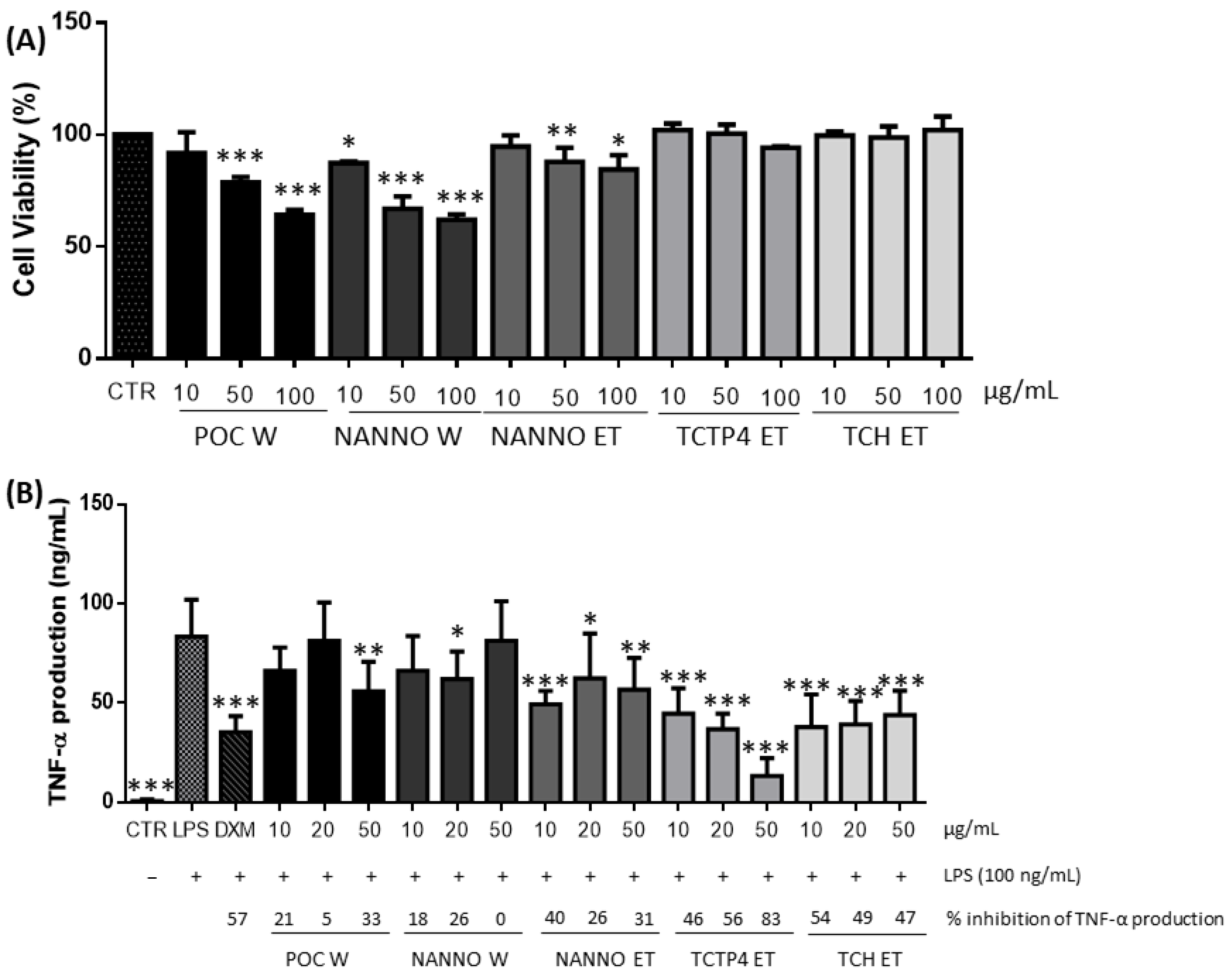
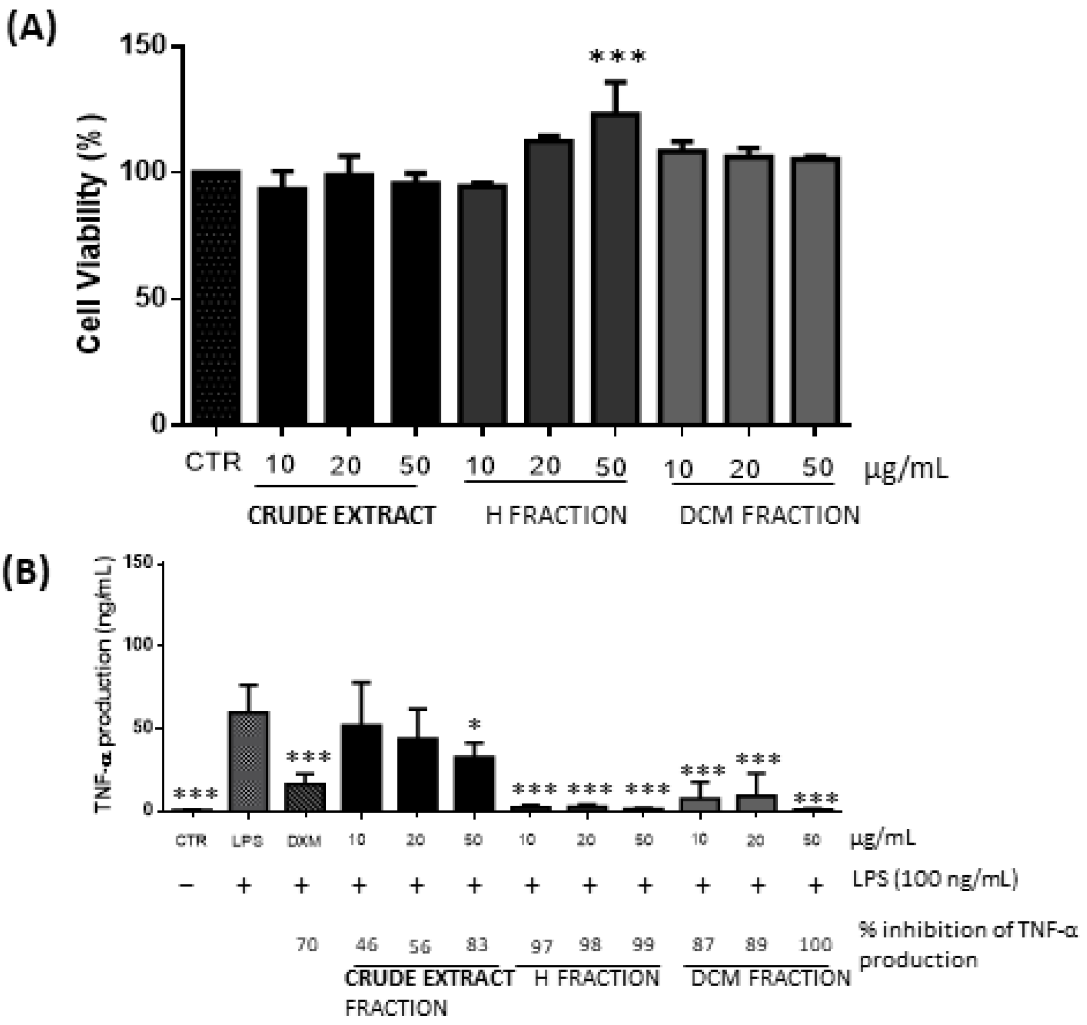
3.1.4. Anti-Inflammatory Activity

Cell viability of the POC W, NANNO W, NANNO ET, TCTP4 ET and TCH ET extracts at 10, 50 and 100 µg/mL was tested in Mac THP-1 cells. All ethanol extracts revealed not to have a cytotoxic effect on Mac THP-1 cells, as cell viability remained over 80%. The NANNO and POC water extracts showed a reduction in cell viability to 60% at concentrations over 50 μg/mL (Figure 1A). Considering these results, the anti-inflammatory assays were performed with 10, 20 and 50 μg/mL for all extracts. DMSO was shown not to affect cell viability (data not shown). A clear downregulation of the TNF-α production occurred in the LPS-stimulated Mac THP-1 cells pretreated with TCTP4 ET and TCH ET extracts at all concentrations tested (p < 0.001) (Figure 1B). Pretreatments with NANNO ET and POC W extracts showed significant TNF-α downregulation at concentrations of 50 μg/mL (p < 0.01) and 10 μg/mL (p < 0.001), and 50 μg/mL (p < 0.01), respectively (Figure 1B), whereas NANNO W extract showed no significant decrease in TNF-α production (Figure 1B). Since the TCTP4 ET extract showed a dose-dependent decrease in TNF-α production, presenting the highest percentage of TNF- α inhibition of 83.0 ± 9.4% at 50 μg/mL, an effect even higher than the positive anti-inflammation control dexamethasone (DXM) (57.0 ± 8.6%) (p < 0.001), this extract was selected for further fractionation. The resulting four fractions (hexane, dichloromethane, ethyl acetate and water/ethanol) of the TCTP4 ET extract were evaluated for cell viability (Figure 2A) and anti-inflammatory activity (Figure 2B) in Mac THP-1 cells. Cell viability was shown to be above 90% for all fractions at all concentrations tested (10, 20, and 50 μg/mL) (Figure 2A). The hexane (H) and dichloromethane (DCM) fractions displayed high TNF-α inhibition in Mac THP-1 cells, with both fractions presenting similar TNF-α levels as the control non-stimulated cells and showing even higher TNF-α inhibition than dexamethasone (DXM), at all concentrations tested (Figure 2B). These results clearly indicate a strong and promising anti-inflammatory potential of hexane and dichloromethane fractions of the TCTP4 ET extract in LPS-stimulated Mac THP-1 cells.
Water extracts presented no significant anti-inflammatory activity, possibly due to the extracted compounds being mainly polar and ionic and therefore presenting low anti-inflammatory effect (besides polysaccharides) [70].
The high TNF α inhibitory effect obtained for the hexane and dichloromethane fractions of the TCTP4 ET extract may be related to the presence of compounds with anti-inflammatory activity, such as phenolic compounds, pigments, and non-polar lipids, which are commonly extracted with hexane and DCM and have reported anti-inflammatory activity [13].

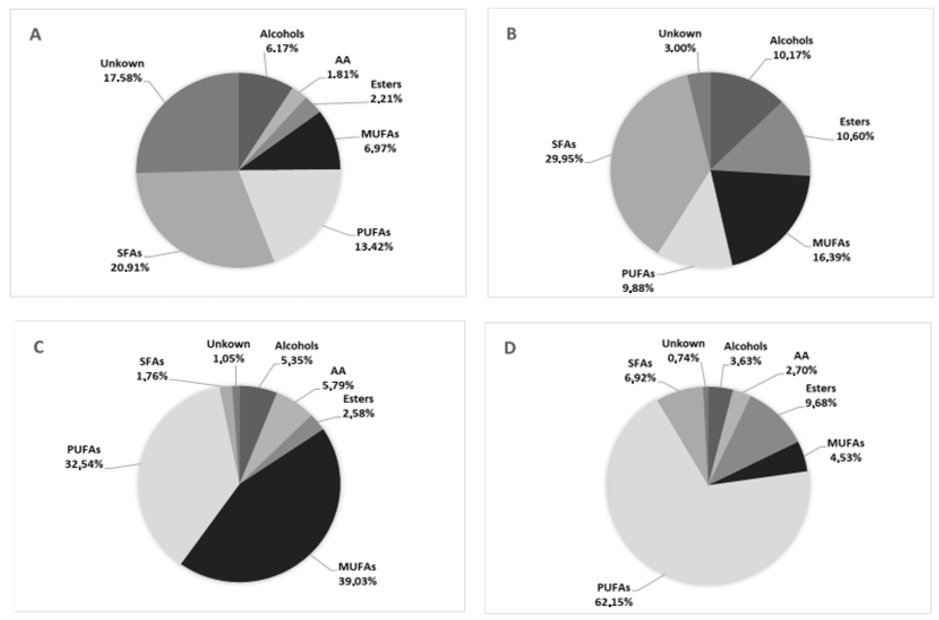
3.2. Chemical Characterization of Promising Microalgal Extracts’ Fractions

The most active fractions at each bioactive assay (hexane fractions of POC ET and SKE ET due to neuroprotective activity; dichloromethane fraction of PHA ET for anti-tumoral activity and hexane fraction of TCTP4 ET for anti-inflammatory activity), were analyzed by GC-MS to tentatively identify compounds that may be responsible for the activities tested. The hexane fractions of ethanol extracts from Porphyridium sp. and Skeletonema sp. generated a total of 45 and 29 peaks in the GC-MS chromatogram, with 40 and 23 peaks having been satisfactorily identified, respectively. The analysis of the hexane fraction of ethanol extract of Tetraselmis striata CTP4, revealed 43 compounds with 42 compounds being successfully identified. Finally, the analysis of the dichloromethane fraction from the ethanol extract of Phaeodactylum tricornutum revealed a total of 52 peaks, of which 33 were successfully identified. The complete list and chemical classes of the identified compounds are presented in Figure 3 and Table 8.
Eight main chemical compound classes were identified, most of them of fatty acids, namely polyunsaturated fatty acids, saturated fatty acids, monounsaturated fatty acids, long-chain aliphatic hydrocarbons (alkanes and alkenes), long-chain alcohols, esters, ethers and sterols (Figure 3).
Fatty acid composition varied greatly in the analyzed fractions and species. Whereas saturated fatty acids were the main compounds in hexane fractions of both Porphyridium sp. and Skeletonema sp., PUFAs were always the most abundant compounds in the analyzed fractions of Phaeodactylum tricornutum and Tetraselmis striata CTP4, which is in accordance with previous studies [71,72,73,74]. Microalgae are reportedly good sources of long-chain fatty acids, specially of PUFA. These compounds are probably responsible for a significant portion of the antioxidant, metal-chelating, anti-tumoral and anti-inflammatory activities exhibited by the fractions, considering that PUFA are reported to possess these bioactive properties and are well extracted with hexane and DCM [68,69]. Furthermore, PUFAs have antiapoptotic activity and may be useful in delaying or preventing inflammatory, metabolic, neurodegenerative (by reducing neuroinflammation and preserving dopaminergic neurons), and neoplastic diseases, by modulating the production of inflammatory cytokines and pro-resolving or protective lipid mediators [75,76].
Alcohols were the second most abundant compounds across all species, with special focus on 3, 7, 11, 15-tetramethyl-2-hexadecen-1-ol (Phytol), which have reported antimicrobial and anti-inflammatory action [77]. Some of the algal extracts examined contained significant amounts of long-chain aliphatic hydrocarbons, mostly in the form of n-alkanes (Table 8). It is reported that these types of compounds are produced naturally as a defense mechanism to protect the organism (mainly plants, insects, and cyanobacteria) from environmental threats [78], and despite some reports about their antioxidant, antibacterial and anti-cancer activity existing, overall evidence regarding their biological properties is still scarce [79,80]. These compounds ranged from 18 to 20 carbons in Porphyridium sp., 17 to 20 carbons in Tetraselmis striata CTP4 and 18 to 54 carbons P. tricornutum. The only cycloalkene compound was found in Tetraselmis striata CTP4, which have reported anti-inflammatory, antimicrobial, antibiotic, antibacterial, antitumor and antiviral activity and is also being used in the development of new pharmaceuticals [81].
Minor amounts of phenolic compound such as alpha-tocopherol (vitamin E) and butylated hydroxytoluene (BHT) were also identified in the microalgal fractions and both are known to have antioxidant properties [82]. All the microalgal fractions had small amounts of ethers (mainly tetrahydropyrans), which have reported diverse biological and pharmaceutical properties including antimicrobial, antitumor and anti-proliferative properties and have the potential to be used in the treatment of neurodegenerative diseases such as Alzheimer’s disease [83].
Sterols occur naturally in plants and algae, but sterol composition may vary widely between species. In most algae, campesterol, stigmasterol, ergosterol and fucosterol are generally the most abundant sterols. In our case, brassicasterol was the main phytosterol in Phaeodactylum tricornutum, chloresterol in Skeletonema sp. and dehydrocholesterol in Porphyridium sp.. Phytosterols have been shown to provide a variety of health benefits in humans, including immunomodulatory, anticancer, anti-inflammatory, and antioxidant properties [84]. The hexane fraction of Tetraselmis striata CTP4 had octadecadienoic acid, which was previously reported to reduce the expression of cytokines and proinflammatory mediators and therefore having anti-inflammatory potential [85].
Despite the identified compounds having some reported antioxidant, metal-chelating, anti-tumoral, and anti-inflammatory activity, it is likely that other compounds, which could not be identified in this preliminary phytochemical analysis, are also responsible for the bioactivities of these extracts, as many other bioactive compounds (e.g., pigments, sphingolipids, glycerophospholipids, and glycerolipids) have been reported by other studies [66,86].

4. Conclusions

This study showed that several industrialy produced microalgal species contain compounds with antioxidant, calcium chelating, anti-inflammatory, and cytotoxic effects (on HepG2 cells) with possible pharmaceutical applications without any need for genetic engineering. Four species deserve some future attention, namely: Phaeodactylum tricornutum with significant antiproliferative effect on HepG2 cells; Skeletonema sp. and Porphyridium sp. with antioxidant effects coupled with metal chelating effect for iron, copper and calcium; and Tetraselmis striata CTP4 with anti-inflammatory activity by decreasing the TNF-α production in LPS stimulated Mac THP-1 cells. The chemical characterization of the hexane fractions of the ethanol extracts of Porphyridium sp., Skeletonema sp. and Tetraselmis striata CTP4 and dichloromethane fraction from the ethanol extract of Phaeodactylum tricornutum showed compounds from different chemical classes, most of them of fatty acids, followed by long-chain aliphatic hydrocarbons (alkanes and alkenes), long-chain alcohols, esters, ethers and sterols. Most of these molecules have reported biological activities and could potentially be (at least partially) responsible for cytotoxicity effects against HepG2 cells, antioxidant, anti-inflammatory, and calcium chelating activities. Small variations in bioactivities of crude extracts were identified in between batches, so an optimization of culture conditions may be beneficial to improve the chemical composition of the algal biomass.
Overall, we were successful in identifying prospective sources of biologically active compounds that could be used to counterbalance the effects of oxidative stress and inflammation, which are inevitably linked to many diseases that plague our modern-day societies, and therefore play an important role in health promotion. However, before considering human consumption, the in vivo effectiveness and the safety of these extracts should be assessed. Additional research is also required to isolate the active compound(s), and confirm their identity and structure. This would allow the study of the bioactive compounds’ metabolism, providing clues for strain improvement via manipulation of microalgal cultivation conditions. This would allow the producers to increase the productivity of the bioactive compounds and enhance the bioactivity of the extracts.

Author Contributions

Conceptualization, C.S.B.V., D.C.S., K.N.G., J.V. and L.B.; methodology, C.S.B.V., D.C.S., K.N.G., J.V. and L.B.; formal analysis, M.S., F.K., S.T.U. and I.-M.K.; investigation, M.S., F.K., S.T.U., I.-M.K., C.S.B.V., D.C.S., K.N.G., J.V. and L.B.; resources, J.V. and L.B.; data curation, M.S., C.S.B.V., D.C.S., K.N.G., J.V. and L.B.; writing—original draft preparation, M.S., F.K., S.T.U. and I.-M.K.; writing—review and editing, M.S., C.S.B.V., D.C.S., J.V. and L.B.; supervision, C.S.B.V., D.C.S., K.N.G., J.V. and L.B.; project administration, C.S.B.V., D.C.S., J.V. and L.B.; funding acquisition, C.S.B.V., D.C.S., J.V. and LB. All authors have read and agreed to the published version of the manuscript.

Funding

The present work was funded by the Portuguese national budget and the UIDB/04326/2020, UIDP/04326/2020 and LA/P/0101/2020 grants, and the transitional provision DL57/2016/CP1361/CT0006 of the Foundation for Science and Technology (FCT). We would also like to acknowledge 055 ALGARED + 05 INTERREG V-A—España-Portugal, ALGAE4IBD project (grantAgreement/EC/H2020/101000501/EU) and AAC No. 41/ALG/2020—Project No. 072583—NUTRISAFE.

Institutional Review Board Statement

Not applicable.

Informed Consent Statement

Not applicable.

Data Availability Statement

Not applicable.

Acknowledgments

To NECTON S.A. for the kind supply of the Porphyridium sp., Nannochloropsis sp., Tisochrysis lutea, Phaeodactylum tricornutum, Skeletonema sp., Spirulina, Haematococcus pluvialis, and Tetraselmis chui strain used in this study. To ALLMICROALGAE—Natural Products, S.A. for providing the biomass of Tetraselmis striata CTP4.

Conflicts of Interest

The authors declare no conflict of interest.

References

  1. Budreviciute, A.; Damiati, S.; Sabir, D.K.; Onder, K.; Schuller-Goetzburg, P.; Plakys, G.; Katileviciute, A.; Khoja, S.; Kodzius, R. Management and prevention strategies for non-communicable diseases (NCDs) and their risk factors. Front. Public Health 2020, 8, 574111. [Google Scholar] [CrossRef] [PubMed]
  2. Pham-Huy, L.A.; He, H.; Pham-Huy, C. Free radicals, antioxidants in disease and health. Int. J. Biomed. Sci. IJBS 2008, 4, 89–96. [Google Scholar] [PubMed]
  3. Hussain, T.; Tan, B.; Yin, Y.; Blachier, F.; Tossou, M.C.B.; Rahu, N. Oxidative stress and inflammation: What polyphenols can do for us? Oxid. Med. Cell. Longev. 2016, 2016, 7432797. [Google Scholar] [CrossRef] [PubMed] [Green Version]
  4. Arulselvan, P.; Fard, M.T.; Tan, W.S.; Gothai, S.; Fakurazi, S.; Norhaizan, M.E.; Kumar, S.S. Role of antioxidants and natural products in inflammation. Oxid. Med. Cell. Longev. 2016, 2016, 5276130. [Google Scholar] [CrossRef] [PubMed] [Green Version]
  5. Lee, Y.-M.; Song, B.C.; Yeum, K.-J. Impact of volatile anesthetics on oxidative stress and inflammation. BioMed Res. Int. 2015, 2015, 242709. [Google Scholar] [CrossRef]
  6. Dales, J.-P.; Desplat-Jégo, S. Metal imbalance in neurodegenerative diseases with a specific concern to the brain of multiple sclerosis patients. Int. J. Mol. Sci. 2020, 21, 9105. [Google Scholar] [CrossRef]
  7. Prakash, A.; Dhaliwal, G.K.; Kumar, P.; Majeed, A.B.A. Brain biometals and Alzheimer’s disease—Boon or bane? Int. J. Neurosci. 2017, 127, 99–108. [Google Scholar] [CrossRef]
  8. Sheikh, S.; Safia; Haque, E.; Mir, S.S. Neurodegenerative diseases: Multifactorial conformational diseases and their therapeutic interventions. J. Neurodegener. Dis. 2013, 2013, 563481. [Google Scholar] [CrossRef] [Green Version]
  9. Ferdous, U.T.; Yusof, Z.N.B. Medicinal prospects of antioxidants from algal sources in cancer therapy. Front. Pharmacol. 2021, 12, 593116. [Google Scholar] [CrossRef]
  10. Rocha, J.; Peixe, L.; Gomes, N.C.M.; Calado, R. Cnidarians as a Source of new marine bioactive compounds—An overview of the last decade and future steps for bioprospecting. Mar. Drugs 2011, 9, 1860–1886. [Google Scholar] [CrossRef]
  11. Wahab, M.; Zahoor, M.; Muhammad Salman, S.; Kamran, A.W.; Naz, S.; Burlakovs, J.; Kallistova, A.; Pimenov, N.; Zekker, I. Adsorption-membrane hybrid approach for the removal of azithromycin from water: An attempt to minimize drug resistance problem. Water 2021, 13, 1969. [Google Scholar] [CrossRef]
  12. Morais, E.G.; Cristofoli, N.L.; Maia, I.B.; Magina, T.; Cerqueira, P.R.; Teixeira, M.R.; Varela, J.; Barreira, L.; Gouveia, L. Microalgal systems for wastewater treatment: Technological trends and challenges towards waste recovery. Energies 2021, 14, 8112. [Google Scholar] [CrossRef]
  13. Montero-Lobato, Z.; Vázquez, M.; Navarro, F.; Fuentes, J.L.; Bermejo, E.; Garbayo, I.; Vílchez, C.; Cuaresma, M. Chemically-Induced production of anti-inflammatory molecules in microalgae. Mar. Drugs 2018, 16, 478. [Google Scholar] [CrossRef] [PubMed] [Green Version]
  14. Caporgno, M.P.; Mathys, A. Trends in microalgae incorporation into innovative food products with potential health benefits. Front. Nutr. 2018, 5, 58. [Google Scholar] [CrossRef]
  15. Rotter, A.; Bacu, A.; Barbier, M.; Bertoni, F.; Bones, A.M.; Cancela, M.L.; Carlsson, J.; Carvalho, M.F.; Cegłowska, M.; Dalay, M.C.; et al. A new network for the advancement of marine biotechnology in Europe and beyond. Front. Mar. Sci. 2020, 7, 278. [Google Scholar] [CrossRef]
  16. Jareonsin, S.; Pumas, C. Advantages of heterotrophic microalgae as a host for phytochemicals production. Front. Bioeng. Biotechnol. 2021, 9, 628597. [Google Scholar] [CrossRef]
  17. Kumar, G.; Shekh, A.; Jakhu, S.; Sharma, Y.; Kapoor, R.; Sharma, T.R. Bioengineering of microalgae: Recent advances, perspectives, and regulatory challenges for industrial application. Front. Bioeng. Biotechnol. 2020, 8, 914. [Google Scholar] [CrossRef]
  18. Moreira, D.; Pires, J.C.M. Atmospheric CO2 capture by algae: Negative carbon dioxide emission path. Bioresour. Technol. 2016, 215, 371–379. [Google Scholar] [CrossRef]
  19. Bagwell, C.E.; Abernathy, A.; Barnwell, R.; Milliken, C.E.; Noble, P.A.; Dale, T.; Beauchesne, K.R.; Moeller, P.D.R. Discovery of bioactive metabolites in biofuel microalgae that offer protection against predatory bacteria. Front. Microbiol. 2016, 7, 516. [Google Scholar] [CrossRef] [Green Version]
  20. Cragg, G.M.; Newman, D.J. Natural products: A continuing source of novel drug leads. Biochim. Biophys. Acta 2013, 1830, 3670–3695. [Google Scholar] [CrossRef] [Green Version]
  21. Ebrahimzadeh, M.A.; Khalili, M.; Dehpour, A.A. Antioxidant activity of ethyl acetate and methanolic extracts of two marine algae, Nannochloropsis oculata and Gracilaria gracilis—An in vitro assay. Braz. J. Pharm. Sci. 2018, 54, 1. [Google Scholar] [CrossRef] [Green Version]
  22. Conde, T.A.; Neves, B.F.; Couto, D.; Melo, T.; Neves, B.; Costa, M.; Silva, J.; Domingues, P.; Domingues, M.R. Microalgae as sustainable bio-factories of healthy lipids: Evaluating fatty acid content and antioxidant activity. Mar. Drugs 2021, 19, 357. [Google Scholar] [CrossRef] [PubMed]
  23. Gangadhar, K.N.; Pereira, H.; Rodrigues, M.J.; Custódio, L.; Barreira, L.; Malcata, F.X.; Varela, J. Microalgae-based unsaponifiable matter as source of natural antioxidants and metal chelators to enhance the value of wet Tetraselmis chuii biomass. Open Chem. 2016, 14, 299–307. [Google Scholar] [CrossRef] [Green Version]
  24. Ibrahim, M.R.; Hamouda, R.A.; Tayel, A.A.; Al-Saman, M.A. Anti-cholesterol and antioxidant activities of independent and combined microalgae aqueous extracts in vitro. Waste Biomass Valorization 2021, 12, 4845–4857. [Google Scholar] [CrossRef]
  25. Casas-Arrojo, V.; Decara, J.; de Los Ángeles Arrojo-Agudo, M.; Pérez-Manríquez, C.; Abdala-Díaz, R.T. Immunomodulatory, antioxidant activity and cytotoxic effect of sulfated polysaccharides from Porphyridium cruentum. (S.F.Gray) Nägeli. Biomolecules 2021, 11, 488. [Google Scholar] [CrossRef]
  26. Nacer, W.; Baba Ahmed, F.Z.; Merzouk, H.; Benyagoub, O.; Bouanane, S. Evaluation of the anti-inflammatory and antioxidant effects of the microalgae Nannochloropsis gaditana in streptozotocin-induced diabetic rats. J. Diabetes Metab. Disord. 2020, 19, 1483–1490. [Google Scholar] [CrossRef]
  27. Saranya, C.; Hemalatha, A.; Chermapandi, P.; Anantharaman, P. Evaluation of antioxidant properties, total phenolic and carotenoid content of Chaetoceros calcitrans, Chlorella salina and Isochrysis galbana. Int. J. Curr. Microbiol. App.Sci. 2014, 3, 365–377. [Google Scholar]
  28. Neumann, U.; Derwenskus, F.; Flaiz Flister, V.; Schmid-Staiger, U.; Hirth, T.; Bischoff, S.C. Fucoxanthin, A carotenoid derived from Phaeodactylum tricornutum exerts antiproliferative and antioxidant activities in vitro. Antioxidants 2019, 8, 183. [Google Scholar] [CrossRef] [Green Version]
  29. Custódio, L.; Justo, T.; Silvestre, L.; Barradas, A.; Duarte, C.V.; Pereira, H.; Barreira, L.; Rauter, A.P.; Alberício, F.; Varela, J. Microalgae of different phyla display antioxidant, metal chelating and acetylcholinesterase inhibitory activities. Food Chem. 2012, 131, 134–140. [Google Scholar] [CrossRef]
  30. Bhattacharjee, M. Pharmaceutically valuable bioactive compounds of algae. Asian J. Pharm. Clin. Res. 2016, 9, 43–47. [Google Scholar] [CrossRef]
  31. Manrique, R.; Moreno, J.L.; Villagracia, A.R.; Ubando, A.; Kasai, H.; Arboleda, N.; David, M.; Culaba, A. Effects of salinity on the CO2 permeation across lipid bilayer for microalgae biofixation: A molecular dynamics study. J. Appl. Phycol. 2018, 30, 55–61. [Google Scholar] [CrossRef]
  32. Ismael, A.; Henriques, M.S.C.; Marques, C.; Rodrigues, M.; Barreira, L.; Paixão, J.A.; Fausto, R.; Cristiano, M.L.S. Exploring saccharinate-tetrazoles as selective Cu(II) ligands: Structure, magnetic properties and cytotoxicity of copper(II) complexes based on 5-(3-aminosaccharyl)-tetrazoles. RSC Adv. 2016, 6, 71628–71637. [Google Scholar] [CrossRef]
  33. Araújo, N.; Viegas, C.; Pontes, J.F.; Marreiros, C.; Raimundo, P.; Macedo, A.L.; Alves de Matos, A.; Grenha, A.; Vermeer, C.; Costa Simes, D. Nanoencapsulation as a novel delivery approach for therapeutic applications of gla-rich protein (GRP). Ann. Med. 2021, 53, S23–S24. [Google Scholar] [CrossRef]
  34. Araújo, N.; Viegas, C.S.B.; Zubía, E.; Magalhães, J.; Ramos, A.; Carvalho, M.M.; Cruz, H.; Sousa, J.P.; Blanco, F.J.; Vermeer, C.; et al. Amentadione from the alga Cystoseira usneoides as a novel osteoarthritis protective agent in an ex vivo co-culture OA model. Mar. Drugs 2020, 18, 624. [Google Scholar] [CrossRef] [PubMed]
  35. Pintać, D.; Majkić, T.; Torović, L.; Orčić, D.; Beara, I.; Simin, N.; Mimica–Dukić, N.; Lesjak, M. Solvent selection for efficient extraction of bioactive compounds from grape pomace. Ind. Crops Prod. 2018, 111, 379–390. [Google Scholar] [CrossRef]
  36. Do, Q.D.; Angkawijaya, A.E.; Tran-Nguyen, P.L.; Huynh, L.H.; Soetaredjo, F.E.; Ismadji, S.; Ju, Y.-H. Effect of extraction solvent on total phenol content, total flavonoid content, and antioxidant activity of Limnophila aromatica. J. Food Drug Anal. 2014, 22, 296–302. [Google Scholar] [CrossRef] [Green Version]
  37. Herrero, M.; Martín-Álvarez, P.J.; Señoráns, F.J.; Cifuentes, A.; Ibáñez, E. Optimization of accelerated solvent extraction of antioxidants from Spirulina platensis microalga. Food Chem. 2005, 93, 417–423. [Google Scholar] [CrossRef] [Green Version]
  38. Aminina, N.M.; Karaulova, E.P.; Vishnevskaya, T.I.; Yakush, E.V.; Kim, Y.-K.; Nam, K.-H.; Son, K.-T. Characteristics of polyphenolic content in brown algae of the Pacific coast of Russia. Mol. Basel Switz. 2020, 25, 3909. [Google Scholar] [CrossRef]
  39. Foo, S.C.; Yusoff, F.M.; Ismail, M.; Basri, M.; Yau, S.K.; Khong, N.M.H.; Chan, K.W.; Ebrahimi, M. Antioxidant capacities of fucoxanthin-producing algae as influenced by their carotenoid and phenolic contents. J. Biotechnol. 2017, 241, 175–183. [Google Scholar] [CrossRef]
  40. Zullaikah, S.; Jessinia, M.C.P.; Rinaldi; Yasmin, M.; Rachimoellah, M.; Wu, D.W. Lipids extraction from wet and unbroken microalgae Chlorella vulgaris using subcritical water. Mater. Sci. Forum 2019, 964, 103–108. [Google Scholar] [CrossRef]
  41. Ardiles, P.; Cerezal-Mezquita, P.; Salinas-Fuentes, F.; Órdenes, D.; Renato, G.; Ruiz-Domínguez, M.C. Biochemical composition and phycoerythrin extraction from red microalgae: A comparative study using green extraction technologies. Processes 2020, 8, 1628. [Google Scholar] [CrossRef]
  42. Chandrasekaran, R.; Barra, L.; Carillo, S.; Caruso, T.; Corsaro, M.M.; Dal Piaz, F.; Graziani, G.; Corato, F.; Pepe, D.; Manfredonia, A.; et al. Light modulation of biomass and macromolecular composition of the diatom Skeletonema marinoi. J. Biotechnol. 2014, 192, 114–122. [Google Scholar] [CrossRef] [PubMed]
  43. Kranthi, K.R.; Sardar, U.R.; Bhargavi, E.; Devi, I.; Bhunia, B.; Tiwari, O.N. Advances in exopolysaccharides based bioremediation of heavy metals in soil and water: A critical review. Carbohydr. Polym. 2018, 199, 353–364. [Google Scholar] [CrossRef]
  44. Priatni, S.; Ratnaningrum, D.; Warya, S.; Audina, E. Phycobiliproteins production and heavy metals reduction ability of Porphyridium sp. IOP Conf. Ser. Earth Environ. Sci. 2018, 160, 012006. [Google Scholar] [CrossRef]
  45. Pratiwi, D.C.; Pratiwi, N.; Yona, D.; Sasmita, R.D.; Pratiwi, I.A. Microalgae Skeletonema costatum for Cd and Cu remediation. Asian J. Water Environ. Pollut. 2020, 17, 43–48. [Google Scholar] [CrossRef]
  46. Canabady-Rochelle, L.L.S.; Harscoat-Schiavo, C.; Kessler, V.; Aymes, A.; Fournier, F.; Girardet, J.-M. Determination of reducing power and metal chelating ability of antioxidant peptides: Revisited methods. Food Chem. 2015, 183, 129–135. [Google Scholar] [CrossRef]
  47. Shi, Y.; Jiang, W.; Auckloo, B.N.; Wu, B. Several classes of natural products with metal ion chelating ability. Curr. Org. Chem. 2015, 19, 1935–1953. [Google Scholar] [CrossRef]
  48. Gulcin, İ.; Alwasel, S.H. Metal ions, metal chelators and metal chelating assay as antioxidant method. Processes 2022, 10, 132. [Google Scholar] [CrossRef]
  49. Kheeree, N.; Kuptawach, K.; Puthong, S.; Sangtanoo, P.; Srimongkol, P.; Boonserm, P.; Reamtong, O.; Choowongkomon, K.; Karnchanatat, A. Discovery of calcium-binding peptides derived from defatted lemon basil seeds with enhanced calcium uptake in human intestinal epithelial cells, caco-2. Sci. Rep. 2022, 12, 4659. [Google Scholar] [CrossRef]
  50. Ge, M.; Zhang, J.; Chen, S.; Huang, Y.; Chen, W.; He, L.; Zhang, Y. Role of calcium homeostasis in Alzheimer’s Disease. Neuropsychiatr. Dis. Treat. 2022, 18, 487–498. [Google Scholar] [CrossRef]
  51. Pchitskaya, E.; Popugaeva, E.; Bezprozvanny, I. Calcium signaling and molecular mechanisms underlying neurodegenerative diseases. Cell Calcium 2018, 70, 87–94. [Google Scholar] [CrossRef] [PubMed]
  52. Vizetto-Duarte, C.; Custódio, L.; Acosta, G.; Lago, J.H.G.; Morais, T.R.; Bruno de Sousa, C.; Gangadhar, K.N.; Rodrigues, M.J.; Pereira, H.; Lima, R.T.; et al. Can macroalgae provide promising anti-tumoral compounds? A closer look at Cystoseira tamariscifolia as a source for antioxidant and anti-hepatocarcinoma compounds. PeerJ 2016, 4, e1704. [Google Scholar] [CrossRef] [PubMed]
  53. Castell, J.V.; Jover, R.; Martnez-Jimnez, C.P.; Gmez-Lechn, M.J. Hepatocyte cell lines: Their use, scope and limitations in drug metabolism studies. Expert Opin. Drug Metab. Toxicol. 2006, 2, 183–212. [Google Scholar] [CrossRef]
  54. Saide, A.; Damiano, S.; Ciarcia, R.; Lauritano, C. Promising Activities of marine natural products against hematopoietic malignancies. Biomedicines 2021, 9, 645. [Google Scholar] [CrossRef] [PubMed]
  55. Lopes, S.M.M.; Sousa, E.P.; Barreira, L.; Marques, C.; Rodrigues, M.J.; Pinho e Melo, T.M.V.D. Synthesis and anti-cancer activity of chiral tetrahydropyrazolo[1,5-a]pyridine-fused steroids. Steroids 2017, 122, 16–23. [Google Scholar] [CrossRef] [PubMed]
  56. Morales, S.A.T.; Aguilar, M.G.d.; Pereira, R.C.G.; Duarte, L.P.; Sousa, G.F.; Oliveira, D.M.d.; Evangelista, F.C.G.; Sabino, A.P.; Viana, R.O.; Alves, V.S.; et al. Constituents from roots of Maytenus distichophylla, antimicrobial activity and toxicity for cells and Caenorhabditis elegans. Quím. Nova 2020, 43, 1066–1073. [Google Scholar] [CrossRef]
  57. López-Lázaro, M. A simple and reliable approach for assessing anticancer activity in vitro. Curr. Med. Chem. 2015, 22, 1324–1334. [Google Scholar] [CrossRef]
  58. Zhong, L.; Li, Y.; Xiong, L.; Wang, W.; Wu, M.; Yuan, T.; Yang, W.; Tian, C.; Miao, Z.; Wang, T.; et al. Small molecules in targeted cancer therapy: Advances, challenges, and future perspectives. Signal Transduct. Target. Ther. 2021, 6, 1–48. [Google Scholar] [CrossRef]
  59. Schüler, L.M.; Gangadhar, K.N.; Duarte, P.; Placines, C.; Molina-Márquez, A.M.; Léon-Bañares, R.; Sousa, V.S.; Varela, J.; Barreira, L. Improvement of carotenoid extraction from a recently isolated, robust microalga, Tetraselmis sp. CTP4 (Chlorophyta). Bioprocess Biosyst. Eng. 2020, 43, 785–796. [Google Scholar] [CrossRef]
  60. Schüler, L.; Greque de Morais, E.; Trovão, M.; Machado, A.; Carvalho, B.; Carneiro, M.; Maia, I.; Soares, M.; Duarte, P.; Barros, A.; et al. Isolation and characterization of novel Chlorella vulgaris mutants with low chlorophyll and improved protein contents for food applications. Front. Bioeng. Biotechnol. 2020, 8, 469. [Google Scholar] [CrossRef]
  61. Schüler, L.M.; Bombo, G.; Duarte, P.; Santos, T.F.; Maia, I.B.; Pinheiro, F.; Marques, J.; Jacinto, R.; Schulze, P.S.C.; Pereira, H.; et al. Carotenoid biosynthetic gene expression, pigment and n-3 fatty acid contents in carotenoid-rich Tetraselmis striata CTP4 strains under heat stress combined with high light. Bioresour. Technol. 2021, 337, 125385. [Google Scholar] [CrossRef] [PubMed]
  62. Almutairi, A.W. Improvement of chemical composition of Tisochrysis lutea grown mixotrophically under nitrogen depletion towards biodiesel production. Molecules 2020, 25, 4609. [Google Scholar] [CrossRef] [PubMed]
  63. Andrianasolo, E.H.; Haramaty, L.; Vardi, A.; White, E.; Lutz, R.; Falkowski, P. Apoptosis-inducing galactolipids from a cultured marine diatom, Phaeodactylum tricornutum. J. Nat. Prod. 2008, 71, 1197–1201. [Google Scholar] [CrossRef] [PubMed] [Green Version]
  64. Chang, F.-Z.; Wang, Q.; Zhang, Q.; Chang, L.-L.; Li, W. Omega-3 polyunsaturated fatty acid inhibits the malignant progression of hepatocarcinoma by inhibiting the wnt/β-catenin pathway. Eur. Rev. Med. Pharmacol. Sci. 2018, 22, 4500–4508. [Google Scholar] [CrossRef]
  65. Hussein, H.A.; Abdullah, M.A. Anticancer compounds derived from marine diatoms. Mar. Drugs 2020, 18, 356. [Google Scholar] [CrossRef]
  66. Gangadhar, K.N.; Rodrigues, M.J.; Pereira, H.; Gaspar, H.; Malcata, F.X.; Barreira, L.; Varela, J. Anti-hepatocellular carcinoma (HepG2) activities of monoterpene hydroxy lactones isolated from the marine microalga Tisochrysis lutea. Mar. Drugs 2020, 18, 567. [Google Scholar] [CrossRef]
  67. Le Goff, M.; Delbrut, A.; Quinton, M.; Pradelles, R.; Bescher, M.; Burel, A.; Schoefs, B.; Sergent, O.; Lagadic-Gossmann, D.; Le Ferrec, E.; et al. Protective action of Ostreococcus tauri and Phaeodactylum tricornutum extracts towards benzo[a]pyrene-induced cytotoxicity in endothelial cells. Mar. Drugs 2020, 18, 3. [Google Scholar] [CrossRef] [Green Version]
  68. Cayot, N.; Lafarge, C.; Bou-Maroun, E.; Cayot, P. Substitution of carcinogenic solvent dichloromethane for the extraction of volatile compounds in a fat-free model food system. J. Chromatogr. A 2016, 1456, 77–88. [Google Scholar] [CrossRef]
  69. Li, Y.; Ghasemi Naghdi, F.; Garg, S.; Adarme-Vega, T.C.; Thurecht, K.J.; Ghafor, W.A.; Tannock, S.; Schenk, P.M. A Comparative study: The impact of different lipid extraction methods on current microalgal lipid research. Microb. Cell Factories 2014, 13, 14. [Google Scholar] [CrossRef] [Green Version]
  70. Xia, D.; Qiu, W.; Wang, X.; Liu, J. Recent advancements and future perspectives of microalgae-derived pharmaceuticals. Mar. Drugs 2021, 19, 703. [Google Scholar] [CrossRef]
  71. Ulusoy Erol, H.B.; Menegazzo, M.L.; Sandefur, H.; Gottberg, E.; Vaden, J.; Asgharpour, M.; Hestekin, C.N.; Hestekin, J.A. Porphyridium cruentum grown in ultra-filtered swine wastewater and its effects on microalgae growth productivity and fatty acid composition. Energies 2020, 13, 3194. [Google Scholar] [CrossRef]
  72. Sharmin, T.; Monirul Hasan, C.M.; Aftabuddin, S.; Rahman, M.A.; Khan, M. Growth, fatty acid, and lipid composition of marine microalgae Skeletonema costatum available in bangladesh coast: Consideration as biodiesel feedstock. J. Mar. Biol. 2016, 2016, e6832847. [Google Scholar] [CrossRef] [Green Version]
  73. Branco-Vieira, M.; Martin, S.S.; Agurto, C.; Santos, M.A.; Freitas, M.A.V.; Caetano, N.S. Analyzing Phaeodactylum tricornutum lipid profile for biodiesel production. Energy Procedia 2017, 136, 369–373. [Google Scholar] [CrossRef]
  74. Cardoso, C.; Pereira, H.; Franca, J.; Matos, J.; Monteiro, I.; Pousão-Ferreira, P.; Gomes, A.; Barreira, L.; Varela, J.; Neng, N.; et al. Lipid composition and some bioactivities of 3 newly isolated microalgae (Tetraselmis sp. IMP3, Tetraselmis sp. CTP4, and Skeletonema sp.). Aquac. Int. 2020, 28, 711–727. [Google Scholar] [CrossRef]
  75. Calviello, G.; Su, H.-M.; Weylandt, K.H.; Fasano, E.; Serini, S.; Cittadini, A. Experimental evidence of -3 polyunsaturated fatty acid modulation of inflammatory cytokines and bioactive lipid mediators: Their potential role in inflammatory, neurodegenerative, and neoplastic diseases. BioMed Res. Int. 2013, 2013, e743171. [Google Scholar] [CrossRef] [Green Version]
  76. Mori, M.A.; Delattre, A.M.; Carabelli, B.; Pudell, C.; Bortolanza, M.; Staziaki, P.V.; Visentainer, J.V.; Montanher, P.F.; Del Bel, E.A.; Ferraz, A.C. Neuroprotective effect of omega-3 polyunsaturated fatty acids in the 6-OHDA model of Parkinson’s disease is mediated by a reduction of inducible nitric oxide synthase. Nutr. Neurosci. 2018, 21, 341–351. [Google Scholar] [CrossRef] [PubMed]
  77. El-Fayoumy, E.A.; Shanab, S.M.M.; Gaballa, H.S.; Tantawy, M.A.; Shalaby, E.A. Evaluation of antioxidant and anticancer activity of crude extract and different fractions of Chlorella vulgaris axenic culture grown under various concentrations of copper ions. BMC Complement. Med. Ther. 2021, 21, 51. [Google Scholar] [CrossRef]
  78. Kang, M.-K.; Nielsen, J. Biobased production of alkanes and alkenes through metabolic engineering of microorganisms. J. Ind. Microbiol. Biotechnol. 2017, 44, 613–622. [Google Scholar] [CrossRef] [Green Version]
  79. Enema, O.; Umoh, U.; Thomas, P.; Adesina, S.; Eseyin, O. Phytochemical and antioxidant studies of leaf of Tetrapleura tetraptera (schum and thon) taubert (mimosaceae). Brifish J. Pharm. Med. Res. 2019, 4, 1865–1875. [Google Scholar] [CrossRef]
  80. Parthasarathy, A.; Jayalakshmi, M.; Pushpabharathi, N.; Varadharaj, V. Identification of bioactive components in Enhalus acoroides seagrass extract by gas chromatography–mass spectrometry. Asian J. Pharm. Clin. Res. 2018, 11, 313. [Google Scholar] [CrossRef]
  81. Salaün, J. Cyclopropane derivatives and their diverse biological activities. In Small Ring Compounds in Organic Synthesis VI; de Meijere, A., Ed.; Topics in Current Chemistry; Springer: Berlin/Heidelberg, Germany, 2000; Volume 207, pp. 1–67. ISBN 978-3-540-66471-0. [Google Scholar]
  82. Yehye, W.A.; Abdul Rahman, N.; Alhadi, A.A.; Khaledi, H.; Weng, N.S.; Ariffin, A. Butylated hydroxytoluene analogs: Synthesis and evaluation of their multipotent antioxidant activities. Molecules 2012, 17, 7645–7665. [Google Scholar] [CrossRef] [PubMed] [Green Version]
  83. Mahdavi, S.M.; Habibi, A.; Dolati, H.; Shahcheragh, S.M.; Sardari, S.; Azerang, P. Synthesis and antimicrobial evaluation of 4H-pyrans and schiff bases fused 4H-pyran derivatives as inhibitors of Mycobacterium bovis (BCG). Iran. J. Pharm. Res. IJPR 2018, 17, 1229–1239. [Google Scholar] [PubMed]
  84. Luo, X.; Su, P.; Zhang, W. Advances in microalgae-derived phytosterols for functional food and pharmaceutical applications. Mar. Drugs 2015, 13, 4231–4254. [Google Scholar] [CrossRef] [PubMed]
  85. Kang, M.-C.; Ham, Y.-M.; Heo, S.-J.; Yoon, S.-A.; Cho, S.-H.; Kwon, S.-H.; Jeong, M.S.; Jeon, Y.-J.; Sanjeewa, K.; Yoon, W.-J.; et al. Anti-inflammation effects of 8-Oxo-9-octadecenoic acid isolated from Undaria peterseniana in lipopolysaccharide-stimulated macrophage cells. EXCLI J. 2018, 17, 775–783. [Google Scholar] [CrossRef] [PubMed]
  86. Bigagli, E.; D’Ambrosio, M.; Cinci, L.; Niccolai, A.; Biondi, N.; Rodolfi, L.; Dos Santos Nascimiento, L.B.; Tredici, M.R.; Luceri, C. A Comparative in vitro evaluation of the anti-inflammatory effects of a Tisochrysis lutea extract and fucoxanthin. Mar. Drugs 2021, 19, 334. [Google Scholar] [CrossRef] [PubMed]
Figure 1. (A) Cell Viability (%) of macrophage-differentiated THP-1 (Mac THP-1) cells after incubation with Porphyridium sp. water (POC W), Nannochloropsis sp. water (NANNO W) and ethanol (NANNO ET), Tetraselmis striata CTP4 ethanol (TCTP4 ET) and Tetraselmis chui ethanol (TCH ET) microalgal crude extracts at 10, 50 and 100 µg/mL for 48 h, assessed through the MTS assay. (B) Anti-inflammatory activity of the microalgal crude extracts defined in panel (A), in LPS-stimulated Mac THP-1 cells. Evaluation of the inflammatory marker TNF-α was performed by ELISA in the cell culture media of Mac THP-1 cells treated for 24 h with the microalgae crude extracts at 10, 20 and 50 µg/mL, and then stimulated with LPS (100 ng/mL) for a further 24 h. Dexamethasone (DXM) (2 µM) was used as a positive anti-inflammatory control and non-stimulated cells (CTR) as controls to LPS stimulation (LPS). Data are representative of three independent experiments, and presented as mean ± SD. Two-way Anova and multiple comparisons were achieved with the Dunnett’s test and presented relatively to the control untreated cells in (A), and LPS-stimulated cells in (B). Statistical significance was defined as p ≤ 0.05 (*), p ≤ 0.01 (**), and p ≤ 0.001 (***).
Applsci 12 05877 g001
Figure 2. (A) Cell Viability (%) of macrophage-differentiated THP-1 (Mac THP-1) cells after incubation with hexane (H) and dichloromethane (DCM) fractions of the ethanol crude extract from Tetraselmis striata CTP4 at 10, 20 and 50 µg/mL for 48 h, assessed through the MTS assay. (B) Anti-inflammatory activity of the crude extract and fractions defined in the panel A in LPS-stimulated Mac THP-1 cells. Evaluation of the inflammatory marker TNF-α was performed by ELISA in the cell culture media of Mac THP-1 cells treated for 24 h with the crude extract and fractions at 10, 20 and 50 µg/mL, and then stimulated with LPS (100 ng/mL) for a further 24 h. Dexamethasone (DXM) (2 µM) was used as a positive anti-inflammatory control and non-stimulated cells (CTR) as controls to LPS stimulation (LPS). Data are representative of three independent experiments, and presented as mean ±SD. Two-way ANOVA and multiple comparisons were achieved with the Dunnett’s test and presented relatively to the control untreated cells in (A), and LPS-stimulated cells in (B). Statistical significance was defined as p ≤ 0.05 (*), and p ≤ 0.001 (***).
Applsci 12 05877 g002
Figure 3. Main chemical compound classes identified in the ethanol microalgal extracts by GC/MS. (A) Hexane fraction of Porphyridium sp.; (B) Hexane fraction of Skeletonema sp.; (C) Dichloromethane fraction of Phaeodactylum tricornutum and (D) Hexane fraction of Tetraselmis striata CTP4. PUFAs, polyunsaturated fatty acids; AA, alkanes and alkenes; SFAs, saturated fatty acids; MUFAs, monounsaturated fatty acids.
Applsci 12 05877 g003
Table 1. DPPH radical scavenging activity (IC50: mg/mL) of water (W), ethanol (ET) and ethyl acetate (EA) extracts of different microalgae species at 1 mg/mL.
Microalga/Positive ControlExtractDPPH Scavenging Activity (%)IC50 (mg/mL)
Porphyridium sp. (POC)W87.5 ± 10.00.535 ± 0.001 d
ET59.1 ± 1.00.566 ± 0.001 d
EA105 ± 30.524 ± 0.001 d
Nannochloropsis sp. (NANNO)W51.0 ± 5.2n.d.
ET80.3 ± 2.30.539 ± 0.030 d
EA81.7 ± 2.20.990 ± 0.024 b
Tetraselmis striata CTP4 (TCTP4)W23.2 ± 3.7n.d.
ET58.1 ± 1.51.17 ± 0.01 a
EA37.8 ± 3.2n.d.
Tisochrysis lutea (TIL)W42.2 ± 3.3n.d.
ET41.6 ± 0.2n.d.
EA97.8 ± 2.10.794 ± 0.001 c
Phaeodactylum tricornutum
(PHA)
W26.8 ± 2.2n.d.
ET61.8 ± 1.30.575 ± 0.004 d
EA94.9 ± 3.30.452 ± 0.002 d
Skeletonema sp. (SKE)W24.1 ± 3.1n.d.
ET71.1 ± 1.20.478 ± 0.054 d
EA73.3 ± 7.250.447 ± 0.060 d
Spirulina
(SPR)
ET71.5 ± 0.30.923 ± 0.001 b
Tetraselmis chui (TCH)ET66.8 ± 1.01.14 ± 0.01 a
Haematococcus pluvialis (HPL)ET103 ± 580.408 ± 0.007 d
BHT *-94.1 ± 2.8n.d.
* Positive control, BHT, 0.1 mg/mL. n.d. = not determined. N = 3. Presented values are mean with standard deviation. Extracts labelled with different superscript letters are significantly different (p < 0.05).
Table 2. Redox-metal chelating activity (CA) for copper (Cu2+) and iron (Fe2+) of water (W), ethanol (ET) and ethyl acetate (EA) extracts of different microalgae species tested at 1 mg/mL (%) and IC50 (mg/mL) of the most active extracts.
MicroalgaExtract% Cu2+ CAIC50 (mg/mL) for Cu2+CA% Fe2+ CAIC50 (mg/mL) for Fe2+CA
Porphyridium sp. (POC)W52.5 ± 3.1n.d.34.7 ± 0.2 n.d.
ET62.5 ± 0.30.144 ± 0.004 a 71.7 ± 0.10.0854 ± 0.0140 a
EA82.4 ± 2.00.337 ± 0.013 c 47.6 ± 0.5n.d.
Nannochloropsis sp. (NANNO)W72.0 ± 0.30.621 ± 0.018 e83.2 ± 0.10.279 ± 0.014 c
ET43.5 ± 0.6n.d.19.7 ± 0.5n.d.
EA3.6 ± 0.40n.d.4.03 ± 0.1n.d.
Tetraselmis striata CTP4 (TCTP4)W31.6 ± 0.7n.d.96.6 ± 0.7 a0.385 ± 0.003 d
ET54.4 ± 6.5n.d.3.23 ± 0.15n.d.
EA39.0 ± 5.6n.d.33.2 ± 0.4n.d.
Tisochrysis lutea (TIL)W72.6 ± 0.40.184 ± 0.003 a79.0 ± 1.90.403 ± 0.013 d
ET85.5 ± 0.10.228 ± 0.008 b75.0 ± 0.10.207 ± 0.006 b
EA67.8 ± 0.50.297 ± 0.009 c42.7 ± 1.0n.d.
Phaeodactylum tricornutum
(PHA)
W62.8 ± 1.5n.d.75.2 ± 0.90.508 ± 0.005 e
ET74.4 ± 0.60.358 ± 0.008 c 58.8 ± 1.4 n.d.
EA65.0 ± 0.90.737 ± 0.025 f 11.8 ± 0.1n.d.
Skeletonema sp. (SKE)W74.0 ± 0.50.434 ± 0.012 d 63.8 ± 0.9n.d.
ET87.1 ± 0.10.195 ± 0.007 a 53.4 ± 0.1n.d.
EA76.3 ±0.10.487 ± 0.030 d 42.4 ± 0.6n.d.
Spirulina (SPR)W74.0 ± 0.5n.d.1.71 ± 0.21n.d.
ET60.0 ± 1.7n.d.6.04 ± 0.62n.d.
EA52.3 ± 0.1n.d.4.46 ± 0.27n.d.
Tetraselmis chui (TCH)W19.8 ± 4.8n.d.4.90 ± 0.23n.d.
ET54.4 ± 6.5n.d.16.6 ± 0.7n.d.
EA39.0 ± 5.6n.d.15.8 ± 0.3n.d.
Haematococcus pluvialis (HPL)W20.3 ± 0.3n.d.1.90 ± 1.20n.d.
ET51.9 ± 0.2n.d.6.60 ± 0.73n.d.
EA59.5 ± 0.1n.d.4.71 ± 0.48n.d.
EDTA *-91.0 ± 0.3n.d.96.1 ± 0.2n.d.
* Positive control, EDTA, 0.1 mg/mL. n.d. = not determined. Presented values are mean with standard deviation and extracts with different superscript letters are significantly different from each other (p < 0.05). n = 3.
Table 3. Redox-metal chelating activity (CA) for copper (Cu2+) and iron (Fe2+) of hexane (H), dichloromethane (DCM) and ethyl acetate (EA) fractions of ethanol extracts from Porphyridium sp. and Skeletonema sp. tested at 1 mg/mL (%) and IC50 (mg/mL).
Microalgal ExtractFractionating
Solvent
% Cu2+ CAIC50 (mg/mL) for Cu2+CA% Fe2+ CAIC50 (mg/mL) for Fe2+CA
Porphyridium sp.
(POC ET)
Crude ET extract74.1 ± 0.60.272 ± 0.010 b 77.4 ± 0.10.047 ± 0.002 a
H81.2 ± 0.10.046 ± 0.004 a85.2 ± 0.30.026 ± 0.001 a
DCM61.1 ± 1.10.648 ± 0.043 c61.1 ± 1.0n.d.
EA46.6 ± 1.20.912 ± 0.023 d46.6 ± 1.20.384 ± 0.019 c
Skeletonema sp.
(SKE ET)
Crude ET extract83.4 ± 0.40.240 ±0.010 b83.5 ± 1.60.055 ± 0.002 a
H92.6 ± 0.30.036 ± 0.001 a97.9 ± 0.30.024 ± 0.001 a
DCM61.4 ± 0.10.725 ± 0.038 c61.4 ± 0.10.386 ± 0.015 c
EA51.0 ± 0.10.917 ± 0.056 d51.0 ± 0.10.246 ± 0.018 b
n.d. = not determined. Presented values are mean with standard deviation and extracts with different superscript letters are significantly different from each other (p < 0.05). n = 3.
Table 4. Calcium chelating activity (Ca2+ CA) of water (W), ethanol (ET) and ethyl acetate (EA) extracts of different microalgae species tested at 0.6 mg/mL (%) and IC50 (mg/mL) of the most active extracts.
MicroalgaExtract% Ca2+ CAIC50 (mg/mL) for Ca2+ CA
Porphyridium sp.
(POC)
W28.6 ± 0.01n.d.
ET75.8 ± 0.080.0519 ± 0.0019 a
EA67.3 ± 1.300.150 ± 0.004 c
Nannochloropsis sp. (NANNO)W13.2 ± 1.48n.d.
ET15.5 ± 0.59n.d.
EA7.98 ± 0.15n.d.
Tetraselmis striata CTP4 (TCTP4)W58.6 ± 1.50n.d.
ET54.7 ± 1.07n.d.
EA12.3 ± 0.26n.d.
Tisochrysis lutea
(TIL)
W6.67 ± 0.46n.d.
ET52.8 ± 0.63n.d.
EA38.0 ± 0.12n.d.
Phaeodactylum tricornutum
(PHA)
W47.4 ± 0.49n.d.
ET35.2 ± 0.97n.d.
EA48.1 ± 0.50n.d.
Skeletonema sp.
(SKE)
W24.3 ± 0.75n.d.
ET87.8 ± 1.660.0638 ± 0.0038 a
EA86.7 ± 1.580.0906 ± 0.0001 b
Spirulina
(SPR)
W7.12 ± 0.30n.d.
ET48.8 ± 0.37n.d.
EA19.4 ± 0.30n.d.
Tetraselmis chui
(TCH)
W23.1 ± 1.98n.d.
ET48.9 ± 0.44n.d.
EA57.0 ± 1.98n.d.
Haematococcus pluvialis
(HPL)
W8.12 ± 0.76n.d.
ET0.70 ± 0.12n.d.
EA2.40 ± 0.76n.d.
EGTA *-85.9 ± 0.42n.d.
* Positive control, EGTA, 0.06 mg/mL. Presented values are mean with standard deviation and extracts with different superscript letters (a, b, c, d) are significantly different from each other (p < 0.05). ET = Ethanol; H = Hexane; DCM = dichloromethane; EA = ethyl acetate. n = 3.
Table 5. Calcium chelating activity (Ca2+ CA) of hexane (H), dichloromethane (DCM) and ethyl acetate (EA) fractions of ethanol extracts from Porphyridium sp. and Skeletonema sp. tested at 0.6 mg/mL (%) and IC50 (mg/mL).
Microalgal ExtractFractionating
Solvent
% Ca2+ CAIC50 (mg/mL) for Ca2+ CA
Porphyridium sp.
(POC ET)
Crude ET extract80.0 ± 0.30.0663 ± 0.0050 b
H95.4 ± 0.10.0281 ± 0.0001 a
DCM52.5 ± 0.10.277 ± 0.016 d
EA62.5 ± 0.10.189 ± 0.069 c
Skeletonema sp.
(SKE ET)
Crude ET extract88.6 ± 0.70.0850 ± 0.0030 b
H97.1 ± 0.10.0113 ± 0.0001 a
DCM80.0 ± 0.50.104 ± 0.003 b
EA81.5 ± 0.90.0844 ± 0.0025 b
Presented values are mean with standard deviation and extracts with different superscript letters are significantly different from each other (p < 0.05). ET = Ethanol; H = Hexane; DCM = dichloromethane; EA = ethyl acetate. n = 3.
Table 6. In vitro cytotoxicity activity (IC50: μg/mL) and Selectivity Index of ethanol and ethyl acetate extracts from Phaeodactylum tricornutum, Porphyridium sp., Skeletonema sp., Nannochloropsis sp., Tetraselmis striata CTP4 and Tisochrysis lutea, and fractions of ethanol extract from Phaeodactylum tricornutum on HepG2 and S17 cell lines.
Cell LineSelectivity Index
MicroalgaeExtractHepG2S17THP-1HepG2 vs. S17THP1 vs. S17
Phaeodactylum tricornutum
(PHA)
EA34.6 ± 3.5 a107 ± 7.0 cn.d.3.09 ± 0.12 bn.d.
Porphyridium sp. (POC)EA42.3 ± 2.7 b>125 cn.d.>2.7 an.d.
Tisochrysis lutea
(TIL)
EA44.7 ± 3.1 b79.2 ± 2.7 bn.d.1.77 ± 0.09 cn.d.
Skeletonema sp.
(SKE)
EA37.2 ± 3.6 a34.8 ± 5.4 an.d.n.sn.d.
Nannochloropsis sp.
(NANNO)
EA>125 a>125 cn.d.n.sn.d.
Tetraselmis striata CTP4
(TCTP4)
EA>125 a>125 cn.d.n.sn.d.
Phaeodactylum tricornutum
(PHA)
ET19.4 ± 2.2 c85.6 ± 4.4 c102.0 ± 7.004.40 ± 0.15 a0.844 ± 0.072 a
Tisochrysis lutea
(TIL)
ET67.8 ± 2.6 a104 ± 4.0 bn.d.1.53 ± 0.08 cn.d.
Etoposide-29.0 ± 3.2 c45.4 ± 0.5 a0.924 ± 0.2411.56 ± 0.11 c49.4 ± 1.2 b
Results are expressed as mean ± SD (n = 3). Different superscript letters within each column represent significant differences (p < 0.05) between the species and the positive control (etoposide). EA = ethyl acetate; ET = ethanol; H = hexane; DCM = Dichloromethane; EA = ethyl acetate; W + ET = water + ethanol; n.d. = not determined; n.s = non-selective (Selectivity Index equal or less than one).
Table 7. In vitro cytotoxicity activity (IC50: μg/mL) and Selectivity Index of fractions from ethanol extract from Phaeodactylum tricornutum, on HepG2 and S17 cell lines.
Microalgal ExtractFractioning SolventHepG2S17THP-1HepG2 vs. S17THP1 vs. S17
Phaeodactylum tricornutum
(PHA ET)
Crude ET extract39.8 ± 4.3 b>125 b>125 d>3.14 bn.s
H>125 a>125 b>125 dn.sn.s
DCM27.5 ± 1.6 c>125 b22.3 ± 1.8 c>4.54 a>5.60 b
EA84.2 ± 3.7 a>125 b81.9 ± 2.0 b1.48 ± 0.13 c1.53 ± 0.15 a
W + ET>125 a>125 b>125 dn.sn.s
Etoposide 29.0 ± 3.2 c45.4 ± 0.5 a0.924 ± 0.241 a1.56 ± 0.11 c49.4 ± 1.2 c
Results are expressed as mean ± SD (n = 3). Different superscript letters within each column represent significant differences (p < 0.05) between the fractions and the positive control (etoposide). EA = ethyl acetate; ET = ethanol; H = hexane; DCM = Dichloromethane; EA = ethyl acetate; W + ET = water + ethanol; n.d. = not determined; n.s = non-selective (Selectivity Index equal or less than one).
Table 8. Chemical characterization by GC-MS of compounds presents in the most bioactive fractions of the ethanol extracts of Porphyridium sp. (hexane fraction), Skeletonema sp. (hexane fraction), Phaeodactylum tricornutum (dichloromethane fraction) and Tetraselmis striata CTP4 (hexane fraction).
Relative Abundance (%)
CompoundsPorphyridium sp.
(POC ET)
-Hexane Fraction
Skeletonema sp. (SKE ET)
-Hexane Fraction
Phaeodactylum tricornutum
(PHA ET)
-Dichloromethane Fraction
Tetraselmis striata CTP4 (TCTP4)
-Hexane Fraction
Cyclohexanol, 1-butyl 2.61
2H-Pyran, 2-[(5cyclopropyl idene pentyl)oxy]tetrahydro 2.01
3,7,11,15-Tetramethyl-2-hexadecen-1-ol 0.887.015.351.51
4-Hexen-3-ol, 2,5-dimethyl 0.80
2,3-Dimethyl-undec-1-en-3-ol 0.73
Phytol 0.59 1.52
2-Octene-2-ol, 2-methoxy 0.31
9,12-Octadecadieol 0.15
1-Tetradecanol, 14-chloro 0.10
3-[2-pentenyl]-4-methyl-tetrahydrofuran-2-one0.11 0.48
Octadecane 1.3
Tetratetracontane 0.92
Nonadecane 0.86
9,12-Octadecadien chloride, (Z,Z) 0.16
9-Eicosyne 0.79 1.51
2-hydroxy-2-methylbutane-1,4-dioic acid 1.00
Tetrahydropyranyl ether of citronellol 10.601.62.96
2-Pentadecanone, 6,10,14-trimethyl 3.07
trans-13-Octadecenoic acid 3.21
Methyl (11R,12R,13S)-(Z)-12,13-epoxy-11-ol-9-octadecenoic acid 2.14
8-Octadecenoic acid 1.90
15-Tetracosenoic acid, (Z) 0.82
9-Hexadecenoic acid 19.61.61
n-Hexadecanoic acid 17.4
10-Octadecenoic acid 0.58
10-Undecenoic acid 1.15
hexadecenic acid 0.59
9-Hexadecenoic acid, (Z) 3.482.47
11-Octadecenoic acid 2.045.85
9-Octadecenoic acid (Z) 0.88 1.45
13-Methyltetradecanoic acid 0.33
9-eicosenoic acid 0.24 1.18
2,4-bis(1,1-dimethylethyl) Phenol/2,4-Di-tertbutylphenol 0.35
Phosphoric acid, monododecyl ester 0.20
5,8,11,14,17-Eicosapentaenoic acid(EPA) 2.8916.70.37
7,10-Hexadecadienoic acid 2.17
6,9,12,15-octadecatetraenoate (Stearidonic acid) 5.19
Octadecatrienoic acid
9,12-Hexadecadienoic acid 1.95
Heneicosapentaenoic Acid 0.84
Arachidonic acid 0.35
9,12-Octadecadienoic acid (Z,Z) 7.22 6.12
11,13-Eicosadienoic acid 1.98
cis-11,14-Eicosadienoic acid 0.94
8,11,14-Eicosatrienoic acid, (Z,Z,Z) 0.87 0.3
4,7,10,13,16,19-Docosahexaenoic acid 0.66 0.67
6,9,12-Hexadecatrienoic acid 0.364.821.9813.24
γ-Linolenic acid 0.29 7.23.4
5,8,11,14-Eicosatetraenoic acid, (all-Z) 0.21 0.522.01
cis-13,16-Docasadienoic acid 0.20
cis-7,10,13,16-Docosatetraenoic acid 0.19
4,7,10,13,16,19-Docosahexaenoic acid, (all-Z) 0.15 2.35
cis-7,10,13,16-Docosatetraenoic acid 0.13
12,15-Octadecadienoic acid 0.12
4,7,10,13,16-Docosapentaenoic acid 0.10
Tridecanoic acid 16.80
Methyl stearate 1.42
Tetracosanoic acid 0.39
Pentadecanoic acid, 14-methyl 0.810.38
Eicosanoic acid 1.38
Heptadecanoic acid 2.79
Valeric acid 1.15
Myristoleic acid 0.82
Stearic acid 0.68
Heptadecenoic acid 0.84
Hexadecanoic acid 15.800.52
Tetradecanoic acid 3.59 0.37
Stearoic acid 0.92
Tetradecanoic acid, 12-methyl 0.32
Pentadecanoic acid 0.289.01 0.27
Chloresterol 6.66
24-methylcholesta-5,22-dien-3β-ol (Brassicasterol) 1.21
Ergosta-5,22-dien-3-ol, acetate (Brassicasterol acetate) 0.76
Cholesta-5,22-dien-3-ol, (3β) 2.56
Tetrapentacontane, 1,54-dibromo- 3.04
dl-α-Tocopherol 1.160.34
Pregna-5,9(11)-dien-20-ol-3-one ethylene ketal 1.030.34
Tetrahydro-2H-pyran 0.98
3-Hexadecyne 0.53
trans-octadecadienoic acid 6.12
3,6,6-trimethyl-2-Norpinanol 2.96
2-methyl-5-pentyl-tetrahydrofuran 2.96
Tetrahydropyran 12-tetradecyn-1-ol ether 0.6
3,28-bis[(tetrahydro) Lup-20(29)-en-21-ol 0.6
(1-Methoxy-pentyl)-cyclopropane 0.6
17-chloro7-Heptadecene 0.59
5.alpha.-Androstan-3-one, 17.beta.-hydroxy-4.alpha.-methyl-, 0.48
Cholestan-3-one, 4,4-dimethyl-, cyclic 3 0.48
Total identified area51.5178.0086.9683.46
Unknown compound 0.191.001.050.74
Unresolved mixture of compounds 17.201.00
Publisher’s Note: MDPI stays neutral with regard to jurisdictional claims in published maps and institutional affiliations.

