Article

Heme Dependent Catalase Conditionally Contributes to Oxygen Tolerance of Tetragenococcus halophilus Strains Isolated from Soy Sauce Moromi

1
Key Laboratory of Industrial Biotechnology, Ministry of Education, School of Biotechnology, Jiangnan University, Wuxi 214122, China
2
Science Center for Future Foods, Jiangnan University, Wuxi 214122, China
3
Engineering Research Center of Ministry of Education on Food Synthetic Biotechnology, Jiangnan University, Wuxi 214122, China
4
Jiangsu Province Engineering Research Center of Food Synthetic Biotechnology, Jiangnan University, Wuxi 214122, China
5
National Engineering Laboratory for Cereal Fermentation Technology, Jiangnan University, Wuxi 214122, China
6
The Key Laboratory of Carbohydrate Chemistry and Biotechnology, Ministry of Education, Jiangnan University, Wuxi 214122, China
*
Author to whom correspondence should be addressed.
Appl. Sci. 2022, 12(16), 8039; https://doi.org/10.3390/app12168039
Submission received: 27 June 2022 / Revised: 27 July 2022 / Accepted: 9 August 2022 / Published: 11 August 2022
(This article belongs to the Special Issue New Technologies on Microbiology of Traditionally Fermented Food)

Abstract

:
Tetragenococcus halophilus strains are the halophilic lactic acid bacteria (LAB) that are present in microbial communities during soy sauce or other hyperosmotic foods’ fermentation. This species contributes to the formation of volatiles in fermented foods but may experience harsh conditions such as oxidative stress and osmotic stress during fermentation. The characterization of the oxygen tolerance of T. halophilus and elaboration of its antioxidant mechanism are important for the selection of suitable LAB for food fermentation. In this work, the growth of T. halophilus strains isolated from soy sauce moromi under both aerobic and anaerobic conditions was compared, and the function of their antioxidant enzymes was investigated. These strains showed differences in oxidation resistance, and they all produce antioxidant enzymes including superoxide dismutase, peroxidase and glutathione reductase. Interestingly, genes encoding catalase (CAT) are present in the genome of T. halophilus strains, though some of them are pseudogenes. Catalase produced by T. halophilus belongs to the heme-dependent CAT, and its activity could only be detected in the presence of heme under aerobic condition. The CAT from T. halophilus conditionally contributes to resistance to hydrogen peroxide and oxidative stress. These results elucidated the possible antioxidant mechanism of T. halophilus and revealed the differences in the oxidative stress tolerance of T. halophilus strains.

1. Introduction

Halophilic lactic acid bacteria (LAB) are important bacteria that are involved in the process of food fermentation [1,2]. They play important roles in the formation of volatiles and the enhancement of the aroma of fermented foods [3,4]. Tetragenococcus halophilus is a species of halophilic or hyperosmotic lactic acid bacteria (LAB) that is present in highly osmotic food products such as fermented fish, soy sauce and sugar thick juice [5]. T. halophilus was previously classified as Pediococcus, known as Pediococcus halophilus or Pediococcus soyae. It has recently been reclassified and designated as Tetragenococcus halophilus [6]. T. halophilus hydrolyzes proteins even in high-salt environments, and the addition of T. halophilus in fish sauce fermentation significantly increases the total amino acids and free amino acids, especially glutamic acid, contents in fish sauce [7]. The combination of T. halophilus with alkaline protease or flavor protease in fermented foods helps to increase the content of both volatiles and free amino acids, thus improves the flavor of food [7,8]. Cooperation with yeasts such as Zygosaccharomyces rouxii and Candida, T. halophilus improves the quality of fermented foods and shortens the fermentation period [9,10,11]. T. halophilus was also found to be able to degrade aflatoxin B1 (AFB1) or repress cadaverine formation during food fermentation [12,13]. However, in the process of food fermentation, T. halophilus experiences challenges including acidic, oxidative and osmotic stresses [14]. The tolerance to these stressful conditions of T. halophilus has a great impact on its performance during food fermentation.
The mechanisms of T. halophilus in response to osmotic and acidic stresses have been investigated by transcriptomic and proteomic analyses. The production of functional molecular chaperones GroESL, DnaK, and ClpB by T. halophilus promotes its adaptation to stressful conditions [15,16]. Synthesis of functional proteins and the existence of post-transcriptional modification in T. halophilus upon osmotic stress were found to be related to its salinity adaptation [17]. Moreover, T. halophilus accumulates osmoprotectants through the transportation of carnitine and glycine betaine or the conversion of choline to glycine betaine, in order to resist osmotic stress [18]. In response to acidic stress, T. halophilus increases the content of unsaturated fatty acids in the cell membrane [19]. Amino acids were found to be helpful for stabilizing cell membranes and protein structures and avoiding cell persecution by stress factors. It was found that glutamic acid and arginine also protect LAB from acidic stress [20,21]. Apart from osmotic and acidic stress, T. halophilus experiences oxidative stress during food fermentation as well. For instance, occasional aeration of the moromi during soy sauce fermentation is required to mix the contents and to promote the growth of functional aerobes such as yeasts. LAB are often classified as facultative anaerobic bacteria for lacking catalase and an active electron transport (ET) chain [22]. Aeration retards the growth of LAB and even leads to cells’ death and DNA degradation [23]. Thus, both oxygen and peroxide are harmful to the cells of facultative anaerobes including T. halophilus. The antioxidant mechanism of the LAB Oenococcus oeni was found to be associated with the DPPH and ROS scavenging capability, the presence of the glutathione system, inhibition of ascorbic oxidation, and linoleic acid oxidation (TLA) abilities [24]. Some LAB strains are capable of chelating iron ions, scavenging ROS or possessing reducing activity to enhance their antioxidant capacity [25]. Antioxidant enzymes also play important roles in oxidative stress tolerance in LAB. Enzymes that are related to oxidative stress in LAB including oxygen-consuming enzymes (NADH oxidases, NOX; pyruvate oxidase, POX; lactate oxidase, LOX) help to enhance oxidative stress [23]. Catalase rarely exists in LAB, but pseudocatalase and heme-dependent catalase are present in lactobacilli strains, and they contribute to H2O2 resistance [23]. Genes encoding these antioxidant enzymes are found in the genome of T. halophilus, but their relations with resistance to oxidative stress have not been identified.
This work aims at clarifying the oxygen tolerance diversity of T. halophilus strains isolated from soy sauce moromi, identifying antioxidant enzymes in T. halophilus, and elaborating the possible mechanism of these enzymes in the protection of T. halophilus against oxidative stress.

2. Materials and Methods

2.1. Strains and Culture Conditions

T. halophilus strains were originally isolated from soy sauce moromi [26]. They were grown at 30 °C in MRS medium supplemented with 10% NaCl, pH 7.2. The medium was supplemented with 20 mg/L heme for the detection of the activity of catalase. The strains were statically cultured, aerobically cultivated in a shaker (200 rpm), or anaerobically cultivated in an MGC AnaeroPack jar (Mitsubishi Gas Chemical Company, Tokyo, Japan) at 30 °C.

2.2. Detection of Growth of T. halophilus under Different Conditions

To investigate the antioxidant capacity of T. halophilus, strains R23, R21, C25, R8, and R44 were grown under static conditions to the exponential phase and then switched to either aerobic or anaerobic conditions and incubated for 0–4 h before plating on MRS agar. Growth rates and growth ratios were calculated according to the equations below:
Growth rate = (viable cell counts) T4/(viable cell counts) T0
Growth ratio = (growth rate) aerobic/(growth rate) anaerobic

2.3. Determination of Antioxidant Enzymes Activities

T. halophilus was incubated aerobically in MRS medium at 30 °C. The cells were collected by centrifugation, and afterwards were washed with 20 mmol L−1 phosphate buffer saline (PBS, pH 7.0). Then 1 mL cell suspension was broken by grinding in liquid nitrogen, and cell debris was removed by centrifugation. The supernatant was used as the crude enzyme for the determination of antioxidant enzymes’ activity. The activities of superoxide dismutase (SOD) and glutathione reductase were determined using the total superoxide dismutase assay kit and the glutathione reductase assay kit (Beyotime Biotechnology, China), respectively. Peroxidase activity was determined by the oxidation of guaiacol [27]. In the reaction system, 80 μL of 50 mmol L−1 PBS, 40 μL of 2% H2O2, 40 μL of 50 mmol L−1 guaiacol and 40 μL enzyme solution or PBS (the blank) were added. The mixture was incubated at 34 °C for 3 min, and then its absorbance at 470 nm was measured every minute for five minutes. A change of A470 equal to 0.01 per minute was defined as one enzyme activity unit (U).
For the detection of catalase activity, strains were grown aerobically in MRS medium supplemented with 10% (w/v) NaCl and 20 mg L−1 heme at 30 °C. The cells were then harvested by centrifugation and washed before being resuspended in 20 mmol L−1 PBS (pH 7.0). Cells were broken as described previously. Catalase activity was determined by adding 0.5 mL of 3% (w/v) H2O2 to 1 mL of the cell extract (crude enzyme) and recording effervescence as the positive reaction [28].

2.4. H2O2 Challenge Experiment

T. halophilus strains were inoculated into MRS medium (containing 10% NaCl), with or without 20 mg L−1 heme, and cultured at 30 °C under aerobic or static conditions. The cells were cultured to logarithmic phase, washed with 0.85% NaCl, and then suspended in 20 mmol L−1 H2O2, treated at 30 °C for 2 h before plating them on MRS agar. Viable cells were counted, and the survival rate was calculated.

2.5. Characterization of cat Gene in T. halophilus

The genomic DNA of T. halophilus strains was obtained using the TIANamp bacteria DNA kit. Primers CAT1 (5′-TGCTCGTTTTTCTACAGTTGCCG-3′) and CAT2 (5′-CGTCCATCCGTCCTTGTCCATCG-3′) were used for amplification of the cat gene. The PCR was carried out as: 5 min at 95 °C, followed by 30 cycles of 30 s at 95 °C, 30 s at 54 °C, and 1 min at 72 °C.

2.6. Effect of Catalase Activity on Strain’s Growth

T. halophilus strains R23, R21, C25, R8, and R44 were inoculated into MRS medium, (10% NaCl) with or without 20 mg L−1 heme, and cultured at 30 °C under aerobic or static conditions. The OD600 was measured at different time intervals to detect the growth of the cells.

2.7. Statistical Analysis

All experiments were repeated three times. SPSS 26 was used for data processing and significance analysis. Values were considered significantly different when p < 0.05. Adobe Illustrator 2022 and Origin 2022 were used for drawing figures.

3. Results

3.1. Tolerance of T. halophilus Strains to Oxidative Stress

The growth ratio of strains cultured under aerobic and anaerobic conditions was used to investigate the tolerance of T. halophilus to oxidative stress. As shown in Figure 1, five T. halophilus strains exhibited different tolerances to oxidative stress. T. halophilus C25 had the highest growth ratio, indicating the best capability to tolerate oxidative stress. Growth ratios of strains R8 and R44 were greater than 0.6, demonstrating that these two strains were able to grow in the aerobic environment, too. Cultivation under aerobic conditions significantly affected the growth of strain R23 (the growth ratio was less than 0.4), indicating the least tolerance to oxidative stress among these strains.

3.2. Antioxidant Enzymes Activities Analysis

Many bacteria solve the toxicity of oxygen using their defense system, including antioxidant enzymes such as superoxide dismutase (SOD), glutathione reductase (GR), peroxidase (POD) and catalase. Thus, the antioxidant enzymes produced by T. halophilus strains were analyzed to find explanations for the differences in antioxidant capability (Figure 2). The highest activities of POD and SOD were detected in C25, and the lowest activity of all tested antioxidant enzymes was detected in R23. This was consistent with their tolerance to oxidative stress. However, the activity of both SOD and GR was quite high in the less oxidative stress-tolerant strain R21. This indicated that the tolerance of T. halophilus to oxygen stress might be less related to the activity of SOD and GR than other antioxidant enzymes. Genes encoding oxygen-consuming enzymes (NADH oxidases, NOX; pyruvate oxidase, POX; lactate oxidase, LOX) are present in the genome of T. halophilus. These enzymes may form reactive oxygen species (ROS) such as H2O2 when they consume O2. Thus, the tolerance of T. halophilus to oxidative stress is related to its resistance to H2O2, and the efficient removal of ROS by POD or catalase is probably critical.

3.3. Resistance of T. halophilus to Hydrogen Peroxide

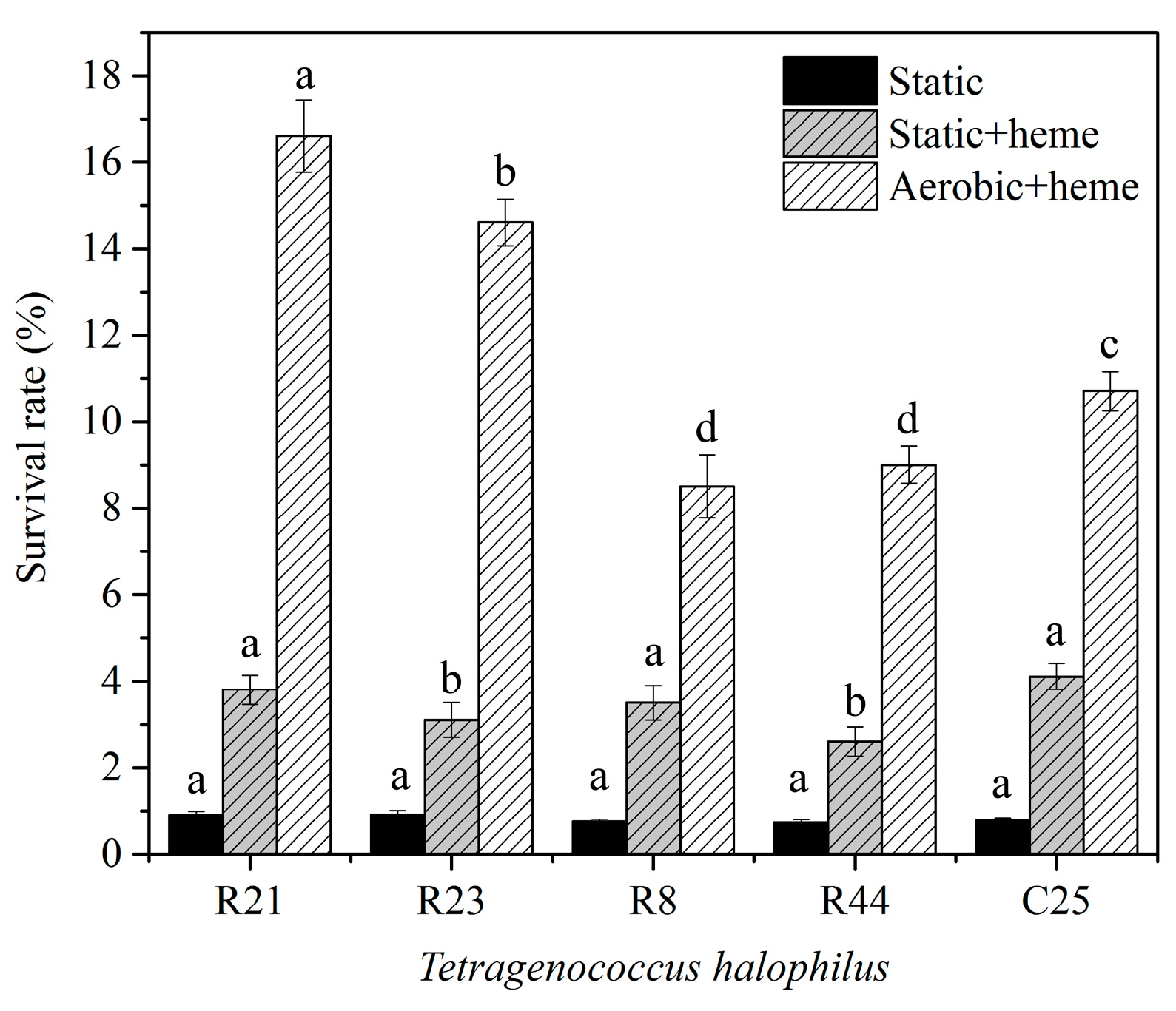
The resistance of T. halophilus to hydrogen peroxide was shown in Figure 3. T. halophilus cultivated statically could not survive in the presence of 20 mmol L−1 H2O2. With the addition of heme during cultivation, the survival rate of all tested strains increased. When T. halophilus strains were grown aerobically with the addition of heme, the survival rates of cells in H2O2 dramatically increased. The survival rates of R21 and R23 challenged with H2O2 increased by 18 and 16-fold compared with static cultivation. For R8, R44 and C25, their survival rates increased by about 10 to 13-fold. These results showed that the resistance to H2O2 of T. halophilus could be conditionally improved and was various for different strains. Moreover, the resistance to H2O2 of T. halophilus was not correlated with their capability of oxygen tolerance.

3.4. Characterization of Catalase Produced by T. halophilus

Catalase is an enzyme that efficiently catalyzes the conversion of H2O2 to water and oxygen. It protects cells against H2O2 toxicity and improves antioxidant capacity [29]. Lactobacillus is generally catalase-negative, while T. halophilus showed capability to resist H2O2 (Figure 3), and gene encode catalase was present in the genome of T. halophilus. Our work demonstrated that all tested T. halophilus strains had the cat gene, but only those from R21 and R23 were active when heme was present (Table 1). Sequence analysis suggested that cat genes in T. halophilus could be divided into two categories: the active cat and the pseudogene cat, which had a stop codon in the middle of the gene (Figure A1). Although cat genes from these two categories shared a similarity of about 97%, they function differently in resistance to H2O2 (Figure 3).

3.5. The Effect of Catalase on the Growth of T. halophilus Strains

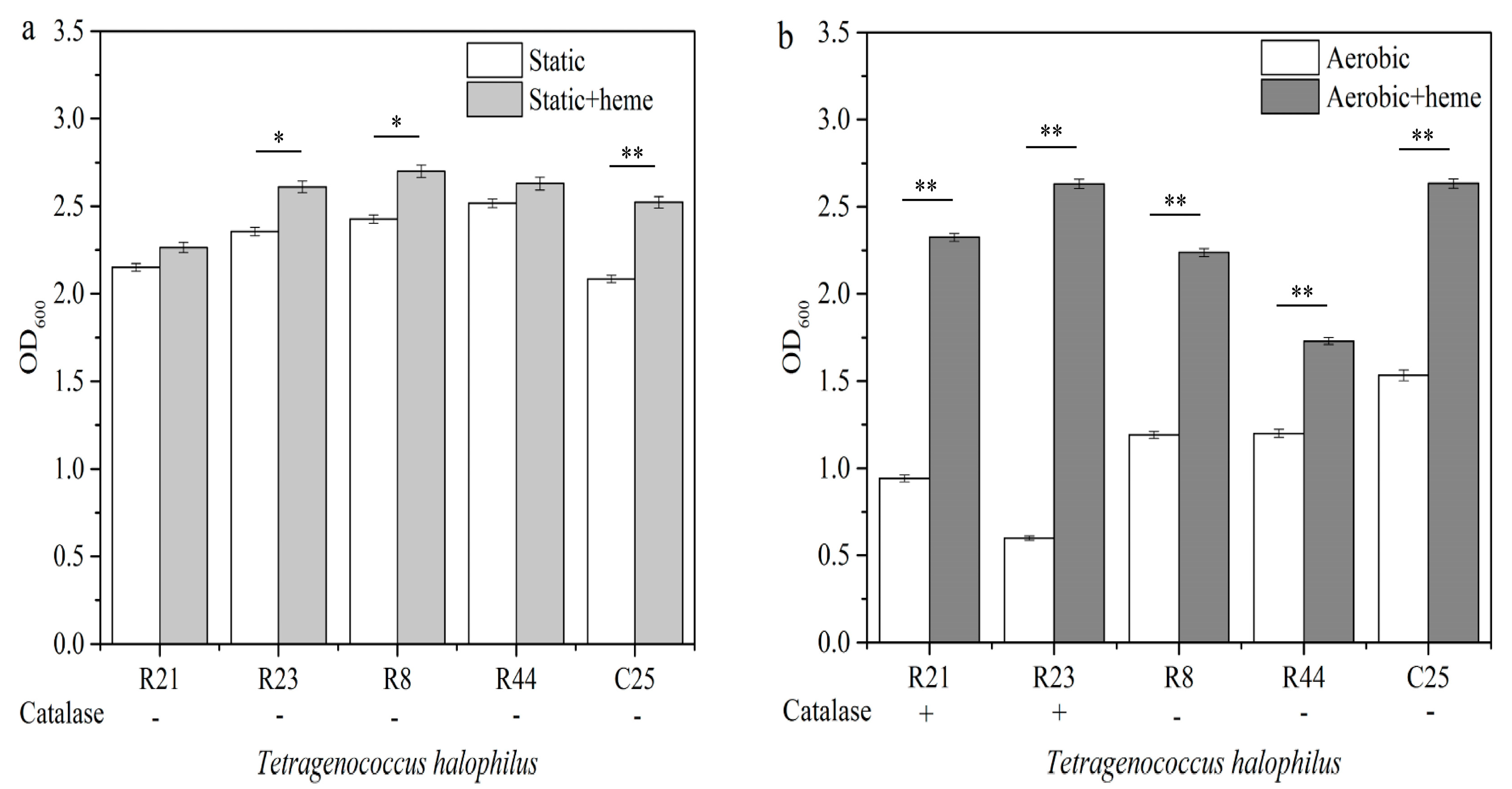
Functional catalase contributes to the resistance of T. halophilus strains to H2O2. We assumed that catalase might contribute to oxygen tolerance. Figure 4a showed that with the addition of heme, T. halophilus grew slightly better than the control when statically cultivated, and no catalase activity was detected under this condition. Thus, T. halophilus grew well regardless of the presence or absence of heme when cultivated statically. However, T. halophilus grew poorly when grown under aerobic conditions, while the growth of these strains under aerobic conditions was dramatically improved when heme was present (Figure 4b). Moreover, the growth of CAT active strains R21 and R23 under aerobic conditions in the presence of heme were increased by 1.5 times and 3.5 times. It was even better than they cultivated statically. Our results demonstrated that both active CAT and pseudo-CAT (or other heme-dependent antioxidant enzymes) in T. halophilus contribute to the tolerance to oxygen and H2O2 in the presence of heme, and the active CAT is more efficient in its protection of T. halophilus against oxidative stress.

4. Discussion

T. halophilus is a functional LAB that contributes to increasing the content of volatiles and thus to improving of flavor in fermented foods [30]. T. halophilus strains isolated from food origins exhibited differences in biological properties such as substrate utilization, growth performance under multiple-stress and formation of volatile compounds [5,31,32]. The outcome of the employment of this LAB in food fermentation may be attributed to their tolerance to different stress conditions. The salt tolerance and acid resistance mechanisms of T. halophilus were investigated previously [19,33]. In this work, we found that heme-dependent CAT and pseudo-CAT are produced by T. halophilus, and they contribute to tolerance of oxidative stress and H2O2 with the induction of aerobic cultivation. The finding of their differences in resistance to oxidative stress and the elaboration of the possible mechanism provide theoretical criteria for the selection of suitable strains that can be used in food fermentation.
Superoxide dismutase usually takes the front line in protecting cells from reactive oxygen species (ROS). This enzyme is important for the protection of bacteria against various types of oxidative stresses [34]. Other enzyme systems, such as the glutathione system and thioredoxin system, also detoxify hydroperoxide radicals. The glutathione system modulates the activities of E. coli transcriptional regulators that respond to oxidative stress, and glutathione reductase is implicated in the response to chemical oxidative stresses [34,35]. The deletion of glutathione reductase in Streptococcus pneumoniae led to an increased sensitivity to superoxide [36]. In most lactic acid bacteria, the degradation of hydrogen peroxide and other peroxides is generally catalyzed by peroxidase [37,38]. In this work, T. halophilus strains showed different tolerance to oxidative stress (Figure 1), and their antioxidant capacity is associated with peroxidase activity (Figure 2). Moreover, increased resistance of T. halophilus to H2O2 was achieved with an aerobic cultivation induction and presence of heme (Figure 3), indicating the function of a heme-dependent enzyme in tolerance to oxidative stress.
Catalase is the enzyme catalyzing the decomposition of hydrogen peroxide. It is an important enzyme that protects the cell from oxidative damage caused by ROS. LAB are generally considered to be devoid of catalase activity, since they are not able to synthesize heme. However, some species of LAB exhibit catalase activity. Catalase from LAB generally groups into two types. The first is heme-dependent catalase, which is induced by adding heme, produced by some lactobacilli, Pediococcus, Enterococcus and Leuconostoc strains. The other one is the heme-independent catalase or pseudocatalase, which is produced by some strains of Leuconostoc, Pediococcus, Enterococcus and Lactiplantibacillus plantarum [39]. Our results demonstrated that T. halophilus produces heme-dependent CAT and pseudo-CAT (Table 1). Both of them improved the oxygen resistance and H2O2 tolerance of T. halophilus strains. It was found that the presence of heme extended the growth period of Lactococcus lactis during aerated growth and improved long-term survival. This indicated that the respiratory metabolism in L. lactis may be better adapted to respiration than to traditional fermentative metabolism [40]. Our results showed that the addition of heme slightly improved the growth of T. halophilus under static conditions, and activated catalase dramatically improved its growth under aerobic conditions (Figure 4). This indicated a cultivation or growth optimization of T. halophilus through the respiration metabolism.

Author Contributions

Conceptualization, F.F. and J.L.; methodology, B.W.; validation, F.F., J.L. and B.W.; formal analysis, J.L.; investigation, J.L.; resources, G.D.; data curation, J.C.; writing—original draft preparation, F.F. and B.W.; writing—review and editing, F.F.; visualization, G.D.; supervision, F.F. and J.C.; project administration, J.C.; funding acquisition, F.F. All authors have read and agreed to the published version of the manuscript.

Funding

This research was funded by the National Natural Science Foundation of China, grant number 32172182.

Institutional Review Board Statement

Not applicable.

Informed Consent Statement

Not applicable.

Data Availability Statement

The data presented in this study are available on request from the corresponding author.

Conflicts of Interest

The authors have no conflict of interest to declare.

Appendix A

Figure A1. Alignment of catalase amino acid sequence. Sequence alignment of the catalase was done with BioEdit 7.0 using the ClustalW method. Residues highlighted in orange indicate differences in the amino acid sequence of the catalase from T. halophilus. The presence of internal stop codon was shown in frame.
Applsci 12 08039 g0a1

References

  1. Zhang, C.; Zhang, J.; Liu, D. Biochemical changes and microbial community dynamics during spontaneous fermentation of Zhacai, a traditional pickled mustard tuber from China. Int. J. Food Microbiol. 2021, 347, 109199. [Google Scholar] [CrossRef] [PubMed]
  2. Rodpai, R.; Sanpool, O.; Thanchomnang, T.; Wangwiwatsin, A.; Sadaow, L.; Phupiewkham, W.; Boonroumkaew, P.; Intapan, P.M.; Maleewong, W. Investigating the microbiota of fermented fish products (Pla-ra) from different communities of northeastern Thailand. PLoS ONE 2021, 16, e0245227. [Google Scholar] [CrossRef] [PubMed]
  3. Devanthi, P.V.P.; Gkatzionis, K. Soy sauce fermentation: Microorganisms, aroma formation, and process modification. Food Res. Int. 2019, 120, 364–374. [Google Scholar] [CrossRef]
  4. Gao, X.; Liu, E.; Zhang, J.; Yang, L.; Huang, Q.; Chen, S.; Ma, H.; Ho, C.T.; Liao, L. Accelerating aroma formation of raw soy sauce using low intensity sonication. Food Chem. 2020, 329, 127118. [Google Scholar] [CrossRef] [PubMed]
  5. Justé, A.; Van, T.S.; Verreth, C.; Cleenwerck, I.; De, V.P.; Lievens, B.; Willems, K.A. Characterization of Tetragenococcus strains from sugar thick juice reveals a novel species, Tetragenococcus osmophilus sp. nov., and divides Tetragenococcus halophilus into two subspecies, T. halophilus subsp. halophilus subsp. nov. and T. halophilus subsp. flandriensis subsp. nov. Int. J. Syst. Evol. Microbiol. 2012, 62, 129–137. [Google Scholar] [CrossRef] [Green Version]
  6. Collins, M.D.; Williams, A.M.; Wallbanks, S. The phylogeny of Aerococcus and Pediococcus as determined by 16S rRNA sequence analysis: Description of Tetragenococcus gen. nov. FEMS Microbiol. Lett. 1990, 58, 255–262. [Google Scholar] [CrossRef] [PubMed]
  7. Udomsil, N.; Rodtong, S.; Tanasupawat, S.; Yongsawatdigul, J. Proteinase-producing halophilic lactic acid bacteria isolated from fish sauce fermentation and their ability to produce volatile compounds. Int. J. Food Microbiol. 2010, 141, 186–194. [Google Scholar] [CrossRef] [PubMed]
  8. O’Toole, D.K. Chapter Two—The role of microorganisms in soy sauce production. In Advances in Applied Microbiology; Gadd, G.M., Sariaslani, S., Eds.; Academic Press: Cambridge, MA, USA, 2019; Volume 108, pp. 45–113. [Google Scholar]
  9. Devanthi, P.V.P.; Linforth, R.; Onyeaka, H.; Gkatzionis, K. Effects of co-inoculation and sequential inoculation of Tetragenococcus halophilus and Zygosaccharomyces rouxii on soy sauce fermentation. Food Chem. 2018, 240, 1–8. [Google Scholar] [CrossRef]
  10. Yao, S.; Zhou, R.; Jin, Y.; Huang, J.; Wu, C. Effect of co-culture with Tetragenococcus halophilus on the physiological characterization and transcription profiling of Zygosaccharomyces rouxii. Food Res. Int. 2019, 121, 348–358. [Google Scholar] [CrossRef] [PubMed]
  11. He, G.; Huang, J.; Liang, R.; Wu, C.; Zhou, R. Comparing the differences of characteristic flavour between natural maturation and starter culture for Mucor-type Douchi. Int. J. Food Sci. Technol. 2016, 51, 1252–1259. [Google Scholar] [CrossRef]
  12. Kim, K.H.; Lee, S.H.; Chun, B.H.; Jeong, S.E.; Jeon, C.O. Tetragenococcus halophilus MJ4 as a starter culture for repressing biogenic amine (cadaverine) formation during saeu-jeot (salted shrimp) fermentation. Food Microbiol. 2019, 82, 465–473. [Google Scholar] [CrossRef] [PubMed]
  13. Li, J.; Huang, J.; Jin, Y.; Wu, C.; Shen, D.; Zhang, S.; Zhou, R. Aflatoxin B1 degradation by salt tolerant Tetragenococcus halophilus CGMCC 3792. Food Chem. Toxicol. 2018, 121, 430–436. [Google Scholar] [CrossRef]
  14. Zhang, H.; Xu, J.H.; Chen, Q.; Wang, H.; Kong, B.H. Physiological, morphological and antioxidant responses of Pediococcus pentosaceus R1 and Lactobacillus fermentum R6 isolated from harbin dry sausages to oxidative stress. Foods 2021, 10, 1203. [Google Scholar] [CrossRef]
  15. Sugimoto, S.; Saruwatari, K.; Higashi, C.; Tsuruno, K.; Matsumoto, S.; Nakayama, J.; Sonomoto, K. In vivo and in vitro complementation study comparing the function of DnaK chaperone systems from halophilic lactic acid bacterium Tetragenococcus halophilus and Escherichia coli. Biosci. Biotechnol. Biochem. 2008, 72, 811–822. [Google Scholar] [CrossRef] [Green Version]
  16. Sugimoto, S.; Yoshida, H.; Mizunoe, Y.; Tsuruno, K.; Nakayama, J.; Sonomoto, K. Structural and functional conversion of molecular chaperone ClpB from the gram-positive halophilic lactic acid bacterium Tetragenococcus halophilus mediated by ATP and stress. J. Bacteriol. 2006, 188, 8070–8078. [Google Scholar] [CrossRef] [PubMed] [Green Version]
  17. Lin, J.; Liang, H.; Yan, J.; Luo, L. The molecular mechanism and post-transcriptional regulation characteristic of Tetragenococcus halophilus acclimation to osmotic stress revealed by quantitative proteomics. J. Proteom. 2017, 168, 1–14. [Google Scholar] [CrossRef] [PubMed]
  18. Robert, H.; Le Marrec, C.; Blanco, C.; Jebbar, M. Glycine betaine, carnitine, and choline enhance salinity tolerance and prevent the accumulation of sodium to a level inhibiting growth of Tetragenococcus halophila. Appl. Environ. Microbiol. 2000, 66, 509–517. [Google Scholar] [CrossRef] [PubMed] [Green Version]
  19. Denich, T.J.; Beaudette, L.A.; Lee, H.; Trevors, J.T. Effect of selected environmental and physico-chemical factors on bacterial cytoplasmic membranes. J. Microbiol. Meth. 2003, 52, 149–182. [Google Scholar] [CrossRef]
  20. Teixeira, J.S.; Seeras, A.; Sanchezmaldonado, A.F.; Zhang, C.; Su, M.S.; Gänzle, M.G. Glutamine, glutamate, and arginine-based acid resistance in Lactobacillus reuteri. Food Microbiol. 2014, 42, 172–180. [Google Scholar] [CrossRef] [PubMed]
  21. Gong, L.; Ren, C.; Xu, Y.; Kivisaar, M. GlnR negatively regulates glutamate-dependent acid resistance in Lactobacillus brevis. Appl. Environ. Microbiol. 2020, 86, e02615–e02619. [Google Scholar] [CrossRef]
  22. Ianniello, R.G.; Zotta, T.; Matera, A.; Genovese, F.; Parente, E.; Ricciardi, A. Investigation of factors affecting aerobic and respiratory growth in the oxygen-tolerant strain Lactobacillus casei N87. PLoS ONE 2016, 11, e0164065. [Google Scholar] [CrossRef] [PubMed]
  23. Feng, T.; Wang, J. Oxidative stress tolerance and antioxidant capacity of lactic acid bacteria as probiotic: A systematic review. Gut Microbes 2020, 12, 1801944. [Google Scholar] [CrossRef] [PubMed]
  24. Su, J.; Wang, T.; Li, Y.Y.; Li, J.; Zhang, Y.; Wang, Y.; Wang, H.; Li, H. Antioxidant properties of wine lactic acid bacteria: Oenococcus oeni. Appl. Microbiol. Biotechnol. 2015, 99, 5189–5202. [Google Scholar] [CrossRef]
  25. Lin, M.; Yen, C. Antioxidative ability of lactic acid bacteria. J. Agric. Food Chem. 1999, 47, 1460–1466. [Google Scholar] [CrossRef]
  26. Wang, B.; Chen, J.; Du, G.; Fang, F. Classification and characteristics of Tetragenococcus halophilus derived from moromi. Acta Microbiol. Sin. 2018, 58, 1826–1838. [Google Scholar]
  27. Maehly, A.C.; Chance, B. The assay of catalases and peroxidases. Methods Biochem. Anal. 1954, 1, 357. [Google Scholar] [PubMed]
  28. Gürtler, M.; Gänzle, M.G.; Wolf, G.; Hammes, W.P. Physiological diversity among strains of Tetragenococcus halophilus. Syst. Appl. Microbiol. 1998, 21, 107–112. [Google Scholar] [CrossRef]
  29. Matés, J.M. Effects of antioxidant enzymes in the molecular control of reactive oxygen species toxicology. Toxicology 2000, 153, 83–104. [Google Scholar] [CrossRef]
  30. Jeong, D.W.; Heo, S.; Lee, J.H. Safety assessment of Tetragenococcus halophilus isolates from doenjang, a Korean high-salt-fermented soybean paste. Food Microbiol. 2017, 62, 92–98. [Google Scholar] [CrossRef]
  31. Juste, A.; Lievens, B.; Frans, I.; Marsh, T.L.; Klingeberg, M.; Michiels, C.W.; Willems, K.A. Genetic and physiological diversity of Tetragenococcus halophilus strains isolated from sugar- and salt-rich environments. Microbiology 2008, 154, 2600–2610. [Google Scholar] [CrossRef] [Green Version]
  32. Wu, C.; Liu, C.; He, G.; Huang, J.; Zhou, R. Characterization of a multiple-stress tolerance Tetragenococcus halophilus and application as starter culture in Chinese horsebean-chili-paste manufacture for quality improvement. Food Sci. Technol. Int. Tokyo 2013, 19, 855–864. [Google Scholar] [CrossRef] [Green Version]
  33. Liu, L.; Si, L.; Meng, X.; Luo, L. Comparative transcriptomic analysis reveals novel genes and regulatory mechanisms of Tetragenococcus halophilus in response to salt stress. J. Ind. Microbiol. Biotechnol. 2015, 42, 601–616. [Google Scholar] [CrossRef]
  34. Staerck, C.; Gastebois, A.; Vandeputte, P.; Calenda, A.; Larcher, G.; Gillmann, L.; Papon, N.; Bouchara, J.P.; Fleury, M.J.J. Microbial antioxidant defense enzymes. Microb. Pathog. 2017, 110, 56–65. [Google Scholar] [CrossRef] [PubMed]
  35. Köhsler, M.; Leitsch, D.; Mbouaka, A.L.; Wekerle, M.; Walochnik, J. Transcriptional changes of proteins of the thioredoxin and glutathione systems in Acanthamoeba spp. under oxidative stress—An RNA approach. Parasite 2022, 29, 24. [Google Scholar] [CrossRef] [PubMed]
  36. Sikanyika, M.; Aragão, D.; McDevitt, C.A.; Maher, M.J. The structure and activity of the glutathione reductase from Streptococcus pneumoniae. Acta Crystallogr. F Struct. Biol. Commun. 2019, 75, 54–61. [Google Scholar] [CrossRef] [Green Version]
  37. Sun, C.; Benlekbir, S.; Venkatakrishnan, P.; Wang, Y.H.; Hong, S.J.; Hosler, J.; Tajkhorshid, E.; Rubinstein, J.L.; Gennis, R.B. Structure of the alternative complex III in a supercomplex with cytochrome oxidase. Nature 2018, 557, 123–126. [Google Scholar] [CrossRef]
  38. Talwalkar, A.; Kailasapathy, K.; Hourigan, J.; Peiris, P.; Arumugaswamy, R. An improved method for the determination of NADH oxidase in the presence of NADH peroxidase in lactic acid bacteria. J. Microbiol. Meth. 2003, 52, 333. [Google Scholar] [CrossRef]
  39. Nanasombat, S.; Treebavonkusol, P.; Kittisrisopit, S.; Jaichalad, T.; Phunpruch, S.; Kootmas, A.; Nualsri, I. Lactic acid bacteria isolated from raw and fermented pork products: Identification and characterization of catalase-producing Pediococcus pentosaceus. Food Sci. Biotechnol. 2017, 26, 173–179. [Google Scholar] [CrossRef]
  40. Pedersen, M.B.; Garrigues, C.; Tuphile, K.; Brun, C.; Vido, K.; Bennedsen, M.; Møllgaard, H.; Gaudu, P.; Gruss, A. Impact of aeration and heme-activated respiration on Lactococcus lactis gene expression: Identification of a heme-responsive operon. J. Bacteriol. 2008, 190, 4903–4911. [Google Scholar] [CrossRef] [Green Version]
Figure 1. Growth of T. halophilus strains under aerobic and anaerobic conditions. Growth ratio: the growth rate of T. halophilus strains cultivated anaerobically divided by that cultivated aerobically. Different lowercase letters indicate significant differences among individual groups (p < 0.05).
Applsci 12 08039 g001
Figure 2. Comparison of the antioxidant enzymes produced by T. halophilus strains. Different lowercase letters indicate significant differences among different groups (p < 0.05).
Applsci 12 08039 g002
Figure 3. Hydrogen peroxide tolerance of T. halophilus strains under different conditions. Different lowercase letters indicate significant differences among individual groups (p < 0.05).
Applsci 12 08039 g003
Figure 4. Effect of heme on growth of T. halophilus strains under (a) static condition and (b) aerobic condition. −/+: negative/positive activity of catalase detected under corresponding conditions. Asterisks mean significant differences among different groups (*, p < 0.05; **, p < 0.01).
Applsci 12 08039 g004
Table 1. Comparison of the catalase activity of different T. halophilus strains.
StrainR23R21C25R44R8
Catalase activity++
Interruption of cat geneNoNoYesYesYes
+, active; −, inactive.
Publisher’s Note: MDPI stays neutral with regard to jurisdictional claims in published maps and institutional affiliations.