
Multifunctional MEN-Doped Adhesives: Strengthening, Bond Quality Evaluation, and Variations in Magnetic Signal with Environmental Exposure


Abstract
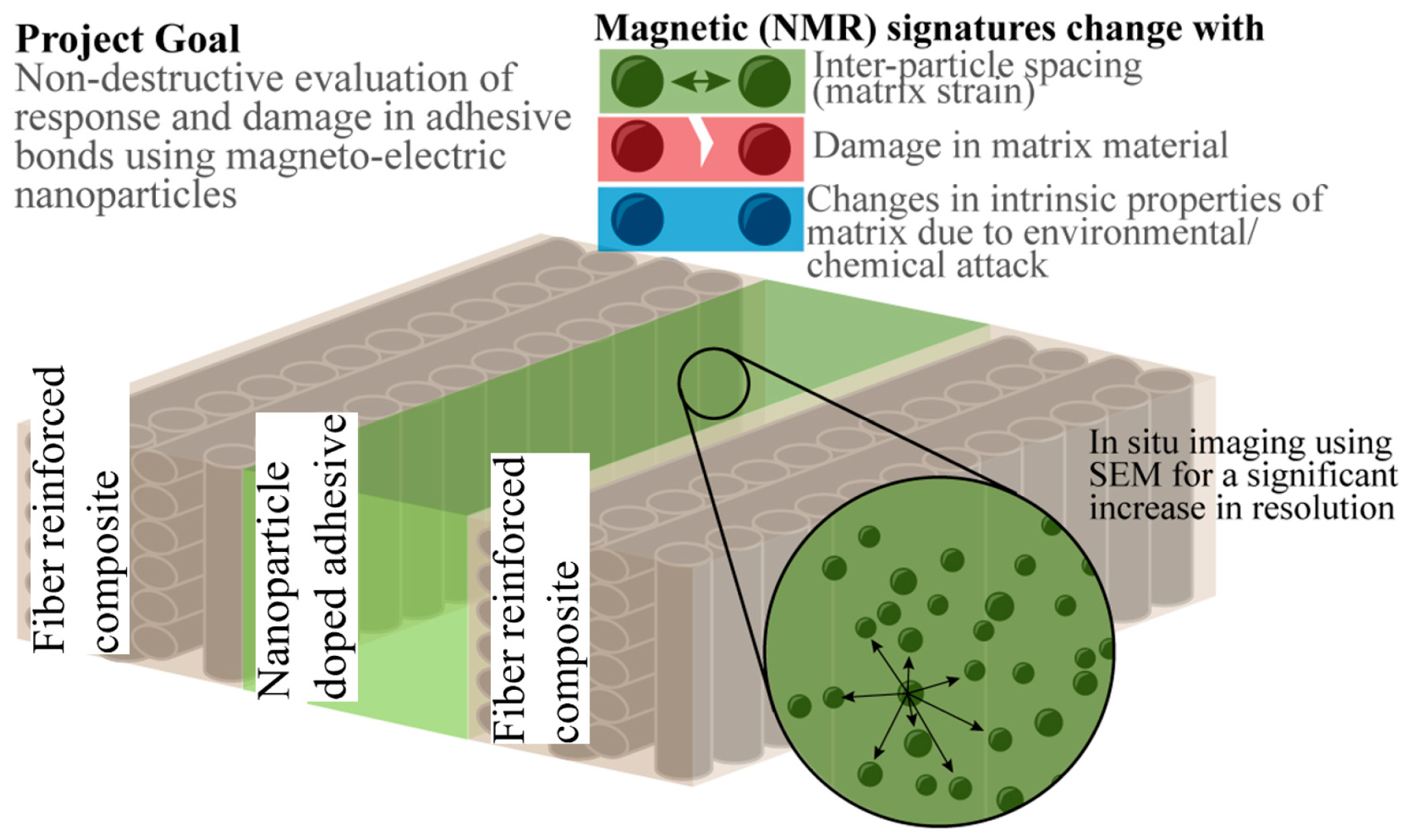
:Featured Application
Abstract
1. Introduction
2. Materials and Methods
3. Results
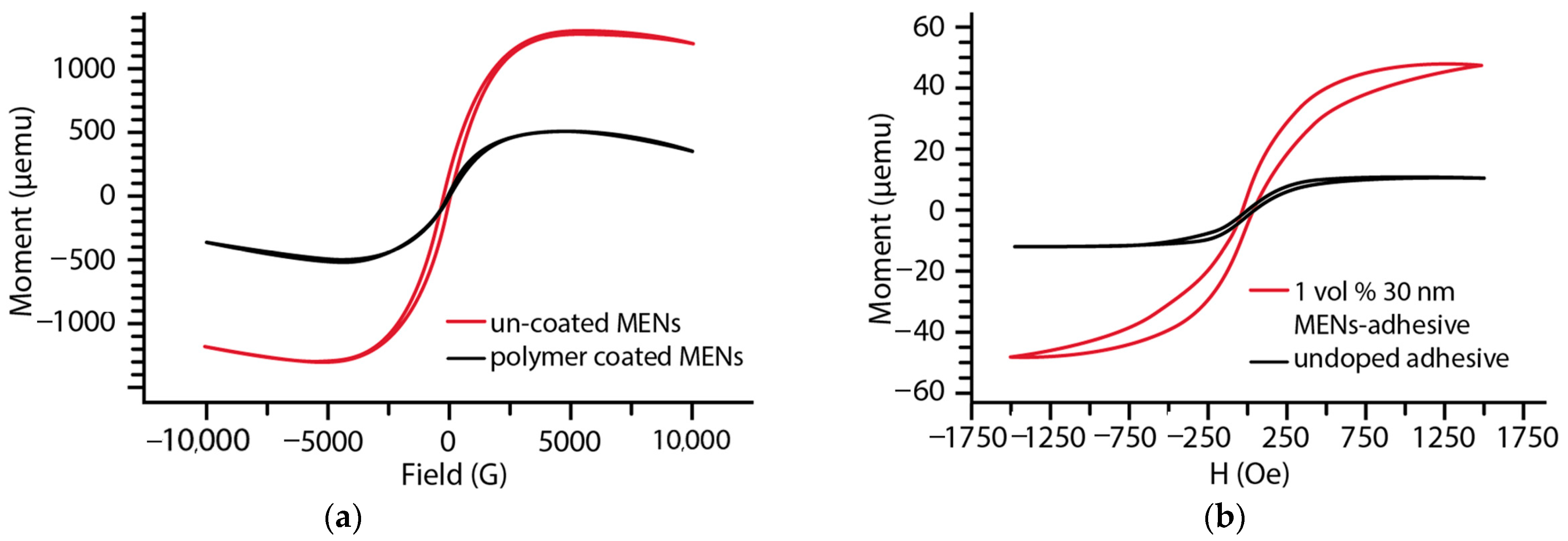
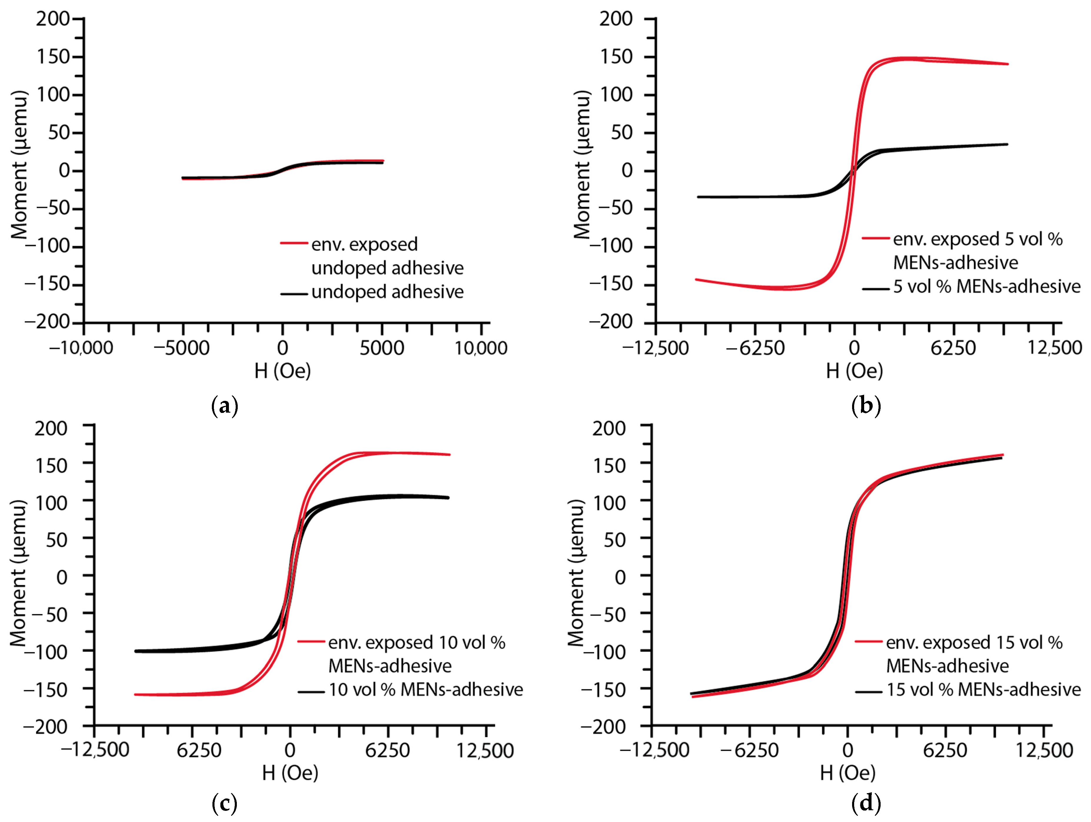
3.1. Magnetic Moment on MEN-Doped Adhesive
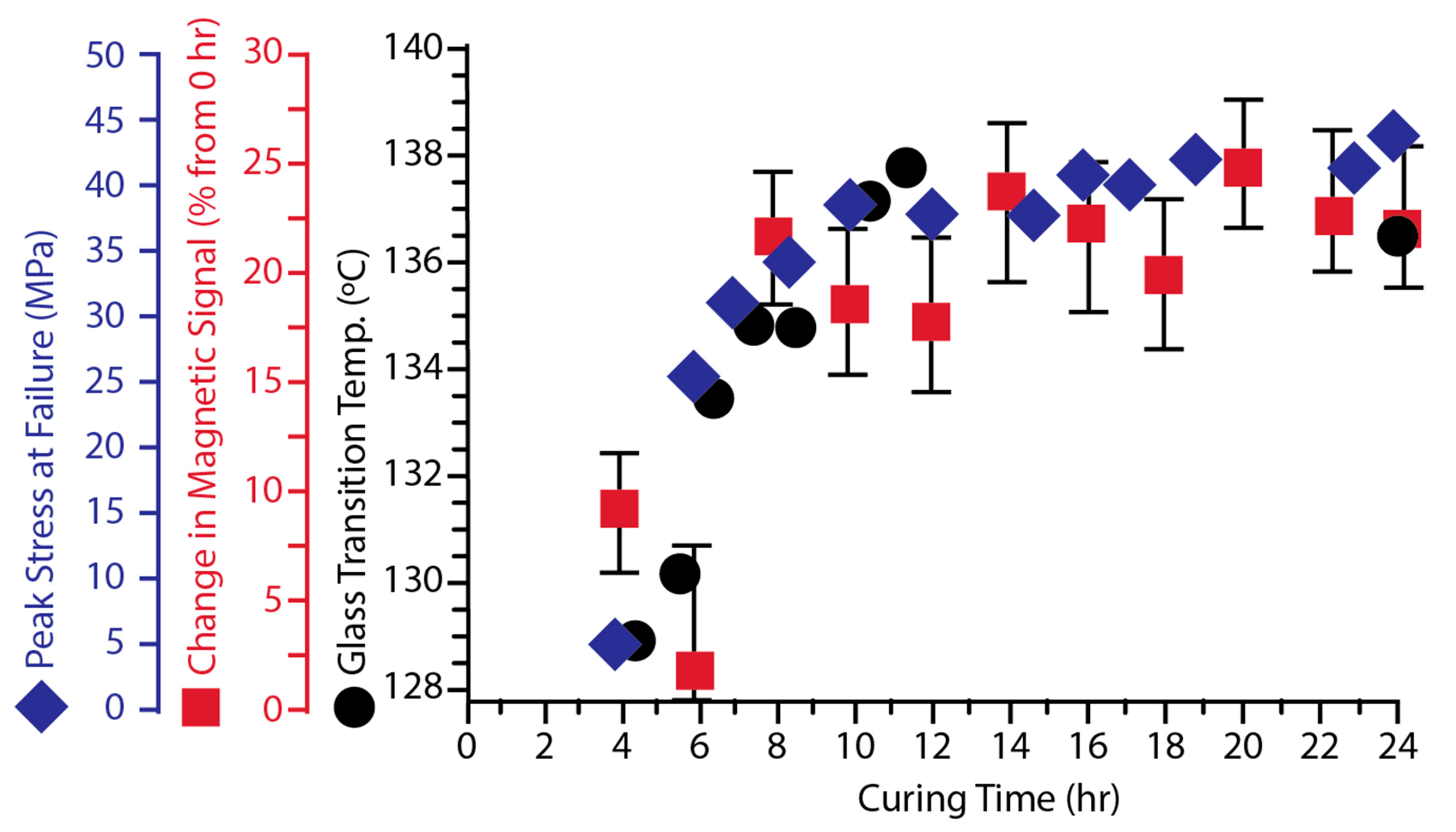
3.2. Monitoring of the Curing Process Using the B–H Looper
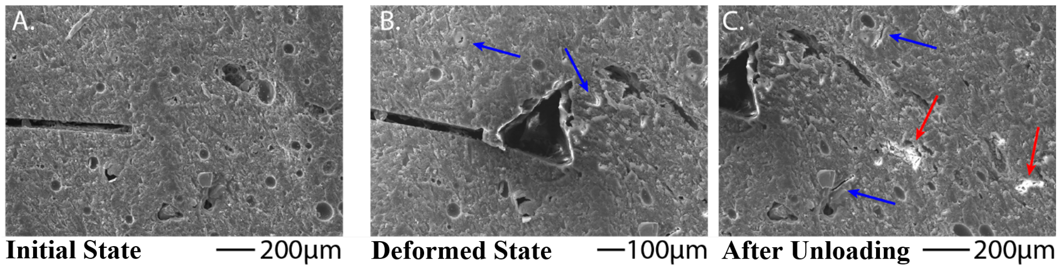
3.3. Single Lap Shear
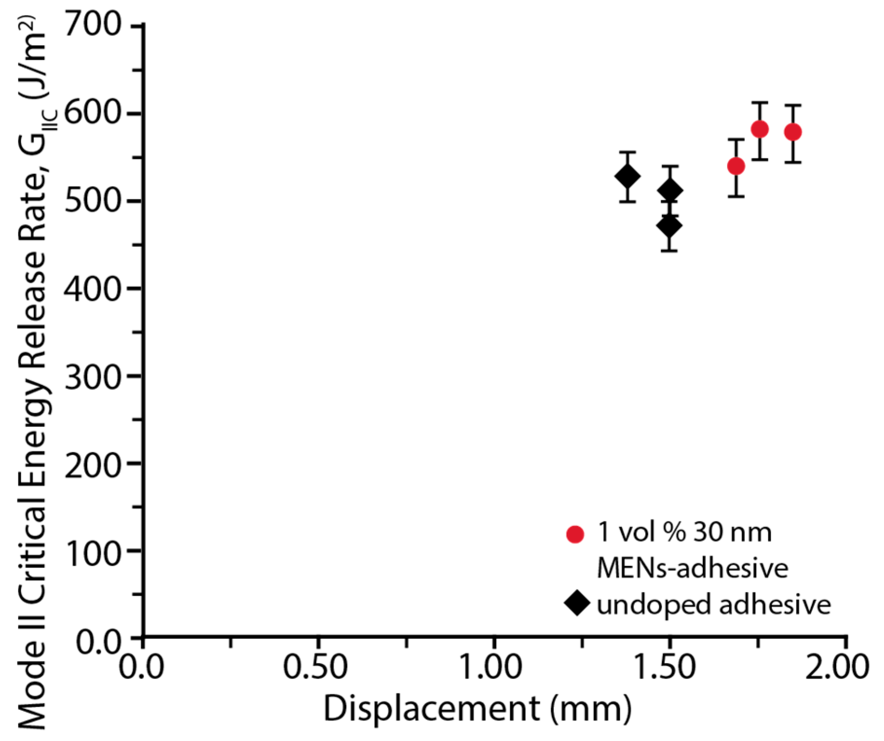
3.4. End Notch Flexure (ENF)
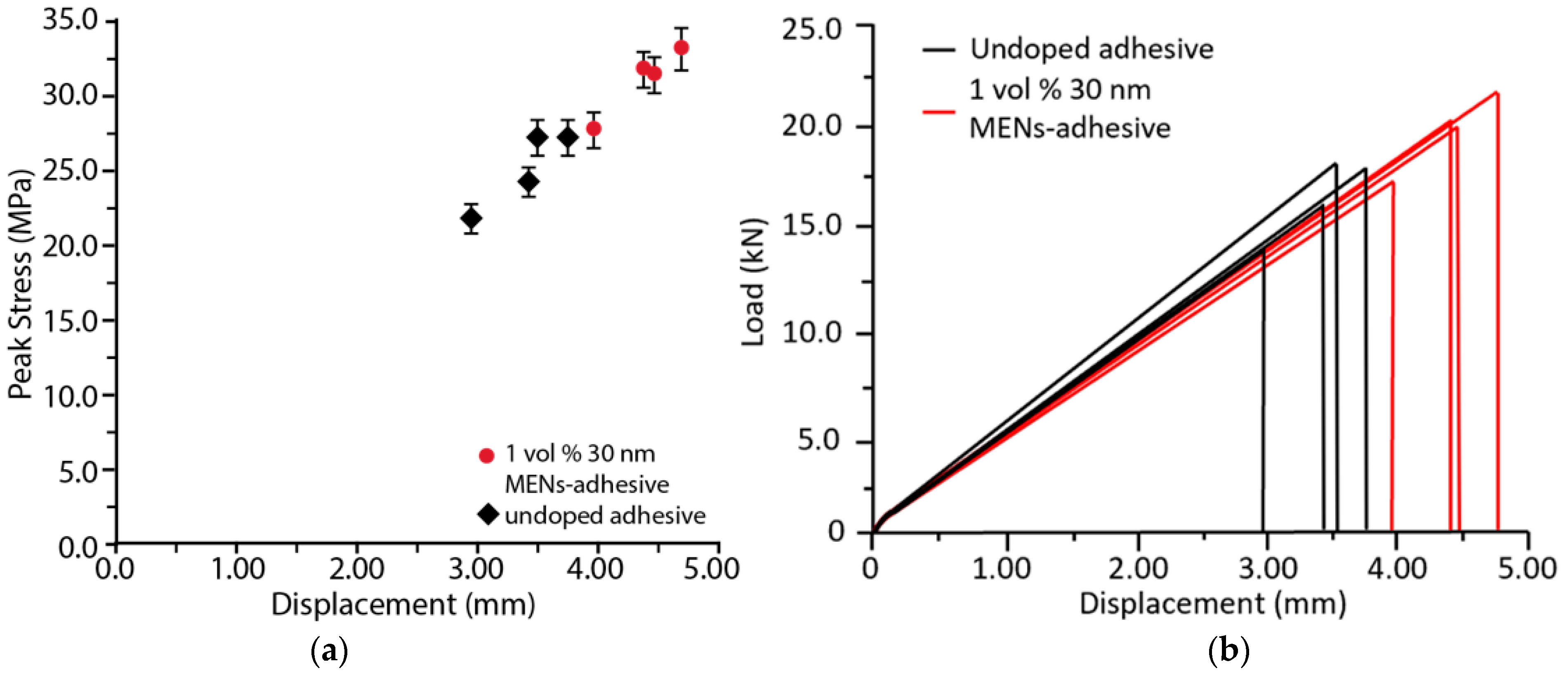
3.5. Tensile Testing
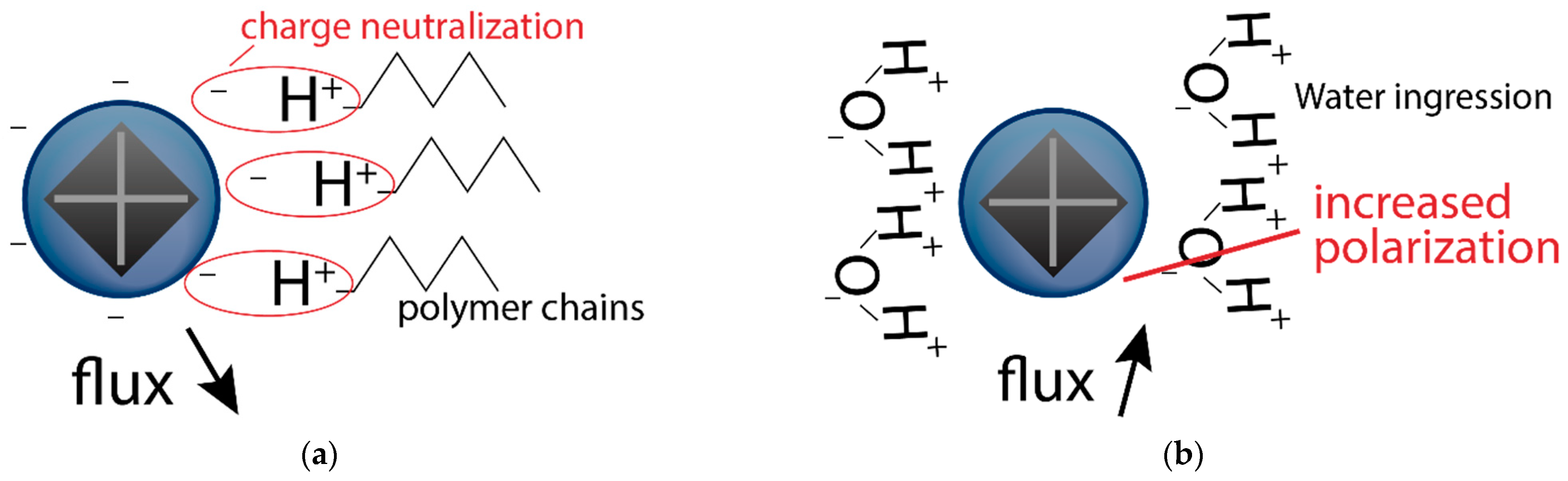
3.6. Environmental Exposure
4. Discussion
5. Conclusions
Supplementary Materials
Author Contributions
Funding
Institutional Review Board Statement
Informed Consent Statement
Data Availability Statement
Acknowledgments
Conflicts of Interest
References
- Soutis, C. Carbon fiber reinforced plastics in aircraft construction. Mater. Sci. Eng. A 2005, 412, 171–176. [Google Scholar] [CrossRef]
- Barbero, E.J. Introduction to Composite Materials Design; CRC Press: Boca Raton, FL, USA, 2011; pp. 1–26. [Google Scholar]
- Pitta, S.; Roure, F.; Crespo, D.; Rojas, J.I. An experimental and numerical study of repairs on composite substrates with composite and aluminum doublers using riveted, bonded, and hybrid joints. Materials 2019, 12, 2978. [Google Scholar] [CrossRef] [PubMed]
- Federal Aviation Administration. Best Practice in Adhesive-Bonded Structures and Repairs; DOT/FAA/AR-TN06/57; FAA: Washington, DC, USA, 2019.
- Piehl, M.J.; Bossi, R.H.; Blohowiak, K.Y.; Dilligan, M.A.; Grace, W.B. Efficient certification of bonded primary structures. In Proceedings of the SAMPE 2013, Long Beach, CA, USA, 6–9 June 2013. [Google Scholar]
- Dillard, D. Advances in Structural Adhesive Bonding; Woodhead Publishing: Sawston, UK, 2016; pp. 286–287. [Google Scholar]
- Giurgiutiu, V.; Zagrai, A.; Bao, J.J. Piezoelectric wafer embedded active sensors for aging aircraft structural health monitoring. Struct. Health Monit. 2022, 1, 41–61. [Google Scholar] [CrossRef]
- Aggelis, D.G.; Barkoula, N.M.; Matikas, T.E.; Paipetis, A.S. Acoustic structural health monitoring of composite materials: Damage identification and evaluation in cross ply laminates using acoustic emission and ultrasonics. Compos. Sci. Technol. 2012, 72, 1127–1133. [Google Scholar] [CrossRef]
- Park, H.S.; Lee, H.M.; Adeli, H.; Lee, I. A new approach for health monitoring of structures: Terrestrial laser scanning. Comput.-Aided Civ. Infrastruct. Eng. 2006, 22, 19–30. [Google Scholar] [CrossRef]
- Tokura, Y.; Seki, S. Multiferroics with Spiral Spin Orders. Adv. Mat. 2010, 22, 1554–1565. [Google Scholar] [CrossRef]
- El-Alaily, T.M.; El-Nimr, M.K.; Saafan, S.A.; Kamel, M.M.; Meaz, T.M.; Assar, S.T. Construction and calibration of a low cost and fully automated vibrating sample magnetometer. J. Magn. Magn. Mater. 2015, 386, 25–30. [Google Scholar] [CrossRef]
- Martins, P.; Lanceros-Méndez, S. Polymer-Based Magnetoelectric Materials. Adv. Funct. Mater. 2013, 23, 3371–3385. [Google Scholar] [CrossRef]
- Liu, W.; Miroshnichenko, A.E.; Neshev, D.N.; Kivshar, Y.S. Broadband unidirectional scattering by magneto-electric core–shell nanoparticles. ACS Nano 2012, 6, 5489–5497. [Google Scholar] [CrossRef] [PubMed]
- Nair, M.; Guduru, R.; Liang, P.; Hong, J.; Sagar, V.; Khizroev, S. Externally controlled on-demand release of anti-HIV drug using magneto-electric nanoparticles as carriers. Nat. Commun. 2013, 4, 1707. [Google Scholar] [CrossRef] [PubMed]
- Guduru, R.; Liang, P.; Runowicz, C.; Nair, M.; Atluri, V.; Khizroev, S. Magneto-electric nanoparticles to enable field-controlled high-specificity drug delivery to eradicate ovarian cancer cells. Sci. Rep. 2013, 3, 2953. [Google Scholar] [CrossRef] [PubMed]
- Pridgen, E.M.; Langer, R.; Farokhzad, O.C. Biodegradable, polymeric nanoparticle delivery systems for cancer therapy. Nanomedicine 2007, 197, 56–58. [Google Scholar] [CrossRef] [PubMed]
- ASTM D5868-01; Standard Test Method for Lap Shear Adhesion for Fiber Reinforced Plastic (FRP) Bonding. ASTM International: West Conshohocken, PA, USA, 2004.
- ASTM Standard D7905/D7905M-14; Standard Test Method for Determination of The Mode II Interlaminar Fracture Toughness of Unidirectional Fiber-Reinforced Polymer Matrix Composites. ASTM International: West Conshohocken, PA, USA, 2014.
- Rodzinski, A.; Guduru, R.; Liang, P.; Hadjikhani, A.; Stewart, T.; Stimphil, E.; Runowicz, C.; Cote, R.; Altman, N.; Datar, R.; et al. Targeted and controlled anticancer drug delivery and release with magnetoelectric nanoparticles. Sci. Rep. 2016, 6, 20867. [Google Scholar] [CrossRef]
- ASTM D638-03; Standard Test Method for Tensile Properties of Plastics. ASTM International: West Conshohocken, PA, USA, 2012.
- Ku, H. Notes on the Use of Propagation of Error Formulas. J Res. Natl. Bur. Stand. C. Eng. Instrum. 1996, 70, 4. [Google Scholar]
- Yang, K.Z. MENs Doped Adhesive and Influence on Fracture Toughness. Master’s Thesis, Florida International University, Miami, FL, USA, 2016. [Google Scholar] [CrossRef]
- Graham, C.D. High-sensitivity magnetization measurements. J. Mater. Sci. Technol. 2000, 16, 97–101. [Google Scholar]
- Dubon, J.; Seisdedos, G.; Ontiveros, M.; Boesl, B.; McDaniel, D. Bond Quality Evaluation Using Adhesive Doped with Magneto-Electric Nanoparticles. Am. Soc. Compos. 2021. [Google Scholar] [CrossRef]
- Wetzel, B.; Rosso, P.; Haupert, F.; Friedrich, K. Epoxy nanocomposites–fracture and toughening mechanisms. Eng. Fract. Mech. 2006, 73, 2375–2398. [Google Scholar] [CrossRef]
- Hsieh, T.H.; Kinloch, A.J.; Masania, K.; Taylor, A.C.; Sprenger, S. The mechanisms and mechanics of the toughening of epoxy polymers modified with silica nanoparticles. Polymer 2010, 51, 6284–6294. [Google Scholar] [CrossRef]
- Johnsen, B.B.; Kinloch, A.J.; Mohammed, R.D.; Taylor, A.C.; Sprenger, S. Toughening mechanisms of nanoparticle-modified epoxy polymers. Polymer 2007, 48, 530–541. [Google Scholar] [CrossRef]
- Boesl, B.; Bourne, G.R.; Sankar, B.V. In situ multiscale analysis of fracture mechanisms in nanocomposites. Compos. Part B 2011, 42, 1157–1163. [Google Scholar] [CrossRef]
- Seisdedos, G.; Hernandez, B.; Dubon, J.; Ontiveros, M.; Boesl, B.; McDaniel, D. Non-Destructive Evaluation of Mechanical Damage of Adhesives Using Magneto-Electric Nanoparticles. Am. Soc. Compos. 2021. [Google Scholar] [CrossRef]
- Zheng, H.; Wang, J.; Lofland, S.E.; Ma, Z.; Mohaddes-Ardabili, L.; Zhao, T.; Salamanca-Riba, L.; Shinde, S.R.; Ogale, S.B.; Bai, F.; et al. Multiferroic BaTiO3-CoFe2O4 Nanostructures. Science 2004, 303, 661–663. [Google Scholar] [CrossRef] [PubMed]
- Nautiyal, P.; Boesl, B.; Agarwal, A. In-Situ Mechanics of Materials; Springer International Publishing: Cham, Switzerland, 2020. [Google Scholar] [CrossRef]


| Sample Type/Sample # | Peak Tensile Stress (MPa) | Std. Dev. (MPa) | Avg. Stress (MPa) | ||||
|---|---|---|---|---|---|---|---|
| 1 | 2 | 3 | 4 | 5 | |||
| Baseline (2R:1H ratio) | 38.09 | 36.61 | 36.10 | 38.44 | 35.00 | 1.28 | 36.85 |
| Doped (5 vol% MENs) | 38.18 | 36.32 | 42.56 | 40.58 | 43.84 | 2.76 | 40.30 |
Publisher’s Note: MDPI stays neutral with regard to jurisdictional claims in published maps and institutional affiliations. |
© 2022 by the authors. Licensee MDPI, Basel, Switzerland. This article is an open access article distributed under the terms and conditions of the Creative Commons Attribution (CC BY) license (https://creativecommons.org/licenses/by/4.0/).
Share and Cite
Dubon, J.; Seisdedos, G.; Watring, D.; Pajon, M.; Khizroev, S.; McDaniel, D.; Boesl, B. Multifunctional MEN-Doped Adhesives: Strengthening, Bond Quality Evaluation, and Variations in Magnetic Signal with Environmental Exposure. Appl. Sci. 2022, 12, 8238. https://doi.org/10.3390/app12168238
Dubon J, Seisdedos G, Watring D, Pajon M, Khizroev S, McDaniel D, Boesl B. Multifunctional MEN-Doped Adhesives: Strengthening, Bond Quality Evaluation, and Variations in Magnetic Signal with Environmental Exposure. Applied Sciences. 2022; 12(16):8238. https://doi.org/10.3390/app12168238
Chicago/Turabian StyleDubon, Juliette, Gonzalo Seisdedos, Dillon Watring, Mauricio Pajon, Sakhrat Khizroev, Dwayne McDaniel, and Benjamin Boesl. 2022. "Multifunctional MEN-Doped Adhesives: Strengthening, Bond Quality Evaluation, and Variations in Magnetic Signal with Environmental Exposure" Applied Sciences 12, no. 16: 8238. https://doi.org/10.3390/app12168238