Article

Anthraquinone Rhein Exhibits Antibacterial Activity against Staphylococcus aureus

by
Federica Dell’Annunziata
1,†,
Veronica Folliero
1,†,
Francesca Palma
1,
Valeria Crudele
2,
Emiliana Finamore
2,
Giuseppina Sanna
3,
Aldo Manzin
3,
Anna De Filippis
1,
Massimiliano Galdiero
1 and
Gianluigi Franci
4,5,*
1
Department of Experimental Medicine, University of Campania “Luigi Vanvitelli”, 80138 Naples, Italy
2
Section of Microbiology and Virology, University Hospital “Luigi Vanvitelli”, 80138 Naples, Italy
3
Microbiology and Virology Unit, Department of Biomedical Sciences, University of Cagliari, 09042 Monserrato, Italy
4
Department of Medicine, Surgery and Dentistry “Scuola Medica Salernitana”, University of Salerno, 84081 Baronissi, Italy
5
Clinical Pathology and Microbiology Unit, San Giovanni di Dio e Ruggi D’Aragona University Hospital, 84126 Salerno, Italy
*
Author to whom correspondence should be addressed.
These authors contributed equally to this work.
Appl. Sci. 2022, 12(17), 8691; https://doi.org/10.3390/app12178691
Submission received: 4 August 2022 / Revised: 22 August 2022 / Accepted: 29 August 2022 / Published: 30 August 2022

Abstract

:
Staphylococcus aureus (S. aureus) represents an important pathogen of clinical relevance, causing a wide variety of symptoms. The broad distribution of multidrug-resistant strains necessarily demands new antibacterial agents for the treatment of S. aureus infections. The aim of this study was to assess the antibacterial activity of plant-derived compounds, pure 4,5″-dihydroxy-anthraquinone-2-carboxylic acid (Rhein), against standard and clinical isolated S. aureus strains. The hemolysis and 3-(4,5-dimethylthiazol-2-yl)-2,5-diphenyl tetrazolium bromide (MTT) assays were used to determine the cytotoxicity on human erythrocytes and bronchial epithelial cells after treatment with Rhein. The antibacterial effect was assessed via disk diffusion test, broth microdilution methods, time-killing assays and live–dead evaluation (50–0.39 µg/mL). Rhein effect on the hemolytic activity of α-toxin and catalase were estimated. Moreover, crystal violet (CV) assay evaluated its impact on biofilm biomass. The compound exhibited 50% cytotoxic concentration (CC50) and 50% hemolysis concentration (EC50) of 43.6 and >50 µg/mL, respectively. The minimum inhibitory concentration (MIC) of Rhein was 12.5 µg/mL for all tested strains, exerting bacteriostatic action. MIC and sub-MIC concentrations of Rhein significantly reduced hemolytic and catalase activities, impairing the major virulence factors of S. aureus strains. Rhein also reduced biofilm biomass in a dose-dependent manner, reaching rates of about 50% eradication at a dose of 50 µg/mL. These findings suggest that Rhein could represent a promising therapeutic option for the treatment of S. aureus infections.

1. Introduction

Staphylococcus aureus (S. aureus) represents an important opportunistic pathogen, able of adapting rapidly to adverse environmental conditions and evading the immune system [1,2]. It causes a wide range of clinical manifestations, ranging from skin and soft tissue infections to systemic and toxin-mediated affections with high prevalence [3,4]. S. aureus infection abides a major and prevalent reason of morbidity and mortality in hospitalized patients, despite the antibiotic treatment and prevention of infectious diseases [5]. Each year in Europe, approximately 25,000 hospitalized patients die from a severe resistant bacterial infection, and a substantial part of them is due to S. aureus [6]. This strain causes blood infections with an incidence of 20–50 cases per 100,000 inhabitants per year, with mortality levels of around 10–30% [7]. Methicillin-resistant S. aureus (MRSA) has been recognized as a global priority pathogen by the World Health Organization, causing growing concern around the world [8,9]. Conventional antibiotics are no longer suitable against most MRSA due to the rapid development of antibiotic resistance [10]. Glycopeptides and latest-generation antibiotics, such as linezolid, daptomycin and the newly available fifth-generation cephalosporins, still show high efficiency against resistant S. aureus, although the development of strains resistant to these antibiotics is clearly on the rise [11]. The prevalence of Vancomycin-resistant S. aureus was 2% before 2006, 5% in 2006–2014 and 7% in 2015–2020, showing a 3.5-fold increase between 2006 and 2020 [12]. Moreover, surveillance studies reported a global resistance rate of S. aureus to Daptomycin and Linezolid of 0.3% [13,14]. The emergence of resistant strains is often faster than the development of new antibiotics. Moreover, the number of Food and Drug Administration approved antibiotics has been reduced in the last three decades, limiting the treatment possibilities of resistant bacteria [11,15]. Therefore, antibacterial agents with new modes of action and alternative pharmacological targets are urgently needed. Natural products are an important source of novel antimicrobial agents, and their use is associated with the following advantages: (i) low cytotoxicity; (ii) reduced propensity to develop resistance; (iii) high chemical diversity; and iv) material of abundant and cheap origin [16,17]. In recent years, many plant-derived compounds have been studied for their antibacterial potential against S. aureus strains. Joray et al. reported the antibacterial potential of (Z, Z)-5-(trideca-4,7-dienyl) resorcinol against S. aureus strains with MIC values of 8 µg/mL [18]. Obiang-Obounou et al. demonstrated that Sanguinarine possesses strong antibacterial activity against S. aureus, recording MIC values of 6.25 µg/mL [19]. Additionally, Zhang et al. proved the antibacterial effect of Berberine against MRSA with MIC values of 128 μg/mL [20]. Rhein is an anthraquinone, constituent of several medicinal plants, such as Rheum palmatum L., Cassia tora L., Polygonum multiflorum Thunb. and Aloe barbadensis Miller (Figure 1) [21]. Numerous evidence reports the hepatoprotective, neuroprotective, antioxidant, antitumor, antidiabetic and anti-inflammatory role of this natural product [22]. Furthermore, Rhein’s antibacterial potential has been proven against different Gram-positive and Gram-negative pathogens, such as Streptococcus mutans, Propionibacterium acnes, Pseudomonas aeruginosa, etc. [23,24,25,26]. The present study aims to evaluate the antibacterial potential of Rhein against standard and clinical isolated S. aureus strains in the planktonic and biofilm state.

2. Materials and Methods

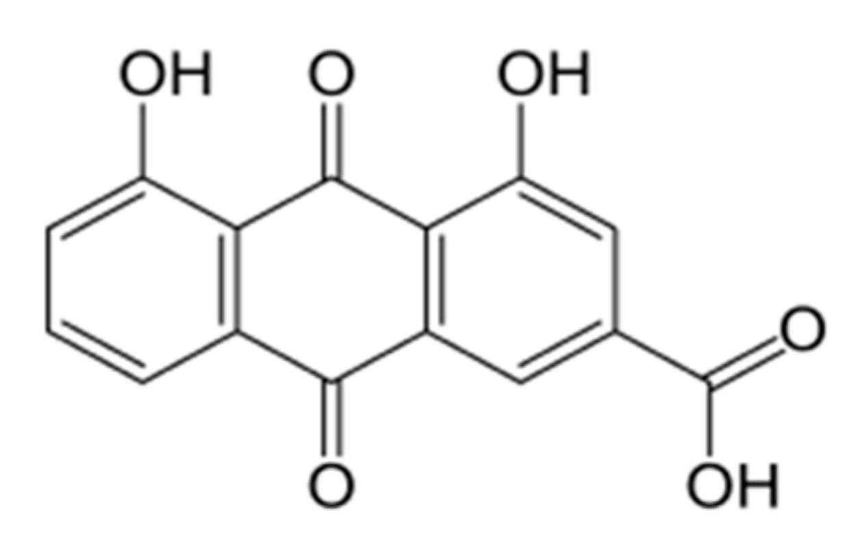
2.1. Compound Preparation

Rhein (purity 99%) was acquired from Sigma-Aldrich (St. Louis, MI, USA) and dissolved in 100% dimethyl sulfoxide (DMSO) (Sigma-Aldrich, St. Louis, MI, USA) at 2 mg/mL.

2.2. Characterization of the Bacterial Strains

In the present study, the tested bacteria were the standard S. aureus strains (ATCC 6538, ATCC 1167) and the respective clinical isolates, including multisensitive, quinolone macrolides, methicillin-resistant strains and beta-lactamase producers (C1-5) (Table 1). The standard strain was purchased from the American Type Culture Collection (ATCC, Manassas, VA, USA), whereas the clinical isolates were isolated from different anatomical districts at the Laboratory of Microbiology of the Luigi Vanvitelli University Hospital. The clinical isolates were isolated from different anatomical districts (blood, skin, pharynx, bladder and eye) at the Laboratory of Microbiology of the Luigi Vanvitelli University Hospital. Samples were plated on Columbia CNA Agar with 5% Sheep Blood (BioMerieux, Marcy-l’Étoile, France) and incubated overnight at 37 °C. Bacterial identification and susceptibility tests were conducted using the Phoenix BD system (Becton Dickinson, Franklin Lakes, NJ, USA). In short, a 0.5 McFarland (ID) bacterial inoculum was set up, using a Phoenix spectrophotometer. A 25 μL volume of ID was added to Phoenix AST Broth with Phoenix AST Indicator. ID and AST were loaded into the Phoenix panels and subsequently filed into the Phoenix system. After 16 h of incubation, bacterial identification and relative antibiograms were obtained.

2.3. Bacterial Growth Conditions

S. aureus strains were grown in Mueller Hinton (MH) and Luria Bertani (LB) media (Oxoid, Basingstoke, NH, USA) at 37 °C under aerobic conditions. To achieve a bacterial inoculum suitable for antibacterial tests, fresh colonies of each bacterial strain were inoculated in MH and LB broth and incubated at 37 °C under stirring at 180 rpm overnight. The inoculum was diluted in a fresh medium and incubated until the exponential phase was reached. Dilutions were performed to obtain the bacterial load suitable for the assays (2 × 10⁵ CFU/mL).

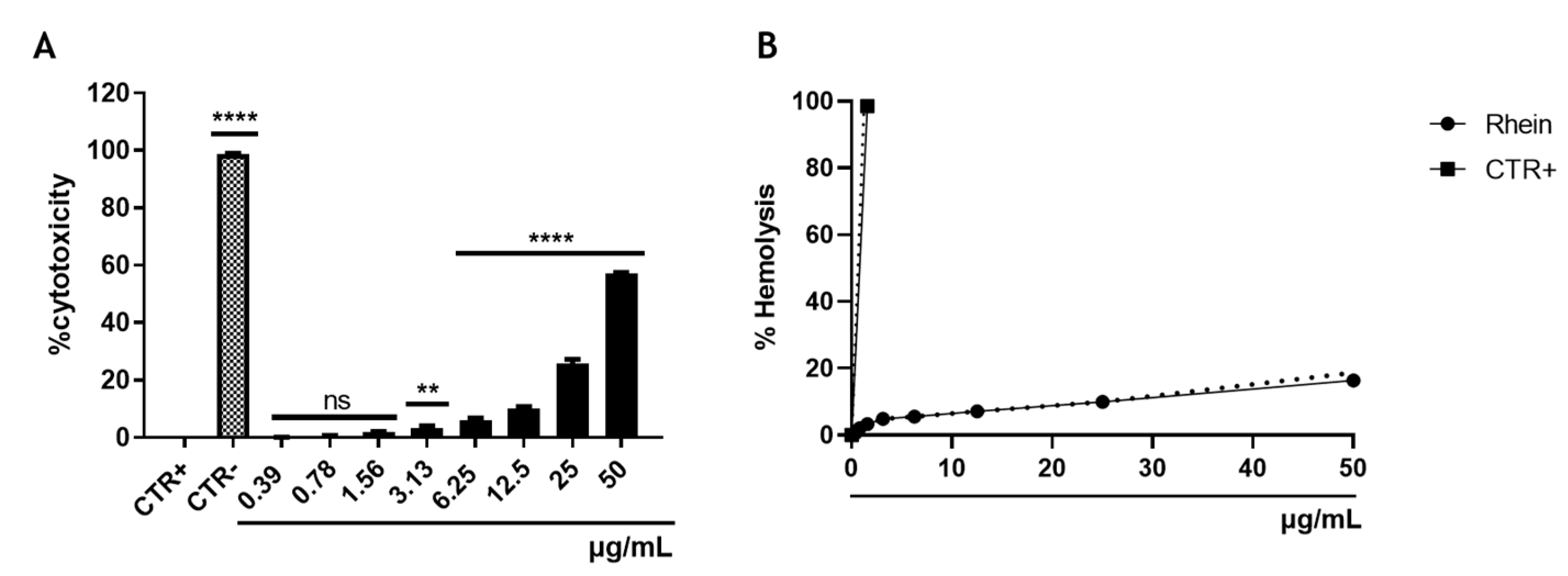
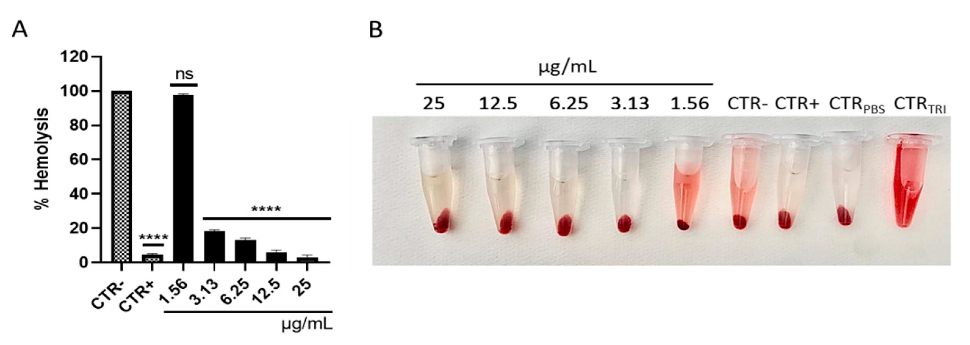
2.4. Hemolysis Assay

Rhein was tested on human erythrocytes derived from a healthy individual with 0 negative blood. The human blood sample was centrifuged at 1500 g for 5 min and the plasma was removed. The pellet was washed 5 times with the TBS solution (50 Mm Tris-HCl at pH 7.6 and 0.15 M NaCl) and diluted 10-fold using the same solution. The assay was conducted in a 96-well microplate (Thermo Scientific, Waltham, MA, USA). A volume of 50 µL of red cell suspension was added to 50 µL of the compound in the range of concentrations of 50–0.39 µg/mL and incubated at 37 °C for 1 h. The compound solvent and 0.1% Triton X-100 were used as the CTR- and CTR+ control, respectively. Finally, the plate was centrifuged at 1500 g for 5 min at room temperature, and 50 μL of supernatant from each well was transferred to a new 96-well plate and used to obtain the absorbance of the hemoglobin released at 540 nm. The hemolysis rate was calculated using the following formula:
%   Hemolysis = Abs   540   nm   of   the   test   sample   Abs   540   nm   of   CTR ( Abs   540   nm   of   CTR + Abs   540   nm   of   CTR × 100

2.5. Cell Cytotoxicity Assay

The cytotoxicity of Rhein towards human bronchial epithelial cells (BEAS-2B cells) was evaluated by the 3-(4,5-dimethylthiazol-2-yl)-2,5-diphenyl tetrazolium bromide (MTT) assay. Cells were seeded at a density of 2 × 10⁴ cells/well of a 96-well plate and incubated at 37 °C with 5% CO2 in a moist environment. BEAS-2B cells were exposed to increasing concentrations of Rhein (50–0.39 μg/mL) for 20 h. The compound solvent and 100% DMSO constituted the CTR- and CTR+, respectively. After exposure to the compound, 100 μL of MTT solution (Sigma-Aldrich, St. Louis, MI, USA) (0.3 mg/mL) was added to each well for 3 h at 37 °C. Thereafter, the solution was removed, and the formazan crystals were solubilized with 100 μL of 100% DMSO. The absorbance at 570 nm was measured by a microplate reader (Tecan, Männedorf, Switzerland), and the percentage of cytotoxicity was obtained according to the following formula:
%   Cytotoxicity = 100 100 ×   Abs   570   nm   of   test   sample Abs   570   nm   of   CTR +

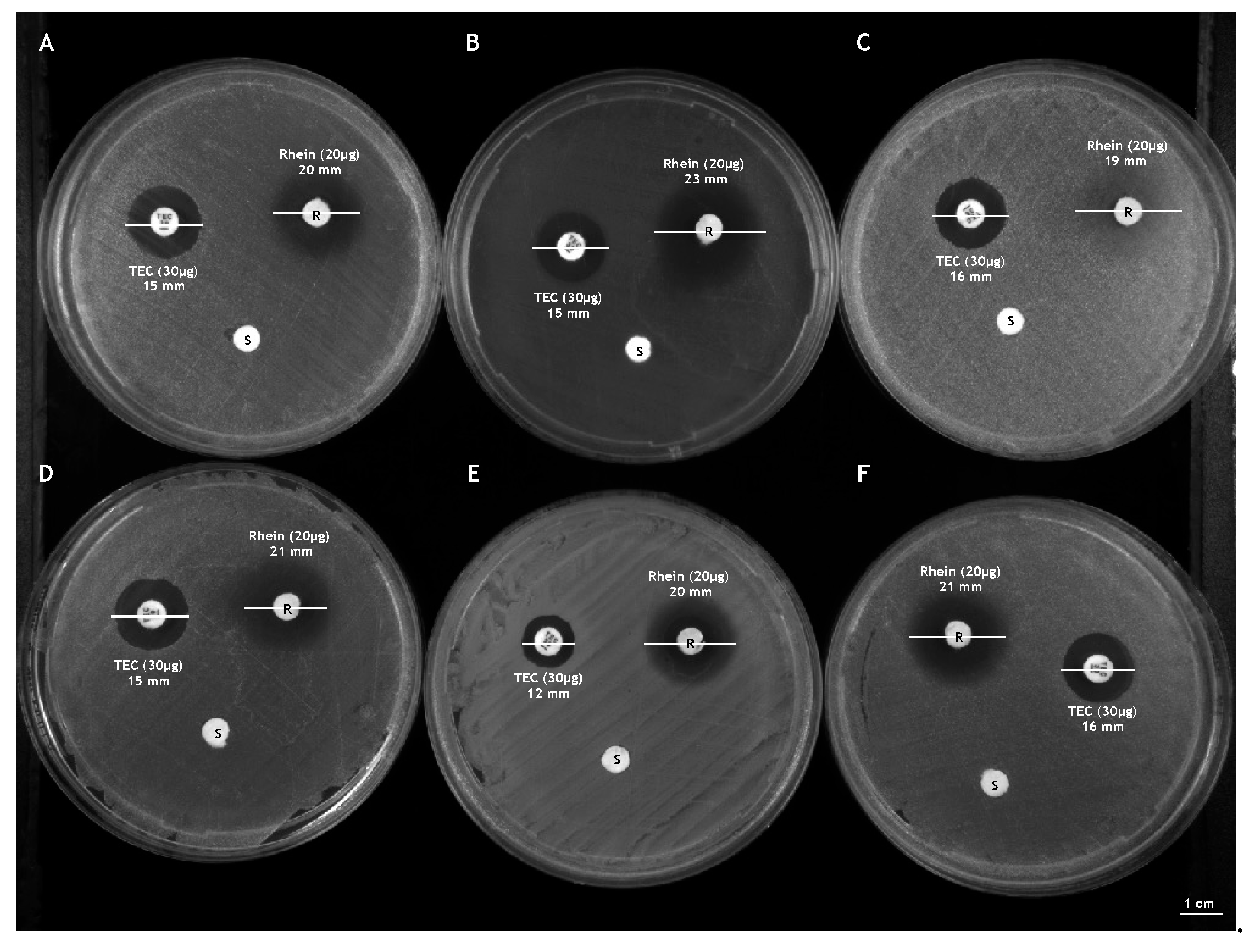
2.6. Kirby-Bauer Disk Diffusion Test

A preliminary antibacterial evaluation of Rhein was performed through the Kirby-Bauer disk diffusion test. Briefly, all bacterial inocula of 0.5 McFarland were evenly plated on Mueller Hinton agar plates (Oxoid, Basingstoke, NH, USA). A paper disk was impregnated with 20 μg of Rhein and placed on an agar plate. A Teicoplanin disk (30 μg) and a disc containing compound solvent were used as CTR+ and CTR−, respectively. The plates were incubated at 37 °C overnight and the diameters of the inhibition zones were measured. The antibacterial potential of the compound was expressed in millimeters (diameter of the inhibition area ± SD).

2.7. Antibacterial Susceptibility Assays

The broth microdilution method was used to determine the MIC values of Rhein against standard and clinical isolated S. aureus strains, according to the Clinical and Laboratory Standards Institute (CLSI). Assays were carried out in 96-well plates (BD Biosciences) for a final test volume of 100 μL. The compound was diluted to obtain concentrations from 50 to 0.39 µg/mL and a bacterial inoculum of 2 × 10⁵ CFU/mL was prepared. The latter was incubated with the test compounds at 37 °C for 20 h under agitation at 180 rpm overnight. Vancomycin and solvent compound were used as CTR+ and CTR−, respectively. The turbidity was measured via a microplate reader (Tecan, Männedorf, Swiss). The percentage of growth inhibition was achieved using the following formula:
%   Growth   inhibition = 100 100 ×   Abs   600   nm   of   the   test   sample Abs   600   nm   of   CTR

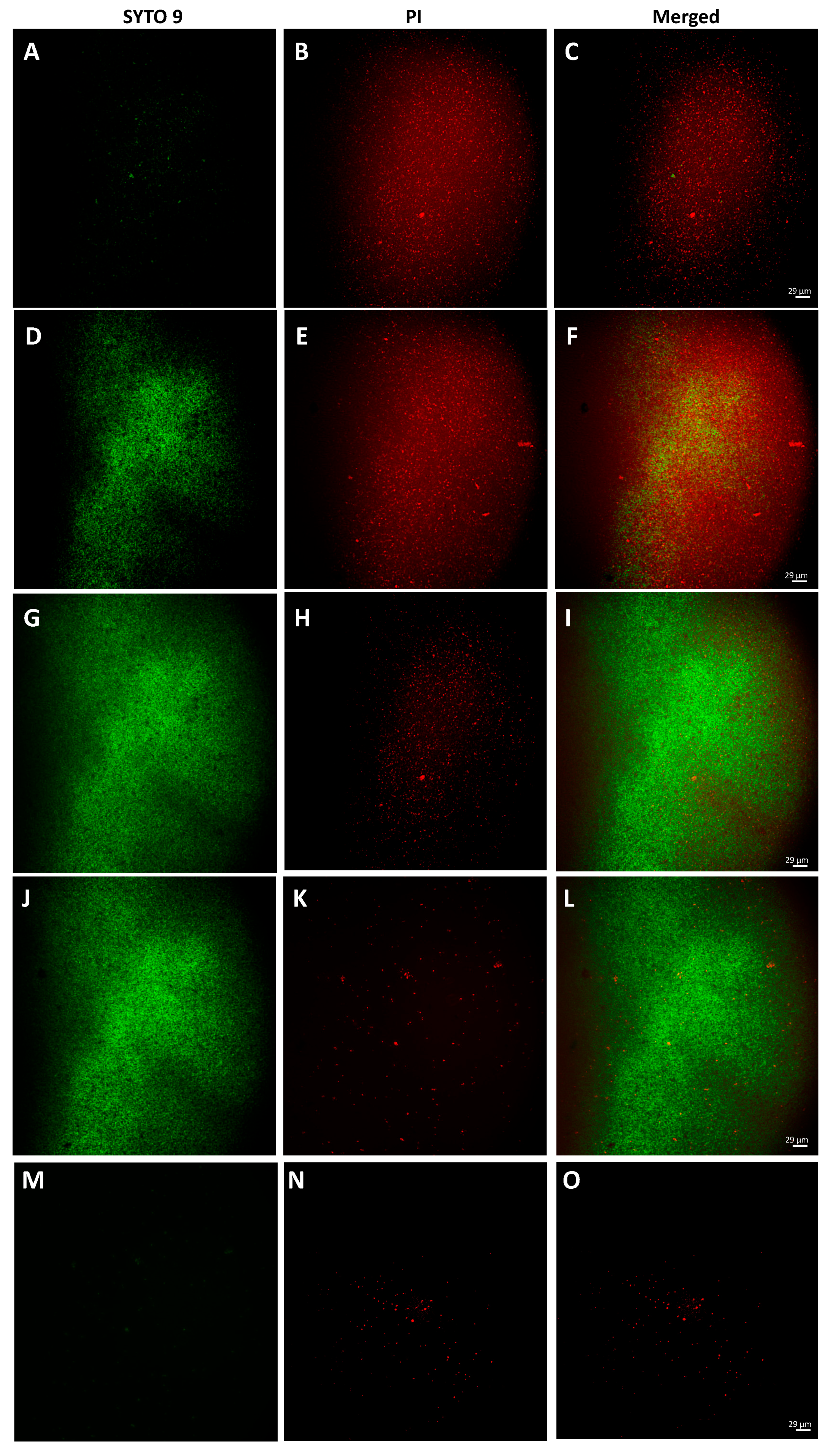
2.8. Bacterial Live/Dead Assay

Fluorescence microscopy (Nikon ECLIPSE Ti2-U, Amsterdam, The Netherlands) was used to visualize live and dead cells using the LIVE/DEAD BacLight Bacterial Viability Kit (Life Technologies, Carlsbad, CA, USA). The assay uses a mixture of SYTO 9 fluorescent dyes and propidium iodide (PI). SYTO 9 green, fluorescent dye crosses intact and damaged cell membranes binds nucleic acids and causes cells to become fluorescent green when excited by a laser with a wavelength of 485 nm. Furthermore, PI red fluorescent dye only passes damaged membranes and binds with greater affinity to nucleic acids than SYTO 9. In accordance with the manufacturer’s instructions, a volume of 1.5 μL of both Syto-9 and PI was added to each well of the 96-well plate (1 × 10⁵ CFU/mL). The plate was incubated in the dark at room temperature for 15 min and then subjected to analysis. Images were acquired via fluorescence microscope with beam settings for FITC, TRITC and merged.

2.9. Determination of the Selectivity Index (SI)

The SI of the Rhein was calculated through the ratio between EC50 and MIC50. Values greater than 3 render the compound rising for in vivo investigation.

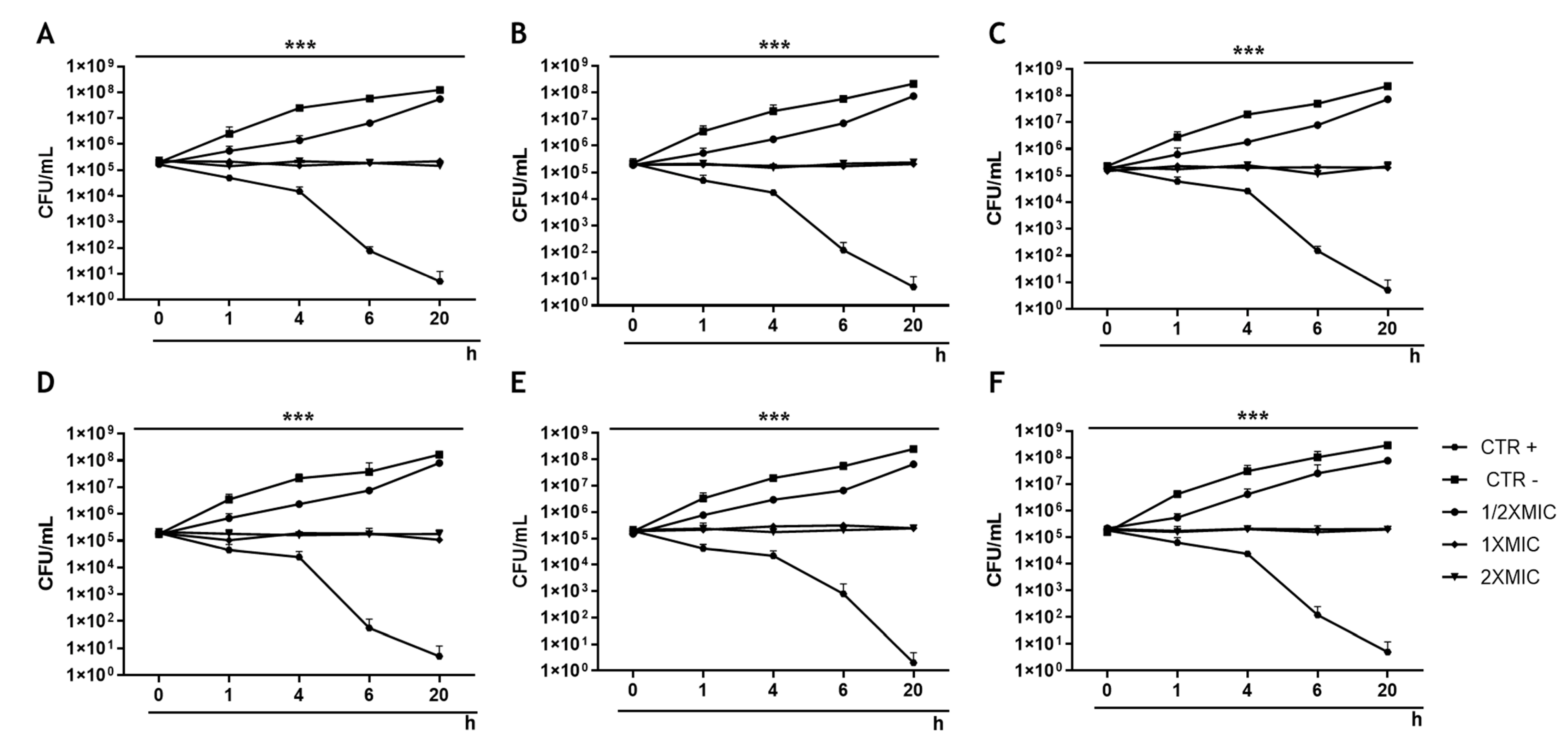
2.10. Time-Killing Assays

The antibacterial effect was further assessed through the time–kill curve examinations. Concentrations of ½ × MIC (6.25 µg/mL), 1 × MIC (12.5 µg/mL) and 2 × MIC (25 µg/mL) were set up for a final volume of 2 mL/tube. Bacteria treated with vancomycin and the compound solvent were used as CTR+ and CTR−, respectively. A bacterial suspension of 2 × 10⁵ CFU/mL was added to each tube and incubated at 37 °C. Aliquots of 100 µL were collected at times 0 and after 1, 4, 6 and 20 h of exposure and serially diluted in 1× phosphate-buffered saline (1XPBS). The dilutions were plated on MH agar, and the plates were incubated at 37 °C overnight. Colonies were counted and values were reported in CFU/mL.

2.11. Measurement of S. aureus Hemolytic Activity

Hemolytic activity of α-toxin was evaluated in response to Rhein treatment. The bacterial inoculum of S. aureus ATCC 6538 with a load of 2 × 10⁵ CFU/mL was prepared and treated with Rhein at concentrations of 4 × MIC (50 µg/mL), 2 × MIC (25 µg/mL), 1 × MIC (12.5 µg/mL), ½ × MIC (6.25 µg/mL) and ¼ × MIC (3.13 µg/mL) for 20 h. After incubation, the treated and untreated bacterial suspensions with the compound were centrifuged at 12,000 rpm for 5 min. A 100 µL volume of the supernatant was collected and incubated with 25 µL of previously washed human erythrocytes at 37 °C for 1 h. The supernatant derived from the untreated and vancomycin-treated bacterial culture were used as CTR− and CTR+, respectively. Erythrocytes treated with 1XPBS represented technical CTR−, while 0.1% Triton X-100 was used as CTR+. The suspensions were centrifuged at 3000 rpm for 5 min and the supernatants were examined, evaluating the absorbance at 450 nm. The hemolysis rate was calculated by comparison with CTR− (100% hemolysis).

2.12. H2O2 Sensitivity Assay

The effect of Rhein on S. aureus catalase production was evaluated by the H2O2 sensitivity test. A 0.5 McFarland bacterial inoculum was prepared using a Phoenix spectrophotometer. The bacterial suspension was uniformly seeded with a sterile swab on agar MH, supplemented with and without compound at MIC concentration. A sterile filter paper disc (Hi-Media, Maharashtra, India) was soaked with 20 µL of 0.18 % H2O2 and placed on the inoculated plate. The latter was incubated at 37 °C for 16 h, and the clearance zone diameters were measured and compared.

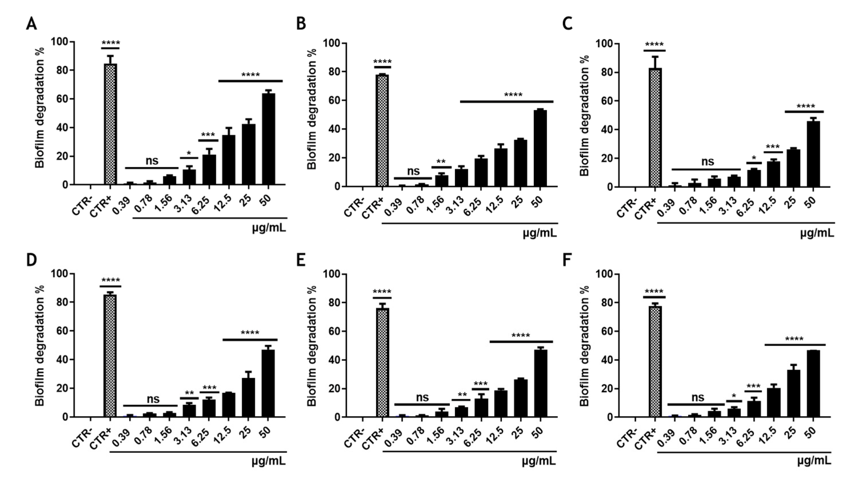
2.13. Biofilm Degradation Assay

The ability of Rhein to degrade mature biofilms was investigated by the crystal violet (CV) test. A bacterial suspension of 2 × 10⁸ CFU/mL in LB associated with 1% glucose was prepared, and a 100 µL aliquot was added to each well of a 96 well plate. The latter was incubated at 37 °C for 24 h in a static condition to allow the formation of a mature biofilm. After incubation, the planktonic cells were removed, the biomass washed with 1XPBS and treated with Rhein at the reported concentrations (0.39 to 50 μg/mL). Biofilms treated with solvent compound and vancomycin were, respectively, CTR͂− and CTR+ for the disruption of the biofilm. After 20 h of treatment, the biofilm was washed with 1XPBS, and the biomass was stained with 100 µL of 0.01% CV for 30 min at room temperature with stirring. The dye was removed, and the matrix was washed with 1XPBS. A 98% ethanol solution was used to solubilize the matrix for 40 min at room temperature under stirring. The absorbance measurement at 570 nm was obtained using a microplate reader (Tecan, Männedorf, Swiss) [27]. The minimum biofilm eradication concentration (MBEC) was calculated according to the following formula:
%   Biofilm   degradation = 1 Abs   570   nm   of   the   test   sample Abs   570   nm   of   CTR × 100

2.14. Statistic Analysis

The assays were conducted in biological and technical triplicate, and data were expressed as mean ± standard deviation (SD). The values of 50 and 90% bacterial growth inhibitory concentration (MIC50 and MIC90), 50% cytotoxic concentration (CC50) and 50% hemolysis concentration (EC50) were obtained from the dose–effect curves by analysis of non-linear regression using Graph Pad Prism 9.0 software (San Diego, CA, USA). The significance of the difference between the treated samples and the CTR− was obtained with the Dunnett test as post hoc by the Graph Pad Prism 9.0 software (San Diego, CA, USA). The p-value < 0.05 was regarded as significant.

3. Results

3.1. Cytotoxicity of Rhein

Rhein cytotoxicity was assessed on BEAS-2B cells and erythrocytes via MTT and hemolysis tests, respectively. The impact of Rhein on BEAS-2B cells is shown in Figure 2A. Rhein induced cytotoxicity through a dose-dependent trend. The compound induced a death rate of 57.2% at the highest concentration tested and exhibited a CC50 value of 43.6 µg/mL. The 100% DMSO, used as CTR+, caused a cytotoxicity rate of 98.7% (Figure 2A). The hemolysis assay showed that the natural product exhibited less than 16.4% hemolytic activity at a concentration of 50 µg/mL. The recorded hemolysis rate is below 10% at concentrations equal to and less than 25 µg/mL. Therefore, EC50 value was greater than 50 µg/mL. The 0.1% Triton X-100, used as a positive control (CTR+), resulted in an average hemolysis rate of 99% (Figure 2B).

3.2. Effect of Rhein on the Growth of S. aureus

Rhein’s antibacterial activity was assessed by Kirby-Bauer disk diffusion, plate microdilution, LIVE/DEAD and time-killing tests. All methods showed an alteration in the growth of S. aureus in response to treatment with Rhein. The diameters of inhibition induced by 20 μg of Rhein were 20 ± 0.21, 23 ± 0.36, 19 ± 0.34, 21 ± 0.38, 20 ± 0.26 and 21 ± 0.26 mm for S. aureus ATCC 6538, multisensitive, methicillin, quinolone, macrolides resistant strains and beta-lactamase producer, respectively. A lower inhibition area was induced by 30 μg of Teicoplanin (12–16 ± 0.39 mm). Contrarily, no halo of inhibition was shown by treating the bacteria with the compound solvent (Figure 3). Through the plate microdilution method, S. aureus strains were sensitive to the compound at concentrations equal to and greater than 12.5 μg/mL. No significant changes in bacterial growth were found at concentrations below 1.56 μg/mL. The values of MIC50 and MIC90 were 4.83 and 9.20 μg/mL, 4.35 and 9.31 μg/mL, 4.45 and 8.45 μg/mL, 4.64 and 9.27 μg/mL, 4.32 and 9.16 μg/mL and 3.22 and 8.13 μg/mL for S. aureus ATCC 6538, multisensitive, methicillin, quinolone, macrolides resistant strains and beta-lactamase producer, respectively. The calculated SI values were greater than 10 for all strains, suggesting Rhein as a favorable compound for in vivo investigations (Figure 4). Rhein’s antibacterial property was further investigated through the LIVE/DEAD assay. To assess Rhein-induced cell damage, the standard S. aureus strain was stimulated with the compound at concentrations of 25 (2 × MIC), 12.5 (1 × MIC), 6.25 (½ × MIC) ug/mL for 20 h and analyzed by fluorescence microscope after exposure with PI and SYTO 9. The bacterial populations treated with Rhein at concentrations of 25 and 12.5 μg/mL exhibited a deep red color compared with the untreated bacteria, confirming the dose-dependent cell damage obtained in the previous assay. A decrease in cell damage and an increase in live cells were found in response to treatment with Rhein at a concentration of 6.25 µg/mL. No viable cells were found after treatment with Vancomycin, used as CTR+ (Figure 5). Rhein action kinetics were evaluated through time-killing tests. Bacterial exposure to Rhein ½ × MIC did not cause significant changes in the growth curve compared with untreated bacteria after 20 h. Bacterial growth block occurred by treating the bacteria with 1 × MIC and 2 × MIC of Rhein, indicating bacteriostatic action (Figure 6).

3.3. Rhein Effect on the Main Virulence Factors of S. aureus

Staphylococcal hemolysins and catalase are important virulence factors that promote bacterial invasion. The effect of Rhein on alpha-hemolysin and catalase of S. aureus ATCC 6538 was evaluated through a hemolytic activity test and H2O2 sensitivity assay, respectively. The results show that the hemolytic activity of S. aureus significantly decreased in response to treatment with Rhein up to the concentration of 3.13 µg/mL compared with CTR-. In detail, a 1-, 5.3-, 7.1-, 14.3- and 25-fold reduction in hemolytic activity occurred by treating the bacterial strain with 1.56, 3.13, 6.25, 12.5 and 25 µg/mL of Rhein, respectively (Figure 7). Treatment with Rhein increases the susceptibility of S. aureus to H2O2. The halo of inhibition resulting from sensitivity to H2O2 in the plate with 6.25 and 3.13 µg/mL was 36 ± 0.31 and 32 ± 0.36 mm. An increase in H2O2 sensitivity of 0.96- and 1.16-fold occurred in response to treatment with Rhein at concentrations of 3.13 and 6.25 µg/mL, respectively (Figure 8).

3.4. Effect of Rhein on S. aureus Biofilm Biomass

Due to the antibiotic-resistant nature of biofilms, conventional antibiotics are inefficient for treating biofilm-associated infections. Therefore, new compounds capable of disrupting the biofilm are needed to render the bacteria non-tolerant to the treatment. The results show that Rhein significantly reduced the biomass of mature biofilms in the concentration range of 50 to 3.13 µg/mL. In detail, treatment with Rhein at 50 µg/mL disrupted S. aureus ATCC 1167, multisensitive, quinolone, macrolides and methicillin-resistant strains and beta-lactamase producers biofilm biomass by 63.8, 52.9, 50, 47.1, 47.4 and 46.9%, respectively. A matrix reduction of 42.7, 32.7, 26.4, 27.3, 26.4 and 33.1 % occurred for the same strains after exposure to 25 µg/mL of Rhein. The compound at MIC concentration caused the degradation of the biofilm produced by the standard multisensitive S. aureus ATCC 1167, by the multisensitive, quinolone, macrolides and methicillin-resistant and producing beta-lactamase strains of 34.7, 26.7, 17.8, 27.3, 18.9 and 20.5%, respectively. Residual biomass greater than 78.8 % remained after exposure with the compound at the dose of 6.25 µg/mL. Biofilm disintegration of less than 12.3% occurred after treatment with Rhein at 3.13 µg/mL (Figure 9).

4. Discussion

S. aureus is one of the most common pathogens responsible for community and hospital-acquired infections [28,29]. Currently, the increase in S. aureus antibiotic resistance is threatening the effective use of the latest-generation antibiotics such as linezolid, daptomycin and glycopeptides [30]. Therefore, research and development of new antibacterial agents to limit the spread of S. aureus antibiotic resistance has become a necessity [31]. Natural products are a rich source of antimicrobial compounds. They have an important role in the discovery of antimicrobial leads and have contributed to the development of most drugs currently in use [32]. Our study assessed the antimicrobial potential of the natural compound, Rhein, against S. aureus strains. The antibacterial assays through the Kirby-Bauer disk diffusion test allowed to obtain preliminary data on the antibacterial efficiency of the compound. In our study, Rhein exhibited remarkable bacteriostatic activity against standard and clinical isolated S. aureus strains with MIC90 values ranging from 8.13 to 9.31 μg/mL. Based on antibacterial data and light cytotoxic activity on human erythrocytes at the highest tested concentration (16.4%), the calculated SI values were greater than 10, indicating this compound is suitable for in vivo studies. A significant amount of evidence has reported the antibacterial activity of Rhein on different bacterial strains. Our previous study showed that this compound altered the growth of S. mutans, showing MIC90 values of 5.69 µg/mL. Moreover, SEM data revealed that the natural product affected the integrity of the wall, inducing the formation of irregular, rough and non-uniform surfaces [23]. Yu et al. proved that Rhein exhibited a bacteriostatic action on S. aureus at a concentration of 8 μg/mL, impairing the gene expression involved in anaerobic respiration, fermentation, iron-regulated surface determinants and ribonucleotide reductase systems [33]. Additionally, Wu et al. documented the antibacterial efficiency of Rhein against Salmonella enterica, detecting MIC values of 250 μg/mL [34]. The cited studies show a varied repertory of Rhein antibacterial features, presumably due to the different bacterial strains tested, growth conditions and assays used.
Staphylococcal hemolysins and catalases represent important virulence factors that participate in and promote the bacterial invasion process [35]. Alpha-toxin is a monomer released during exponential growth and is recognized as the main virulence factor of S. aureus. It oligomerizes in the host erythrocyte membranes, causing the formation of pores and subsequent osmotic lysis and cell death [36]. On the other side, catalase contributes to the intracellular persistence of S. aureus within macrophages during infection by inactivating H2O2 in the phagocytosis process [37]. During the infectious process, host tissue damage is induced by virulence factors secreted during bacterial replication [38,39]. Moreover, numerous bacterial toxins stimulate the immune system, resulting in the release of cytokines that cause further tissue damage [40]. Currently, exposure to antibacterial agents leads to the elimination of pathogens but not the determinants of virulence [41]. Although bacterial clearance does occur, eliminating pathogens is often insufficient to prevent tissue damage. Identifying antibacterial agents that affect the release and/or activity of toxins and enzymes is necessary to block host damage upon elimination of the etiological agent [42]. Our finding showed that Rhein alters the hemolytic capacity of S. aureus up to the concentration of 3.13 μg/mL. Furthermore, the catalase activity was reduced by 0.96 times at the same concentration. No evidence was reported on the impact of Rhein on alpha-hemolysin and catalase activity. To date, this compound has been shown to compromise the system of iron-regulated surface determinants, responsible for the intracellular accumulation of heme molecules [33].
Mature biofilm is considered a highly organized ecosystem that contributes to the onset of chronic infections through the continuous release of planktonic bacterial forms and virulence factors. Biofilm bacteria have a 10 times higher survival rate than planktonic bacteria [43]. Antibiotic tolerance can be reversed through the breakdown of the extracellular matrix and the release of bacteria, which will then be available for antibacterial treatment [44]. Our results report Rhein’s ability to disrupt mature biofilms with an average rate of 31.4 and 24% at 2 × MIC and 1 × MIC concentrations. No studies have reported data on the degradation of mature S. aureus biofilm. Our past results has reported Rhein’s ability to disrupt S. mutans biofilm with minimal biofilm eradication concentrations inducing 50% values of 6.31 μg/mL [23].
Several evidence has demonstrated the impact of Rhein on the activity of NADH type II: quinone oxidoreductase (NDH-2) [45]. This enzyme catalyzes the oxidation of NADH and the reduction in membrane quinone, resulting in the formation of NAD+ [46]. Nguyen et al. proved that treatment of Cutibacterium acnes with Rhein (6.25 µg/mL) completely inhibited the activity of NDH-2 [24]. This increased the (NADH)/(NAD+) ratio, with a negative impact on the glycolytic pathway and the production of ATP and pyruvate [47]. Goodwine et al. documented the role of pyruvate in the biofilm structure of Pseudomonas aeruginosa. The decrease in pyruvate caused a loss of biomass with consequent alteration of biofilm integrity [48]. Schurig-Briccio et al. showed that the hemolytic and catalase activities of S. aureus require NDH-2. NDH-2 knockout strains showed drastically reduced hemolytic activity compared with the wild-type strain due to the failing production of α-toxin. Moreover, NDH-2 knockout reduced the susceptibility to hydrogen peroxide compared with wild-type conditions [49].
Our findings reported the effective bacteriostatic activity of Rhein against standard and clinical isolated S. aureus. This strain invades and replicates within many cell types, evading the immune system through the action of α-hemolysin and catalase. The first results in the death of host cells, promoting host damage, whereas catalase allows bacterial survival in macrophages, avoiding the phagocytosis process. Our studies showed that Rhein at sub-MIC concentrations interferes with the action of α-hemolysin and catalase, thereby reducing the virulence and pathogenicity of S. aureus. Moreover, biofilm formation protects S. aureus strains against antibiotics and hosts’ defense molecules. Interestingly, Rhein disrupted the biofilm matrix, rendering the bacteria susceptible to antibiotic treatment and the action of immunity determinants. These results support Rhein as a potential antibacterial agent with antivirulence and antibiofilm activity. Therefore, Rhein could represent a suitable therapy strategy for the treatment of S. aureus infections. Additional investigation will be needed to better understand the mechanism underlying Rhein’s action against S. aureus.

Author Contributions

Conceptualization and writing—review and editing, V.F.; methodology, F.D.; software, F.P.; validation, G.S. and A.M.; visualization, E.F. and V.C.; visualization, A.D.F.; project administration, M.G. and G.F. All authors have read and agreed to the published version of the manuscript.

Funding

This research received no external funding.

Institutional Review Board Statement

Not applicable.

Informed Consent Statement

Not applicable.

Data Availability Statement

Not applicable.

Conflicts of Interest

The authors declare no conflict of interest.

References

  1. Horn, C.M.; Kielian, T. Crosstalk between Staphylococcus aureus and Innate Immunity: Focus on Immunometabolism. Front. Immunol. 2021, 11, 621750. [Google Scholar] [CrossRef] [PubMed]
  2. de Jong, N.W.M.; van Kessel, K.P.M.; van Strijp, J.A.G. Immune Evasion by Staphylococcus aureus. Microbiol. Spectr. 2019, 7. [Google Scholar] [CrossRef] [PubMed]
  3. Raineri, E.J.M.; Altulea, D.; van Dijl, J.M. Staphylococcal trafficking and infection—From ‘nose to gut’ and back. FEMS Microbiol. Rev. 2021, 46, fuab041. [Google Scholar] [CrossRef] [PubMed]
  4. Tong, S.Y.C.; Davis, J.S.; Eichenberger, E.; Holland, T.L.; Fowler, V.G., Jr. Staphylococcus aureus Infections: Epidemiology, Pathophysiology, Clinical Manifestations, and Management. Clin. Microbiol. Rev. 2015, 28, 603–661. [Google Scholar] [CrossRef] [PubMed]
  5. Cheung, G.Y.C.; Bae, J.S.; Otto, M. Pathogenicity and virulence of Staphylococcus aureus. Virulence 2021, 12, 547–569. [Google Scholar] [CrossRef] [PubMed]
  6. Álvarez, A.; Fernández, L.; Gutiérrez, D.; Iglesias, B.; Rodríguez, A.; García, P. Methicillin-Resistant Staphylococcus aureus in Hospitals: Latest Trends and Treatments Based on Bacteriophages. J. Clin. Microbiol. 2019, 57, e01006-19. [Google Scholar] [CrossRef] [PubMed]
  7. Diekema, D.J.; Hsueh, P.-R.; Mendes, R.E.; Pfaller, M.A.; Rolston, K.V.; Sader, H.; Jones, R.N. The Microbiology of Bloodstream Infection: 20-Year Trends from the SENTRY Antimicrobial Surveillance Program. Antimicrob. Agents Chemother. 2019, 63, e00355-19. [Google Scholar] [CrossRef]
  8. Asokan, G.V.; Ramadhan, T.; Ahmed, E.; Sanad, H. WHO Global Priority Pathogens List: A Bibliometric Analysis of Medline-PubMed for Knowledge Mobilization to Infection Prevention and Control Practices in Bahrain. Oman Med. J. 2019, 34, 184–193. [Google Scholar] [CrossRef]
  9. Dweba, C.C.; Zishiri, O.T.; El Zowalaty, M. Methicillin-resistant Staphylococcus aureus: Livestock-associated, antimicrobial, and heavy metal resistance. Infect. Drug Resist. 2018, 11, 2497–2509. [Google Scholar] [CrossRef]
  10. Guo, Y.; Song, G.; Sun, M.; Wang, J.; Wang, Y. Prevalence and Therapies of Antibiotic-Resistance in Staphylococcus aureus. Front. Cell. Infect. Microbiol. 2020, 10, 107. [Google Scholar] [CrossRef] [Green Version]
  11. Terreni, M.; Taccani, M.; Pregnolato, M. New Antibiotics for Multidrug-Resistant Bacterial Strains: Latest Research Developments and Future Perspectives. Molecules 2021, 26, 2671. [Google Scholar] [CrossRef] [PubMed]
  12. Wu, Q.; Sabokroo, N.; Wang, Y.; Hashemian, M.; Karamollahi, S.; Kouhsari, E. Systematic review and meta-analysis of the epidemiology of vancomycin-resistance Staphylococcus aureus isolates. Antimicrob. Resist. Infect. Control 2021, 10, 1–13. [Google Scholar] [CrossRef] [PubMed]
  13. Shariati, A.; Dadashi, M.; Chegini, Z.; Van Belkum, A.; Mirzaii, M.; Khoramrooz, S.S.; Darban-Sarokhalil, D. The global prevalence of Daptomycin, Tigecycline, Quinupristin/Dalfopristin, and Linezolid-resistant Staphylococcus aureus and coagulase–negative staphylococci strains: A systematic review and meta-analysis. Antimicrob. Resist. Infect. Control 2020, 9, 1–20. [Google Scholar] [CrossRef] [PubMed]
  14. Bender, J.K.; Cattoir, V.; Hegstad, K.; Sadowy, E.; Coque, T.M.; Westh, H.; Hammerum, A.M.; Schaffer, K.; Burns, K.; Murchan, S.; et al. Update on prevalence and mechanisms of resistance to linezolid, tigecycline and daptomycin in enterococci in Europe: Towards a common nomenclature. Drug Resist. Updat. 2018, 40, 25–39. [Google Scholar] [CrossRef]
  15. Dell’Annunziata, F.; Folliero, V.; Giugliano, R.; De Filippis, A.; Santarcangelo, C.; Izzo, V.; Daglia, M.; Galdiero, M.; Arciola, C.; Franci, G. Gene Transfer Potential of Outer Membrane Vesicles of Gram-Negative Bacteria. Int. J. Mol. Sci. 2021, 22, 5985. [Google Scholar] [CrossRef] [PubMed]
  16. Vaou, N.; Stavropoulou, E.; Voidarou, C.; Tsigalou, C.; Bezirtzoglou, E. Towards Advances in Medicinal Plant Antimicrobial Activity: A Review Study on Challenges and Future Perspectives. Microorganisms 2021, 9, 2041. [Google Scholar] [CrossRef]
  17. Silver, L.L. Challenges of Antibacterial Discovery. Clin. Microbiol. Rev. 2011, 24, 71–109. [Google Scholar] [CrossRef]
  18. Joray, M.B.; González, M.L.; Palacios, S.M.; Carpinella, M.C. Antibacterial Activity of the Plant-Derived Compounds 23-Methyl-6-O-desmethylauricepyrone and (Z,Z)-5-(Trideca-4,7-dienyl)resorcinol and Their Synergy with Antibiotics against Methicillin-Susceptible and -Resistant Staphylococcus aureus. J. Agric. Food Chem. 2011, 59, 11534–11542. [Google Scholar] [CrossRef]
  19. Obiang-Obounou, B.W.; Kang, O.-H.; Choi, J.-G.; Keum, J.-H.; Kim, S.-B.; Mun, S.-H.; Shin, D.-W.; Kim, K.W.; Park, C.-B.; Kim, Y.-G.; et al. The mechanism of action of sanguinarine against methicillin-resistant Staphylococcus aureus. J. Toxicol. Sci. 2011, 36, 277–283. [Google Scholar] [CrossRef]
  20. Zhang, X.; Sun, X.; Wu, J.; Wu, Y.; Wang, Y.; Hu, X.; Wang, X. Berberine Damages the Cell Surface of Methicillin-Resistant Staphylococcus aureus. Front. Microbiol. 2020, 11, 621. [Google Scholar] [CrossRef]
  21. Wang, D.; Wang, X.-H.; Yu, X.; Cao, F.; Cai, X.; Chen, P.; Li, M.; Feng, Y.; Li, H.; Wang, X. Pharmacokinetics of Anthraquinones from Medicinal Plants. Front. Pharmacol. 2021, 12, 638993. [Google Scholar] [CrossRef] [PubMed]
  22. Zhou, Y.-X.; Xia, W.; Yue, W.; Peng, C.; Rahman, K.; Zhang, H. Rhein: A Review of Pharmacological Activities. Evid. Based Complement. Altern. Med. 2015, 2015, 578107. [Google Scholar] [CrossRef] [PubMed]
  23. Folliero, V.; Dell’Annunziata, F.; Roscetto, E.; Amato, A.; Gasparro, R.; Zannella, C.; Casolaro, V.; De Filippis, A.; Catania, M.R.; Franci, G.; et al. Rhein: A novel antibacterial compound against Streptococcus mutans infection. Microbiol. Res. 2022, 261, 127062. [Google Scholar] [CrossRef] [PubMed]
  24. Nguyen, A.T.; Kim, K.-Y. Rhein inhibits the growth of Propionibacterium acnes by blocking NADH dehydrogenase-2 activity. J. Med Microbiol. 2020, 69, 689–696. [Google Scholar] [CrossRef]
  25. Peerzada, Z.; Kanhed, A.M.; Desai, K.B. Effects of active compounds from Cassia fistula on quorum sensing mediated virulence and biofilm formation in Pseudomonas aeruginosa. RSC Adv. 2022, 12, 15196–15214. [Google Scholar] [CrossRef]
  26. Dell’Annunziata, F.; Martora, F.; Della Pepa, M.E.; Folliero, V.; Luongo, L.; Bocelli, S.; Guida, F.; Mascolo, P.; Campobasso, C.P.; Maione, S.; et al. Postmortem interval assessment by MALDI-TOF mass spectrometry analysis in murine cadavers. J. Appl. Microbiol. 2021, 132, 707–714. [Google Scholar] [CrossRef]
  27. Pironti, C.; Dell’Annunziata, F.; Giugliano, R.; Folliero, V.; Galdiero, M.; Ricciardi, M.; Motta, O.; Proto, A.; Franci, G. Comparative analysis of peracetic acid (PAA) and permaleic acid (PMA) in disinfection processes. Sci. Total Environ. 2021, 797, 149206. [Google Scholar] [CrossRef]
  28. Zheng, X.; Fang, R.; Wang, C.; Tian, X.; Lin, J.; Zeng, W.; Zhou, T.; Xu, C. Resistance Profiles and Biological Characteristics of Rifampicin-Resistant Staphylococcus aureus Small-Colony Variants. Infect. Drug Resist. 2021, 14, 1527–1536. [Google Scholar] [CrossRef]
  29. Taylor, T.A.; Unakal, C.G. Staphylococcus aureus. In StatPearls; StatPearls Publishing: Treasure Island, FL, USA, 2022. [Google Scholar]
  30. Lade, H.; Kim, J.-S. Bacterial Targets of Antibiotics in Methicillin-Resistant Staphylococcus aureus. Antibiotics 2021, 10, 398. [Google Scholar] [CrossRef]
  31. Saha, M.; Sarkar, A. Review on Multiple Facets of Drug Resistance: A Rising Challenge in the 21st Century. J. Xenobiotics 2021, 11, 197–214. [Google Scholar] [CrossRef]
  32. Schneider, Y. Bacterial Natural Product Drug Discovery for New Antibiotics: Strategies for Tackling the Problem of Antibiotic Resistance by Efficient Bioprospecting. Antibiotics 2021, 10, 842. [Google Scholar] [CrossRef] [PubMed]
  33. Yu, L.; Xiang, H.; Fan, J.; Wang, D.; Yang, F.; Guo, N.; Jin, Q.; Deng, X. Global transcriptional response of Staphylococcus aureus to Rhein, a Natural Plant Product. J. Biotechnol. 2008, 135, 304–308. [Google Scholar] [CrossRef] [PubMed]
  34. Food Research and Technology, the Hershey Company, PA, USA; Wu, X. Antibacterial activities of rhubarb extract and the bioactive compounds against Salmonella. Int. J. Nutr. Sci. Food Technol. 2015, 1, 1–9. [Google Scholar] [CrossRef]
  35. Thammavongsa, V.; Kim, H.K.; Missiakas, D.; Schneewind, O. Staphylococcal manipulation of host immune responses. Nat. Rev. Genet. 2015, 13, 529–543. [Google Scholar] [CrossRef] [PubMed]
  36. Tam, K.; Torres, V.J. Staphylococcus aureus Secreted Toxins and Extracellular Enzymes. Microbiol. Spectr. 2019, 7. [Google Scholar] [CrossRef] [PubMed]
  37. Das, D.; Bishayi, B. Contribution of Catalase and Superoxide Dismutase to the Intracellular Survival of Clinical Isolates of Staphylococcus aureus in Murine Macrophages. Indian J. Microbiol. 2010, 50, 375–384. [Google Scholar] [CrossRef] [PubMed]
  38. Singh, V.; Phukan, U.J. Interaction of host and Staphylococcus aureus protease-system regulates virulence and pathogenicity. Med. Microbiol. Immunol. 2018, 208, 585–607. [Google Scholar] [CrossRef]
  39. Folliero, V.; Dell’Annunziata, F.; Roscetto, E.; Cammarota, M.; De Filippis, A.; Schiraldi, C.; Catania, M.R.; Casolaro, V.; Perrella, A.; Galdiero, M.; et al. Niclosamide as a Repurposing Drug against Corynebacterium striatum Multidrug-Resistant Infections. Antibiotics 2022, 11, 651. [Google Scholar] [CrossRef]
  40. Marshall, J.S.; Warrington, R.; Watson, W.; Kim, H.L. An introduction to immunology and immunopathology. Allergy Asthma Clin. Immunol. 2018, 14, 1–10. [Google Scholar] [CrossRef]
  41. Hotinger, J.A.; Morris, S.T.; May, A.E. The Case against Antibiotics and for Anti-Virulence Therapeutics. Microorganisms 2021, 9, 2049. [Google Scholar] [CrossRef]
  42. Tomlinson, K.L.; Prince, A.S.; Lung, T.W.F. Immunometabolites Drive Bacterial Adaptation to the Airway. Front. Immunol. 2021, 12, 790574. [Google Scholar] [CrossRef] [PubMed]
  43. Muhammad, M.H.; Idris, A.L.; Fan, X.; Guo, Y.; Yu, Y.; Jin, X.; Qiu, J.; Guan, X.; Huang, T. Beyond Risk: Bacterial Biofilms and Their Regulating Approaches. Front. Microbiol. 2020, 11, 928. [Google Scholar] [CrossRef] [PubMed]
  44. Khatoon, Z.; McTiernan, C.D.; Suuronen, E.J.; Mah, T.-F.; Alarcon, E.I. Bacterial biofilm formation on implantable devices and approaches to its treatment and prevention. Heliyon 2018, 4, e01067. [Google Scholar] [CrossRef] [PubMed]
  45. Kim, D.; Kim, K.-Y. Combinatorial treatment of sophoraflavanone G and rhein with ampicillin, oxacillin, or oxytetracycline synergistically increased antibacterial activity against oral bacteria. Rev. Med Microbiol. 2021, 32, 211–218. [Google Scholar] [CrossRef]
  46. Sena, F.V.; Sousa, F.M.; Oliveira, A.S.F.; Soares, C.M.; Catarino, T.; Pereira, M.M. Regulation of the mechanism of Type-II NADH: Quinone oxidoreductase from S. aureus. Redox Biol. 2018, 16, 209–214. [Google Scholar] [CrossRef]
  47. Lencina, M.; Franza, T.; Sullivan, M.J.; Ulett, G.C.; Ipe, D.S.; Gaudu, P.; Gennis, R.B.; Schurig-Briccio, L.A. Type 2 NADH Dehydrogenase Is the Only Point of Entry for Electrons into the Streptococcus agalactiae Respiratory Chain and Is a Potential Drug Target. mBio 2018, 9, e01034-18. [Google Scholar] [CrossRef]
  48. Goodwine, J.; Gil, J.; Doiron, A.; Valdes, J.; Solis, M.; Higa, A.; Davis, S.; Sauer, K. Pyruvate-depleting conditions induce biofilm dispersion and enhance the efficacy of antibiotics in killing biofilms in vitro and in vivo. Sci. Rep. 2019, 9, 3763. [Google Scholar] [CrossRef]
  49. Schurig-Briccio, L.A.; Solorzano, P.K.P.; Lencina, A.M.; Radin, J.N.; Chen, G.Y.; Sauer, J.; Kehl-Fie, T.E.; Gennis, R.B. Role of respiratory NADH oxidation in the regulation of Staphylococcus aureus virulence. EMBO Rep. 2020, 21, e45832. [Google Scholar] [CrossRef]
Figure 1. Chemical structure of Rhein.
Applsci 12 08691 g001
Figure 2. Rhein cytotoxicity on BEAS-2B cell line, ****: p-value < 0.0001, **: p-value 0.0012, ns: not significant (A); and human erythrocytes. The dashed line indicates the DS trend (B).
Applsci 12 08691 g002
Figure 3. Inhibition zone against S. aureus ATCC 6538 (A), multisensitive (B), resistant to beta-lactams (C), resistant to quinolones (D), constitutively resistant to macrolides (E) and resistant to methicillin (F) strains.
Applsci 12 08691 g003
Figure 4. Antimicrobial activity of Rhein against S. aureus ATCC 6538, ****: p-value < 0.0001, ***: p-value 0.0006 *: p-value 0.0148 (A); multisensitive, ****: p-value < 0.0001, ns: not significant (B); resistant to beta-lactams, ****: p-value < 0.0001, ***: p-value 0.0001, **: p-value 0.0032 (C); resistant to quinolones, ****: p-value < 0.0001, **: p-value 0.0019, ns: not significant (D); constitutively resistant to macrolides, ****: p-value < 0.0001, **: p-value 0.0016, ns: not significant (E); resistant to methicillin strains, ****: p-value < 0.0001, *: p-value 0.0267 (F).
Applsci 12 08691 g004
Figure 5. LIVE/DEAD BacLight staining after treatment of S. aureus with Rhein analyzed under a fluorescence microscope. (AC) treatment with 25 μg/mL; (DF) treatment with 12.5 μg/mL; (GI) treatment with 6.25 μg/mL; (JL) bacteria not treated with Rhein; (MO) bacteria treated with Vancomycin (CTR+).
Applsci 12 08691 g005
Figure 6. Killing kinetics of Rhein on S. aureus ATCC 6538 (A), multisensitive (B), resistant to beta-lactams (C), resistant to quinolones (D), constitutively resistant to macrolides (E) and resistant to methicillin (F). ***: p-value = 0.0001.
Applsci 12 08691 g006
Figure 7. Efficacy of Rhein in inhibiting the hemolytic activity of S. aureus. (A) Quantitative analysis, ****: p-value < 0.0001, ns: non-significant; (B) qualitative analysis.
Applsci 12 08691 g007
Figure 8. Rhein effect on S. aureus catalase production. (A) untreated bacteria; (B) treatment with 6.25 μg/mL; (C) treatment with 3.13 μg/mL.
Applsci 12 08691 g008
Figure 9. Impact of 20-h treatment with Rhein on mature biofilms of S. aureus ATCC 1167, ****: p-value < 0.0001, ***: p-value 0.0003, *: p-value 0.0323; ns: non-significant (A); multisensitive, ****: p-value < 0.0001, **: p-value 0.0012, ns: not significant (B); resistant to beta-lactams, ****: p-value < 0.0001, ***: p-value 0.0002, *: p-value 0.0036, ns: not significant (C); resistant to quinolones, ****: p-value < 0.0001, ***: p-value 0.0003, **: p-value 0.0046, ns: not significant (D); constitutively resistant to macrolides, ****: p-value < 0.0001, ***: p-value 0.0001, **: p-value 0.0076, ns: no significant (E); resistant to methicillin strains, ****: p-value < 0.0001, ***: p-value 0.0003, *: p-value 0.0392, ns: not significant (F).
Applsci 12 08691 g009
Table 1. Characteristics of the bacterial strains used in this study.
Bacterial SpeciesStrain CodeResistance PhenotypeAnatomical District
S. aureusATCC 6538MultisensitiveStandard strain
S. aureusATCC 1167MultisensitiveStandard strain
S. aureusC1MultisensitiveEye
S. aureusC2Beta-lactamase producerBladder
S. aureusC3Constitutive resistance to macrolidesSkin
S. aureusC4Quinolone resistancePharynx
S. aureusC5Methicillin resistanceBlood
Publisher’s Note: MDPI stays neutral with regard to jurisdictional claims in published maps and institutional affiliations.

