Article

Antioxidant, Anticancer, Antibacterial, Antibiofilm Properties and Gas Chromatography and Mass Spectrometry Analysis of Manuka Honey: A Nature’s Bioactive Honey

1
Department of Medical Laboratory Science, College of Applied Medical Sciences, University of Ha’il, Hail 55476, Saudi Arabia
2
Department of Pharmacology and Toxicology, College of Pharmacy, University of Ha’il, Hail 55211, Saudi Arabia
3
Department of Medical Laboratory Sciences, Faculty of Applied Medical Sciences, King Abdulaziz University, Jeddah 22254, Saudi Arabia
4
Hematology Research Unit, King Fahd Medical Research Center, King Abdulaziz University, Jeddah 22254, Saudi Arabia
5
Molecular Diagnostics and Personalized Therapeutics Unit, University of Ha’il, Hail 55476, Saudi Arabia
6
College of Applied Medical Sciences, University of Ha’il, Hail 55476, Saudi Arabia
7
Laboratory of Chemical, Pharmaceutical and pharmacological Development of Drugs, Faculty of Pharmacy, University of Monastir, Monastir 5000, Tunisia
8
Department of Medical Microbiology, Qunfudah Faculty of Medicine, Umm Al-Qura University, Al-Qunfudah 21961, Saudi Arabia
*
Authors to whom correspondence should be addressed.
Appl. Sci. 2022, 12(19), 9928; https://doi.org/10.3390/app12199928
Submission received: 28 August 2022 / Revised: 26 September 2022 / Accepted: 28 September 2022 / Published: 2 October 2022
(This article belongs to the Special Issue Bioactive Potential of Plants)

Abstract

:
Honey has a history of medical use and is known as bio-alternative therapy. This research assessed the phytochemical and biological activity of the medical grade manuka honey (MH). Gas chromatography–mass spectrometry (GC–MS) was chosen to investigate bioactive compounds of the MH. The DPPH and ABTS free radical scavenging and beta-carotene antioxidant activities as well as the antibacterial and antibiofilm effects against S. aureus, B. subtilis, E. coli and P. aeruginosa were all determined. Furthermore, to gauge anticancer properties of MH, a MTT assay was opted towards three cell lines, including HCT-116 (colon), A549 (lung) and MCF-7 (breast) cancer cells. The GC–MS analysis of the tested MH revealed the identification of various chemical constituents belonging to the fatty acids, phenols, and esters. The MH was found to have higher reducing power activity (DPPH IC50: 7.36; ABTS IC50: 4.49 mg/mL) than the beta-carotene bleaching power (IC50: 37.51 mg/mL). Similarly, the MH was noted to be more active against the planktonic and biofilm of Gram-positive bacteria. Furthermore, a dose-dependent anticancer potential was observed, although a significant anticancer potential was pointed out against the MCF-7 and A549 cell conforming to the IC50 values of 9.05 and 9.37 mg/mL, respectively. This study’s results have highlighted the MH’s chemical composition with significant bioactivities.

1. Introduction

In natural medicine, honey is known as having been a therapeutic agent since ancient time. Honey has been widely used as a treatment for multiple illnesses. The majority of those claims were validated following scientifically modulated experiments [1,2]. Honey was utilized as a relief for sunburns and sores, treating ulcers and reducing throat inflammation and coughing [3]. In recent times, honey is also being used for the treatment of bacterial and rotaviral gastrointestinal tract (GI) infections, such as gastric ulcerations, gastritis and duodenitis [4,5]. Numerous studies have reported the potential anti-inflammatory [6], antioxidant [7,8], antibacterial [9] and antidiabetic [10] activities of honey. Honey has the properties of naturally sweetening food materials. This fluid of a chemically diverse nature is a by-product of ambrosia extracts from flowers. The existence of organic acids, phenolic acid enzymes, bioactive peptides and organic acids in honey contributes to a pronounced capacity in honey to cure and treat diseases of various etiologies [11]. Manuka honey (MH), compared to other honeys, constitutes a higher number of polyphenolic compounds and an even higher antioxidant capacity [12].
MH is native of New Zealand or Australia, a work of pollinator bumble bees extracting the juices of Manuka myrtle (Leptospermum scoparium) [12]. It exerts strong bactericidal and bacteriostatic effects [13,14] thanks to its composition of polyphenols, such as flavonoids and benzoic acids; of note here are cinnamic acid and methylglyoxal (MGO) [12]. MGO is found in other kinds of honey in very minute quantities. However, the concentration of MGO in MH may reach up to 800 mg/kg, whereas the level of MGO is comparatively low in other commercially available honeys, ranging from 100 to 550 mg/kg, giving the fact that the quantity of the MGO is a determinant of a honey’s antibacterial properties. MGO potentially inhibits Bacillus subtilis and Staphylococcus aureus. Even after neutralization of MGO, the bactericidal properties of MH remain effective as it contains other components that are not well elucidated yet, which would prevent the growth of pathogenic bacteria [15] that are associated with a wide range of infections, including Pseudomonas aeruginosa and Escherichia coli [16,17]. Therefore, the current understanding behind the key function of MH is directed toward possessing a high quantity of a mixture of compounds including fructose, glucose, sucrose, hydrogen peroxide, MGO, bee-defensin-1 peptide, as well as the acidity and certain other undiscovered substances [15,18,19]. A recently identified glycoside in MH known previously as methyl syringate (MSYR), and now named as Leptosin, is also found to contribute to the antimicrobial activity in a concentration-dependent manner and may serve as a specific marker of MH [20].
Despite all previous efforts to define the chemical components associated with the biological activities of MH, there are limited reports describing its metabolites that are linked with its broad spectrum of antimicrobial activities. Consequently, current study aims to explore and validate the chemical composition along with different biological activities including antimicrobial, antibiofilm and anticancer of MH using an array of chemical and biological assays.

2. Results and Discussion

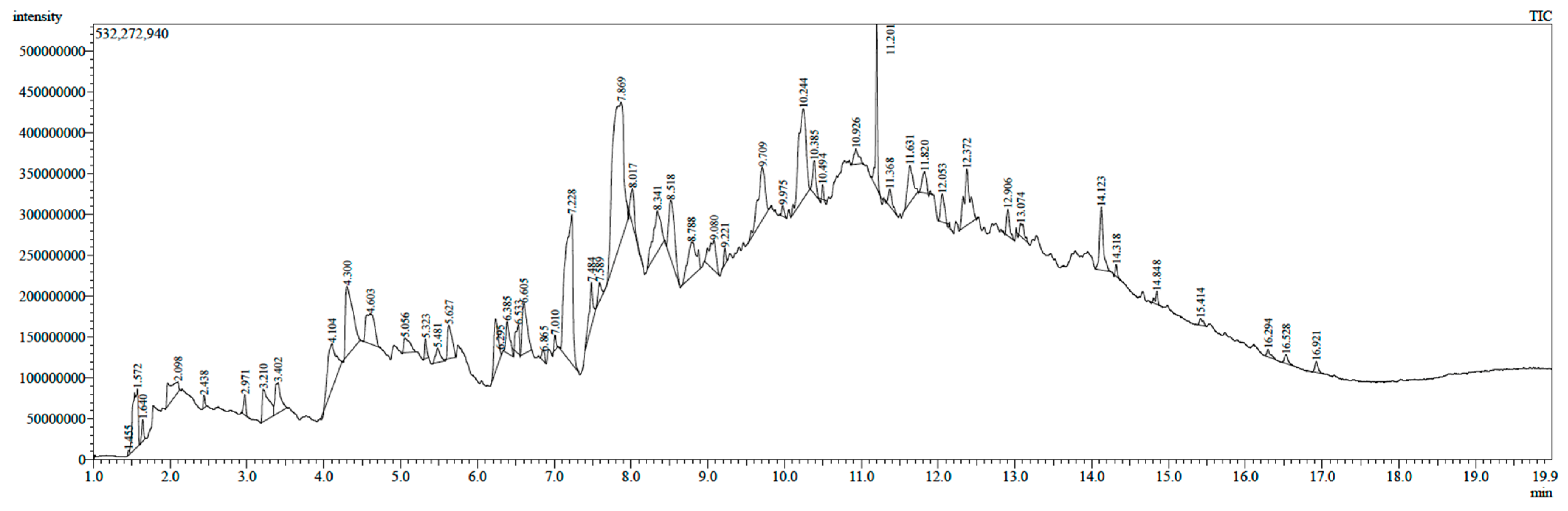
2.1. Gas Chromatography–Mass Spectrometry Analysis

Phenols and flavonoids are frequently recorded bioactive phytochemicals that may potentially be used to develop novel active drugs [21]. In plants, the possession of those phytochemicals is attributed to the biological activities [22]. Similarly, the presence of these phytochemicals along with other chemical compositions in the honey depends on various factors, ranging from the source of the flower to geography and entomology [23]. Phytochemicals are known to be significant components in ensuring plant defense against multiple factors, ranging from microorganisms, environmental stress as well as to interspecies protections. These phytochemicals have been incorporated in therapeutic works for ages [24]. Hence, the initial step in this study was to screen for potentially active chemical compounds in MH. This was achieved by GC–MS analysis (Figure 1) to highlight the main bioactive compounds within the tested MH sample (Table 1).
Overall, GC-MS analysis has revealed the presence of 37 different phytoconstituents belonging to various chemical classes were tentatively identified. Compounds which were identified in larger percentages within the tested MH sample are as follows: benzofuranone (16%), methylethylamino thiazole (10%), propenal nitrophenyl (3.7 %), heptyl caprylate (3.3%) and furan-2-yl ethan 1-amine (3.2 %). The remaining compounds constitute of 63% of the tested MH, each of which is present with less than 3%. These different chemical constituents belong to diverse, yet important classes of phytochemicals, such as phenol, sesquiterpenes, fatty acids, methyl esters and others. The presence of varied bioactive components in the MH sample might justify its use for the treatment of numerous diseases [25]. For example, Benzofuran derivatives are well known for its multiple biological activities, including anticancer, antibacterial and antioxidation [26]. Methylethylamino thiazole has also shown to have an antibacterial activity that has shown high level of interaction with bacterial proteins [27]. Furthermore, the geographical location from which the honey is originated plays a major role in its composition. The climatic conditions, such as humidity, sunlight, chemical nature of soil and other factors would determine the concentration of phytochemicals, including carbohydrates, phenolic compounds and volatile compounds in the honey. Thus, it is very logical to assume that the components of any honey are different, depending on the place of origin. In part, this may also be attributed mostly to the difference in compositions of pollen or nectar that influences the chemical composition of the honey [28,29].

2.2. Antioxidant Activity

Several pathophysiology caused by oxidation and free radicals including inflammatory, degenerative, cancerous, obesity, dementia and chronic diseases like diabetes [30,31,32,33]. While natural antioxidants usually impede the process of oxidation by interaction with free radicals [21,30,31,32,33], but it might not be the case for honey since it contains mixture of compounds which possess various mechanism of actions. For such a reason, it is difficult to estimate antioxidant capability of honey by one assay, and hence, multiple antioxidant experiments are required to obtain more accurate data from different angles [34]. The antioxidant potential of the MH sample was assessed in this research using free radical scavenging (DPPH, ABTS) and beta-carotene bleaching antioxidant tests (Table 2). MH sample showed considerable radical scavenging properties (DPPH IC50: 7.36 mg/mL; ABTS IC50: 4.49 mg/mL) and a moderate beta-carotene bleaching potential (IC50: 37.51 mg/mL). Different chemical components are responsible for this potent antioxidant activity. Each one of the performed assays investigates different mechanisms of antioxidant activities (ABTS and/or DPPH compared to bleaching assay) which were achieved due to phytochemical compounds present in the tested MH confirming that MH possesses higher radical scavenging properties than bleaching properties. Collectively, the obtained data from the antioxidants experiments in this study are in concordance with preceding studies that presented a robust positive relationship between the bioactive phytochemical compounds and their antioxidant affect [35,36].
Honeys are considered as strong antioxidant agents that can protect cell and its components against the destructive effects of various stressful factors by acting as modulator of free radicals [37,38]. MH contains a high number of identified and yet to be elucidated phenolic compounds [39,40]. These phenols effectively reduce the existence and number of free radicals, thus provide a fitting antioxidant capacity [41,42]. Consequently, this bioactive characteristic of honey that is often used as a reference method [37] to investigate the antioxidant capability of multiple types of honey from various geographical locations. In this context, studies form Algeria, Germany, Scotland and Saudi Arabia showed that MH was found to present with higher phenolic contents and exhibited increased antioxidant activity compared to acacia, portobello, and wild carrot honeys [19,38]. Comparable outcomes were acquired with the Malaysian honey [41] and Tualang honey [60]. An electronic paramagnetic resonance has also been used to study the MH’s transitory action against superoxide anion radicals [43,44]. In these two studies, obtained outcomes provided that the alleviating role of MH could be very much due to the presence of methyl syringate [45]. Former in-vivo model experiments have proved the activity of MH in halting the damages caused by oxidation in young and middle-aged rodents hepatocytes [46]. To achieve these advantages, antioxidant enzyme activities (like catalase) might be modulated, and the high antioxidant activity associate with phenolic content could be potentially used as therapeutic agent in this respect [46]. The current antioxidant data suggested that MH can be used as an alternative medicinal treatment to boost physiological oxidative status.

2.3. Antibacterial and Antibiofilm Activity

The potential antibacterial activity of honey were associated with its physicochemical properties just as much as various chemical components are derived from the plant secretions, pollens, propolis and from bee that is collecting it [47]. MH exhibited a relatively increased antimicrobial activity against Gram-positive bacteria compared to Gram-negative bacteria (Table 3). Currently, tested sample of MH showed a higher antimicrobial effect towards Gram-positive than negative bacterial strains. This is in agreement with previous reports where generally Gram-positive bacteria exhibited higher level of sensitivity towards MH than Gram-negative bacterial isolates [12,48].
The intrinsic property of honey to work against pathogenic bacteria is the essence of multiple factors. These factors either work individually or collectively to achieve the required function/s. The major factors that are playing an important role would include, phenolic compounds, H2O2, honey pH and the osmotic pressure produced by honey itself [49]. It was claimed that the prominent antibacterial efficacy of MH is mainly linked to the quantity of MGO present in the honey [40]. A study explored the antimicrobial effect of MH has shown its activity against various bacterial species, including Streptococcus pyogenes, Streptococcus mutans, Staphylococcus aureus, Enterobacter cloacae, Proteus mirabilis and Pseudomonas aeruginosa [46].
Biofilm formation can project great hindrance against antibiotics, as they furiously remain stable and would not be eradicated in severe infections [50]. The majority of anti-bacterial and anti-biofilm agents are known to be present in most medicinal plants and their compounds [51]. Even though all extracts have strong anti-bacterial potential, the lowest anti-biofilm potential of extracts must be discovered in the current investigation. The MH sample’s biofilm percentage inhibition was assessed (Table 4), and ½ MIC values were also calculated (Table 4). A similar pattern of these findings was obtained in the case of anti-bacterial activity was noted, and the MH sample exerted more anti-biofilm potential against S. aureus and B. subtilis, with percentage of inhibition as 67.13 and 56.52 %, respectively. Recent research has shown that metabolites, such as alkaloids, tannins, polyphenols, flavonoids, steroids and terpenoids are effective anti-bacterial and anti-biofilm agents. Even the function of catechin and polyphenol fractions as powerful biofilm inhibitors has been investigated [52]. The presence of these compounds in MH could be the reason for the observed anti-biofilm activity in the current study.

2.4. Anticancer Potential

Various concentrations of MH were tested against three cancer cell lines (HCT-116, MCF-7, A549), and their cytotoxic effects were observed using MTT assays. The percentage of cell viability at different concentrations of the MH sample and the IC50 values were determined (Table 5).
Anticancer activity of MH against tested cell lines are surmised in Figure 2. A dose-dependent anti-cancer potential was observed, as well as a substantial anticancer potential against the MCF-7 and A549 cell lines, and IC50 values were as 9.05 and 9.37 mg/mL, respectively. Aside from its antibacterial and antioxidant characteristics, honey has been found in recent research to have features that could slow the multiplication of malignant cells [45,53,54]. The anti-cancerous activity can occur via the adoption of one of the following routes: (1) depolarizing the mitochondrial membrane of cancer cells, hence causing apoptosis, (2) cyclooxygenase-2 inhibition by components, such as flavonoids, (3) releasing cytotoxic H2O2 and (4) scavenging the reactive oxygen species (ROS). It has also been associated with the phytochemical compounds [55]. On human breast cancer (MCF-7), colorectal carcinoma (CT26) and murine melanoma (B16.F1), MH has been shown to have a dose and time-dependent anti-proliferative effect [37]. It is primarily has this effect through mitochondrial membrane apoptosis [56]. The initiator caspase-9 is triggered, which causes the executioner caspase-3 to be activated [55]. It also promotes apoptosis by activating poly(ADP-ribose)polymerase protein (PARP), which causes the loss of Bcl-2 expression and fragmentation of DNA [57]. In a mouse melanoma model, MH significantly lowered overall tumor volume and boosted death of these cells [58]. A study by Forbes-Hernández and colloques used rats suffering from inflammatory bowel disease, and after applying HM, colonic inflammation was decreased, lipid peroxidation level was lowered, and the antioxidant activities were increased [55]. These findings justify the observed cytotoxicity potential of the tested MH sample.

3. Materials and Methods

3.1. Honey Sample

A medical grade manuka honey sample (250 g) was purchased from Manuka Guard (Otara, New Zealand). The honey was stored at room temperature and cultured on Mueller-Hinton Agar (MHA) to ensure absence of contamination.

3.2. Gas Chromatography–Mass Spectrometry Analysis

To analyze the chemical components of the MH sample, gas chromatography and mass spectrometry (GC–MS) were employed. For the GC–MS analysis, a TSQ Quantum mass spectrometer and a Thermo Trace GC Ultra gas chromatograph were used [59]. The mass detector was run in full-scan mode at 0.132 scans per second at 70 eV ionization energy throughout a mass range of 40–500 Daltons. Thermo TR-5MS fused silica capillary column was utilized in combination with a 5% phenyl polysilphenylene-siloxane stationary phase, and it had a length of 0.25 mm and a film thickness of 0.25 μm. The oven temperature was raised slowly from 40 °C to 300 °C over the course of the first 10 min, with an injector temperature of 250 °C. With a flow rate of 1 mL/min and a split flow rate of 25 mL/min, helium was used as the carrier gas, producing a split ratio of 25. The transfer line’s temperature stayed at 250 °C all the time. By comparing the mass spectra of the analyzed MH to multiple relevant libraries, including Wiley Library and the National Institute of Standards and Technology (NIST) as well as data from previously published studies, the majority of the present chemicals were identified accordingly.

3.3. Antioxidant Assays

3.3.1. DPPH Scavenging Activity

The antioxidant potential of the MH sample was established with regard to its intrinsic scavenging capacity versus DPPH free radicals [60]. Various concentrations ranged between 1 and 100 µg/mL of the MH sample were assorted into designated tubes. Briefly, each tube has 2 mL of DPPH solution (6 × 10⁻⁵ M) dissolved in dimethyl sulfoxide (DMSO). These were further assorted during incubation in the dark for a period of 1 h. Upon termination of incubation, the absorbance attained 517 nm following previous protocol [61]. To calculate the results, the following formula was used:
DPPH scavenging activity (%) = (A0 − A1)/A0 × 100
A0 = absorbance of the control. A1 = absorbance of the sample.

3.3.2. ABTS Scavenging Assay

The free-radical-scavenging activity of the MH sample was determined by ABTS radical cation decolorization assay [62]. Cation decolorization of ABTS+ radical cation is accomplished using a reaction mixture of 7 mM ABTS and 2.45 mM potassium persulfate. The reaction mixture was then stored at ambient temperature for 16 h in the dark. The resulting combination was then diluted with ABTS solution and methanol (MeOH) to yield an absorbance of 0.70 ± 0.02 units at 734 nm. A 50 µL MH sample was combined with 1 mL ABTS+ solution that was then kept at room temperature for 30 min. The absorbance of the reaction mixture was measured at 734 nm. The ABTS scavenging percentage inhibition was calculated as follows:
ABTS radical scavenging activity (%) = [(Abscontrol − Abssample)/(Abscontrol)] × 100
where Abs control is the absorbance of ABTS radical + MeOH; Abs sample is the absorbance of ABTS radical + sample extract/standard.

3.3.3. Beta-Carotene Assays

This test measures the obstruction of decomposition of beta-carotene by oxidation. The oxidative by product of linoleic acid causes discoloration of beta-carotenes when the emulsion of the two is solubilized in concentrations of 25 µL and 0.5 mg beta-carotene in 1 mL chloroform. Tween 40 in 200 mg (w/v) was added to the mixture followed by adding 100 mL of oxygenated distilled water. Approximately 350 µL of extracts solubilized in MeOH (2 mg/mL) were added to reach 2.5 mL of emulsion. The procedure is then performed using MeOH and H2O as negative controls. The absorbance at 490 nm was measured at regular intervals from one hour to 24 h of incubation at room temperature in the dark.
The percentage of inhibition of β-carotene decomposition in the MH sample’s antioxidant potential was measured as follows:
AA% = ABS test/ABS BHT × 100
AA%: Percentage of the antioxidant activity. ABS test: Absorbance in the presence of the MH sample (test). ABS BHT: Absorbance in the presence of positive control BHT.

3.4. Antibacterial Activity

Bacterial strains used for antibacterial activity were obtained from the Microbial Type Culture Collection (MTCC) and were cultivated overnight at 37 °C in Muller-Hinton Agar (MHA). These bacterial isolates include Escherichia coli, (E. coli) (MTCC 9537), Bacillus subtilis (B. subtilis) (MTCC 121), Pseudomonas aeruginosa (P. aeruginosa) (MTCC 741) and Staphylococcus aureus (S. aureus) (MTCC 96). The antibacterial activity of the MH sample was determined using the agar well diffusion method [63]. By adjusting the culture’s turbidity using 0.9% of sterile saline solution, the concentration of bacteria was adjusted to 0.5 McFarland standard 108 CFU/mL. Then, each prepared solution of tested bacterial strains was inoculated into designated agar plate using sterile swabs. Next, approximately 60 µL of the MH sample (1 mg/mL) were injected into wells that were constructed previously by using a sterile cork borer (6 mm). These plates were then incubated at 37 °C for 24 h. Inhibitory zones were measured in millimeter (mm) and three replicates for each strain were conducted. Chloramphenicol (1 mg/mL) and Muller–Hinton broth were employed as positive and negative controls, respectively.

3.5. Antibiofilm Assay

The potential effect of the MH sample toward biofilm formation was assessed using a modified version of a previously reported method [64]. A growing culture of each bacterial strain (107 cells/mL) was inoculated into 96-well microtiter plates made of Mueller–Hinton Broth (MHB) and 1% glucose to form biofilms. Planktonic cells were removed, and the wells were gently rinsed three times with normal saline at the conclusion of the 24 h incubation at 37 °C. Half of minimum inhibitory concentration (MIC) of MH was added (approximately 200 µL) to the wells and then incubated at the abovementioned conditions. The absorbance was monitored each hour for 24 h using 492 nm wavelength. The MHB of each bacterial strain was utilized as a biofilm growth control. The percentage of inhibition in the biofilm was calculated using the equation below:
[(OD (control) − OD (test)/OD (control)] × 100

3.6. Anticancer Assay (MTT Assay)

The anticancer activity of the MH sample was investigated against different human cancer cell lines, including colon (HCT-116), breast (MCF-7), and lung (A549), which were acquired from the National Centre for Cell Science (NCCS). Cells were grown in a 25 cm flask containing Dulbecco’s Modified Eagle’s Medium (DMEM) supplemented with 10% Fetal Bovine Serum (FBS) and gentamicin and was then overnight incubated at 37 °C in 5% CO2 environment. Cells were grown to 80 percent confluence before being seeded at a density of more than 1 × 10⁵ cells per well in 96-well plates and incubated in the described abovementioned growth conditions. Cells were then stained with Trypan Blue (0.4%), and viable cell count was determined using a hemocytometer. Cells were then treated with different concentrations of MH sample (1–100 µg/mL) for 24 h. Cells were washed with phosphate-buffered solution (PBS) and subjected to 100 µL of MTT solution 5 mg/mL followed by incubation for 4 h. Next, the medium was then removed, and formazan crystals were solubilized with 100 µL of DMSO. The amount of formazan crystal was determined by measuring the absorbance at 570 nm using an ELISA microplate reader, following theses protocols [65,66,67,68]. All assays were performed at least three times and 50% cytotoxic concentration (IC50) was calculated [69,70].

4. Conclusions

The current study highlights the GC–MS phytochemical and biological effects of manuka honey. The tested samples were found to contain various bioactive compounds that might be potentially responsible for the observed antioxidant effect in MH. Furthermore, the observed antibacterial and antibiofilm effects of the MH would suggest a promising and naturally alternative source for easily and readily available antibacterial agents. Obtained data for the cytotoxicity assays indicated that MH exerted a dose-dependent anti-cancer potential against HCT-116, MCF-7 and A549 cell lines. Forthcoming studies that further investigate the molecular mechanistic and pharmacogenetics of MH with different pathogens and cancer cells are needed to scrutinize the validity and efficacy of MH.

Author Contributions

Conceptualization, A.S.B. and A.A. (Abdu Aldarhami); Formal analysis—Antimicrobial activity, A.S.B.; Formal analysis—Antioxidant activity, J.K.; Formal analysis—Anticancer activity, M.N.A., H.Q. and N.K.B., Formal analysis—Antibiofilm activity, A.S.B., A.A. (Abdulwahab Alamri) and A.A. (Abdu Aldarhami). Investigation, A.S.B., A.A. (Abdulwahab Alamri); M.N.A., H.Q., A.A.A., J.A., N.K.B., J.K., A.A. (Abdu Aldarhami) and A.A. (Ahmed Alafnan); Funding acquisition, A.S.B.; Visualization, A.S.B., A.A. (Abdulwahab Alamri), M.N.A., H.Q., A.A.A., J.A and N.K.B.; Writing—original draft preparation, A.S.B. and A.A. (Abdulwahab Alamri); Writing—review and editing, A.S.B., A.A. (Abdulwahab Alamri), M.N.A., H.Q., A.A.A., J.A., N.K.B., J.K., A.A. (Abdu Aldarhami) and A.A. (Ahmed Alafnan). All authors have read and agreed to the published version of the manuscript.

Funding

This research has been funded by the Scientific Research Deanship at University of Ha’il—Saudi Arabia via a project number (RG-21081).

Institutional Review Board Statement

Not applicable.

Informed Consent Statement

Not applicable.

Data Availability Statement

All data that support the findings of this study are available within the article.

Acknowledgments

All authors thank the Scientific Research Deanship at University of Ha’il-Saudi Arabia for their grant through a project number (RG-21081).

Conflicts of Interest

The authors declare no conflict of interest.

References

  1. Duru, C.E.; Duru, I.A. Phytochemical evaluation and health risk assessment of honey from an Apiary in Amizi, Ikuano local government area, Abia State, Nigeria. Sci. Afr. 2021, 13, e00885. [Google Scholar] [CrossRef]
  2. Bazaid, A.S.; Aldarhami, A.; Gattan, H.; Aljuhani, B. Saudi Honey: A Promising Therapeutic Agent for Treating Wound Infections. Cureus 2021, 13, e18882. [Google Scholar] [CrossRef] [PubMed]
  3. Gunther, R.T. The Greek Herbal of Dioscorides; The University of Chicago Press: Chicago, IL, USA, 1959. [Google Scholar]
  4. Al Somal, N.; Coley, K.; Molan, P.; Hancock, B. Susceptibility of Helicobacter pylori to the antibacterial activity of manuka honey. J. R. Soc. Med. 1994, 87, 9. [Google Scholar] [CrossRef]
  5. Topham, J. Why do some cavity wounds treated with honey or sugar paste heal without scarring? J. Wound Care 2002, 11, 53–55. [Google Scholar] [CrossRef] [PubMed] [Green Version]
  6. Markelov, V.; Trushin, M. Bee venom therapy and low dose naltrexone for treatment of multiple sclerosis. Nepal J. Neurosci. 2006, 3, 71–77. [Google Scholar]
  7. Chua, L.S.; Rahaman, N.L.A.; Adnan, N.A.; Eddie Tan, T.T. Antioxidant activity of three honey samples in relation with their biochemical components. J. Anal. Methods Chem. 2013, 2013, 313798. [Google Scholar] [CrossRef] [PubMed]
  8. Aljadi, A.; Kamaruddin, M. Evaluation of the phenolic contents and antioxidant capacities of two Malaysian floral honeys. Food Chem. 2004, 85, 513–518. [Google Scholar] [CrossRef]
  9. Arawwawala, M.; Hewageegana, S. Health benefits and traditional uses of honey: A review. J. Apither. 2017, 2, 9–14. [Google Scholar] [CrossRef] [Green Version]
  10. Al-Waili, N.S. Natural honey lowers plasma glucose, C-reactive protein, homocysteine, and blood lipids in healthy, diabetic, and hyperlipidemic subjects: Comparison with dextrose and sucrose. J. Med. Food 2004, 7, 100–107. [Google Scholar] [CrossRef]
  11. Ahmad, R.S.; Hussain, M.B.; Saeed, F.; Waheed, M.; Tufail, T. Phytochemistry, metabolism, and ethnomedical scenario of honey: A concurrent review. Int. J. Food Prop. 2017, 20, S254–S269. [Google Scholar] [CrossRef] [Green Version]
  12. Gośliński, M.; Nowak, D.; Kłębukowska, L. Antioxidant properties and antimicrobial activity of manuka honey versus Polish honeys. J. Food Sci. Technol. 2020, 57, 1269–1277. [Google Scholar] [CrossRef] [PubMed]
  13. Marshall, S.M.; Schneider, K.R.; Cisneros, K.V.; Gu, L. Determination of antioxidant capacities, α-dicarbonyls, and phenolic phytochemicals in Florida varietal honeys using HPLC-DAD-ESI-MS n. J. Agric. Food Chem. 2014, 62, 8623–8631. [Google Scholar] [CrossRef] [PubMed]
  14. Alzahrani, H.A.; Alsabehi, R.; Boukraâ, L.; Abde-llah, F.; Bellik, Y.; Bakhotmah, B.A. Antibacterial and antioxidant potency of floral honeys from different botanical and geographical origins. Molecules 2012, 17, 10540–10549. [Google Scholar] [CrossRef] [PubMed] [Green Version]
  15. Kwakman, P.H.; Te Velde, A.A.; De Boer, L.; Vandenbroucke-Grauls, C.M.; Zaat, S.A. Two major medicinal honeys have different mechanisms of bactericidal activity. PLoS ONE 2011, 6, e17709. [Google Scholar] [CrossRef] [Green Version]
  16. Bazaid, A.S.; Barnawi, H.; Qanash, H.; Alsaif, G.; Aldarhami, A.; Gattan, H.; Alharbi, B.; Alrashidi, A.; Al-Soud, W.A.; Moussa, S.; et al. Bacterial Coinfection and Antibiotic Resistance Profiles among Hospitalised COVID-19 Patients. Microorganisms 2022, 10, 495. [Google Scholar] [CrossRef] [PubMed]
  17. Bazaid, A.S.; Aldarhami, A.; Gattan, H.; Barnawi, H.; Qanash, H.; Alsaif, G.; Alharbi, B.; Alrashidi, A.; Eldrehmy, E.H. Antibiogram of Urinary Tract Infections and Sepsis among Infants in Neonatal Intensive Care Unit. Children 2022, 9, 629. [Google Scholar] [CrossRef] [PubMed]
  18. Bogdanov, S.; Tomislav, J.; Sieber, R. PhD and Gallmann, P.J. Am. College Nut. 2008, 27, 677–689. [Google Scholar] [CrossRef]
  19. Schneider, M.; Coyle, S.; Warnock, M.; Gow, I.; Fyfe, L. Anti-microbial activity and composition of manuka and portobello honey. Phytother. Res. 2013, 27, 1162–1168. [Google Scholar] [CrossRef]
  20. Kato, Y.; Umeda, N.; Maeda, A.; Matsumoto, D.; Kitamoto, N.; Kikuzaki, H. Identification of a novel glycoside, leptosin, as a chemical marker of manuka honey. J. Agric. Food Chem. 2012, 60, 3418–3423. [Google Scholar] [CrossRef]
  21. Saleem, H.; Zengin, G.; Khan, K.-U.-R.; Ahmad, I.; Waqas, M.; Mahomoodally, F.M.; Rengasamy, K.R.; Zainol, N.; Abidin, S.A.Z.; Ahemad, N. New insights into the phytochemical composition, enzyme inhibition and antioxidant properties of desert cotton (Aerva javanica (Bum. f) Shult.-Amaranthaceae). Nat. Prod. Res. 2021, 35, 664–668. [Google Scholar] [CrossRef]
  22. Altemimi, A.; Lakhssassi, N.; Baharlouei, A.; Watson, D.G.; Lightfoot, D.A. Phytochemicals: Extraction, Isolation, and Identification of Bioactive Compounds from Plant Extracts. Plants 2017, 6, 42. [Google Scholar] [CrossRef] [PubMed]
  23. Wang, S.; Kirillova, K.; Lehto, X. Travelers’ food experience sharing on social network sites. J. Travel Tour. Mark. 2017, 34, 680–693. [Google Scholar] [CrossRef]
  24. War, A.R.; Paulraj, M.G.; Ahmad, T.; Buhroo, A.A.; Hussain, B.; Ignacimuthu, S.; Sharma, H.C. Mechanisms of plant defense against insect herbivores. Plant Signal. Behav. 2012, 7, 1306–1320. [Google Scholar] [CrossRef] [PubMed] [Green Version]
  25. Yupanqui Mieles, J.; Vyas, C.; Aslan, E.; Humphreys, G.; Diver, C.; Bartolo, P. Honey: An Advanced Antimicrobial and Wound Healing Biomaterial for Tissue Engineering Applications. Pharmaceutics 2022, 14, 1663. [Google Scholar] [CrossRef]
  26. Miao, Y.-H.; Hu, Y.-H.; Yang, J.; Liu, T.; Sun, J.; Wang, X.-J. Natural source, bioactivity and synthesis of benzofuran derivatives. RSC Adv. 2019, 9, 27510–27540. [Google Scholar] [CrossRef] [Green Version]
  27. Althagafi, I.; El-Metwaly, N.; Farghaly, T.A. New series of thiazole derivatives: Synthesis, structural elucidation, antimicrobial activity, molecular modeling and MOE docking. Molecules 2019, 24, 1741. [Google Scholar] [CrossRef] [Green Version]
  28. Alissandrakis, E.; Tarantilis, P.A.; Harizanis, P.C.; Polissiou, M. Comparison of the volatile composition in thyme honeys from several origins in Greece. J. Agric. Food Chem. 2007, 55, 8152–8157. [Google Scholar] [CrossRef]
  29. Awasum, C.A.; Fotzo, S.L.M.; Ndukum, J.A.; Genesis, C.D.; Zoli, A. Gas chromatography-mass spectroscopy analysis and chemical composition of Ngaoundere, Cameroon honey. Am. J. Biosci. Bioeng. 2015, 3, 33–36. [Google Scholar]
  30. Liu, Y.E.; Chen, C.; Wang, X.; Sun, Y.; Zhang, J.; Chen, J.; Shi, Y. An Epigenetic Role of Mitochondria in Cancer. Cells 2022, 11, 2518. [Google Scholar] [CrossRef]
  31. Chen, K.; Zhang, J.; Beeraka, N.M.; Tang, C.; Babayeva, Y.V.; Sinelnikov, M.Y.; Zhang, X.; Zhang, J.; Liu, J.; Reshetov, I.V.; et al. Advances in the Prevention and Treatment of Obesity-Driven Effects in Breast Cancers. Front. Oncol. 2022, 12, 820968. [Google Scholar] [CrossRef]
  32. Fang, X.; Zhang, J.; Roman, R.J.; Fan, F. From 1901 to 2022, how far are we from truly understanding the pathogenesis of age-related dementia? Geroscience 2022, 44, 1879–1883. [Google Scholar] [CrossRef] [PubMed]
  33. Chen, K.; Lu, P.; Beeraka, N.M.; Sukocheva, O.A.; Madhunapantula, S.V.; Liu, J.; Sinelnikov, M.Y.; Nikolenko, V.N.; Bulygin, K.V.; Mikhaleva, L.M.; et al. Mitochondrial mutations and mitoepigenetics: Focus on regulation of oxidative stress-induced responses in breast cancers. Semin. Cancer Biol. 2022, 83, 556–569. [Google Scholar] [CrossRef] [PubMed]
  34. Combarros-Fuertes, P.; Estevinho, L.M.; Dias, L.G.; Castro, J.M.; Tomás-Barberán, F.A.; Tornadijo, M.E.; Fresno-Baro, J.M. Bioactive components and antioxidant and antibacterial activities of different varieties of honey: A screening prior to clinical application. J. Agric. Food Chem. 2018, 67, 688–698. [Google Scholar] [CrossRef] [PubMed] [Green Version]
  35. Khurshid, U.; Ahmad, S.; Saleem, H.; Nawaz, H.A.; Zengin, G.; Locatelli, M.; Mahomoodally, M.F.; Abidin, S.A.Z.; Tousif, M.I.; Ahemad, N. Phytochemical composition and in vitro pharmacological investigations of Neurada procumbens L.(Neuradaceae): A multidirectional approach for industrial products. Ind. Crops Prod. 2019, 142, 111861. [Google Scholar] [CrossRef]
  36. Saleem, H.; Khurshid, U.; Sarfraz, M.; Tousif, M.I.; Alamri, A.; Anwar, S.; Alamri, A.; Ahmad, I.; Abdallah, H.H.; Mahomoodally, F.M. A comprehensive phytochemical, biological, toxicological and molecular docking evaluation of Suaeda fruticosa (L.) Forssk.: An edible halophyte medicinal plant. Food Chem. Toxicol. 2021, 154, 112348. [Google Scholar] [CrossRef]
  37. Patel, S.; Cichello, S. Manuka honey: An emerging natural food with medicinal use. Nat. Prod. Bioprospect. 2013, 3, 121–128. [Google Scholar] [CrossRef] [Green Version]
  38. Alzahrani, H.A.; Boukraâ, L.; Bellik, Y.; Abdellah, F.; Bakhotmah, B.A.; Kolayli, S.; Sahin, H. Evaluation of the antioxidant activity of three varieties of honey from different botanical and geographical origins. Glob. J. Health Sci. 2012, 4, 191. [Google Scholar] [CrossRef] [Green Version]
  39. Stephens, J.M.; Schlothauer, R.C.; Morris, B.D.; Yang, D.; Fearnley, L.; Greenwood, D.R.; Loomes, K.M. Phenolic compounds and methylglyoxal in some New Zealand manuka and kanuka honeys. Food Chem. 2010, 120, 78–86. [Google Scholar] [CrossRef]
  40. Mavric, E.; Wittmann, S.; Barth, G.; Henle, T. Identification and quantification of methylglyoxal as the dominant antibacterial constituent of Manuka (Leptospermum scoparium) honeys from New Zealand. Mol. Nutr. Food Res. 2008, 52, 483–489. [Google Scholar] [CrossRef]
  41. Moniruzzaman, M.; Sulaiman, S.A.; Khalil, M.I.; Gan, S.H. Evaluation of physicochemical and antioxidant properties of sourwood and other Malaysian honeys: A comparison with manuka honey. Chem. Cent. J. 2013, 7, 138. [Google Scholar] [CrossRef] [Green Version]
  42. Jubri, Z.; Rahim, N.B.A.; Aan, G.J. Manuka honey protects middle-aged rats from oxidative damage. Clinics 2013, 68, 1446–1454. [Google Scholar] [CrossRef]
  43. Henriques, A.; Jackson, S.; Cooper, R.; Burton, N. Free radical production and quenching in honeys with wound healing potential. J. Antimicrob. Chemother. 2006, 58, 773–777. [Google Scholar] [CrossRef] [PubMed]
  44. Inoue, K.; Murayama, S.; Seshimo, F.; Takeba, K.; Yoshimura, Y.; Nakazawa, H. Identification of phenolic compound in manuka honey as specific superoxide anion radical scavenger using electron spin resonance (ESR) and liquid chromatography with coulometric array detection. J. Sci. Food Agric. 2005, 85, 872–878. [Google Scholar] [CrossRef]
  45. Fukuda, M.; Kobayashi, K.; Hirono, Y.; Miyagawa, M.; Ishida, T.; Ejiogu, E.C.; Sawai, M.; Pinkerton, K.E.; Takeuchi, M. Jungle honey enhances immune function and antitumor activity. Evid. Based Complement. Altern. Med. 2011, 2011, 908743. [Google Scholar] [CrossRef] [PubMed]
  46. Alvarez-Suarez, J.M.; Gasparrini, M.; Forbes-Hernández, T.Y.; Mazzoni, L.; Giampieri, F. The composition and biological activity of honey: A focus on Manuka honey. Foods 2014, 3, 420–432. [Google Scholar] [CrossRef] [Green Version]
  47. Salonen, A.; Virjamo, V.; Tammela, P.; Fauch, L.; Julkunen-Tiitto, R. Screening bioactivity and bioactive constituents of Nordic unifloral honeys. Food Chem. 2017, 237, 214–224. [Google Scholar] [CrossRef] [Green Version]
  48. Alvarez-Suarez, J.M.; Tulipani, S.; Díaz, D.; Estevez, Y.; Romandini, S.; Giampieri, F.; Damiani, E.; Astolfi, P.; Bompadre, S.; Battino, M. Antioxidant and antimicrobial capacity of several monofloral Cuban honeys and their correlation with color, polyphenol content and other chemical compounds. Food Chem. Toxicol. 2010, 48, 2490–2499. [Google Scholar] [CrossRef]
  49. Alvarez-Suarez, J.M.; Tulipani, S.; Romandini, S.; Bertoli, E.; Battino, M. Contribution of honey in nutrition and human health: A review. Mediterr. J. Nutr. Metab. 2010, 3, 15–23. [Google Scholar] [CrossRef]
  50. Bjarnsholt, T. The role of bacterial biofilms in chronic infections. APMIS 2013, 121, 1–58. [Google Scholar] [CrossRef]
  51. Pinto, M.; Langer, T.M.; Hüffer, T.; Hofmann, T.; Herndl, G.J. The composition of bacterial communities associated with plastic biofilms differs between different polymers and stages of biofilm succession. PLoS ONE 2019, 14, e0217165. [Google Scholar]
  52. Zacchino, S.A.; Butassi, E.; Di Liberto, M.; Raimondi, M.; Postigo, A.; Sortino, M. Plant phenolics and terpenoids as adjuvants of antibacterial and antifungal drugs. Phytomedicine 2017, 37, 27–48. [Google Scholar] [CrossRef] [PubMed]
  53. Ghashm, A.A.; Othman, N.H.; Khattak, M.N.; Ismail, N.M.; Saini, R. Antiproliferative effect of Tualang honey on oral squamous cell carcinoma and osteosarcoma cell lines. BMC Complement. Altern. Med. 2010, 10, 49. [Google Scholar] [CrossRef] [PubMed] [Green Version]
  54. Swellam, T.; Miyanaga, N.; Onozawa, M.; Hattori, K.; Kawai, K.; Shimazui, T.; Akaza, H. Antineoplastic activity of honey in an experimental bladder cancer implantation model: In vivo and in vitro studies. Int. J. Urol. 2003, 10, 213–219. [Google Scholar] [CrossRef] [PubMed]
  55. Forbes-Hernández, T.Y.; Giampieri, F.; Gasparrini, M.; Mazzoni, L.; Quiles, J.L.; Alvarez-Suarez, J.M.; Battino, M. The effects of bioactive compounds from plant foods on mitochondrial function: A focus on apoptotic mechanisms. Food Chem. Toxicol. 2014, 68, 154–182. [Google Scholar] [CrossRef] [PubMed]
  56. Aryappalli, P.; Al-Qubaisi, S.S.; Attoub, S.; George, J.A.; Arafat, K.; Ramadi, K.B.; Mohamed, Y.A.; Al-Dhaheri, M.M.; Al-Sbiei, A.; Fernandez-Cabezudo, M.J. The IL-6/STAT3 signaling pathway is an early target of manuka honey-induced suppression of human breast cancer cells. Front. Oncol. 2017, 7, 167. [Google Scholar] [CrossRef] [Green Version]
  57. Erejuwa, O.O.; Sulaiman, S.A.; Wahab, M.S.A. Effects of honey and its mechanisms of action on the development and progression of cancer. Molecules 2014, 19, 2497–2522. [Google Scholar] [CrossRef] [Green Version]
  58. Fernandez-Cabezudo, M.J.; El-Kharrag, R.; Torab, F.; Bashir, G.; George, J.A.; El-Taji, H.; Al-Ramadi, B.K. Intravenous administration of manuka honey inhibits tumor growth and improves host survival when used in combination with chemotherapy in a melanoma mouse model. PLoS ONE 2013, 8, e55993. [Google Scholar] [CrossRef] [Green Version]
  59. Al-Nemari, R.; Al-Senaidy, A.; Semlali, A.; Ismael, M.; Badjah-Hadj-Ahmed, A.Y.; Ben Bacha, A. GC-MS profiling and assessment of antioxidant, antibacterial, and anticancer properties of extracts of Annona squamosa L. leaves. BMC Complement. Med. Ther. 2020, 20, 296. [Google Scholar] [CrossRef]
  60. Adnan, M.; Patel, M.; Reddy, M.N.; Alshammari, E. Formulation, evaluation and bioactive potential of Xylaria primorskensis terpenoid nanoparticles from its major compound xylaranic acid. Sci. Rep. 2018, 8, 1740. [Google Scholar] [CrossRef] [Green Version]
  61. Orhan, I.; Aslan, M. Appraisal of scopolamine-induced antiamnesic effect in mice and in vitro antiacetylcholinesterase and antioxidant activities of some traditionally used Lamiaceae plants. J. Ethnopharmacol. 2009, 122, 327–332. [Google Scholar] [CrossRef]
  62. Soulef, S.; Seddik, K.; Nozha, M.; Smain, A.; Saliha, D.; Hosni, K. Phytochemical screening and in vivo and in vitro evaluation antioxidant capacity of Fargaria ananassa, Prunus armeniaca and Prunus persica fruits growing in Algeria. Prog. Nutr. 2020, 22, 236–252. [Google Scholar]
  63. Bazaid, A.S.; Forbes, S.; Humphreys, G.J.; Ledder, R.G.; O’Cualain, R.; McBain, A.J. Fatty Acid Supplementation Reverses the Small Colony Variant Phenotype in Triclosan-Adapted Staphylococcus aureus: Genetic, Proteomic and Phenotypic Analyses. Sci. Rep. 2018, 8, 3876. [Google Scholar] [CrossRef] [PubMed] [Green Version]
  64. Lemos, A.S.; Campos, L.M.; Melo, L.; Guedes, M.C.; Oliveira, L.G.; Silva, T.P.; Melo, R.C.; Rocha, V.N.; Aguiar, J.A.; Apolônio, A.C. Antibacterial and antibiofilm activities of psychorubrin, a pyranonaphthoquinone isolated from Mitracarpus frigidus (Rubiaceae). Front. Microbiol. 2018, 9, 724. [Google Scholar] [CrossRef] [PubMed] [Green Version]
  65. Elasbali, A.M.; Al-Soud, W.A.; Al-Oanzi, Z.H.; Qanash, H.; Alharbi, B.; Binsaleh, N.K.; Alreshidi, M.; Patel, M.; Adnan, M. Cytotoxic Activity, Cell Cycle Inhibition, and Apoptosis-Inducing Potential of Athyrium hohenackerianum (Lady Fern) with Its Phytochemical Profiling. Evid. Based Complement. Altern. Med. 2022, 2022, 2055773. [Google Scholar] [CrossRef]
  66. Lombardi, V.R.; Carrera, I.; Cacabelos, R. In vitro screening for cytotoxic activity of herbal extracts. Evid. Based Complement. Altern. Med. 2017, 2017, 2675631. [Google Scholar] [CrossRef]
  67. Qanash, H.; Yahya, R.; Bakri, M.M.; Bazaid, A.S.; Qanash, S.; Shater, A.F.; TM, A. Anticancer, antioxidant, antiviral and antimicrobial activities of Kei Apple (Dovyalis caffra) fruit. Sci. Rep. 2022, 12, 5914. [Google Scholar] [CrossRef]
  68. Reddy, M.N.; Adnan, M.; Alreshidi, M.M.; Saeed, M.; Patel, M. Evaluation of anticancer, antibacterial and antioxidant properties of a medicinally treasured fern Tectaria coadunata with its phytoconstituents analysis by HR-LCMS. Anti Cancer Agents Med. Chem. Former. Curr. Med. Chem. Anti Cancer Agents 2020, 20, 1845–1856. [Google Scholar] [CrossRef]
  69. Al-Rajhi, A.M.; Qanash, H.; Almuhayawi, M.S.; Al Jaouni, S.K.; Bakri, M.M.; Ganash, M.; Salama, H.M.; Selim, S.; Abdelghany, T.M. Molecular Interaction Studies and Phytochemical Characterization of Mentha pulegium L. Constituents with Multiple Biological Utilities as Antioxidant, Antimicrobial, Anticancer and Anti-Hemolytic Agents. Molecules 2022, 27, 4824. [Google Scholar] [CrossRef]
  70. Abdelghany, T.M.; Yahya, R.; Bakri, M.M.; Ganash, M.; Amin, B.H.; Qanash, H. Effect of Thevetia peruviana seeds extract for microbial pathogens and cancer control. Int. J. Pharmacol. 2021, 17, 643–655. [Google Scholar] [CrossRef]
Figure 1. GC–MS chromatogram of the tested manuka honey sample (X-axis: retention time in minutes; Y-axis: intensity).
Applsci 12 09928 g001
Figure 2. Anticancer activity of tested manuka honey towards three cancer cell lines, including colon cancer cells (HCT-116), breast cancer cells (MCF-7), and lung carcinoma epithelial cells (A549).
Applsci 12 09928 g002
Table 1. Gas chromatography–mass spectrometry analysis of tested manuka honey samples.
No.Retention Time (min)Area (%)Tentative Compounds Identification
17.86916.112(3H)-Benzofuranone, hexahydro-3-methylene-
27.22810.615-Methyl-2-ethylamino-2-thiazoline
39.7093.622-Propenal, 3-(2-nitrophenyl)-
48.5183.37Heptyl caprylate
512.3723.232-(Furan-2-yl)ethan-1-amine
61.5722.972-Chloroethyl methyl sulfoxide
78.7882.95Silane, [3-(2,3-epoxypropoxy)propyl]cethoxydim
811.2012.66Benzoic acid, 4-hydroxy-3,5-dimethoxy-, methyl
94.6032.54Thiophene, tetrahydro-2-methyl-
1014.1232.244-[3-(4-Fluorobenzyloxy)propyl]-1H-imidazole
113.212.071-Methylcyclopropanemethanol
1211.6311.953,4,5-Trihydroxybenzyl methyl ether
133.4021.69Propanedioic acid, oxo-, diethyl ester
147.4841.624H-Pyran-4-one, 5-hydroxy-2-(hydroxymethyl
152.0981.57Propane, 1,2-bis(difluoroamino)-2-methyl-
165.6271.41L-Valine, N-ethoxycarbonyl-
1710.3851.02,4-Dimethoxy-5-pyrimidine carboxaldehyde
1812.0530.993,5-Methano-2H-cyclopenta[b]furan-2-one, 6-b
195.0560.883H-Pyrazol-3-one, 2,4-dihydro-5-methyl-
2011.820.87Butyl dimethyl phosphorothioate
2112.9060.771,4-Methanonaphthalene, 6,7-diethyldecahydro-, c
2210.9260.72p-Menthan-3-one, semicarbazone
2313.0740.514,5-Imidazoledimethanol
242.9710.42S-Methyl-L-cysteine, N-(n-propyloxycarbonyl)
251.640.39Oxirane, (fluoromethyl)-
265.3230.372-Furanmethanol, 5-methyl-
2716.9210.33Cirsiumaldehyde
2816.5280.32Succinic acid, dodec-9-yn-1-yl pentyl ester
299.2210.32-Aminoresorcinol
306.8650.272H-Pyran-2-one, 5,6-dihydro-
319.9750.26(6R,7aR)-3,6-Dimethyl-5,6,7,7a-tetrahydrobenz
3214.8480.26Hexadecanoic acid, 2-hydroxy-1-(hydroxymeth
3316.2940.25Octadecanoic acid, 2,3-dihydroxypropyl ester
3415.4140.222-Furaldehyde azine
352.4380.212,3-Dimethylpentanoic acid
3614.3180.17(8S)-Eremophila-7(11)-en-12,8-lactam
3710.9490.081,7-Dimethylxanthine
Table 2. Antioxidant activity of tested manuka honey.
Antioxidant AssaysIC50 Values
Radical Scavenging activityDPPH7.36 ± 0.03
ABTS4.49 ± 0.1
Bleaching assayBeta-carotene37.51 ± 0.64
IC50 results are presented as the mean ± standard deviation. DPPH: 2,2-diphenyl-1-picrylhydrazyl; ABTS: 2,2’-azino-bis 3-ethylbenzothiazoline-6-sulfonic acid.
Table 3. Zone of inhibition of various bacterial isolates treated with manuka honey.
Tested BacteriaTested Honey Sample (10%)
Zone of Inhibition (mm)
Gram-positiveB. subtilis9.54 ± 0.3
S. aureus6.55 ± 0.24
Gram-negativeE. coli4.6 ± 0.25
P. aeruginosa3.7 ± 0.15
All results are presented as the mean ± standard deviation (n = 3).
Table 4. Antibiofilm activity of the manuka honey against biofilm-forming bacterial species.
Tested BacteriaMean% Inhibition
B. subtilisControl0.76 ± 0.0167.13
1/2 MIC0.25 ± 0.01
S. aureusControl0.84 ± 0.0156.52
1/2 MIC0.36 ± 0.05
E. coliControl0.72 ± 0.0348.28
1/2 MIC0.37 ± 0.01
P. aeruginosaControl0.87 ± 0.0142.78
1/2 MIC0.50 ± 0.04
All results are presented as the mean ± standard deviation.
Table 5. Percentage of inhibition, cell viability, and IC50 values of tested manuka honey against various cancer cell lines.
Tested Cell LinesPercentage of Inhibition and Cell ViabilityHoney Concentrations (%)IC50
246810
HCT-116Inhibition rate (%)18.5231.7842.0354.3464.7348.65
Cell viability (%)81.4768.2157.9645.6535.26
MCF-7Inhibition rate (%)12.5323.2234.0442.2357.819.05
Cell viability (%)87.4676.7765.9557.7642.18
A549Inhibition rate (%)9.7315.8628.3137.4349.899.37
Cell viability (%)90.2684.1371.6862.5650.10
IC50 results are presented as the mean of three reads. HCT-116: colon cancer cells; MCF-7: breast cancer cells; A549: lung carcinoma epithelial cells.
Publisher’s Note: MDPI stays neutral with regard to jurisdictional claims in published maps and institutional affiliations.

