Article

Effects of Mini-Implant-Assisted Rapid Palatal Expansion on Incisive Canal Morphology and Tooth–Canal Relationship

1
Department of Orthodontics, School of Dentistry, University of Wonkwang, Iksan 54538, Korea
2
Wonkwang Dental Research Institute, University of Wonkwang, Iksan 54538, Korea
3
Institute of Biomaterials and Implant, University of Wonkwang, Iksan 54538, Korea
4
Postgraduate Orthodontic Program, Arizona School of Dentistry & Oral Health, A.T. Still University, Mesa, AZ 85206, USA
5
Graduate School of Dentistry, Kyung Hee University, Seoul 02447, Korea
*
Author to whom correspondence should be addressed.
These authors contributed equally to this work.
Appl. Sci. 2022, 12(20), 10470; https://doi.org/10.3390/app122010470
Submission received: 19 September 2022 / Revised: 8 October 2022 / Accepted: 14 October 2022 / Published: 17 October 2022

Abstract

:
The objective of this study was to identify the change in incisive canal (IC) morphology and tooth–canal relationship after mini-implant-assisted rapid palatal expansion (MARPE). Pretreatment and posttreatment cone-beam computed tomography images of 30 subjects were retrospectively evaluated. The dimensional and volume changes of the IC after MARPE treatment were evaluated, and the tooth–canal relationship and positional relationship between the maxillary central incisors were additionally compared in the group where the root apex of the maxillary central incisors was higher than the IC oral opening. The mediolateral and labiopalatal widths of the IC were significantly increased in all three levels after MARPE treatment (p < 0.01). The amount of increase was greater in the mediolateral direction than in the labiopalatal direction. The anteroposterior distance from the mesial point of the maxillary central incisors to the anterior margin of the IC was significantly decreased only in the oral opening level in the samples where the apices of the maxillary central incisors were located more superior to the oral opening of the IC (p < 0.05). The mediolateral distance between the mesial points of the maxillary central incisors and the distance between the root apex of the maxillary central incisors significantly increased after MARPE (p < 0.001). However, the distance between the crown tips of the maxillary central incisors did not significantly increase, even after MARPE treatment (p > 0.05). The volume of the IC significantly increased after MARPE treatment (p < 0.001), and the average increase in the total volume of the IC was about 65%. MARPE increased the width and volume of the IC and did not result in a clinically significant change in the root–canal relationship.

1. Introduction

Innovative materials and technologies that improve treatment outcomes while reducing morbidity and biological and surgical times are contentious issues in dentistry [1]. Endosseous implants are used in a variety of ways, from the restoration of missing teeth to the absolute anchor in the orthodontic field. In particular, the intraosseous mini-implant in the field of orthodontics broadened the treatment range of non-surgical orthodontics and increased the effectiveness of orthopedic treatment.
Maxillary transverse deficiency is a common orthodontic condition that may cause posterior crossbite and unstable occlusion [2,3]. Various tooth-borne appliances have been created to correct maxillary constriction since the introduction of the Hass-type rapid palatal expander [4,5,6]. Young, growing patients with a maxillary transverse deficiency can be treated with various tooth-borne appliances because the midpalatal suture is not heavily interdigitated in these people [4,5,7]. However, in non-growing patients, it is difficult to obtain sufficient maxillary expansion with this type of appliance, and it can cause undesirable side effects, including unwanted buccal tipping, root resorption, buccal bone dehiscence, diminished skeletal effects of the expansion, loss of long-term stability and gingival recession [8,9,10].
In order to correct the transverse maxillary deficiency in skeletally mature individuals, surgically assisted rapid palatal expansion was proposed [11]. However, a lot of patients turned down surgical intervention due to high expenses, dangers, and complications, as well as surgical complexity and morbidity.
Lately, mini-implant-assisted rapid palatal expansion (MARPE) has been suggested as an alternative to surgically assisted rapid palatal expansion [12], and many studies have reported successful maxillary skeletal expansion in young adults using MARPE [13,14,15,16].
Orthodontic mini-implants have been recommended as a skeletal anchorage for RPE in order to offer greater orthopedic expansion while minimizing unwanted side effects [15,17]. Due to this, good skeletal expansion can be obtained even in adults with mature sutures.
According to its shape, MARPE can be divided into a bone-borne type that uses only a skeletal anchor and a hybrid type that uses both a skeletal anchor and a tooth anchor.
A maxillary skeletal expander (MSE) is one of the hybrid-type MARPE devices with four mini-implants positioned more posteriorly to maximize the possibility of bicortical engagement into the palatal bone and nasal floor [18]. MARPE increases the possibility of non-extraction treatment by creating extra space [19]. However, in the case of severe crowding or lip protrusion, a significant amount of retraction of the maxillary incisors may be required along with MAPRE.
Recent studies have reported the increased risk of external apical root resorption when roots contact the incisive canal (IC) [20,21,22]. A strong cortical bone surrounds the IC, which is situated posterior to the roots of the central incisor on the median plane of the palatine process of the maxilla [23,24]. It carries the maxillary artery, branches of the trigeminal nerve’s maxillary division, and nasopalatine arteries and nerves [24,25]. The palatal cortical plate is thought to be the anatomical limit of maxillary incisor retraction. The IC, another anatomical component, runs parallel more closely than the palatal cortical plate to the maxillary incisor roots between the central incisor roots in the median plane. Due to its closeness to the maxillary incisors, there is a chance that the IC may be invaded during dental treatments.
Although the dental and skeletal effects of MARPE have been variously studied [15,26,27], there is no study confirming the effects on the IC morphology and tooth–canal relationship.
We hypothesized that if the distance between the maxillary central incisor and IC decreases after MARPE treatment, more attention should be paid to root resorption of the maxillary central incisor due to contact with the IC during maximum anterior retraction after MARPE. Therefore, the specific aim of this study was to identify the change in the IC morphology and tooth–canal relationship after MARPE.

2. Materials and Methods

2.1. Subjects

This retrospective study was approved by the institutional review board of Wonkwang University Dental Hospital (approval no. WKUDHIRB202206-02). A total of 30 patients aged 12 to 55 years (mean age, 20.45 ± 9.49 years, 12 males, 18 females) and treated in Wonkwang University Dental Hospital were included in this study. The inclusion criteria were as follows: (1) patients presenting transverse discrepancy of maxilla and mandible: less than 5 mm difference in maxillary and mandibular intermolar width or less than −2 mm difference in first molar center of resistance distance in maxillary and mandibular arches. (2) MSE as the initial step in therapy, (3) successful midpalatal suture opening, and (4) available cone-beam computed tomography (CBCT) images taken before (T0) and at least 6 months after expansion (T1). The exclusion criteria were as follows: (1) past trauma to the craniofacial region, (2) craniofacial syndrome patients, (3) a history of orthognathic surgery or (4) orthodontic and dentofacial orthopedic therapy in the past, (5) simultaneous facemask treatments and (6) the intermolar widths of the mandible and the maxilla vary by more than 6 mm [18].

2.2. Materials and Treatment Protocol

MSE appliances (MSE II expander, BioMaterials Korea, Seoul, South Korea) and four mini-implants (1.5 mm diameter and 11.0 mm length) were used for maxillary expansion in this study [28] (Figure 1). Type II appliances expand by 0.8 mm in 6 turns, and the body size is 13.5 × 14.5 mm. It is located between the maxillary first molars in the paramidsagittal region. MSE contains four holes and four arms that are soldered to the bands of the maxillary first permanent molars. Four 1.5 × 11.0 mm mini-implants are applied to fenestrate the palatal and nasal floor in order to create higher expanding skeletal stresses. The appliance used in this study had a maximum expansion of 8.0 mm.
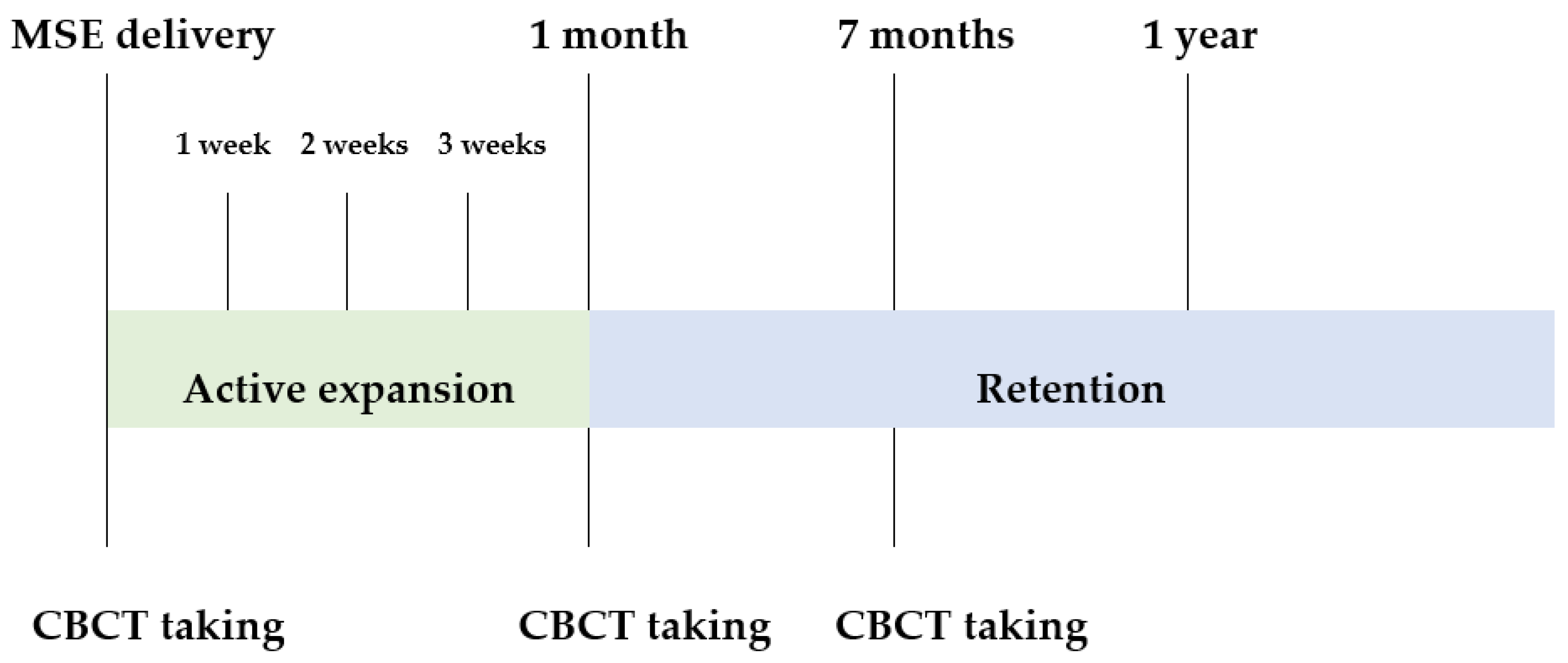
The MSE treatment protocol applied in our clinic is as follows (Figure 2). To fabricate the MSE device, a separation ring is inserted anteriorly and posteriorly to the maxillary first molars. If the separation ring cannot be inserted well due to tight interdental contact, a brass wire is inserted. A band is fitted 7 days after separation. After taking a pickup impression, an MSE is placed as close to the palate as possible on the working model. The MSE arm is soldered to the maxillary first molar band. The activation rate is 1 turn per day for early teens (under 15 years), 2 turns per day for late teens (over 15, under 20 years), and 4 turns per day for adult patients (over 20 years) until diastema appears. Once a diastema was observed, the number of activations was halved, and the expansion continued until sufficient expansion was obtained. After implantation, the patient was asked to visit the clinic every 7 to 10 days to observe whether a diastema had occurred. After the diastema occurs, the number of expansions is reduced by half. Existing opinions on palatal expansion are mainly described based on the cusps of the maxillary and mandibular teeth, and it is recommended to expand enough to overcorrection. Similarly, in adult patients, the amount of expansion can be recommended based on the cusp, but it is necessary to balance the width of the maxilla with a narrow palatal arch and the width of the mandible, which is relatively normal when viewed from the frontal face of the patient.
Active expansion is usually performed for about a month, and thereafter, the MSE is maintained so that bone deposition can occur at the expansion site. It has been reported that sufficient new bone is deposited in the expanded area 3 to 6 months after palatal expansion, and clinically, when observing the median palatine suture using CT in some patients, the new bone generated between the expanded median palatine sutures was reported. It is expected that it will take several months to fully mature. Therefore, unless there is a special reason, it is recommended to maintain the MSE for 6 months to 1 year. Six months after the end of the expansion, a CBCT is taken to check whether the expansion is well maintained and if there is sufficient bone formation.
During CBCT imaging, all patients were instructed to sit upright with their imagined Frankfort horizontal plane parallel to the floor. To ensure their position during the CBCT scan, their heads were secured with a chin cup and ear rods. The CBCT scanner was adjusted to 80 kVp and 7.0 mA for adults and 80 kVp and 3.0 mA for adolescents (Alphard-3030; ASAHI Roentgen IND, Kyoto, Japan). The scan time was 17 s, and the voxel size was 0.39 mm. INFINITT PACS software was used to import the images as digital imaging and communications in medicine files after they had been taken (INFINITT Healthcare Co, Ltd., Seoul, Korea).

2.3. CBCT Landmarks, Reference Planes and Measurements

CBCT multiplanar reconstruction images were reoriented using nasion, right porion and right and left orbitale and were examined using OnDemand 3D software (Cybermed, Seoul, South Korea). Landmarks around the IC and maxillary central incisors were traced on the T0 and T1 CBCT images of each subject. The Frankfort-horizontal (FH) plane served as a horizontal reference plane for the axial plane. It was found that the sagittal plane was parallel to the plane that passed across the anterior and posterior nasal spines and that it was perpendicular to the axial plane.
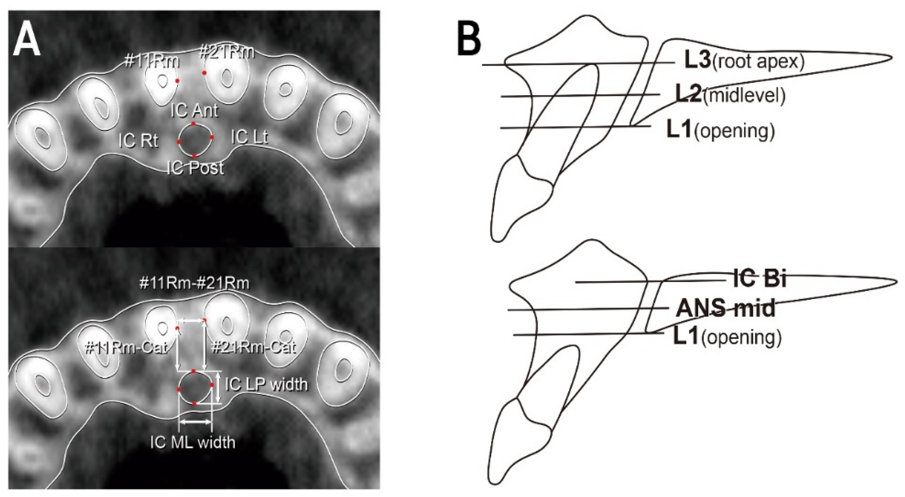
The landmarks and measurements used in this study are described in Figure 3A and Table 1 [22]. When the position of the root apex of the left and right central incisors was vertically different, L3 was set based on the apex located further below.
In ten of the samples, the root apex was located lower than the oral opening of the IC. Therefore, the IC level through the root of the maxillary incisor was not available for these samples. Therefore, in this case, we used another method using ANS and IC nasal bifurcation to measure the IC dimension (Figure 3B, bottom) [29]. The anteroposterior distances between the IC and the maxillary central incisor were not measured. The IC ML width, IC LP width, and the distance between the crown tip of the maxillary central incisors and the distance between the apex of the maxillary central incisors were measured regardless of the vertical level setting.
In the previous study by Cho [22], three indices for measuring the anterior–posterior distance between the maxillary central incisor and the IC were established: Rm–Cat, the distance from Rm to the tangent line through the most anterior point of the incisive canal; Rm–Canal, the distance from Rm to the anterior border of the incisive Canal; and IC Lt (or Rt)–Root, the distance from Cl to the posterior border of the maxillary central incisor root. As a result of the measurement, these three values did not show a significant difference according to the vertical level. Therefore, in our study, Rm–Cat was set as a representative value for measuring the anteroposterior distance between the maxillary central incisor and the IC in consideration of the convenience and accuracy of measurement.
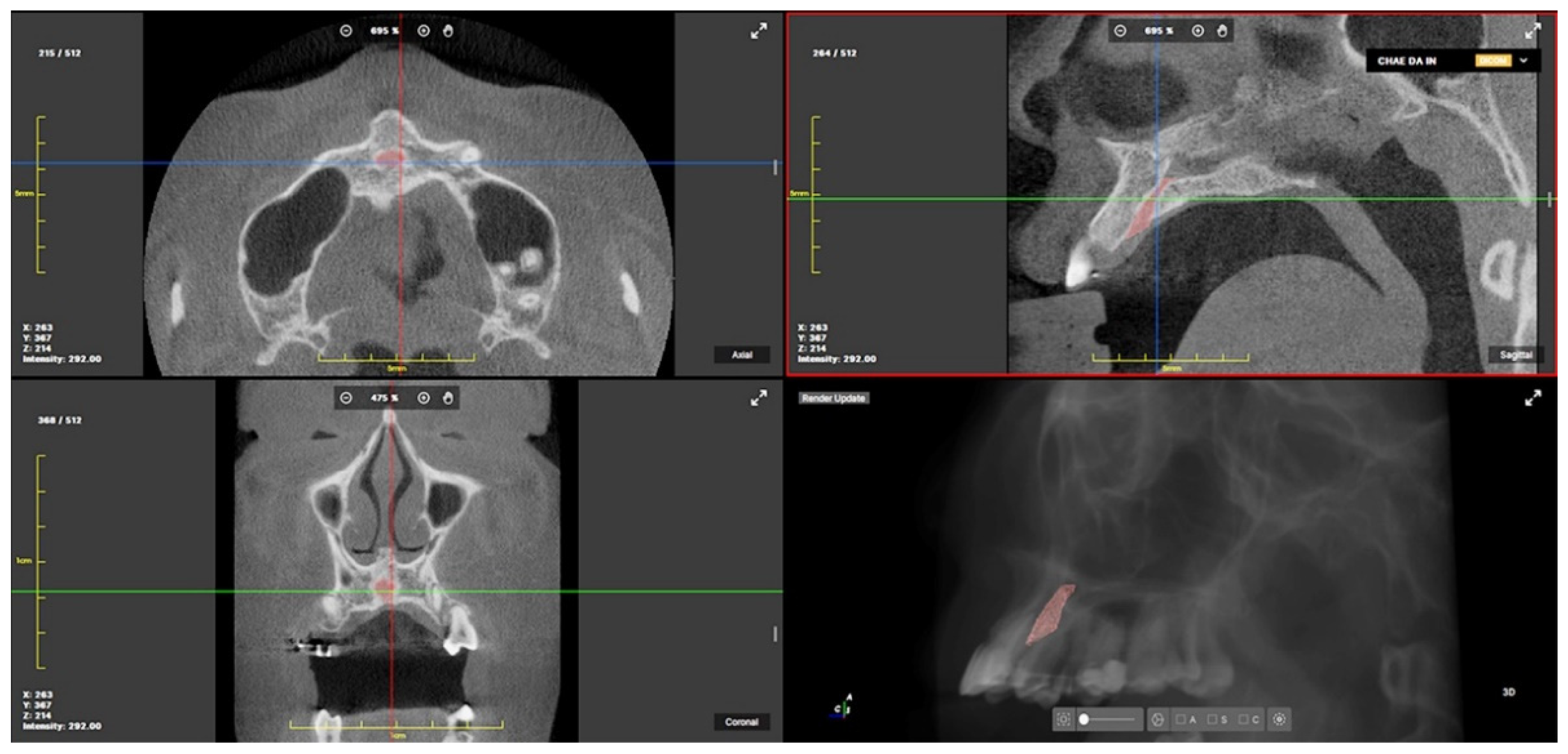
The IC was segmented using semi-automatic 3D segmentation and image analysis software (Medilabel, Ingradient, Seoul, Korea), and 3D volumetric models of the IC were generated from these segmentations (Figure 4). The volume of the IC was automatically calculated in pixels and cubic centimeters.

2.4. Statistical Evaluation

All measurements were re-measured after 2 weeks by the same investigator (UR C). Intraclass correlation coefficients (ICCs) were calculated for reliability analysis. Most of ICCs were over 0.93 (excellent reliability), except for #11Cr-#21Cr (0.787, fair reliability). Kolmogorov–Smirnov and Shapiro–Wilk tests were performed to assess the normality of the data. T0 and T1 data were compared using paired t-tests in parametric data and Wilcoxon signed-rank test in non-parametric data. p < 0.05 was judged to be statistically significant.

3. Results

3.1. Changes in IC Dimensions and Tooth–Canal Relationship before and after MARPE Treatment

Samples in which the apices of the maxillary central incisors were located more superior to the oral opening of the incisive canal are analyzed in Table 2 (n = 20).
Mediolateral and labiopalatal widths of the IC were significantly increased in all three levels after MARPE treatment (p < 0.01), and the amount of increase was greater in mediolateral direction than labiopalatal direction. Anteroposterior distance from the mesial point of maxillary central incisors to the anterior margin of the IC was significantly decreased only in L1 level (p < 0.05).
Ten samples where the root apex of maxillary central incisors was vertically separated from the IC are analyzed in Table 3 (n = 10). Similar to the other 20 samples, the mediolateral and labiopalatal width of the IC significantly increased after MARPE treatment (p < 0.05), except for IC LP width in the L1 level.

3.2. Change in the Positional Relationship between the Maxillary Central Incisors after MARPE Treatment (Table 4)

Mediolateral distance between mesial points of maxillary central incisors was significantly increased in all three levels (p < 0.001). The distance between the root apex of the maxillary central incisors significantly increased after MARPE (p < 0.001), but the distance between the crown tips of the maxillary central incisors did not significantly increase, even after MARPE treatment (p > 0.05).
Table 4. Change in positional relationship between the maxillary central incisors after MARPE treatment (mean ± standard deviation).
MeasurementsT0T1p Value
#11Cr-#21Cr (n = 30)9.74 ± 1.449.43 ± 0.800.258
#11Rm-#21Rm(L1) (n = 20)2.88 ± 1.06 4.86 ± 1.84 0.000 ***
#11Rm-#21Rm(L2) (n = 20)3.25 ± 1.15 5.44 ± 1.75 0.000 ***
#11Rm-#21Rm(L3) (n = 20)3.95 ± 1.12 5.84 ± 1.66 0.000 ***
#11Rt-#21Rt (n = 30)7.17 ± 1.558.94 ± 1.680.000 ***
*** p < 0.001.

3.3. Volume Change in the IC after MARPE Treatment

The volume of the IC was measured for all samples, and the results are described in Table 5. The volume of the IC significantly increased after MARPE treatment (p < 0.001), and the average increase in the total volume of the IC was about 65%.

4. Discussion

The purpose of this study was to investigate whether there was a change in the shape of IC and the relationship between the maxillary central incisor and IC after MARPE treatment.
MARPE has been reported to induce not only maxillary transverse expansion but also three-dimensional changes in the zygomaticomaxillary complex [30]. We hypothesized that the morphology of the IC existing between the left and right maxilla would be changed through MARPE, which would affect the tooth–canal relationship.
Using CBCT images, the regional morphologic changes of the IC in response to MARPE therapy were identified. These changes are comparable to the remodeling pattern and changes of the hard and soft tissues to different stimuli, including orthodontic tooth movement [31,32,33,34,35].
The resolution of reconstructed CBCT images may vary based on a number of factors, including kVa, mA, scan time, reconstruction algorithms, the measurement location of the phantom, etc. [36,37]. A 0.3-mm voxel size is often used with a considerably longer scan time for CBCT images with a broad field of view covering the whole face, as in our CBCT images, to provide higherquality images. CBCT images obtained for orthodontic reasons have been utilized as a reliable method to study orthodontically caused apical root resorption and changes in surrounding tissues, even if the limit of distance measurement is indicated to be twice the voxel size. Overall morphologic alterations, interroot distance, root canal distances, and the volume of the IC following MARPE treatment were all reliably detected in the research.
The mediolateral, labiopalatal width, and volume of the IC increased clearly in all participants who received the MARPE treatment. It is notable that, to our knowledge, this is the first research to identify the remodeling possibilities of IC in response to MARPE treatment.
Since the relationship between the IC and the teeth is more clinically important than the morphological change of the IC itself, the level was set using the roots of the maxillary central incisors. However, in a third of the total samples, IC was positioned at a different vertical level from the maxillary central incisors. Therefore, the plane was set based on the skeletal landmarks in these samples. That is, in about one-third of the patients, there was no risk of root-incisive canal contact due to retraction of the maxillary arch unless intrusion of maxillary anterior teeth was planned.
CBCTs taken 6 months after completion of the expansion were compared with T0 to provide sufficient time for remodeling of the ICs and ossification of the midpalatal sutures. However, some of the samples had an irregular and ambiguous margin in the anterior and posterior area of the IC due to insufficient ossification of midpalatal sutures. In these cases, an imaginary ellipse was created along the lateral wall of the IC, and the anterior and posterior points were marked. The labiopalatal width of the IC was possibly slightly changed due to the cortical ossification of the IC progress.
The mean increase in mediolateral width of the IC was about 2 mm, similar to the interdental distance increase between the maxillary central incisors. This suggests that MARPE did not increase the likelihood of contact between the IC and the roots of the maxillary central incisors by expanding the IC laterally. In patients with maxillary transverse deficiency, most of the central incisors were inclined distally. Therefore, unless the amount of expansion was large, there was little chance that the distance between the roots of the teeth would decrease significantly during orthodontic treatment.
According to our findings, the biological anteroposterior distances between the roots of the central incisors in the maxilla and the incisive canal were about 4–5 mm, which is a little smaller than Cho’s study. Although not statistically significant, the distance between the maxillary central incisor and IC showed a tendency to decrease slightly after treatment compared to before MARPE treatment.
Taking into account the morphologic aspects of the incisive canal, the central incisor roots, the L1 or L2 apical third’s posterior-median aspect of the roots as opposed to the root apex itself is most likely to approximate with the canal after retraction and root movement of the maxilla [22]. However, anterior intrusion occurs in a variety of clinical circumstances, increasing the potential for approximation along the tooth movement. As a result, 3D images may be useful in determining the distance between the incisive canal and the dimensional properties when a substantial maxillary incisor retraction with vertical control is planned.
After MARPE, the anteroposterior distance between the maxillary central incisor and the IC decreased only at the L1 level. One of the most apparent changes related to rapid Maxillary expansion is the formation of a diastema between the maxillary central incisors. The incisors are anticipated to separate roughly half the distance the expansion screw has been extended during active suture opening. Following this separation, the crowns of the incisors converge and make proximal contact. The elastic rebound of the transseptal fibers causes the crowns to mesially tip. The persistent pull of the fibers enables the roots to converge toward their original axial inclinations after the crowns make contact. This cycle takes approximately 4 months [38]. Considering that the distance between the crown points of the maxillary incisors did not increase after MARPE means that the maxillary central incisors moved mesiopalatally toward the diastema. As a result, the decrease in the anterior–posterior distance between the maxillary central incisor and the IC was about 0.5 mm, which was statistically significant but not clinically significant.
The application of maxillary expansion has been extended to nasal obstruction since it has been hypothesized that RME increases nasal breadth and volume [39]. Likewise, considering the effect of rapid maxillary expansion on the surrounding anatomical structures, MARPE may affect the IC volume. The volume of the IC was significantly increased after MARPE. If the increased volume was comprised of fibrous tissue and blood vessels similar to the midpalatal suture in rapid palatal expansion, additional morphological changes would be possible during the remodeling process. Further histological examination is required to confirm this part. It is also worth noting that transient paresthesia of the lower lip was recorded in the mandibular canal in relation to tooth root when in touch with the canal wall after orthodontic tooth movement [40]. Does a temporary or long-lasting loss of sensitivity near the incisal foramen occur as a result of the median dissection of the nasopalatine channels at their point of unification? To fully address this question, additional research is required.
In this study, 30 subjects were divided into a group in which the root and IC of the maxillary central incisor vertically overlap and a group in which they were separated. It would also be worthwhile to investigate the differences between the two types of subjects, such as differences in skeletal patterns, through further research.
There were several limitations in this study. (1) The amount of MSE expansion varied depending on the patient’s orthodontic condition. (2) The age of the patients ranged from the early teens to the 50 s, so the suture maturity was probably different. (3) Due to the wide age distribution of the samples, different expansion protocols were used. (4) Since the alignment of maxillary incisors was initiated between T0 and T1 in many samples for aesthetic reasons, it is not clear whether the treatment outcome was just due to MARPE.

5. Conclusions

In one-third of the samples, the maxillary central incisor root and IC did not overlap vertically. MARPE increased the ML, LP width and volume of the IC but did not result in a clinically significant change in the root–canal relationship.
MARPE did not increase the contact between the maxillary central incisor root and the IC during maximum retraction of the anterior teeth.

Author Contributions

Conceptualization, S.-K.C. and Y.-R.K.; methodology, S.-K.C. and Y.-R.K.; software, J.H.P.; validation, J.H.P., N.-Y.C. and K.-H.K.; formal analysis, Y.-R.K.; investigation, S.-K.C.; resources, K.-H.K.; data curation, N.-Y.C.; writing—original draft preparation, S.-K.C. and Y.-R.K.; writing—review and editing, J.-M.C. and J.H.P.; visualization, J.H.P.; supervision, J.-M.C.; project administration, J.-M.C. All authors have read and agreed to the published version of the manuscript.

Funding

This research received no external funding.

Institutional Review Board Statement

The study was conducted in accordance with the Declaration of Helsinki and approved by the Institutional Review Board at the Wonkwang University Dental Hospital (IRB number WKUDHIRB202206-02).

Informed Consent Statement

Written informed consent was obtained from the patient for publication of this short report and any accompanying images.

Data Availability Statement

The authors declare that the materials are available.

Acknowledgments

This paper was supported by Wonkwang University in 2022.

Conflicts of Interest

The authors declare no conflict of interest.

References

  1. Bennardo, F.; Barone, S.; Vocaturo, C.; Nucci, L.; Antonelli, A.; Giudice, A. Usefulness of Magnetic Mallet in Oral Surgery and Implantology: A Systematic Review. J. Pers. Med. 2022, 12, 108. [Google Scholar] [CrossRef] [PubMed]
  2. McNamara, J.A. Maxillary Transverse Deficiency. Am. J. Orthod. Dentofac. Orthop. Off. Publ. Am. Assoc. Orthod. Its Const. Soc. Am. Board Orthod. 2000, 117, 567–570. [Google Scholar] [CrossRef]
  3. Vandersea, B.A.; Ruvo, A.T.; Frost, D.E. Maxillary Transverse Deficiency–Surgical Alternatives to Management. Oral Maxillofac. Surg. Clin. N. Am. 2007, 19, 351–368. [Google Scholar] [CrossRef] [PubMed]
  4. Haas, A.J. Rapid Expansion of the Maxillary Dental Arch and Nasal Cavity by Opening the Midpalatal Suture. Angle Orthod. 1961, 31, 73–90. [Google Scholar] [CrossRef]
  5. Haas, A.J. The Treatment of Maxillary Deficiency by Opening the Midpalatal Suture. Angle Orthod. 1965, 35, 200–217. [Google Scholar] [CrossRef]
  6. Sandikçiolu, M.; Hazar, S. Skeletal and Dental Changes after Maxillary Expansion in the Mixed Dentition. Am. J. Orthod. Dentofacial Orthop. 1997, 111, 321–327. [Google Scholar] [CrossRef]
  7. Liu, S.; Xu, T.; Zou, W. Effects of Rapid Maxillary Expansion on the Midpalatal Suture: A Systematic Review. Eur. J. Orthod. 2015, 37, 651–655. [Google Scholar] [CrossRef] [Green Version]
  8. Christie, K.F.; Boucher, N.; Chung, C.-H. Effects of Bonded Rapid Palatal Expansion on the Transverse Dimensions of the Maxilla: A Cone-Beam Computed Tomography Study. Am. J. Orthod. Dentofac. Orthop. 2010, 137 (Suppl. S4), S79–S85. [Google Scholar] [CrossRef]
  9. Baysal, A.; Karadede, I.; Hekimoglu, S.; Ucar, F.; Ozer, T.; Veli, I.; Uysal, T. Evaluation of Root Resorption Following Rapid Maxillary Expansion Using Cone-Beam Computed Tomography. Angle Orthod. 2012, 82, 488–494. [Google Scholar] [CrossRef] [Green Version]
  10. Garib, D.G.; Henriques, J.F.C.; Janson, G.; de Freitas, M.R.; Fernandes, A.Y. Periodontal Effects of Rapid Maxillary Expansion with Tooth-Tissue-Borne and Tooth-Borne Expanders: A Computed Tomography Evaluation. Am. J. Orthod. Dentofac. Orthop. Off. Publ. Am. Assoc. Orthod. Its Const. Soc. Am. Board Orthod. 2006, 129, 749–758. [Google Scholar] [CrossRef]
  11. Suri, L.; Taneja, P. Surgically Assisted Rapid Palatal Expansion: A Literature Review. Am. J. Orthod. Dentofac. Orthop. 2008, 133, 290–302. [Google Scholar] [CrossRef]
  12. Wilmes, B.; Nienkemper, M.; Drescher, D. Application and Effectiveness of a Mini-Implant-and Tooth-Borne Rapid Palatal Expansion Device: The Hybrid Hyrax. World J. Orthod. 2010, 11, 323–330. [Google Scholar]
  13. Carlson, C.; Sung, J.; McComb, R.W.; Machado, A.W.; Moon, W. Microimplant-Assisted Rapid Palatal Expansion Appliance to Orthopedically Correct Transverse Maxillary Deficiency in an Adult. Am. J. Orthod. Dentofac. Orthop. 2016, 149, 716–728. [Google Scholar] [CrossRef] [PubMed]
  14. Brunetto, D.P.; Sant’Anna, E.F.; Machado, A.W.; Moon, W. Non-Surgical Treatment of Transverse Deficiency in Adults Using Microimplant-Assisted Rapid Palatal Expansion (MARPE). Dent. Press J. Orthod. 2017, 22, 110–125. [Google Scholar] [CrossRef] [PubMed] [Green Version]
  15. Park, J.J.; Park, Y.-C.; Lee, K.-J.; Cha, J.-Y.; Tahk, J.H.; Choi, Y.J. Skeletal and Dentoalveolar Changes after Miniscrew-Assisted Rapid Palatal Expansion in Young Adults: A Cone-Beam Computed Tomography Study. Korean J. Orthod. 2017, 47, 77–86. [Google Scholar] [CrossRef] [PubMed] [Green Version]
  16. Lim, H.-M.; Park, Y.-C.; Lee, K.-J.; Kim, K.-H.; Choi, Y.J. Stability of Dental, Alveolar, and Skeletal Changes after Miniscrew-Assisted Rapid Palatal Expansion. Korean J. Orthod. 2017, 47, 313–322. [Google Scholar] [CrossRef] [Green Version]
  17. Lee, K.-J.; Park, Y.-C.; Park, J.-Y.; Hwang, W.-S. Miniscrew-Assisted Nonsurgical Palatal Expansion before Orthognathic Surgery for a Patient with Severe Mandibular Prognathism. Am. J. Orthod. Dentofac. Orthop. 2010, 137, 830–839. [Google Scholar] [CrossRef]
  18. Lee, D.-W.; Park, J.H.; Moon, W.; Seo, H.Y.; Chae, J.-M. Effects of Bicortical Anchorage on Pterygopalatine Suture Opening with Microimplant-Assisted Maxillary Skeletal Expansion. Am. J. Orthod. Dentofac. Orthop. 2021, 159, 502–511. [Google Scholar] [CrossRef]
  19. Cunha, A.C.; da Lee, H.; Nojima, L.I.; da C. G. Nojima, M.; Lee, K.-J. Miniscrew-Assisted Rapid Palatal Expansion for Managing Arch Perimeter in an Adult Patient. Dent. Press J. Orthod. 2017, 22, 97–108. [Google Scholar] [CrossRef]
  20. Chung, C.J.; Choi, Y.J.; Kim, K.-H. Approximation and Contact of the Maxillary Central Incisor Roots with the Incisive Canal after Maximum Retraction with Temporary Anchorage Devices: Report of 2 Patients. Am. J. Orthod. Dentofac. Orthop. 2015, 148, 493–502. [Google Scholar] [CrossRef] [Green Version]
  21. Chung, C.J.; Nguyen, T.; Lee, J.-H.; Kim, K.-H. Incisive Canal Remodelling Following Maximum Anterior Retraction Reduces Apical Root Resorption. Orthod. Craniofac. Res. 2021, 24 (Suppl. S1), 59–65. [Google Scholar] [CrossRef] [PubMed]
  22. Cho, E.-A.; Kim, S.-J.; Choi, Y.J.; Kim, K.-H.; Chung, C.J. Morphologic Evaluation of the Incisive Canal and Its Proximity to the Maxillary Central Incisors Using Computed Tomography Images. Angle Orthod. 2016, 86, 571–576. [Google Scholar] [CrossRef] [PubMed] [Green Version]
  23. Mraiwa, N.; Jacobs, R.; Van Cleynenbreugel, J.; Sanderink, G.; Schutyser, F.; Suetens, P.; van Steenberghe, D.; Quirynen, M. The Nasopalatine Canal Revisited Using 2D and 3D CT Imaging. Dentomaxillofac. Radiol. 2004, 33, 396–402. [Google Scholar] [CrossRef] [PubMed]
  24. Liang, X.; Jacobs, R.; Martens, W.; Hu, Y.; Adriaensens, P.; Quirynen, M.; Lambrichts, I. Macro- and Micro-Anatomical, Histological and Computed Tomography Scan Characterization of the Nasopalatine Canal. J. Clin. Periodontol. 2009, 36, 598–603. [Google Scholar] [CrossRef] [Green Version]
  25. Song, W.-C.; Jo, D.-I.; Lee, J.-Y.; Kim, J.-N.; Hur, M.-S.; Hu, K.-S.; Kim, H.-J.; Shin, C.; Koh, K.-S. Microanatomy of the Incisive Canal Using Three-Dimensional Reconstruction of MicroCT Images: An Ex Vivo Study. Oral Surg. Oral Med. Oral Pathol. Oral Radiol. Endodontol. 2009, 108, 583–590. [Google Scholar] [CrossRef] [PubMed]
  26. Zong, C.; Tang, B.; Hua, F.; He, H.; Ngan, P. Skeletal and Dentoalveolar Changes in the Transverse Dimension Using Microimplant-Assisted Rapid Palatal Expansion (MARPE) Appliances. Semin. Orthod. 2019, 25, 46–59. [Google Scholar] [CrossRef] [Green Version]
  27. Salmoria, I.; de Souza, E.C.; Furtado, A.; Franzini, C.M.; Custodio, W. Dentoskeletal Changes and Their Correlations after Micro-Implant-Assisted Palatal Expansion (MARPE) in Adults with Advanced Midpalatal Suture Ossification. Clin. Oral Investig. 2022, 26, 3021–3031. [Google Scholar] [CrossRef]
  28. Sfogliano, L. A Three-Dimensional Analysis to Investigate the Midface Changes with MSE. Master’s Thesis, University of California, Los Angeles (UCLA), Los Angeles, CA, USA, 2019. [Google Scholar]
  29. Asaumi, R.; Kawai, T.; Sato, I.; Yoshida, S.; Yosue, T. Three-Dimensional Observations of the Incisive Canal and the Surrounding Bone Using Cone-Beam Computed Tomography. Oral Radiol. 2010, 26, 20–28. [Google Scholar] [CrossRef]
  30. Song, K.-T.; Park, J.H.; Moon, W.; Chae, J.-M.; Kang, K.-H. Three-Dimensional Changes of the Zygomaticomaxillary Complex after Mini-Implant Assisted Rapid Maxillary Expansion. Am. J. Orthod. Dentofac. Orthop. 2019, 156, 653–662. [Google Scholar] [CrossRef]
  31. Wennström, J.L.; Lindhe, J.; Sinclair, F.; Thilander, B. Some Periodontal Tissue Reactions to Orthodontic Tooth Movement in Monkeys. J. Clin. Periodontol. 1987, 14, 121–129. [Google Scholar] [CrossRef]
  32. Baek, E.S.; Hwang, S.; Choi, Y.J.; Roh, M.R.; Nguyen, T.; Kim, K.-H.; Chung, C.J. Quantitative and Perceived Visual Changes of the Nasolabial Fold Following Orthodontic Retraction of Lip Protrusion. Angle Orthod. 2018, 88, 465–473. [Google Scholar] [CrossRef] [PubMed]
  33. Krishnan, V.; Davidovitch, Z. On a Path to Unfolding the Biological Mechanisms of Orthodontic Tooth Movement-V. 2009. Available online: https://journals.sagepub.com/doi/full/10.1177/0022034509338914 (accessed on 12 September 2022).
  34. Chung, C.J.; Jang, W.; Piers, C.; Lee, D.-W.; Hwang, S.; Kim, K.-H.; Proffit, W.R.; Nguyen, T. Differential Alveolar Bone Modeling after Orthodontic Retraction. J. Am. Dent. Assoc. 2019, 150, 313–320. [Google Scholar] [CrossRef] [PubMed]
  35. Krishnan, V.; Davidovitch, Z. Cellular, Molecular, and Tissue-Level Reactions to Orthodontic Force. Am. J. Orthod. Dentofacial Orthop. 2006, 129, 469.e1–469.e32. [Google Scholar] [CrossRef] [PubMed]
  36. Brüllmann, D.; Schulze, R.K.W. Spatial Resolution in CBCT Machines for Dental/Maxillofacial Applications—What Do We Know Today? Dentomaxillofac. Radiol. 2015, 44, 20140204. [Google Scholar] [CrossRef] [PubMed] [Green Version]
  37. Tsutsumi, K.; Chikui, T.; Okamura, K.; Yoshiura, K. Accuracy of linear measurement and the measurement limits of thin objects with cone beam computed tomography: Effects of measurement directions and of phantom locations in the fields of view. Int. J. Oral Maxillofac. Implants. 2011, 26, 91–100. [Google Scholar]
  38. Kumar, S.A.; Gurunathan, D.; Muruganandham, S.S.; Kumar, S.A. Rapid Maxillary Expansion: A Unique Treatment Modality in Dentistry. J. Clin. Diagn. Res. 2011, 5, 906–911. [Google Scholar]
  39. Doruk, C.; Sökücü, O.; Biçakçi, A.A.; Yilmaz, U.; Taş, F. Comparison of Nasal Volume Changes during Rapid Maxillary Expansion Using Acoustic Rhinometry and Computed Tomography. Eur. J. Orthod. 2007, 29, 251–255. [Google Scholar] [CrossRef] [Green Version]
  40. Krogstad, O.; Omland, G. Temporary Paresthesia of the Lower Lip: A Complication of Orthodontic Treatment. A Case Report. Br. J. Orthod. 1997, 24, 13–15. [Google Scholar] [CrossRef]
Figure 1. (A) MSE II appliances. (B) Initial state. (C) After expansion.
Applsci 12 10470 g001
Figure 2. MSE treatment protocol.
Applsci 12 10470 g002
Figure 3. Assessment of incisive canal (IC) and central incisor relationship. Landmarks and linear measurements. (A). #11Rm, the most medial point of the right maxillary central incisor root; #21Rm, the most medial point of the left maxillary central incisor root; IC Lt, the most lateral point of the left contour of the IC; IC Rt, the most lateral point of the right contour of IC, IC Ant, the most anterior point of the incisive canal; IC Post, the most posterior point of IC; #11Rm-#21Rm, mediolateral distance between #11Rm and #21 Rm; #11Rm-Cat, anteroposterior distance between #11Rm and anterior margin of IC; #21Rm-Cat, the anteroposterior distance between #21Rm and anterior margin of IC; IC LP width, labiopalatal width of IC; IC ML width, mediolateral width of IC (B). Three vertical levels of the incisive canal: L1, the level at which the IC begins on the oral side; L2, midlevel between L1 and L3; L3, root apex level; IC Bi, the level at which the IC bifurcates in the nasal cavity; ANS mid, midlevel between L1 and the level passing ANS. All levels were parallel to the Frankfort horizontal plane.
Applsci 12 10470 g003
Figure 4. Axial, sagittal, coronal view and the result of volume rendering of the IC on pretreatment CBCT (red).
Applsci 12 10470 g004
Table 1. Measurements used in this study (summary of abbreviations).
MeasurementDefinition
#11Rm-#21Rm Mediolateral distance between #11Rm and #21Rm (mm)
#11Rm-CatAnteroposterior distance between #11Rm and anterior margin of IC (mm)
#21Rm-CatAnteroposterior distance between #21Rm and anterior margin of IC (mm)
IC LP widthLabiopalatal width of IC (mm)
IC ML widthMediolateral width of IC (mm)
#11Rt-#21RtMediolateral distance between root apices of #11 and #21 (mm)
#11Cr-#21CrMediolateral distance between crown tips of #11 and #21 (mm)
IC volumeVolume of IC from oral opening to nasal bifurcation (px, cm3)
Table 2. Changes in IC dimensions and tooth–canal relationship after MARPE treatment (mean ± standard deviation).
Measurements
(n = 20)
L1 L2L3
T0T1p ValueT0T1p ValueT0T1p Value
IC ML width3.64 ± 1.47 5.82 ± 1.04 0.000 ***3.71 ± 1.04 5.73 ± 1.46 0.000 ***3.45 ± 0.84 5.77 ± 1.270.000 ***
IC LP width3.27 ± 0.69 3.60 ± 0.73 0.008 **3.09 ± 0.65 3.61 ± 0.50 0.001 **2.90 ± 0.573.38 ± 0.620.004 **
#11 Rm-Cat4.48 ± 1.34 4.03 ± 1.17 0.008 **4.13 ± 1.47 3.95 ± 1.50 0.2184.11 ± 1.473.94 ± 1.450.299
#21 Rm-Cat4.53 ± 1.25 4.02 ± 1.15 0.013 *4.18 ± 1.54 3.90 ± 1.310.1294.07 ± 1.553.87 ± 1.440.182
* p < 0.05, ** p < 0.01, *** p < 0.001. Wilcoxon signed-rank test.
Table 3. Changes in IC dimensions after MARPE treatment in samples where the maxillary central incisors and the IC were vertically separated (mean ± standard deviation).
Measurements
(n = 10)
L1ANS midIC Bi
T0T1p ValueT0T1p ValueT0T1p Value
IC ML width3.97 ± 2.185.97 ± 1.100.028 *3.52 ± 1.525.70 ± 1.280.000 ***4.85 ± 1.536.73 ± 1.560.000 ***
IC LP width2.89 ± 0.722.98 ± 0.630.7122.59 ± 0.712.93 ± 0.880.004 **3.09 ± 1.1093.37 ± 1.130.047 *
* p < 0.05, ** p < 0.01, *** p < 0.001. Wilcoxon signed-rank test.
Table 5. Volume change in the IC after MARPE treatment (mean ± standard deviation).
MeasurementsT0T1p Value
IC volume (px)2269.6 ± 838.73765.8 ± 1096.80.000 ***
IC volume (cm3)0.13 ± 0.0500.22 ± 0.0640.000 ***
*** p < 0.001.
Publisher’s Note: MDPI stays neutral with regard to jurisdictional claims in published maps and institutional affiliations.