Article

Lab-on-Chip Culturing System for Fungi—Towards Nanosatellite Missions

by
Agnieszka Krakos (Podwin)
1,*,
Patrycja Śniadek
1,
Marta Jurga
2,
Marcin Białas
1,
Agata Kaczmarek-Pieńczewska
2,
Krzysztof Matkowski
2,
Rafał Walczak
1 and
Jan Dziuban
1
1
Division of Microsystems, Faculty of Electronics, Photonics and Microsystems, Wroclaw University of Science and Technology, 27 Wybrzeze Wyspianskiego Street, 50-370 Wroclaw, Poland
2
Division of Plant Pathology and Mycology, Department of Plant Protection, Wroclaw University of Environmental and Life Sciences, Plac Grunwaldzki 24a, 50-363 Wroclaw, Poland
*
Author to whom correspondence should be addressed.
Appl. Sci. 2022, 12(20), 10627; https://doi.org/10.3390/app122010627
Submission received: 16 September 2022 / Revised: 11 October 2022 / Accepted: 19 October 2022 / Published: 20 October 2022

Abstract

:
In this paper, a lab-on-chip system dedicated to fungi cultivation in Earth’s gravity and simulated microgravity, being a solution that could be used in future nanosatellite missions, is shown. For the first time, a fully glass lab-on-chip structure enabling the proper environment for cultivation of fungi species—Fusarium culmorum—is presented. Apart from the biological validation of the fungi cultures with the use of the lab-on-chip system, tests were carried out under induced microgravity utilising a Rotary Wall Vessel. Correct functioning of the lab-on-chip system was obtained, enabling the growth of fungi spores both in ground and in simulated microgravity conditions. Interestingly, culturing tests have shown that microgravity stimulates the growth of fungi notably, compared to the ground-based experimentation performed simultaneously. The findings of this study can provide substantial new knowledge on microscopic fungi cultivation in lab-on-chip devices, other soil organisms, as well as a potential behavior of these species in microgravity conditions. Culturing system shown in this work can help mycologists to provide better understanding of microscopic fungi nature and their development mechanisms at a single spore level. This opens the way towards regular usage of microfluidic tools in agriculture and horticulture fields and more importantly, in future research on microscopic fungi in space, e.g., as a part of nanosatellite missions.

1. Introduction

Contemporary techniques of microfluidics find applications in most of the scientific branches, especially in the field of life sciences, where reliable, fast and automated analysis is indispensable [1,2,3]. Rapid development of laboratory-on-chip devices and accompanying new methodologies ensure new insights into biomedical solutions by providing substantial improvements over often complex and labour-intensive protocols [4,5,6]. High throughput screening, manipulation and analysis based on single cell techniques; perspective for very specific LOC designs with simultaneous ease of integration and relatively low costs bring new advances for diagnostics and biomedical engineering [7,8,9,10,11]. However, there are still a few domains in which the potential of LOCs has not been fully recognised, and one of these branches may be considered to be mycology.
In principle, mycology is the biological discipline that covers the study of fungi. This is a kingdom of a vast group of organisms (both macroscopic and microscopic) of great significance, especially in the context of the so-called tree of life [12,13]. Investigation of fungi encompasses not only their taxonomy, genetic and biochemical properties, but also multiple beneficial and harmful effects that have a notable impact on our daily lives [14,15,16].
Fungi can be considered to be fairly dangerous, e.g., some species are pathogenic towards plants or cause human and animal diseases. On that basis, special control and in-depth investigation of both uniform colonies and the mutual relationships with other environmental organisms is significant and necessary. Microscopic fungi also find application in diagnostics and plant health evaluation—i.e., in crops, plants, and rare and precious species—in identification of new fungi species and their genetic mutations, track changes in environmental and impact fungi for other organisms and our daily lives [17].
As was mentioned previously in this paper, the application of miniaturised microfluidic tools for mycology is currently quite limited, but gradually growing in popularity. The first report on the usage of a lab-on-chip for mycology, to the best of the authors’ knowledge, was published in 2010 by M. Held et al. [18]. The paper mentions biological responses of two diverse filamentous fungi confined in miniaturised PDMS mazel-like structures, pointing to specific, previously unknown directional growth of these species.
Further works within this scope appeared in 2016, in which the paper released by C.E. Stanley et al. was especially interesting [19]. Herein, the idea of, so-called, soil-on-a-chip was presented, showing the significance of the development of microfluidic platforms for the study of environmental creatures. The article put forward the interactions of soil-dwelling organisms (e.g., nematodes, bacteria, plants, etc.) with special attention paid to fungi as excellent bioindicators of any physical/chemical changes in the local habitats [20,21,22]. Similar issues were raised by the same scientific group recently in [23] and [24], where L.J. Millet et al. present positive results of the application of PDMS lab-on-chip for stationary co-culture of selected filamentous fungi and bacteria.
There are also other papers that discuss the demand for microfluidics in mycology, but not in the context of environmental studies, but rather as advanced techniques for identification of pathogenic fungi in food or blood samples [25,26,27], utilising modern real-time PCR methods [28] or inertial focussing chip-based platforms [27].
Although some examples of fungi investigation utilising lab-on-chip solutions can be found in the literature, there is still a lack of fully featured, portable and miniaturised instruments that would ensure long-term cultivation of these interesting species that also have the capacity to automatically change the culturing conditions, for instance nourishment flow and temperature, and to constantly monitor the colony. Development of the aforementioned tools is vital not only to improve our understanding of our local surroundings but possibly also to address some novel, near-future challenges of astrobiology and space medicine—a trend that is currently gaining in popularity worldwide [29,30,31,32].
As indicated in [33,34,35], several biological objects exhibit uncommon behaviour in microgravity conditions, but the nature of this behaviour has still not been fully recognised. For instance, enhanced biofilm formation of selected bacteria [35] and formation of good quality protein crystals have recently been observed by NASA and SpacePharma, pointing to a potential new generation of vaccines and antibiotic drugs. Moreover, according to the article [34], most cells (e.g., human cancer cells) create 3D forms—MSCs (multicellular spheroids)—in microgravity, which corresponds strongly with the in vivo environment; thus, microgravity can be treated as a perfect model habitat for investigation. Similarly important is the research concerning plants [36,37] and fungi growth, since the recognition of development dynamics of these species may play a crucial role in our future colonisation aspirations.
Based on all of this, herein we present a fully equipped, ready-to-use microfluidic device, dedicated to culturing cosmopolitan and ubiquitous fungi species, on the example of Fusarium culmorum (Wm.G. Sm.) Sacc. This work is in good agreement with the trend in astrobiological research provided mainly by NASA and ESA [38,39,40,41,42,43,44] focussed on the investigation of the influence of microgravity on widely understood life (bacteria, microbes, cells, etc.) in both simulated and space-based conditions. The application of LOCs in this regard outlines their tremendous potential towards completely new insights into the behaviour of microscale objects in microgravity, compared to the conventional, bulky and relatively expensive approaches.
The paper covers multiple steps that had to be undertaken to fulfil the assumptions of the work. At first, the selection of the biological object was done, with a focus put on its features and morphology. Next, the lab-on-chip structure, equipped with specific microchannels arrangement was proposed, designed and numerically modelled to evaluate the LOC performance. Afterwards, the technological works were employed to obtain the lab-on-chip structure and then, the culturing system was assembled. The article is finished with cell culturing experiments performed in standard laboratory conditions and in simulated microgravity, imitated utilizing Rotary Wall Vessel.

2. Materials and Methods

2.1. Biomedical Experimentation in Space—Tools

Most astrobiological research is generally carried out on the International Space Station (ISS). Apart from the cell cultivation experiments, concerning basically stem and cancer cells proliferation and differentiation [34,41,44], along with some interesting research on colloidal chemistry, protein crystallisation and drug delivery has been performed [45]. Nevertheless, experimentation done on the ISS has some substantial limitations. The lack of an experiment control, long waiting time, IP ownership issues and relatively high costs determines the development of novel technical means for astrobiological investigation, such as microgravity simulators (e.g., Rotary Wall Vessel—RWV, Random Positioning Machine—RPM) and CubeSat-type nanosatellites. Ground-based facilities are important first approaches to define baselines and enable thorough testing of the biological system to address gravity-related issues prior to space experiments. CubeSats in turn may provide space-based tests in a way that is fully controlled by the user.
CubeSat-type nanosatellites [46] have been used for space exploration for circa 10 years. Technically, the primary application of these miniaturised instruments is Earth observation and telecommunication, however, a new trend of their utility in the field of biomedicine has already arisen. As one of the basic features that characterises CubeSats is their limited space for experimentation, covering typically 1U–3U (1U = 10 cm3). However, these and other requirements of nanosatellite systems (low power, low weight, high-level integrity, short response time, automation, etc.) can be perfectly matched by microfluidic platforms which have the capacity to ultimately bring some new functionalities for biomedical laboratory payloads. Remote-controlled, miniaturised instrumentation with on-board results reading and LOCs easily adapted per specific research needs can open the way towards novel, universal solutions applicable for high range biomedical tests in outer space. The only problem that is at present commonly faced by the scientists engaged in this field is the limited access to timely rocket launches, which is highly important in the case of biomedical samples and their potential time-dependent stability. Typically, a 3-month waiting period is required and during this period, the nanosatellite cannot be operated, even in standby mode. Prior to launch, the CubeSats are kept in ambient temperature (17–22 °C) in the satellite hangar and only the “late access” option (additionally paid) provides the opportunity to launch the rocket within circa 24 h after nanosatellite integration. On that basis, biomedical experimentation conducted on a CubeSat board in low-Earth orbit (LEO) requires special protectants and/or specific sample preparation methods. One of the solutions herein may be lyophilisation, which for bacteria, nucleic acids and proteins is a standardised procedure [47], but cells and more specialised structures are still a serious problem [48].
In this paper, our microfluidic platform has been designed for simulated and space-based microgravity research to study long-term culturing of microscopic fungi—F. culmorum. For the purpose of these experiments, a simplified method to obtain powdered fungi has been employed, based on drying the sample in elevated temperature conditions. These and other issues related to the development of the LOC platform are described in detail in the following sections of the article.

2.2. Object of the Study—F. culmorum

Fusarium is ubiquitous, cosmopolitan and one of the most important genera, which can be found in most regions of the world. Fusarium spp. has been of interest to scientists for many years and is encountered in plants, soil, water and organic matter biofilms. In particular, this genus is made up of pathogens that produce thread-like hyphae and penetrate plant surfaces as primary or secondary pathogens [49]. An interesting but also dangerous aspect is the production of toxic secondary metabolites by fungi of the genus Fusarium. The presence of mycotoxins in products of plant origin or fodder has a negative impact on our and animal health [50].
One of the most important fungal Fusarium spp. is F. culmorum. This species produces thick-walled and curved macroconidia. Usually, they are 3–5 septate-type and measure 30–50 μm × 5.0–7.5 μm. Microconidia are absent herein, chlamydospores in turn may occur singly, in chains or in clumps. Macroscale colonies of F. culmorum grow rapidly on potato dextrose agar (PDA). The aerial mycelium is dense, whitish, light yellow or red. The underside is greyish-rose, carmine red or burgundy [49].
A similar method of F. culmorum culture preparation was used in these studies. A fungal colony was cultivated on PDA medium for 14-days at room temperature. Next, utilising a sterile cotton-tipped swab, a mass of spores was transferred from the pure culture surface (without gouging the agar) into a 1.5 mL Eppendorf tube containing 1.0 mL sterile H2O. In order to effectively separate the spores from the mycelium, the tube was centrifuged for 2 min at 6000 RPM. Next, only the spores of F. culmorum were transferred to a new 1.5 mL tube. Then, circa 70 µL of the sample containing the fungi spores was introduced into the lab-on-chip. The lab-on-chip was finally dried for 3 h at 35 °C to achieve the powdered form of the fungi. (When dealing desiccated material, cultivation research in LOCs can be initialised even with a 3-month delay in relation to nanosatellite integration. Based on this, dosing of the life-supporting media can be performed after the launch to the LEO, when the proper position, inclination and communication with the nanosatellite is achieved).

2.3. LOC—Construction and Technology

In each microfluidic project, selection of the proper construction material, technology flow, and lab-on-chip geometry is a key factor. Cell culturing LOCs are typically equipped with microchamber(s)—area for cells growth and microchannel(s), passing through the microchamber(s) and delivering the culturing media for cells feeding. Depending on the technology employed for the lab-on-chip fabrication, some limitations in LOC construction may appear, which can simply modify the ultimate microchamber and microchannel geometry. Other issue is the fact that every microscopic object, e.g., fungi species Fusarium culmorum, has its own characteristics, that may simply determine the most appropriate chip design. The literature on the subject is rich in examples of cell culturing LOCs fabricated out of polymers (e.g., PDMS, COC, PMMA), glass (borosilicate, FOTURAN), as well as utilizing 3D printing method; nevertheless, as mentioned earlier, the papers only to a small extent mention about the LOCs dedicated to fungi cultivation. Moreover, these LOCs are made of PDMS majorly [18,19,23,24,51], which based on recent literature reports [52,53,54] and investigation provided by the authors [55], may face some problems with biocompatibility. For instance, leaching of uncured oligomers from the polymer network to the culture medium may badly affect the cells, making cell culture unreliable. For this reason, in this paper the authors decided to employ all-glass technology for the chip fabrication. Borosilicate glass chosen as a construction material for LOC is characterised by high mechanical and chemical resistance, as well as excellent light transmission in the ultraviolet (UV) to near-infrared (IR) range [56]. Borosilicate glass is also a biocompatible material that does not interact with the tested biological samples [57].
As mentioned earlier, an important factor in LOC design is the construction of the microchannels/microchambers to ensure optimal conditions for cell growth. In addition to providing the necessary nutrients, the proper gas atmosphere is essential for many types of cultured cells and must be guaranteed. In the subject literature, we can find example constructions of microbiological incubation chambers connected with a gas cavity. This closed volume of gas must meet the demand of the microorganisms for gas exchange during culture [58]. However, in this solution, the microorganisms can only use the gas in the cavity, which reduces the culture time. Another example of structures providing an appropriate gas atmosphere for cell cultivation is the use of PDMS membranes [59,60]. Thin membranes made of this material are permeable for gases, which can ensure proper aeration of the culture medium.
Apart from the substantial experience of the group covering development of many structures dedicated to culturing of radically different bio-objects (e.g., freshwater microorganisms, animal oocytes, human cancer and normal cells) [3,54,55,57], the preliminary research on LOC design for microscopic fungi was recently done and presented in the paper [55]. However, the aforementioned research did not include microflow culture—only stationary culture conditions were applied. On that basis, other research had to be undertaken to propose to best LOC design for F. culmorum fungi species, assuming powdered fungi form obtainment (necessary to ensure delayed experimentation on fungi, e.g., due to long waiting for the rocket launch), and autonomous media delivery system. Therefore, in this paper, we have designed a lab-on-chip with a view to efficient delivery of the needful medium, while also providing the appropriate gas atmosphere which is necessary for the proper growth of the fungi culture. As assumed earlier in this work, all-glass technology has been employed for the fabrication of the lab-on-chip.
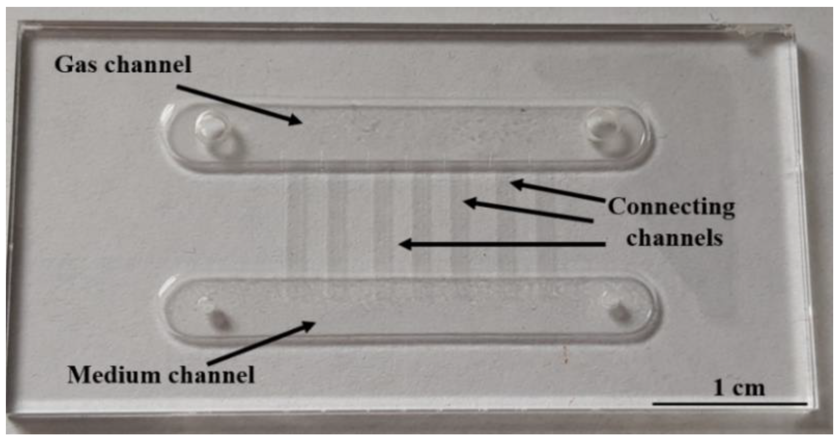
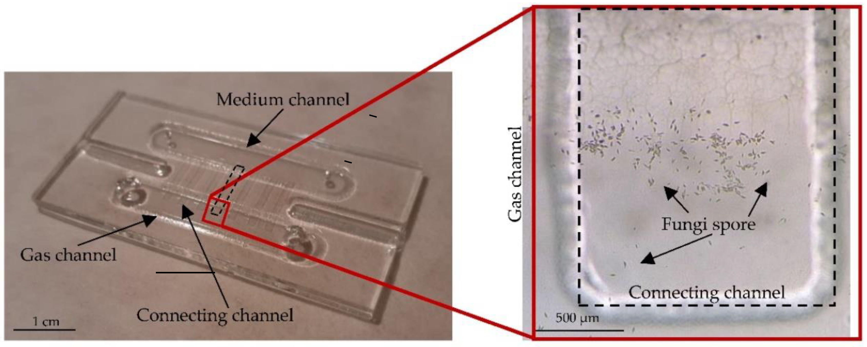
We propose the lab-on-chip (Figure 1) consisting of two glass substrates, hereinafter referred to in the text as the lower and upper substrate. Two channels with dimensions of 3.5 × 30 mm2 and a depth of 550 µm were designed in the lower substrate. In the first channel, the fungi nourishment is supplied (medium channel), while the second channel provides an appropriate gas atmosphere (gas channel).The fungi culture is also established and developed in the gas channel. In the upper glass substrate, the inlet/outlet holes enabling the introduction of the culture medium (diameter 1 mm) and gas diffusion (diameter 2 mm) were designed. A matrix of microchannels connecting the medium and gas channels was also applied in the upper glass substrate (7 connecting channels), whose dimensions are 8.8 × 1 mm2 and the depth is 20 µm [61]. This solution is proposed to ensure the constant gas exchange within the area of the medium channel, simultaneously preventing excessive “flooding” of the gas chamber.
As the translation of macroscale methodologies towards microscale lab-on-chip approach is always not trivial, the matter of dry, “non-immersed” fungi spores is a significant novelty, which may open the way towards other applications, suggesting indirect creation of chemical gradients within the culturing habitat. Moreover, use of all-glass technology is still unpopular with scientists dealing with microfluidics, thus we hope that this work is a good example of its application, which may shortly change the attitude to glass microengineering, by and large.
Computer simulations were carried out to support and initially assess the correctness of the proposed solution. For this purpose, COMSOL Multiphysics 4.3 software was used. It is a software that enables simulation in a very wide range of changes in parameters and materials [62,63,64].
The Laminar Flow module was selected for the simulation. It enables conducting research in a microscale [65,66]. The equations used in this module are the Navier-Stokes, dedicated to conservation of momentum and the continuity equation for conservation of mass. The definition of laminar flow based on the Reynolds number was also used. The following boundary conditions were adopted: at the inlet of the liquid channel (according to the Figure 1), the liquid flow velocity was 200 µL/min, which was set in accordance with the planned in the experiment, while an atmospheric pressure was established at the outlet of the liquid channel and at the inlet and outlet of the gas channel. Water was chosen as the liquid and air as the gas. Next, the project was meshed. A uniform mesh for the entire model was used herein (Figure 2).
As a result, it could be observed that the spread of the liquid in the medium channel was as expected (Figure 3a). At the same time, the correct working of the channels connecting both chambers was confirmed, the task of which is to provide humidity in the gas chamber, but not to flood it with nutrient solution. The gas channel remained almost dry (Figure 3b).
We also carried out simulations with the use of three different number of mesh elements (coarse, normal, fine) at which was measured the point of flow in the liquid and gas channel (Table 1). It can be observed that the increasing number of mesh elements does not have a significant effect on the final result.
On that basis, it can be concluded that the proposed design of the lab-on-chip provides the correct environment for fungi cultivation.
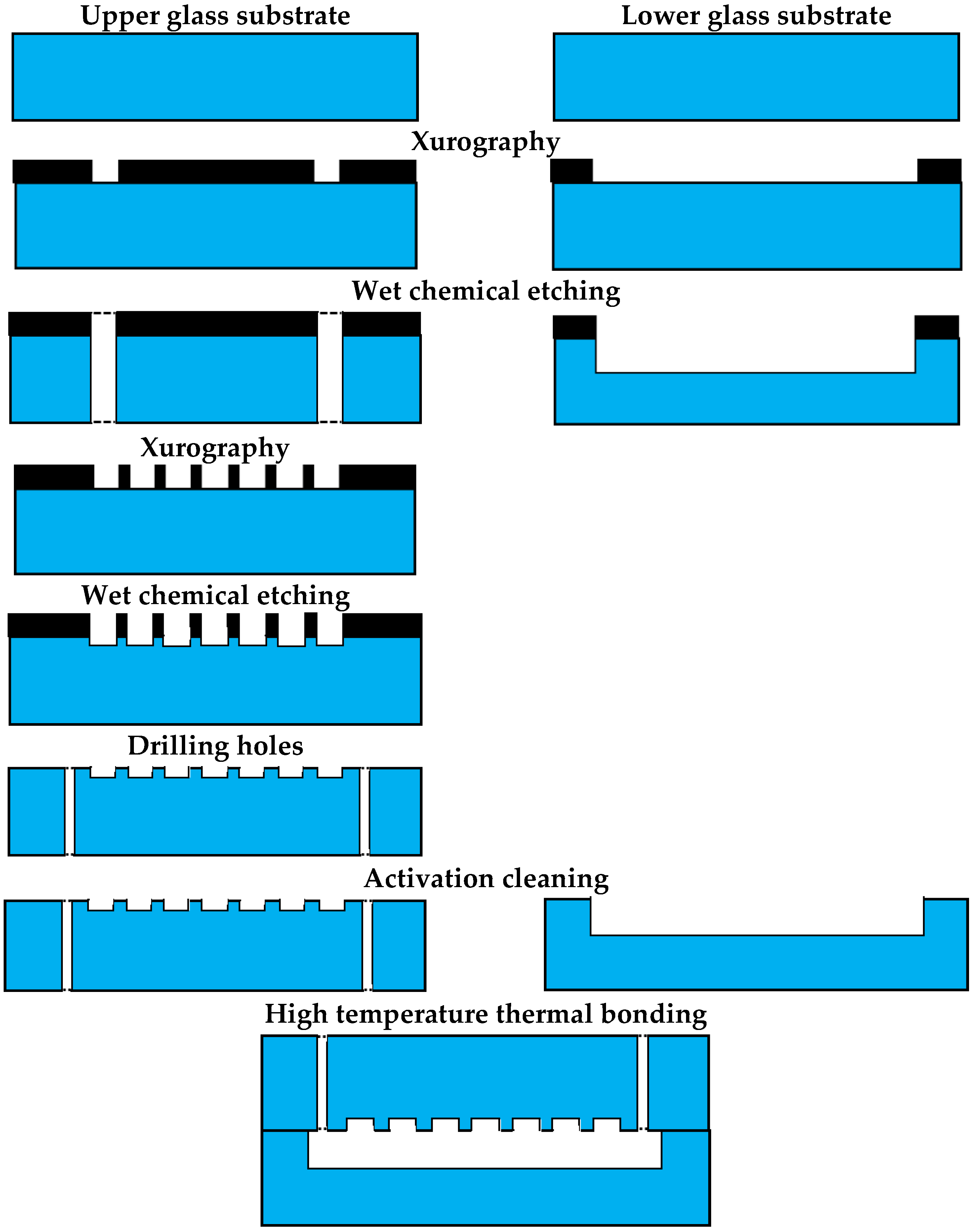
Next, microengineering processes were used to fabricate the lab-on-chip (Figure 4). Two borosilicate glass substrates (BOROFLOAT® 33 Schott, Mainz, Germany) of standard dimensions 50 × 25 × 1.1 mm served as the basic spatial structures. Designs for the masks were prepared according to the geometry of the individual channels utilising the CAD technique. Then, using the CNC method, thin vinyl foil mask patterns (Avery Dennison Graphics Solutions, Mentor, OH, USA) were obtained for each of the glass substrates. The three-dimensional channel structures were formed through the process of wet glass chemical etching in a solution of hydrofluoric acid with additives (40% HF: 65% HNO3, 10:1, v/v). First, 550 µm deep channels (lower substrate) and gas inlet/outlet holes (upper substrate) with a diameter of 2 mm were obtained in the first etching process. Next, a second etching process had to be performed to achieve shallow, connecting channel structures of 20 µm depth in the upper glass substrate. After this process, the masking layer was removed, and medium inlet/outlet holes were made in the upper substrate by precise mechanical drilling. The last stage of technological works was the bonding of the glass substrates. This process was preceded by the appropriate preparation of the substrates, through the procedures of washing and activating (hydrophilising) the surface in a solution of ethanol and Piranha. A durable bond of the substrates was achieved in a furnace under high-temperature conditions (650 °C). The ready-to-use lab-on-chip is shown in Figure 5.

2.4. Microgravity Simulation with RWV

One type of device that can simulate microgravity conditions is the single-axis microgravity simulator (Rotary Wall Vessel—RWV)—Figure 6. Such devices are being used increasingly often to understand how microorganisms react to space conditions [57,59,67]. They allow cells to grow in three dimensions and aggregate them based on natural cellular affinity [68]. These devices ensure that the cells are placed in a constant state of suspension and deprived of shear and turbulent forces [69].
We developed our own laboratory version of the RWV (Figure 6b). The most important features of the device were to assure the application of the container with cells (e.g., lab-on-chip) and automatic adjustment of the rotation speed in the range of 0–50 RPM. Additional requirements, such as small dimensions and weight (fitting inside the incubator) and user-friendly operation, were assumed.
Our RWV is a flat, vertically placed platform with diameter of 122 mm that can rotate at any given speed. Test elements, e.g., a lab-on-chip, can be attached to its surface. It is driven by a small, 3 W DC motor and two stage gear train with a gear ratio of 63:1. The platform itself is the second stage output gear. Speed control is performed electronically in an open-loop system via an LM317 integrated circuit. The lab-on-chip with inoculated fungi culture can be easily attached to the centre of the vertical platform. Based on recent literature reports [41,43,67], a constant speed of 1 rotation per 4 s was applied to simulate microgravity conditions. Our tests were conducted for at least 14 days in order to fulfil the requirements of long-term culture and repeated five times. Every RWV experiment was controlled by a reference culture.

2.5. Culturing System

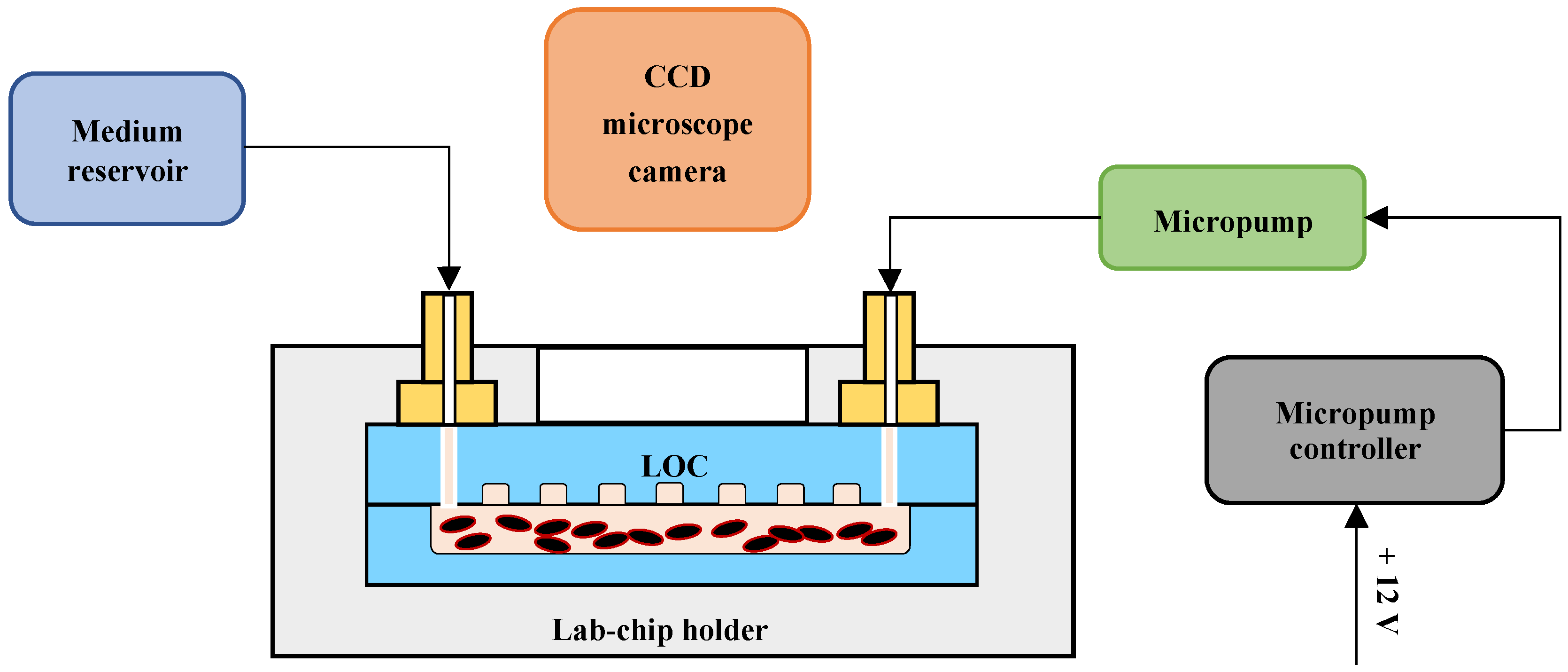
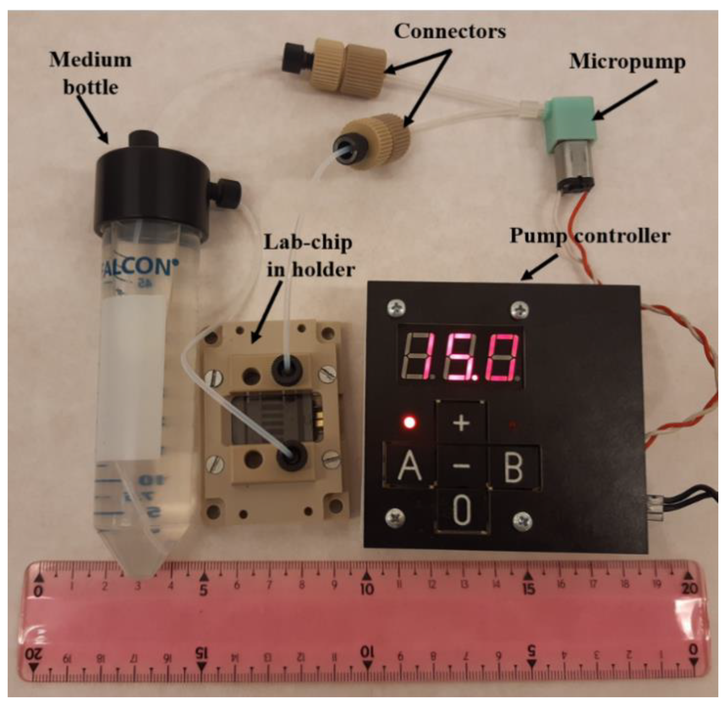
The culturing system consisted of a peristaltic micropump with a dedicated controller, a medium bottle containing sterile distilled water and a lab-on-chip in a packaging with microfluidic connections (Figure 7 and Figure 8). A Takasago peristaltic pump (model: RP-Q1.5S-P45Z-DC3V, Takasago Fluidic Systems, Japan) was used, which permitted flow control in the range of 0.1–0.45 mL/min. A flow management controller was specially designed and made to provide delivery of the medium at programmed intervals.
The pump controller is a standalone unit based on an ATtiny48 microcontroller. It has two independent channels, so one unit can control two different culturing systems. The flow rate is regulated by PWM (pulse width modulation) with base frequency of 125 Hz and resolution of 0.1%. The PWM signal can be also switched on and off for some periods of time, with a maximum of approximately 16 min. on, set with 1 sec. resolution, and 100 h off, set with 6 min. resolution. Additionally, the first run of the pump can last for a different time than the normal mode (on-state) time in order to pre-charge the culturing system. All parameters can be set via 5 keys and a 3-digit LED display. Unit dimensions are approx. 80 × 80 × 20 mm.
In our experiments, a pulse-based medium flow was applied and every 10 h fresh water entered the lab-on-chip microchannel at the minimal flow value (200 µL/min). The entire culturing system was placed in an incubator (ST 3/3 B Smart, Pol-Eko-Aparatura) to ensure homogeneous and optimal fungi growth at a temperature of 27 °C. Observation of the culture development was performed every 24 h, utilising an optical microscope (Leica DM750) with a Leica ICC50 W Microscope camera. Special attention was paid to indicating the start of germination and evaluating the physiological character of the mycelium growth.

3. Results and Discussion

3.1. Fungi Cultivation in Simulated Microgravity

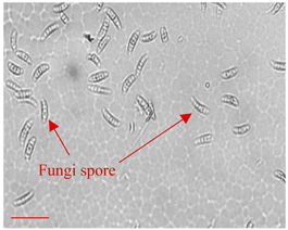
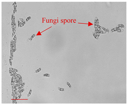
At first, the studies on the influence of microgravity on the culture of fungi F. culmorum were carried out. The fungi spores were placed in the lab-on-chip (Figure 9) according to the procedure described above. The lab-on-chip medium chamber was filled with sterile distilled water and the gas chamber was empty. The medium was changed manually with a pipette every 4 days. All inlets and outlets were covered with parafilm to limit evaporation of the medium while ensuring air supply to the gas chamber. The lab-on-chip was placed in the central part of the RWV which was rotating at a speed of 1 rotation per 4 s. The fungi cultivation lasted for 14 days. Simultaneously, a reference culture was provided on-chip.
Apart from the microscopic observation, growth of F. culmorum was assessed quantitatively, as percentage of microscopic image coverage. For this purpose, on the top of photo, a grid resembling of Thom’s counting chamber was superimposed. Coverage percentage was calculated as a sum of all the grid’s elements where the mycelium was noticed.
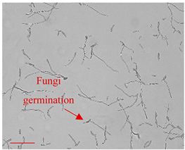
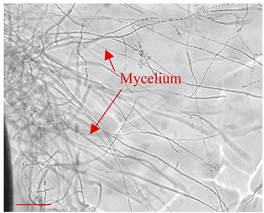
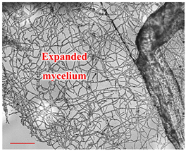
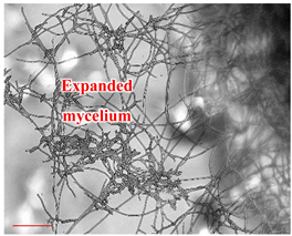
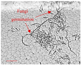
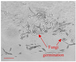
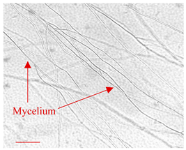
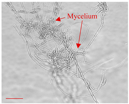
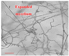
Correct fungi growth was observed for both the RWV and reference cultures (Table 2). The spore germination process started on the third day, which was more visible for the LOC placed in the RWV than for the control LOC. Over the following days, the hyphae grew until the mycelium formed. The mycelium growth in the LOC on the RWV was noticeably more branched, thus the simulated microgravity stimulated the development of the fungi in our experiment. It can be assumed that such cultivation in space would also bring a positive result.

3.2. Fungi Cultivation with Culturing System

Prior to biological experimentation, the lab-on-chip was assembled with the culturing system and partly covered with parafilm (gas inlet and outlet) in order to confine the evaporation of the medium from the channels while also creating a semi-permeable membrane, thanks to which an appropriate gas environment was ensured.
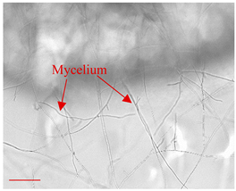
The duration of F. culmorum culture on-chip was up to 14 days. First, the dried fungi spores were introduced into the chip chamber, and next, the area covering mostly the connecting channels was selected for observation as the central, most interesting part of the LOC. On the third day of the culture, germination of the spores could be observed, being the typical time at which this process may appear [49]. In the following days, a mycelium of F. culmorum was found to develop correctly and no changes were observed in the structures of the fungal hyphae compared to the reference culture on the Petri dish. Moreover, a fusiform shape of the mycelium, imitating a cobweb, could be noticed herein, which is a typically occurring pattern for this kind of fungi [49]. Ultimately, a physiological, notable growth of fungi species on-chip could be observed, suggesting appropriate performance of the LOC platform.
The table summarising the fungi development in terrestrial conditions with a view of the defined culture day is shown below (Table 3).

3.3. Fungi Cultivation in Space—Concept

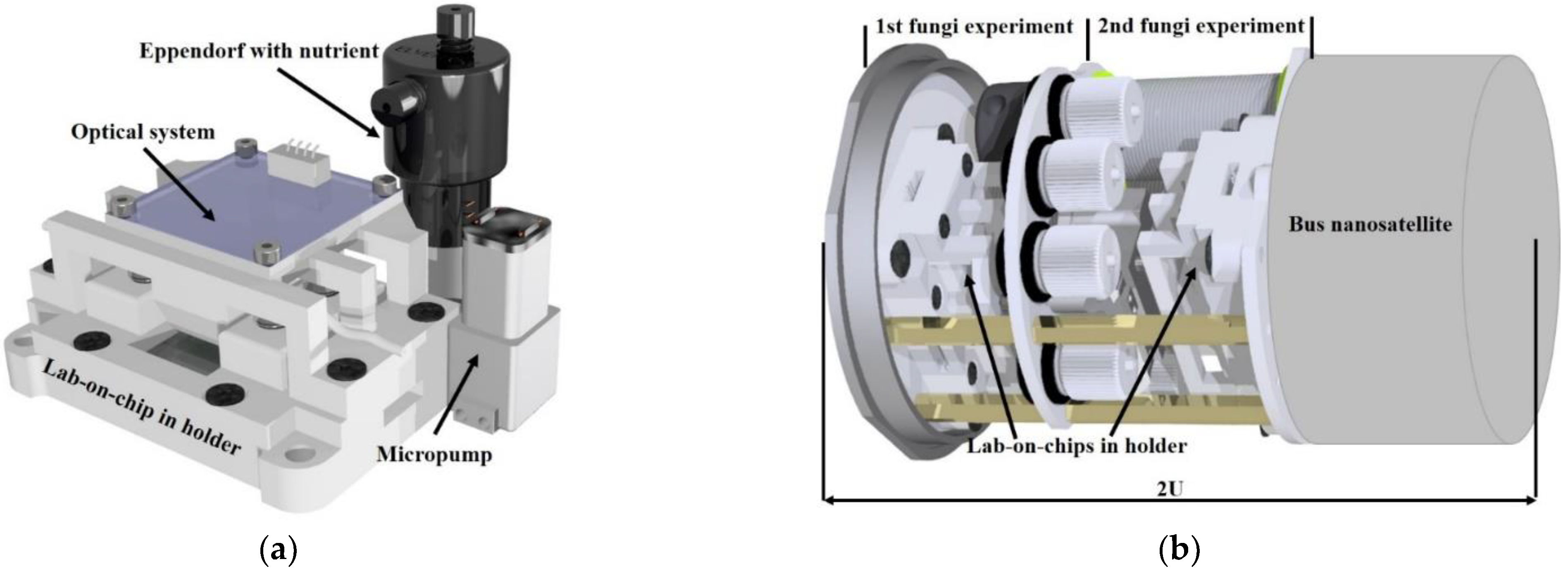
As the results of fungi cultivation in both terrestrial and simulated microgravity conditions were successful, a concept of microfluidic laboratory platform, being a payload solution for the biological nanosatellite mission, has been proposed (Figure 10). The same, all-glass LOCs are the “heart” of the platform, which material and manufacturing techniques fulfil the low outgassing criteria required for the space tools. Apart from the culturing microflow system proposed and tested in this work, a miniaturized CMOS camera equipped with motorized lens and white LED lightning is used herein to acquire the image samples. As the optimal temperature for the fungi growth is 27 °C, flexible PCB heaters maintain the defined value, based on on-chip sensors that work in a closed feedback loop. Thermal aluminum cover (thermos) protects the lab-on-chip platform from the harsh space environment. Moreover, sensors of humidity, temperature, pressure, and radiation are embedded within to constantly monitor the thermos atmosphere. All the experimental data is managed on-board and transmitted via the CAN bus to the nanosatellite module and ultimately, to the ground station. Overall structure of the nanosatellite do not exceed 2U.

4. Conclusions

In the paper, a microfluidic lab-on-chip platform ensuring long-term culturing of the selected microscopic fungi species (F. culmorum) in terrestrial and simulated microgravity conditions is shown. The all-glass lab-on-chip of the described design is proposed to fulfil the demands of the development of this biological object and allow for appropriate mycelium growth for at least 14 days. Complementary modelling of the LOC using the Comsol Multiphysics software is also provided to confirm appropriate LOC performance in the context of medium flow and gas cavity functioning.
The lab-on-chip platform has been equipped with components that assure the use of the culturing system for both terrestrial and microgravity experiments, e.g., as a payload solution for potential astrobiological missions with nanosatellites. Microscopic fungi were dried with the established procedure which permits microflow culture with the desired time-shift and thus potential biological experimentation in Low-Earth-Orbit, even with a 3-month delay.
The positive results of our experimentation encompass both technical and biological outcomes. As microfluidics for mycology is still a mostly unexplored branch, and further, no microscopic fungi (except for yeasts) have ever been investigated in microgravity, we decided to provide this new knowledge on a significant and cosmopolitan fungi representative—F. culmorum. Our research has shown that the culturing platform developed herein is suitable for long-term microflow cultivation of this species. For instance, notable mycelium growth could be observed in laboratory conditions, being consistent with the macroscale reference Petri dish culture and literature reports [49]. Interesting results were also obtained during simulated microgravity studies with the use of a custom-made RWV, which indicate repeatable, enhanced mycelium growth in these conditions compared to the reference culture on-chip.
With a view to the aforementioned results, it can be assumed that our lab-on-chip platform can be used by mycologists in other laboratory studies (e.g., concerning environmental approaches and coculture investigation with plants or soil organisms), but also as a tool for astrobiological research, e.g., conducted in a CubeSat in LEO. This undoubtedly opens new horizons for LOCs utility in agriculture and horticulture fields, making the creation of controlled environments on-chip vital and of indispensable need, especially in the context of single spore insight. Microfluidic approach towards precise investigation of microscopic fungi and other soil dwellings can provide new knowledge on interactions of these species and their mutual interplay (symbiotic/parasitic relations). Mycorrhiza and its mechanisms play an important role in plant cultivation, thus further research in this field can bring new advancement, especially needed with a view to our potential colonization aspirations. Use of lab-on-chip techniques fits the demands of the aforementioned subjects. In addition, numerical modelling of the LOC structures with microflow management is a good practice that performed prior to experimentation can answer most of the scientific questions and describe microfluidic performance thoroughly. Computational work, as shown in this paper, ensures to evaluate the microflow consistency, remaining the LOC functionalities as assumed.
In conclusion, our lab-on-chip platform was fabricated by means of reliable, resistant and biocompatible miniaturised tools, to ensure it would be able to be applied in simulated microgravity instruments, space infrastructure (e.g., International Space Station—ISS) and/or to withstand potential harsh space environments, for instance during space missions with nanosatellites. Based on the findings presented in this article, the first Polish astrobiological mission with a biological nanosatellite has been prepared and launched to study the behaviour of F. culmorum in space.

Author Contributions

Conceptualization, A.K., P.Ś. and J.D.; methodology, A.K., P.Ś., K.M., R.W. and J.D.; software, M.B.; validation, A.K., P.Ś., M.J. and A.K.-P.; investigation, A.K., P.Ś., M.J., M.B. and A.K.-P.; resources, K.M., R.W. and J.D.; data curation, A.K. and P.Ś.; writing—original draft preparation, A.K. and P.Ś; writing—review and editing, A.K., P.Ś. and M.J.; project administration, P.Ś., K.M., R.W. and J.D; funding acquisition, J.D. All authors have read and agreed to the published version of the manuscript.

Funding

The work has been financed by the National Centre for Research and Development, Poland, under project no POIR.04.01.01-00-0010/19 entitled “Bio-nanosatellite using miniaturized lab-on-chip instruments and methodology of conducting bio-medical research with its use in microgravity”.

Institutional Review Board Statement

Not applicable.

Informed Consent Statement

Not applicable.

Data Availability Statement

The authors declare that all data supporting the findings of this study are available within this article or from the corresponding author upon reasonable request.

Conflicts of Interest

The authors declare no conflict of interest in this study.

References

  1. Whitesides, G.M. The origins and the future of microfluidics. Nature 2006, 442, 368–373. [Google Scholar] [CrossRef] [PubMed]
  2. Hol, F.J.H.; Dekker, C. Zooming in to see the bigger picture: Microfluidic and nanofabrication tools to study bacteria. Science 2014, 346, 6208. [Google Scholar] [CrossRef]
  3. Podwin, A.; Lizanets, D.; Przystupski, D.; Kubicki, W.; Śniadek, P.; Kulbacka, J.; Wymysłowski, A.; Walczak, R.; Dziuban, J.A. Lab-on-Chip Platform for Culturing and Dynamic Evaluation of Cells Development. Micromachines 2020, 11, 196. [Google Scholar] [CrossRef] [PubMed] [Green Version]
  4. Reyes, D.R.; van Heeren, H.; Guha, S.; Herbertson, L.; Tzannis, A.P.; Ducrée, J.; Bissig, H.; Becker, H. Accelerating innovation and commercialization through standardization of microfluidic-based medical devices. Lab Chip 2020, 21, 9–21. [Google Scholar] [CrossRef] [PubMed]
  5. Narayanamurthy, V.; Jeroish, Z.E.; Bhuvaneshwari, K.S.; Bayat, P.; Premkumar, R.; Samsuri, F.; Yusoff, M.M. Advances in passively driven microfluidics and lab-on-chip devices: A comprehensive literature review and patent analysis. RSC Adv. 2020, 10, 11652–11680. [Google Scholar] [CrossRef]
  6. Walsh, D.I.; Kong, D.S.; Murthy, S.K.; Carr, P.A. Enabling Microfluidics: From Clean Rooms to Makerspaces. Trends Biotechnol. 2017, 35, 383–392. [Google Scholar] [CrossRef] [Green Version]
  7. Walczak, R.; Śniadek, P.; Dziuban, J.A.; Kluger, J.; Chełmońska-Soyta, A. Supravital fluorometric apoptosis detection in a single mouse embryo using lab-on-a-chip. Lab Chip 2011, 11, 3263–3268. [Google Scholar] [CrossRef]
  8. Aziz, A.U.R.; Geng, C.; Fu, M.; Yu, X.; Qin, K.; Liu, B. The Role of Microfluidics for Organ on Chip Simulations. Bioengineering 2017, 4, 39. [Google Scholar] [CrossRef]
  9. Yang, Y.; Chen, Y.; Tang, H.; Zong, N.; Jiang, X. Microfluidics for Biomedical Analysis. Small Methods 2020, 4, 1900451. [Google Scholar] [CrossRef]
  10. Lee, Y.-S.; Lu, Y.-T.; Chang, C.-M.; Liu, C.-H. Finger-powered cell-sorting microsystem chip for cancer-study applications. Sens. Actuators B Chem. 2022, 370, 132430. [Google Scholar] [CrossRef]
  11. Feng, J.J.; Hedtrich, S. A similarity scaling approach for organ-on-chip devices. Lab Chip 2022, 22, 3663–3667. [Google Scholar] [CrossRef] [PubMed]
  12. Davín, A.A.; Tannier, E.; Williams, T.A.; Boussau, B.; Daubin, V.; Szöllősi, G.J. Gene transfers can date the tree of life. Nat. Ecol. Evol. 2018, 2, 904–909. [Google Scholar] [CrossRef] [PubMed]
  13. Choi, J.J.; Kim, S.-H. A genome Tree of Life for the Fungi kingdom. Proc. Natl. Acad. Sci. USA 2017, 114, 9391–9396. [Google Scholar] [CrossRef] [PubMed] [Green Version]
  14. Satyanarayana, T.; Deshmukh, S.K.; Johri, B.N. Developments in Fungal Biology and Applied Mycology; Springer Nature Singapore Pte Ltd.: Singapore, 2017. [Google Scholar] [CrossRef]
  15. Satyanarayana, T.; Deshmukh, S.K.; Deshpande, M.V. Advancing Frontiers in Mycology & Mycotechnology: Basic and Applied Aspects of Fungi; Springer Nature Singapore Pte Ltd.: Singapore, 2019. [Google Scholar] [CrossRef]
  16. Hamid, A.; Zheng, T.; Yu, X. Fungi Contamination of Drinking Water. Rev. Environ. Contam. Toxicol. 2014, 228, 121–139. [Google Scholar] [CrossRef] [Green Version]
  17. Wilkinson, K.; Grant, W.P.; Green, L.E.; Hunter, S.; Jeger, M.J.; Lowe, P.; Medley, G.F.; Mills, P.; Phillipson, J.; Poppy, G.M.; et al. Infectious diseases of animals and plants: An interdisciplinary approach. Philos. Trans. R. Soc. Lond. Ser. B Biol. Sci. 2011, 366, 1933–1942. [Google Scholar] [CrossRef] [Green Version]
  18. Held, M.; Lee, A.P.; Edwards, C.; Nicolau, D.V. Microfluidics structures for probing the dynamic behaviour of filamentous fungi. Microelectron. Eng. 2010, 87, 786–789. [Google Scholar] [CrossRef]
  19. Stanley, C.E.; Grossmann, G.; Casadevall, X.; de-Mello, A.J. Soil-on-a-Chip: Microfluidic platforms for environmental organismal studies. Lab Chip 2016, 16, 228–241. [Google Scholar] [CrossRef]
  20. Wagg, C.; Schlaeppi, K.; Banerjee, S.; Kuramae, E.E.; Van Der Heijden, M.G.A. Fungal-bacterial diversity and microbiome complexity predict ecosystem functioning. Nat. Commun. 2019, 10, 4841. [Google Scholar] [CrossRef]
  21. Rai, M.; Agarkar, G. Plant–fungal interactions: What triggers the fungi to switch among lifestyles? Crit. Rev. Microbiol. 2016, 42, 428–438. [Google Scholar] [CrossRef]
  22. Jokerst, J.C.; Emory, J.M.; Henry, C.S. Advances in microfluidics for environmental analysis. Analyst 2012, 137, 24–34. [Google Scholar] [CrossRef]
  23. Stanley, C.E.; van der Heijden, M.G.A. Microbiome-on-a-Chip: New Frontiers in Plant-Microbiota Research. Trends Microbiol. 2017, 25, 610–613. [Google Scholar] [CrossRef] [PubMed]
  24. Millet, L.J.; Aufrecht, J.; Labbé, J.; Uehling, J.; Vilgalys, R.; Estes, M.L.; Guennoc, C.M.; Deveau, A.; Olsson, S.; Bonito, G.; et al. Increasing access to microfluidics for studying fungi and other branched biological structures. Fungal Biol. Biotechnol. 2019, 6, 8. [Google Scholar] [CrossRef] [PubMed]
  25. Prabhu, A.; Nandagopal, M.S.G.; Yegneswaran, P.P.; Singhal, H.R.; Mani, N.K. Inkjet printing of paraffin on paper allows low-cost point-of-care diagnostics for pathogenic fungi. Cellulose 2020, 27, 7691–7701. [Google Scholar] [CrossRef]
  26. Burgwyn Fuchs, B.; Eatemadpour, S.; Martel-Foley, J.M.; Stott, S.; Toner, M.; Mylonakis, E. Rapid Isolation and Concentration of Pathogenic Fungi Using Inertial Focusing on a Chip-Based Platform. Front. Cell. Infect. Microbiol. 2019, 9, 27. [Google Scholar] [CrossRef] [Green Version]
  27. Kordzadeh-Kermani, V.; Madadelahi, M.; Ashrafizadeh, S.N.; Kulinsky, L.; Martinez-Chapa, S.O.; Madou, M.J. Electrified lab on disc systems: A comprehensive review on electrokinetic applications. Biosens. Bioelectron. 2022, 214, 114381. [Google Scholar] [CrossRef]
  28. Ur Rahman, H.; Yue, X.; Yu, Q.; Zhang, W.; Zhang, Q.; Li, P. Current PCR-based methods for the detection of mycotoxigenic fungi in complex food and feed matrices. World Mycotoxin J. 2020, 136, 139–150. [Google Scholar] [CrossRef]
  29. Karouia, F.; Peyvan, K.; Pohorille, A. Toward biotechnology in space: High-throughput instruments for in situ biological research beyond Earth. Biotechnol. Adv. 2017, 35, 905–932. [Google Scholar] [CrossRef]
  30. De Middeleer, G.; Leys, N.; Sas, B.; De Saeger, S. Fungi and Mycotoxins in Space—A Review. Astrobiology 2019, 19, 915–926. [Google Scholar] [CrossRef]
  31. Ehrenfreund, P.; Ricco, A.; Squires, D.; Kitts, C.; Agasid, E.; Bramall, N.; Bryson, K.; Chittenden, J.; Conley, C.; Cook, A.; et al. The O/OREOS mission—Astrobiology in low Earth orbit. Acta Astronaut. 2014, 93, 501–508. [Google Scholar] [CrossRef] [Green Version]
  32. Low, L.A.; Giulianotti, M.A. Tissue Chips in Space: Modeling Human Diseases in Microgravity. Pharm. Res. 2020, 37, 8. [Google Scholar] [CrossRef]
  33. Nassef, M.Z.; Melnik, D.; Kopp, S.; Sahana, J.; Infanger, M.; Lützenberg, R.; Relja, B.; Wehland, M.; Grimm, D.; Krüger, M. Breast Cancer Cells in Microgravity: New Aspects for Cancer Research. Int. J. Mol. Sci. 2020, 21, 7345. [Google Scholar] [CrossRef] [PubMed]
  34. Krüger, M.; Melnik, D.; Kopp, S.; Buken, C.; Sahana, J.; Bauer, J.; Wehland, M.; Hemmersbach, R.; Corydon, T.J.; Infanger, M.; et al. Fighting Thyroid Cancer with Microgravity Research. Int. J. Mol. Sci. 2019, 20, 2553. [Google Scholar] [CrossRef] [PubMed] [Green Version]
  35. Kim, W. Spaceflight Promotes Biofilm Formation by Pseudomonas aeruginosa. PLoS ONE 2013, 8, e62437. [Google Scholar] [CrossRef] [PubMed] [Green Version]
  36. Stutte, G.; Monje, O.; Hatfield, R.; Paul, A.; Ferl, R.; Simone, C. Microgravity effects on leaf morphology, cell structure, carbon metabolism and mRNA expression of dwarf wheat. Planta 2006, 224, 1038–1049. [Google Scholar] [CrossRef]
  37. Sathasivam, M.; Hosamani, R.; Swamy, B.; Kumaran, G.S. Plant responses to real and simulated microgravity. Life Sci. Space Res. 2020, 28, 74–86. [Google Scholar] [CrossRef]
  38. Lewis, B.; Hanel, R.; Bhattacharya, S.; Ricco, A.J.; Agasid, E.; Reiss-Bubbenheim, D.; Straume, T.; Parra, M.; Boone, T.; Maria, S.S.; et al. BioSentinel: Monitoring DNA Damage Repair Beyond Low Earth Orbit on a 6U Nanosatellite. In Proceedings of the 28th Annual AIAA/USU Conference on Small Satellites, Moffett Field, CA, USA, 18 March 2014; pp. 1–10. [Google Scholar]
  39. Zea, L. Microbiological Experiments Onboard CubeSats—A Review and Prospects. In Proceedings of the 1st Latin American IAA CubeSat Workshop, Brasília, Brazil, 8–11 December 2014; pp. 1–13. [Google Scholar]
  40. Bizzarri, M.; Monici, M.; Loon, J. How Microgravity Affects the Biology of Living Systems. BioMed Res. Int. 2015, 2015, 863075. [Google Scholar] [CrossRef]
  41. Grimm, D.; Wehland, M.; Corydon, T.J.; Richter, P.; Prasad, B.; Bauer, J.; Egli, M.; Kopp, S.; Lebert, M.; Krüger, M. The effects of microgravity on differentiation and cell growth in stem cells and cancer stem cells. Stem Cells Transl. Med. 2020, 9, 882–894. [Google Scholar] [CrossRef]
  42. Chen, Z.-Y.; Guo, S.; Li, B.-B.; Jiang, N.; Li, A.; Yan, H.-F.; Yang, H.-M.; Zhou, J.-L.; Li, C.-L.; Cui, Y. Effect of Weightlessness on the 3D Structure Formation and Physiologic Function of Human Cancer Cells. BioMed Res. Int. 2019, 2019, 4894083. [Google Scholar] [CrossRef]
  43. Mylabathula, P.L.; Li, L.; Bigley, A.B.; Markofski, M.M.; Crucian, B.E.; Mehta, S.K.; Pierson, D.L.; Laughlin, M.S.; Rezvani, K.; Simpson, R.J. Simulated microgravity disarms human NK-cells and inhibits anti-tumor cytotoxicity in vitro. Acta Astronaut. 2020, 174, 32–40. [Google Scholar] [CrossRef]
  44. Krüger, M.; Kopp, S.; Wehland, M.; Bauer, J.; Baatout, S.; Moreels, M.; Egli, M.; Corydon, T.J.; Infanger, M.; Grimm, D. Growing blood vessels in space: Preparation studies of the SPHEROIDS project using related ground-based studies. Acta Astronaut. 2019, 159, 267–272. [Google Scholar] [CrossRef]
  45. Scott, T.J.; Vonortas, N.S. Microgravity protein crystallization for drug development: A bold example of public sector entrepreneurship. J. Technol. Transf. 2019, 46, 1442–1461. [Google Scholar] [CrossRef]
  46. Poghosyan, A.; Golkar, A. CubeSat evolution: Analyzing CubeSat capabilities for conducting science missions. Prog. Aerosp. Sci. 2017, 88, 59–83. [Google Scholar] [CrossRef]
  47. Zhu, T.; Moussa, E.M.; Witting, M.; Zhou, D.; Sinha, K.; Hirth, M.; Gastens, M.; Shang, S.; Nere, N.; Somashekar, S.C.; et al. Predictive models of lyophilization process for development, scale-up/tech transfer and manufacturing. Eur. J. Pharm. Biopharm. 2018, 128, 363–378. [Google Scholar] [CrossRef] [PubMed]
  48. Han, Y.; Quan, G.B.; Liu, X.Z.; Ma, E.P.; Liu, A.; Jin, P.; Cao, W. Improved preservation of human red blood cells by lyophilization. Cryobiology 2005, 51, 152–164. [Google Scholar] [CrossRef] [PubMed]
  49. Watanabe, T. Pictorial Atlas of Soil and Seed Fungi: Morphologies of Cultured Fungi and Key to Species; CRC Press: Washington, WA, USA, 2011. [Google Scholar] [CrossRef]
  50. Domsch, K.H.; Gams, W.; Anderson, T.-H. Compendium of Soil Fungi, 2nd ed.; Gams, W., Ed.; IHW-Verlag: Freising, Germany, 2007; p. 672. [Google Scholar] [CrossRef]
  51. Hongbin, Y.; Guangya, Z.; Siong, C.F.; Shouhua, W.; Feiwen, L. Novel polydimethylsiloxane (PDMS) based microchannel fabrication method for lab-on-a-chip application. Sens. Actuators B 2009, 137, 754–761. [Google Scholar] [CrossRef]
  52. Regehr, K.J.; Domenech, M.; Koepsel, J.T.; Carver, K.C.; Ellison-Zelski, S.J.; Murphy, W.L.; Schuler, L.A.; Alarid, E.T.; Beebe, D.J. Biological implications of polydimethylsiloxane-based microfluidic cell culture. Lab Chip 2009, 9, 2132–2139. [Google Scholar] [CrossRef]
  53. Halldorsson, S.; Lucumi, E.; Gómez-Sjöberg, R.; Fleming, R.M. Advantages and challenges of microfluidic cell culture in polydimethylsiloxane devices. Biosens. Bioelectron. 2015, 63, 218–231. [Google Scholar] [CrossRef] [PubMed] [Green Version]
  54. Podwin, A.; Kubicki, W.; Dziuban, J. Study of the behavior of Euglena viridis, Euglena gracilis and Lepadella patella cultured in all-glass microaquarium. Biomed. Microdevices 2017, 19, 1–10. [Google Scholar] [CrossRef] [Green Version]
  55. Podwin, A.; Janisz, T.; Patejuk, K.; Szyszka, P.; Walczak, R.; Dziuban, J. Towards microfluidics for mycology—Material and technological studies on LOCs as new tools ensuring investigation of microscopic fungi and soil organisms. Bull. Pol. Acad. Sci. Tech. Sci. 2021, 69, e136212. [Google Scholar] [CrossRef]
  56. Schott Products Catalog. Available online: https://www.schott.com/english/index.html (accessed on 2 January 2022).
  57. Przystupski, D.; Górska, A.; Michel, O.; Podwin, A.; Sniadek, P.; Łapczynski, R.; Saczko, J.; Kulbacka, J. Testing Lab-on-a-Chip Technology for Culturing Human Melanoma Cells under Simulated Microgravity. Cancers 2021, 13, 402. [Google Scholar] [CrossRef]
  58. Garstecki, P.; Ziolkowski, J.; Kanp, P. Microfluidischer Chip with an Unvented Gas Cavity in a Microfluidic Chip. Patent No. WO 2019/185885 A1, 3 October 2019. [Google Scholar]
  59. Brennan, M.D.; Rexius-Hall, M.L.; Elgass, L.J.; Eddington, D.T. Oxygen control with microfluidics. Lab Chip 2014, 14, 4305–4318. [Google Scholar] [CrossRef] [PubMed]
  60. Gao, Y.; Stybayeva, G.; Revzin, A. Fabrication of composite microfluidic devices for local control of oxygen tension in cell cultures. Lab Chip 2018, 19, 306–315. [Google Scholar] [CrossRef] [PubMed]
  61. Śniadek, P.; Kubicki, W.; Podwin, A. Mikroprzepływowe Laboratorium Hodowlane. Patent No. 437619, 20 April 2021. [Google Scholar]
  62. Dartoomi, H.; Khatibi, M.; Ashrafizadeh, S.N. Importance of nanochannels shape on blue energy generation in soft nanochannels. Electrochim. Acta 2022, 431, 141175. [Google Scholar] [CrossRef]
  63. Karimzadeh, M.; Khatibi, M.; Ashrafizadeh, S.N.; Mondal, P.K. Blue energy generation by the temperature-dependent properties in funnel-shaped soft nanochannels. Phys. Chem. Chem. Phys. 2022, 24, 20303–20317. [Google Scholar] [CrossRef]
  64. Grzebyk, T.; Szmajda, T.; Szyszka, P.; Górecka-Drzazga, A.; Dziuban, J. Glow-discharge ion source for MEMS mass spectrometer. Vacuum 2020, 171, 109008. [Google Scholar] [CrossRef]
  65. Hoshyargar, V.; Khorami, A.; Ashrafizadeh, S.N.; Sadeghi, A. Solute dispersion by electroosmotic flow through soft microchannels. Sens. Actuators B Chem. 2018, 255, 3585–3600. [Google Scholar] [CrossRef]
  66. Hoshyargar, V.; Talebi, M.; Ashrafizadeh, S.N.; Sadeghi, A. Hydrodynamic dispersion by electroosmotic flow of viscoelastic fluids within a slit microchannel. Microfluid. Nanofluidics 2018, 22, 4. [Google Scholar] [CrossRef]
  67. Hammond, T.; Hammond, J. Optimized suspension culture: The rotating-wall vessel. Am. J. Physiol.-Ren. Physiol. 2001, 281, F12–F25. [Google Scholar] [CrossRef] [PubMed]
  68. Barrila, J.; Radtke, A.L.; Crabbé, A.; Sarker, S.F.; Herbst-Kralovetz, M.M.; Ott, C.M.; Nickerson, C.A. Organotypic 3D cell culture models: Using the rotating wall vessel to study host–pathogen interactions. Nat. Rev. 2010, 8, 791–801. [Google Scholar] [CrossRef]
  69. Yamaguchi, N.; Roberts, M.; Castro, S.; Oubre, C.; Makimura, K.; Leys, N.; Grohmann, E.; Sugita, T.; Ichijo, T.; Nasu, M. Microbial Monitoring of Crewed Habitats in Space—Current Status and Future Perspectives. Microbes Environ. 2014, 29, 250–260. [Google Scholar] [CrossRef]
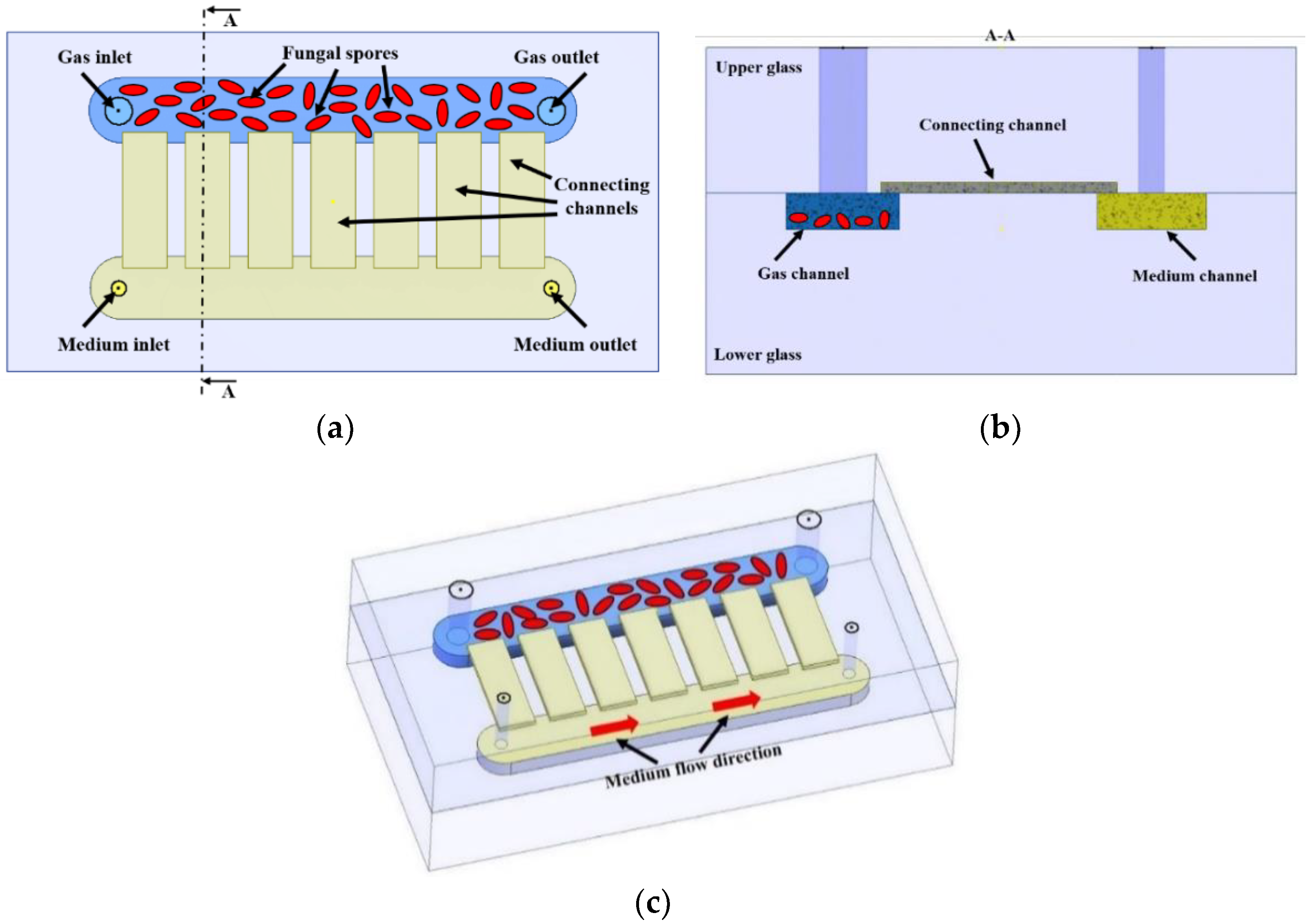
Figure 1. Scheme of the lab-on-chip for fungi culture: (a) top view, (b) cross-section, (c) general view.
Applsci 12 10627 g001
Figure 2. Liquid flow distribution in a lab-on-chip.
Applsci 12 10627 g002
Figure 3. Cross-section through the: (a) liquid channel, (b) gas channel.
Applsci 12 10627 g003
Figure 4. LOC fabrication steps.
Applsci 12 10627 g004
Figure 5. All-glass lab-on-chip for fungi culturing.
Applsci 12 10627 g005
Figure 6. Rotary Wall Vessel: (a) scheme, (b) laboratory version.
Applsci 12 10627 g006
Figure 7. Scheme of the culturing system.
Applsci 12 10627 g007
Figure 8. Culturing system.
Applsci 12 10627 g008
Figure 9. Microscope image of the fungi spores on-chip (F. culmorum) showing the location of the culture relative to the connecting channel.
Applsci 12 10627 g009
Figure 10. Concept of the first Polish nanosatellite with the microfluidic payload: (a) lab-on-chip in the holder, (b) overall 2U structure.
Applsci 12 10627 g010
Table 1. Comparison of flows in the channels depending on the mesh used.
Number of Mesh ElementsFlow in Liquid Channel [m3/s]Flow in Gas Channel [m3/s]
Coarse—3047574.89441 × 10−91.56306 × 10⁻¹³
Normal—6261274.85098 × 10−91.55514 × 10⁻¹³
Fine—13327894.98822 × 10−91.57639 × 10⁻¹³
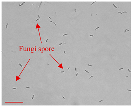
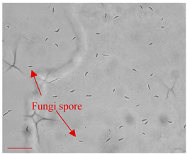
Table 2. Sample photos of cultures conducted on the LOC in simulated microgravity and on the reference LOC. The tests were performed at least 5 times and similar results were obtained. Scale bar—100 µm.
LOC—RWV[%] of CoverageLOC—no RWV[%] of CoverageComments
Day 0Applsci 12 10627 i0015Applsci 12 10627 i0025Dried fungi spores introduced to the LOC.
Day 3Applsci 12 10627 i00320Applsci 12 10627 i00415The beginning of the fungi germination process.
Day 7Applsci 12 10627 i00545Applsci 12 10627 i00620Expansion of the fungus hyphae.
Day 14Applsci 12 10627 i00790Applsci 12 10627 i00854Formation of the mycelium.
Table 3. Summary of the fungi culture development on-chip in comparison with a reference Petri dish culture. The tests were performed at least 5 times and similar, appropriate culturing results were obtained. Scale bar—100 µm.
LOC Platform[%] of CoveragePetri Dish (Control)[%] of CoverageComments
Day 0Applsci 12 10627 i0095Applsci 12 10627 i0105Dried fungi spores of appropriate shape and size introduced to the LOC.
Day 3Applsci 12 10627 i01120Applsci 12 10627 i01215A start of fungi germination process (time consistent with literature data [49]).
Day 7Applsci 12 10627 i01330Applsci 12 10627 i01427Mycelium of fungi under development. Slightly better growth on-chip than in Petri dish.
Day 14Applsci 12 10627 i01545Applsci 12 10627 i01638Appropriate mycelium growth. Long and branched structures of fungal hyphae visible.
Publisher’s Note: MDPI stays neutral with regard to jurisdictional claims in published maps and institutional affiliations.