Article

The Molecular Docking and Inhibition Kinetics of Angiotensin I-Converting Enzyme Inhibitory Peptides Derived from Soft-Shelled Turtle Yolk

by
Nhung Thi Phuong Nong
1,2,
Christoper Caesar Yudho Sutopo
2,
Wei-Ting Hung
3,
Ping-Hsun Wu
4,5,* and
Jue-Liang Hsu
6,7,8,*
1
Department of Basic Science, Thai Nguyen University of Agriculture and Forestry, Quyet Thang Ward, Thai Nguyen 250000, Vietnam
2
Department of Tropical Agriculture and International Cooperation, National Pingtung University of Science and Technology, Pingtung 912301, Taiwan
3
Department of Food Science, National Pingtung University of Science and Technology, Pingtung 91201, Taiwan
4
Division of Nephrology, Department of Internal Medicine, Kaohsiung Medical University Hospital, Kaohsiung Medical University, Kaohsiung 807378, Taiwan
5
Faculty of Medicine, College of Medicine, Kaohsiung Medical University, Kaohsiung 807378, Taiwan
6
Department of Biological Science and Technology, National Pingtung University of Science and Technology, Pingtung 912301, Taiwan
7
Institute of Food Safety Management, National Pingtung University of Science and Technology, Pingtung 912301, Taiwan
8
Research Center for Animal Biologics, National Pingtung University of Science and Technology, Pingtung 912301, Taiwan
*
Authors to whom correspondence should be addressed.
Appl. Sci. 2022, 12(23), 12340; https://doi.org/10.3390/app122312340
Submission received: 24 October 2022 / Revised: 27 November 2022 / Accepted: 28 November 2022 / Published: 2 December 2022
(This article belongs to the Special Issue Food Biochemistry and Effects of Proteins and Peptides)

Abstract

:
The soft-shelled turtle yolk (SSTY) protein hydrolysate contains a potential source of bioactive peptides. Our previous study found that five SSTY peptides (WLQL, LPSW, LPLF, VPGLAL and LVGLPL) showed moderate to high dipeptidyl peptidase IV (DPP-IV) inhibitory activities. This study further investigated their angiotensin-I-converting enzyme (ACE) inhibitory activity. Consequently, WLQL was identified as the most potent ACE inhibitory peptide with a remarkably low IC50 value (16.87 ± 0.54 µM). The Lineweaver–Burk plot analysis was performed for the characterization of the peptide’s inhibition mode and the inhibition kinetics was rationalized using the molecular docking simulation. The result revealed that WLQL would dock into the S1 pockets of ACE, while LPSW interacted with ACE’s secondary binding site. Further evaluation of the peptides’ stability against ACE involved a pre-incubation experiment. After 3 h of pre-incubation with ACE, the four peptides were hydrolyzed into smaller fragments with varying degrees, suggesting that they are substrate-type inhibitors. In contrast, LVGLPL can tolerate hydrolysis by ACE and act as a true inhibitor.

1. Introduction

High blood pressure or hypertension is considered to be 130/80 mmHg (systolic/diastolic blood pressure) in stage 1 and higher than 140/90 mmHg in stage 2 in a patient [1,2]. It has been confirmed as one of the most common chronic health crises, not to mention that it also enhances the risk of developing other metabolic disorders, such as cardiovascular disease and diabetes [3,4]. In addition, hypertensive patients are more vulnerable to developing serious COVID-19 complications, which can lead to an increase in mortality rates [5,6].
The angiotensin-I-converting enzyme (ACE; EC 3.4.15.11) is known to regulate blood pressure and inflammation in the human body through the renin–angiotensin system (RAS). A zinc dipeptide carboxypeptidase contributes to the conversion of decapeptide angiotensin I (Ang I) to octapeptide angiotensin II (Ang II) through the cleavage of the carboxyl-terminal His-Leu dipeptide. Ang II’s primary function is to induce systemic arteriolar contraction, and consequently, high blood pressure is an inevitable result [1,4]. Therefore, the inhibition or inactivation of ACE is one of the most efficient therapeutic strategies for hypertension treatment [4,7]. The high-speed development of pharmaceutical drugs has played an important role in hypertension therapies with increased efficiency; however, their undesirable side effects have also been mentioned in parallel [3,4]. Accordingly, the application of natural peptide therapies has become an attractive alternative to synthetic compounds. A large number of studies concerning the discovery of ACE inhibitory (ACEI) peptides have been carried out on different dietary-derived proteins, including milk proteins [5,8,9] and chicken egg white [10,11].
Previously published evidence of ACEI efficiency from soft-shelled turtles (SST) has been highlighted for egg, meat, and powder hydrolysates [12,13,14]. Consequently, potential ACEI peptides were identified from egg whites and egg yolks of SST [15,16,17]. Recently, dipeptidyl peptidase-IV (DPP-IV) inhibitory peptides were also identified in SST yolk (SSTY) hydrolysates using gastrointestinal enzymes [18]. This suggests that the SST egg is a powerful bioactive peptide source. Multiple-function peptides show promising results in the research of novel peptide therapy, as they could reduce costs and are more convenient for patients undergoing treatment of diseases. Interestingly, the activity of ACE could be inhibited by some DPP-IV inhibitors [19,20]. The DPP-IV inhibitory effect is often associated with the N-terminal of a peptide and the ACE inhibitory activity is affected by the C-terminal of a peptide. Therefore, both terminal positions play significantly important roles in allowing some inhibitors to possess these dual functional properties, while others only have a single impact. The representation of proline (P) and/or hydrophobic amino acids has been commonly reported in the sequence of DPP-IV inhibitory peptides [19,20]. Peptides with hydrophobic amino acid residues (W, F, Y, or P) at the C-terminal position are often associated with potential ACE inhibitory activity [20,21,22].
Therefore, in this study, five DPP-IV inhibitory peptides (LPSW, LPLF, VPGLAL, WLQL, and LVGLPL) previously identified from SSTY hydrolysates were examined for their ACEI effects using in vitro and in silico tests [18]. The ACEI efficiency of these peptides was determined by calculating their IC50 values using synthetic peptides. We used Lineweaver–Burk plots to investigate the peptides’ inhibitory effect on ACE, as well as the interaction mechanism between the peptides and the ACE through docking simulation. Additionally, the overall structural stability of the peptides with ACE was evaluated through an ACE pre-incubation assay.

2. Materials and Methods

2.1. Materials

ACE (from rabbit lungs), hippuryl-L-histidyl-L-leucine (HHL), ferulic acid (FA), and hippuric acid (HA) were acquired from Sigma Chemical Co. (St. Louis, MO, USA). Acetonitrile (ACN), formic acid (FA), boric acid, and piperidine were obtained from J.T. Baker (Phillipsburg, NJ, USA). Trifluoroacetic acid (TFA) was purchased from Alfa Aesar (Lancashire, UK). Deionized water was produced using the PURELAB® water purification system from ELGA LabWater (Lane End, High Wycombe, UK).

2.2. Determination of ACE Inhibitory Activity and Measurement of IC50 Value

The potential peptides were analyzed for their ACE inhibitory activity using Cushman’s method [23] with slight modifications. In this protocol, captopril and hippuryl-L-histidyl-L-leucine (HHL) were utilized as the positive control and substrate, respectively, for the ACE inhibitory assay. The mixture for each assay that contained 10 µL of a sample (at a tested concentration) and 30 µL of 2.5 mM HHL was maintained at 37 °C for 5 min. For the blank and positive control, borate buffer (200 mM borate buffer; 300 mM NaCl, pH 8.3) and captopril were used to replace the sample under the same condition, respectively. Thereafter, 20 µL of ACE (0.05 mU/µL) in borate buffer was added to the pre-incubated mixture and kept at 37 °C for 1 h. For the first 30 min, stationary incubation was employed, followed by the use of a shaker incubator (speed up 200 rpm) for the remaining half hour. Afterward, 60 µL of 1 M HCl was used to quench the reaction. The amount of HA, a product released from HHL hydrolysis, was then detected using HPLC (Hitachi Chromaster, Japan) with a NUCLEODUR C18 HTec column (4.6 mm × 250 mm; 5 µm, C18; Macherey-Nagel, Düren, Germany) and a UV detector at 228 nm. The product of the reaction was monitored under an isocratic elution with 82% mobile phase A (5% ACN and 0.1% TFA in ddH2O) and 18% mobile phase B (95% ACN and 0.1% TFA in ddH2O) at a constant flow rate of 1 mL/min and was maintained for 20 min. The ACE inhibitory activity (%) was calculated using the following formula:
ACEI activity (%) = [1 − (Ainhibitor/Ablank)] × 100%
The HPLC peak areas of HA formed in the ACE-mediated reaction with and without the inhibitor are represented by Ainhibitor and Ablank, respectively. The IC50 value was determined as the inhibitor concentration that was required to inhibit 50% of the ACE activity. Nonlinear regression using Graphpad Prism 5.0 (GraphPad Software, Inc., La Jolla, CA, USA) was performed to calculate the IC50 value with various concentrations of the inhibitor.

2.3. Synthesis of ACE Peptides

Five ACE peptides (WLQL, LPSW, LPLF, VPGLAL and LVGLPL) were obtained from our previous study [18]. After synthesized processing, the sequencing of the peptides was rechecked using LC-MS/MS. Peptides with a purity higher than 95% were achieved by using RP-HPLC purification.

2.4. Determination of Inhibitory Patterns of Synthetic Peptides for ACE

The inhibitory mode of inhibition of ACEI peptides was characterized using several substrate concentrations, as well as varying concentrations of the inhibitor, as described in Section 2.2. The HA produced in the ACE reaction was then measured using a UV–Vis detector at 228 nm during HPLC separation. The Lineweaver–Burk plot provided information about the Km values on the X-axis and Vmax values on the Y-axis intercept for the ACE inhibitory kinetics.

2.5. Stability of ACEI Peptides against ACE

The pre-incubation protocol was applied to evaluate the stability of ACEI peptides against ACE. Based on the previous method [24] with a slight modification, 20 µL of ACEI peptides was pre-incubated with 40 µL of 0.05 mU/µL ACE (in borate buffer) at 37 °C, using a thermostatically controlled incubator for 3 h. Subsequently, 30 µL of the combination solution was added to 30 µL of HHL (2.5 mM) and incubated for another 1 h at 37 °C. The reaction was quenched by adding 60 µL of 1 N HCl. This final solution was separated into two equal portions; one portion was taken to determine the ACE inhibitory peptides through their IC50 value with the same method outlined in Section 2.2. The other portion was loaded into the LC-MS/MS system to monitor the ACEI peptide stability during ACE hydrolysis.

2.6. Molecular Docking to ACE

Discovery Studio Visualizer 3.0 software (Accelrys Software, Cambridge, UK) simulated molecular docking as previously described [7,25]. The software was linked to the dynamic energy calculation program CHARMm (Chemistry at HARvard Macromolecular Mechanics). The crystal structure of human tACE (testicular ACE; 1O86.pdb) in complex with lisinopril was obtained from the Protein Data Bank (with a resolution of 2.0 Å) and then was used as the target structure for molecular docking [26]. Before commencing the docking simulation, lisinopril and all water molecules in the ACE X-ray crystal complex were eliminated, except for zinc and chloride ions. Both the formulation of the peptide conformations and preparation of docking to ACE were performed by the same software and image visualization was carried out by the CHARMm program. To simulate the same in vitro condition, pH 8.3 was set up before the docking procedure. The docking was carried out using cavity detection with a radius of 20 Å centered at the following coordinates: x: 39.07; y: 38.28; z: 50.13. The best binding pose was selected according to the lowest docking score.

2.7. Statistical Analysis

Tukey’s HSD post-hoc test using SPSS 22.0 (SPSS Inc., Chicago, IL, USA) was applied to analyze the differences between means with a p-value lower than 0.05 (p < 0.05). Graphpad Prism 5.0 (GraphPad Software, Inc., La Jolla, CA, USA) was used to determine the IC50 value by nonlinear regression.

3. Results

3.1. Identification of Angiotensin-Converting Enzyme (ACE) Inhibitory Activity

Five DPP-IV inhibitory peptides discovered in our previous study (LPSW, VPGLAL, WLQL, LPLF and LVGLPL) were utilized to identify their ACEI activities [18]. The synthesized peptides were evaluated for their ACE inhibitory activity using an in vitro ACEI assay and an RP-HPLC system. The IC50 value of each peptide was determined using Graphpad Prism 5.0 (GraphPad Software, Inc.) by nonlinear regression of ACEI activities (%) at various concentrations, as indicated in Figure A1, and the results are shown in Table 1. Captopril (1.7 μM), used as the positive control in the in vitro assay, demonstrated an ACEI effect of 99.6% (the result is not shown here). Among these five peptides, WLQL exhibited the highest ACEI effect with the IC50 value of 16.87 ± 0.54 µM, as compared to the remaining peptides (Table 1). The tetrapeptide LPSW (IC50 value of 20.80 ± 0.79 µM) also displayed high ACEI activity. The other peptides, LPLF and VPGLAL, showed moderate IC50 values of 300.08 ± 17.30 µM and 573.00 ± 54.10 µM, respectively, while the peptide LVGLPL showed the lowest ACEI effect with the highest IC50 value of more than 2000 µM (Table 1). Moreover, their IC50 values with the DPP-IV enzyme are also provided in Table 1 [18].
The mode of ACE inhibition was defined using the Lineweaver and Burk analysis. Peptide stability was examined during the pre-incubation assay with ACE for 3 h at 37 °C. After preincubation, the resulting solution was injected into LC-MS to monitor the remaining amount of peptide and the formation of hydrolysis products. The IC50 value (with and without ACE preincubation) was determined using RP-HPLC.
According to previous studies [4,21,22], the activity of ACEI peptides is commonly affected by the following three main interrelated factors: the presence of several specific amino acids, their hydrophobic or hydrophilic nature, and the length of the sequences. Specifically, the highly potent ACEI peptides have aromatic amino acids (tryptophan, tyrosine, and phenylalanine) or hydrophobic amino acids at their C-terminal, which play an important role in ACE inhibitory activity [20,21].
Comparatively, these peptides seem to display promising inhibitory activities against ACE, especially WLQL and LPSW. In a similar manner to our material, eggs from chickens are the focal point in biological activity research due to their good nutritional value and the fact that they are a popular and cheap product. Salim et al. reported that nine ACEI peptides were identified in chicken egg white that contained two to five residues in their sequences, with IC50 values ranging from 1.30 mM to 5.47 mM [10]. Moreover, with an excellent ACE inhibitory effect, the peptide LKYAT (IC50 value of 0.09 μM) identified from chicken egg white is considered as a promising ingredient for functional food [11]. As they possesses four residues in their structures, the three peptides WLQL (IC50: 16.87 µM), LPLF (IC50: 300.08 µM) and LPSW (IC50: 20.80 µM) from the current study seem to exhibit high ACE inhibitory activity, as compared to ELPF (IC50: 4.24 mM) derived from chicken egg white [10], HLHT (IC50: 458 µM) obtained from pearl oyster (Pinctada fucata martensii) protein hydrolysates (HLHT and GWA with the IC50 values of 458.06 ± 3.24 µM and 109.25 ± 1.45 µM, respectively) [27], and KYKA (5.63 µM) from spent hen muscle proteins [28]. Furthermore, other ACEI peptides were also derived from various natural sources, such as marine macroalga Ulva intestinalis hydrolysates (HLHT and GWA with the IC50 values of 458.06 ± 3.24 µM and 109.25 ± 1.45 µM, respectively), MELVLR (IC50: 236.85 µM) from marine hydrolysates [29], and naked oat globulin hydrolysates (SSYYPFK, IC50: 91.82 µM) [3].
Furthermore, the appearance of a branched-chain aliphatic amino acid (isoleucine, leucine, or valine) at the N-terminal end of peptides can improve ACE inhibitory activity [20,30]. Hydrophobic amino acids can ameliorate the efficiency of ACEI peptides [20,22]. These amino acid characteristics can be identified in almost all of these peptides (WLQL, LPSW, LPLF, VPGLAL, and LVGLPL), and all of them showed prominent ACE inhibitory effects, except LVGLPL. In the literature, it was reported that a Leu (L) residue at the N-terminal of ACEI peptides was determined in egg white hydrolysates (LKYKA, IC50: 0.09 µM [11] and LPR, IC50: 1.30 mM [10]) and spent hen muscle (LKYKA, IC50: 0.054 µM and LKY, IC50: 1.91 µM) [28], in addition to our current study (LPSW, IC50: 20.80 µM; LPLF, IC50: 300.08 µM; and LVGLPL, IC50: >2000 µM). Interestingly, LPSW has tryptophan at its C-terminal, leucine at its N-terminal, and proline in its sequence. It matches the hypothesis related to potential ACEI peptides. Moreover, the results of in vitro experiments have also proven its effects on ACE inhibitory activity (Figure A1 and Table 1). In our previous study, other peptides that contained LPSW in their sequence, such as AKLPSW, also played a role in ACEI influence with an IC50 value of 15.3 µM [17], which indicates higher ACEI activity than LPSW (IC50 value of 20.80 µM). With an aromatic residue also at the C terminal, GVGSPY derived from pearl oyster hydrolysates that contain Tyr (Y) displayed great ACEI activity with an IC50 value of 10 µM [21]. In a similar manner, VRY and VRY from spent hen muscle hydrolysates showed excellent ACEI activity with an IC50 value of 13.19 µM and 1.91 µM, respectively [28].
Currently, various publications have reported dual or multiple bioactivities in the same individual peptide, leading to the growing trend of discovering potentially novel peptides. This result can create opportunities for treating several diseases with one inhibitor. This will motivate developments to decrease the cost of care, as well as the anxiety of patients surrounding the consumption of too many kinds over a long treatment period. Recently, dipeptidyl peptidase-IV (DPP-IV) inhibitors were reported to possess the ability to inhibit ACE because they share a common metabolic pathway and features [19,31]. For instance, ten peptides (CF, KM, ELPF, AM, ADHPF, LPR, PR, FR, PRM, and GR) derived from chicken egg white ovalbumin have shown their ability to inhibit ACE and DPP-IV enzymes simultaneously; however, their efficacy is still moderate according to their IC50 values, which range from 1.82 to 5.47 mM and 1.43 to 9.92 mM [10]. Compared to our previous study and present studies from other research teams, the four peptides (LPLF, WLQL, LPSW, and VPGLAL) derived from SSTY hydrolysates seem to have higher inhibitory activity for ACE and DPP-IV, with IC50 values ranging from 16.87 to 573 µM and 269.7 to 463.6 µM, respectively (Table 1) [18]. At the molecular level, these peptides exhibit high activity for both ACE and DPP-IV, which is probably due to the presence of proline in their sequences at the cleavage sites [19]. This indicates that proline could play a crucial role in ACE and DPP-IV inhibitory activities. The proline residue is present at the same position in the peptide sequences of LPSW and LPLF; however, LPLF has only moderate effects on DPP-IV and ACE inhibition. Interestingly, LPSW displayed the greatest DPP-IV inhibitory activity, recording an IC50 value of 269.7 µM, and the second-highest ACEI activity with an IC50 value of 20.8 µM. In conclusion, the novel peptides derived from SSTY hydrolysates using gastrointestinal enzymes in this study showed potent ACE inhibitory properties.

3.2. Kinetic Study with ACE

To assess the inhibition mechanism of these ACEI peptides, the inhibitory patterns of LPSW, WLQL, VPGLAL, and LPLF against the ACE were determined using Lineweaver–Burk plot analysis based on different substrate concentrations of HHL (hippuryl-L-histidyl-L-leucine) with or without the presence of inhibitors. As shown in Figure 1, three different concentrations of peptides were used for the 1/(S) and 1/(V) values, which represent the reciprocal substrate concentration and velocity, respectively. The mechanism modes of WLQL, VPGLAL, and LPLF were identified as competitive inhibitiors against the ACE based on the Vmax and Km values (Table 2 and Figure 1B–D). Under these conditions, the Km values increased with an increased concentration of peptides, while the Vmax values seemed to remain unchanged with and without inhibitors [32]. This suggests that these peptides can directly bind to the active sites of the ACE, preventing the enzyme from binding to their substrate. In contrast, the constant Km and decreased Vmax values indicated that LPSW acted as a non-competitive ACE inhibitor (Table 2 and Figure 1A). Theoretically, these peptides could interact with the secondary binding sites of the ACE, and form an ACE–peptide complex. This complex could inhibit HHL from binding to the ACE.
The modes of inhibitory activity for ACEI peptides have been categorized into competitive, non-competitive, and mixed competitive inhibitors. Until now, the number of non-competitive peptides that inhibit ACE has significantly increased. For instance, ACEI peptides derived from SSTY hydrolysates (AKLPSW) [17], black cumin seed hydrolysates (VTPVGVPKW) [25] and Sanhuang chicken hydrolysates (IPIPATKT) [33] were all found to be in the non-competitive category. In contrast, ACE inhibitors, such as peptides from pearl oyster shells (Pinctada fucata) [21], Phascolosoma esculenta [34], and Caulerpa lentillifera [24], were reported as competitive inhibitors against ACE.
The characteristics of the amino acids in the ACEI peptides are associated with their mode of inhibition against ACE. The appearance of Trp at the C-terminal of LPSW (present study), AKLPSW [17], and VTPVGVPKW [25] may be connected to non-competitive inhibitors. However, most ACEI dipeptides that possess Trp at their C-terminal act as competitive inhibitors [35,36]. The number of amino acids in peptides may significantly contribute to their inhibitory activity patterns, for instance, long sequences for non-competitive inhibitor peptides and short chains for competitive ones [18,33].

3.3. Stability of ACEI Peptides against ACE

Based on the interaction with ACE during the pre-incubation test, ACE inhibitory peptides can be organized into the following three categories: true inhibitors, real substrates, and pro-drugs [23,37]. True inhibitors are not hydrolyzed and maintain their ACE inhibitory activity. Both real substrates and pro-drugs are hydrolyzed by the ACE, leading to the release of inactive or less active fragments for the former, and highly active fragments for the latter.
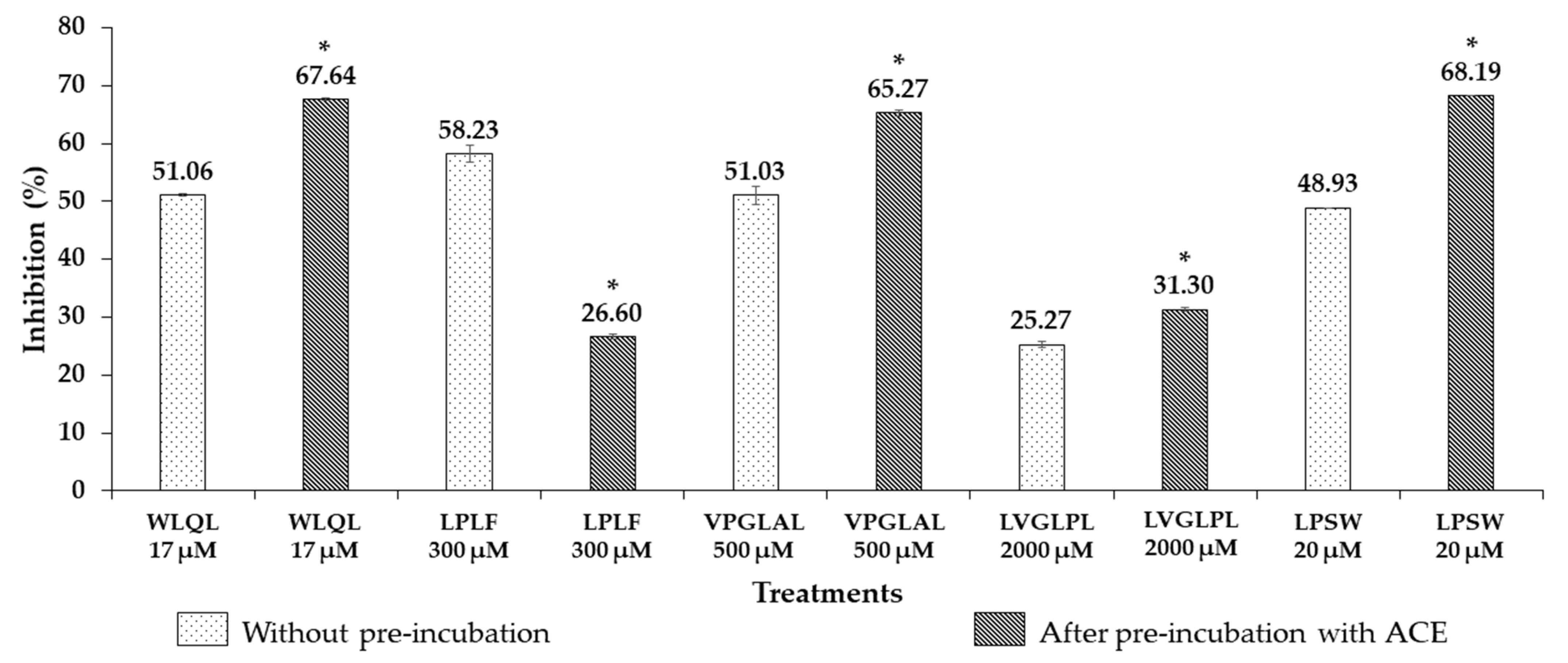
Following 3 h of incubation with ACE at 37 °C, the ACE inhibitory activity of the following peptides WLQL, LPLF, VPGLAL, LVGLPL, and LPSW was compared with samples without pre-incubation. The inhibitory activity (%) for LPSW increased from 48.93% (without pre-incubation) to 68.19% (after pre-incubation with ACE), compared with the same concentration of 20 µM (Figure A2). Moreover, the IC50 value of LPSW changed significantly following pre-incubation with ACE (without pre-incubation: 20.80 ± 0.79 µM; after pre-incubation: 13.39 ± 0.88 µM) (Table 1). Based on the LC-MS analysis, two small fragments, LP and SW, were recognized as the hydrolysis products of LPSW by ACE (Figure 2), which suggests that LPSW is a pro-drug.
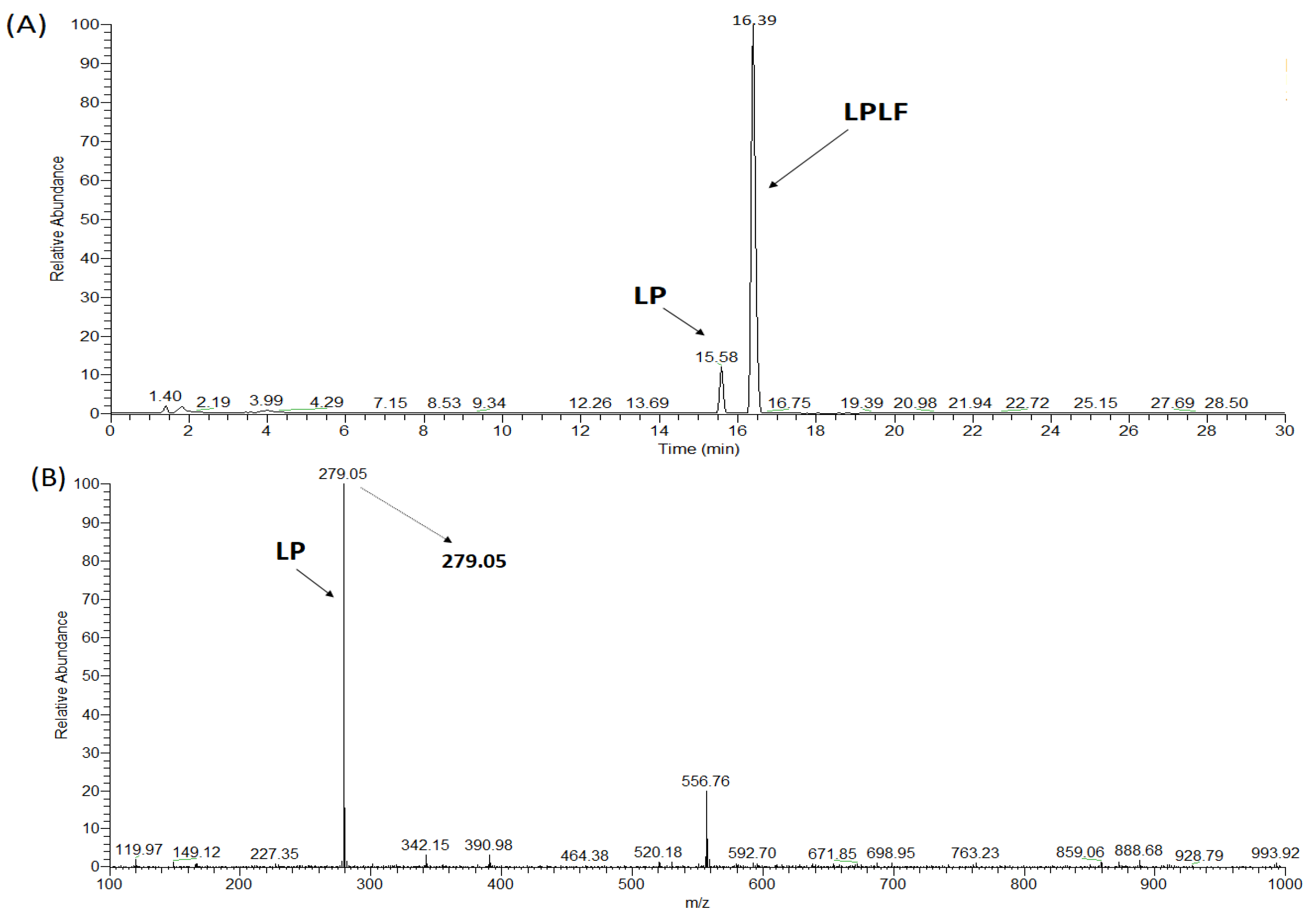
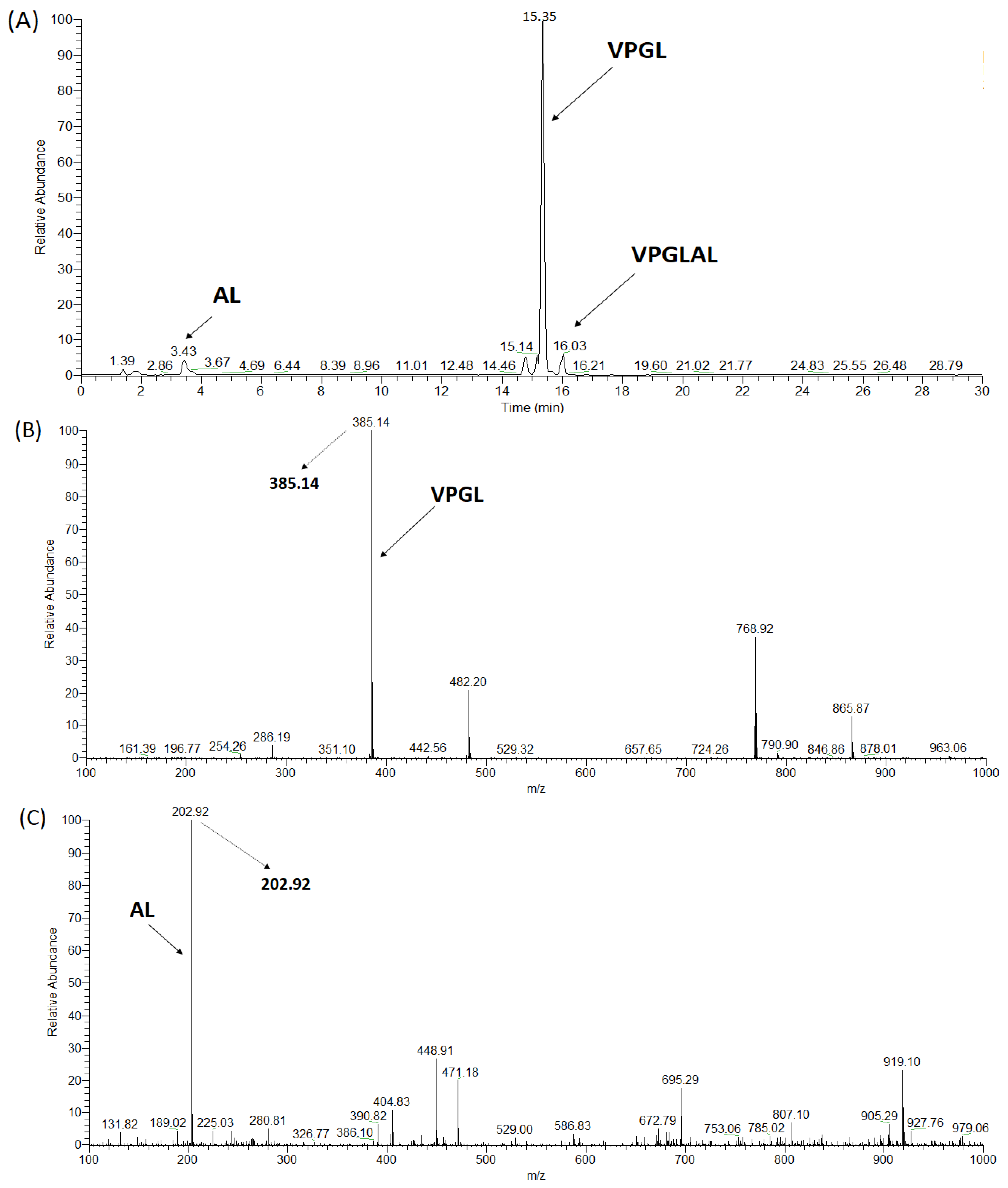
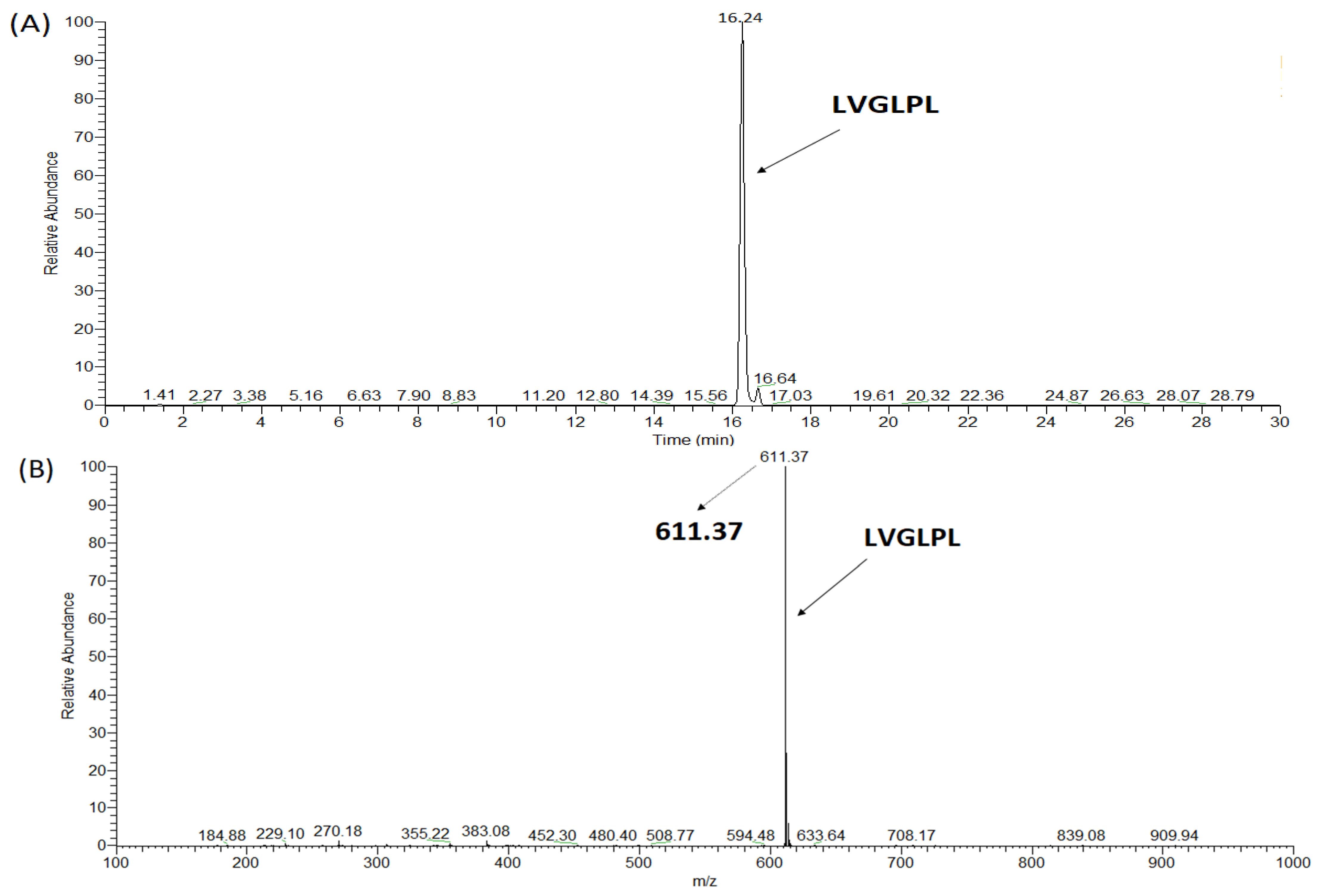
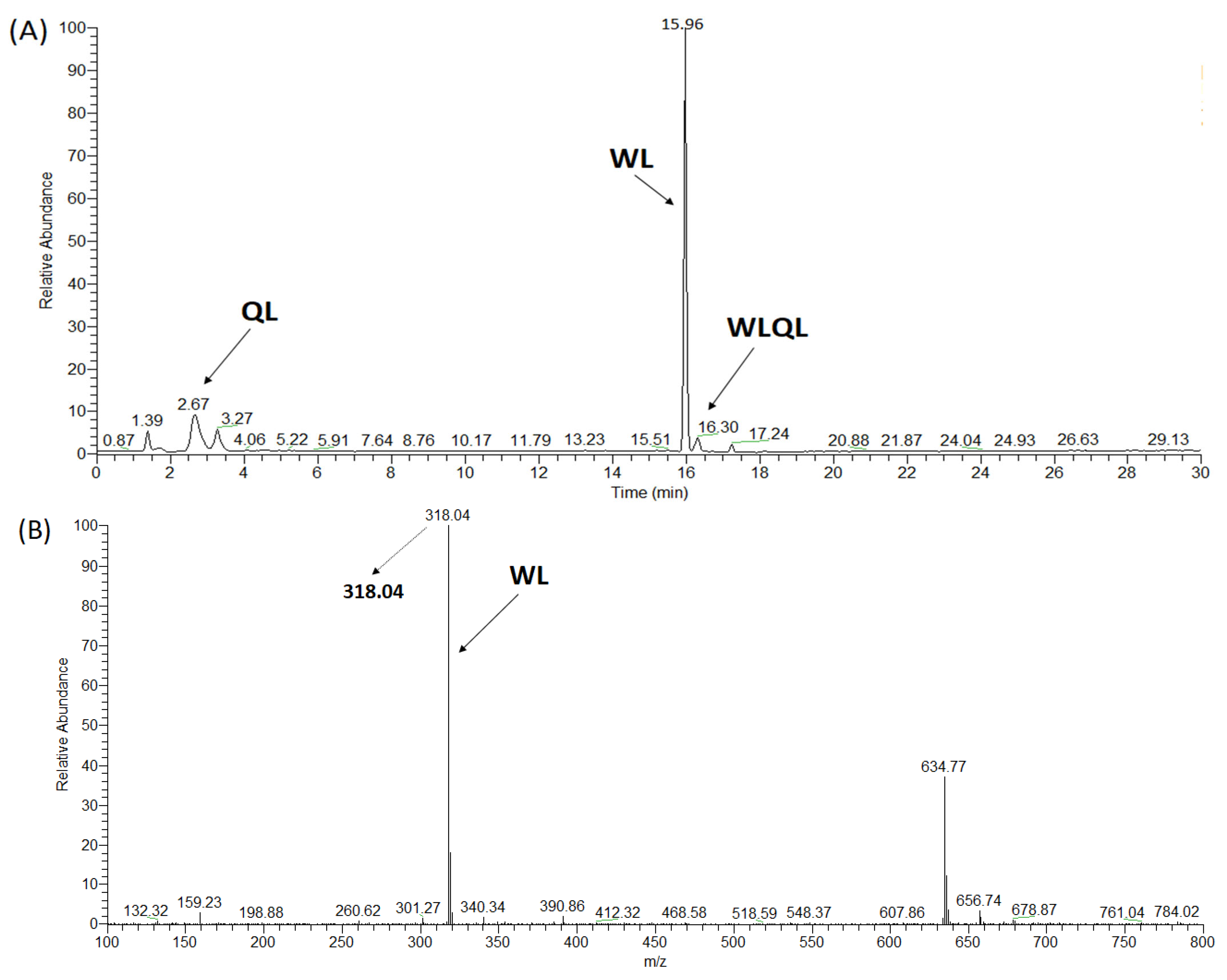
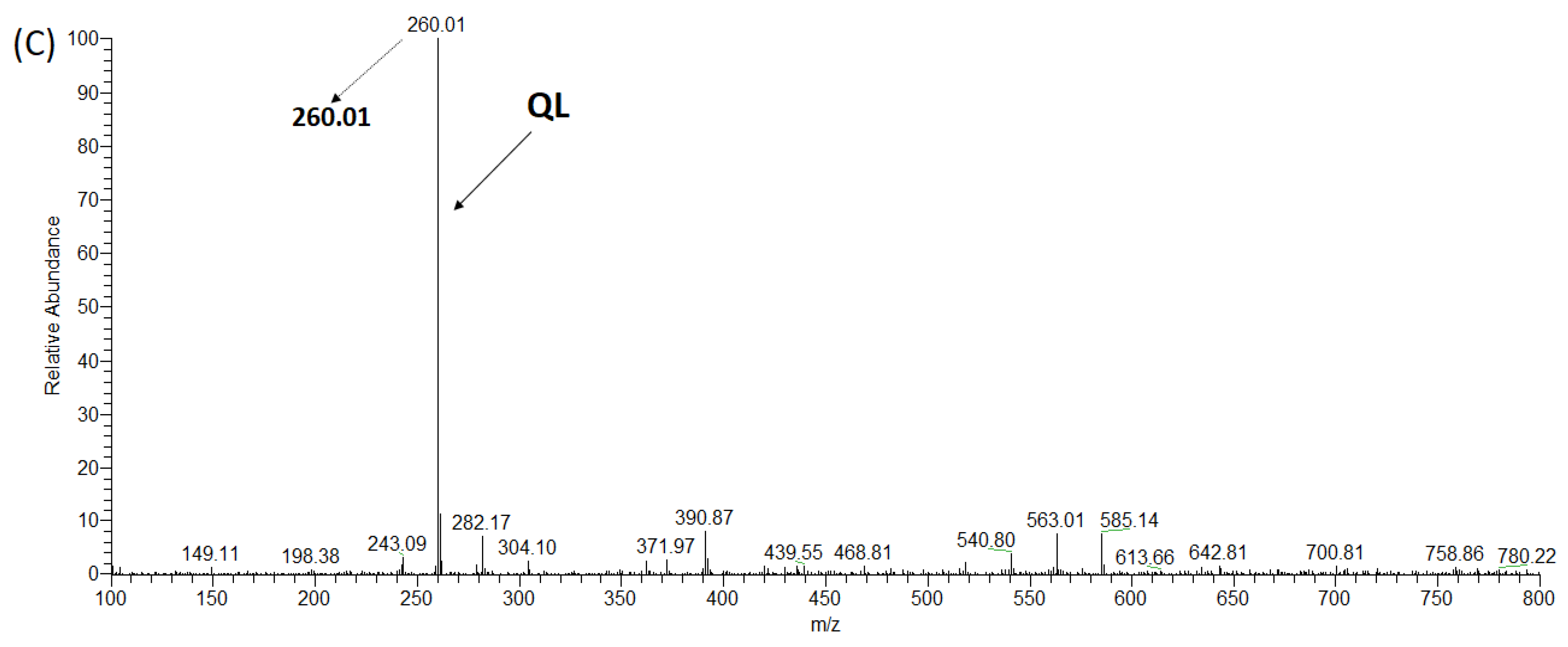
Similar to LPSW, both WLQL and VPGLAL were classified as pro-drugs of ACE. As expected, WLQL was cleavaged into small fragments, such as WL and QL (Figure 3), while VPGLAL was hydrolyzed into two peptides, AL and VPGL (Figure A4). In contrast, the ACE inhibition activity (%) of VPGLAL was notably enhanced from 51.03% to 65.27% after the pre-incubation test (at a concentration of 500 µM) (Figure A2). The tetrapeptide WLQL produced an IC50 value that showed a downward trend from 16.87 ± 0.54 µM without ACE pre-incubation to 8.5 ± 0.86 µM after pre-incubation with ACE (Table 1). According to the results of the LC-MS spectrum and stability analysis via pre-incubation with ACE, it was confirmed that LPLF was a real substrate, while LVGLPL was a true inhibitor (Figure A2, Figure A3 and Figure A5). A similar situation occurred with the pro-drug-type LPLF, which was also affected by ACE, resulting in the release of small fragments of LP and LF (Figure A3). However, after pre-incubation, the new cleavage products of 300 µM LPLF showed reduced inhibitory activity that fell from 58.23% (without pre-incubation) to 26.6% (Figure A2), which implies that LPLF is a real substrate. As observed in Figure A2 and Figure A5, LVGLPL was not hydrolyzed by ACE and its ACEI activity was not significantly changed (from 25.27% to 31.30% at the level of 2000 µM).
Peptides designated as pro-drugs, real substrates, or true inhibitors are important for in vivo or even clinical applications. Fujita’s study showed that LKPNM derived from fish protein acts as a pro-drug-type ACE inhibitor [38]. Following this, the pentapeptides IKPVQ, IKPVA, and IKPHL were reported as pro-drug substrates, while IKPVK, IKPVR, and IKPFR were reported as real substrates for ACE [37]. Both FDGIP and AIDPVRA obtained from sea grape protein were proven to possess ACEI activity, with the former as a true inhibitor and the latter as a real substrate [24]. Recently, two novel potent ACEI peptides (FRVW and LPYY) isolated from Pinctada fucata meat hydrolysates were classified as pro-drug substrates [39]. Furthermore, peptides acquired from Manchego cheese [40], Cassia obtusifolia seeds [7], and black cumin seed hydrolysates [25] also represent examples of true inhibitors of ACE.

3.4. Molecular Docking Study

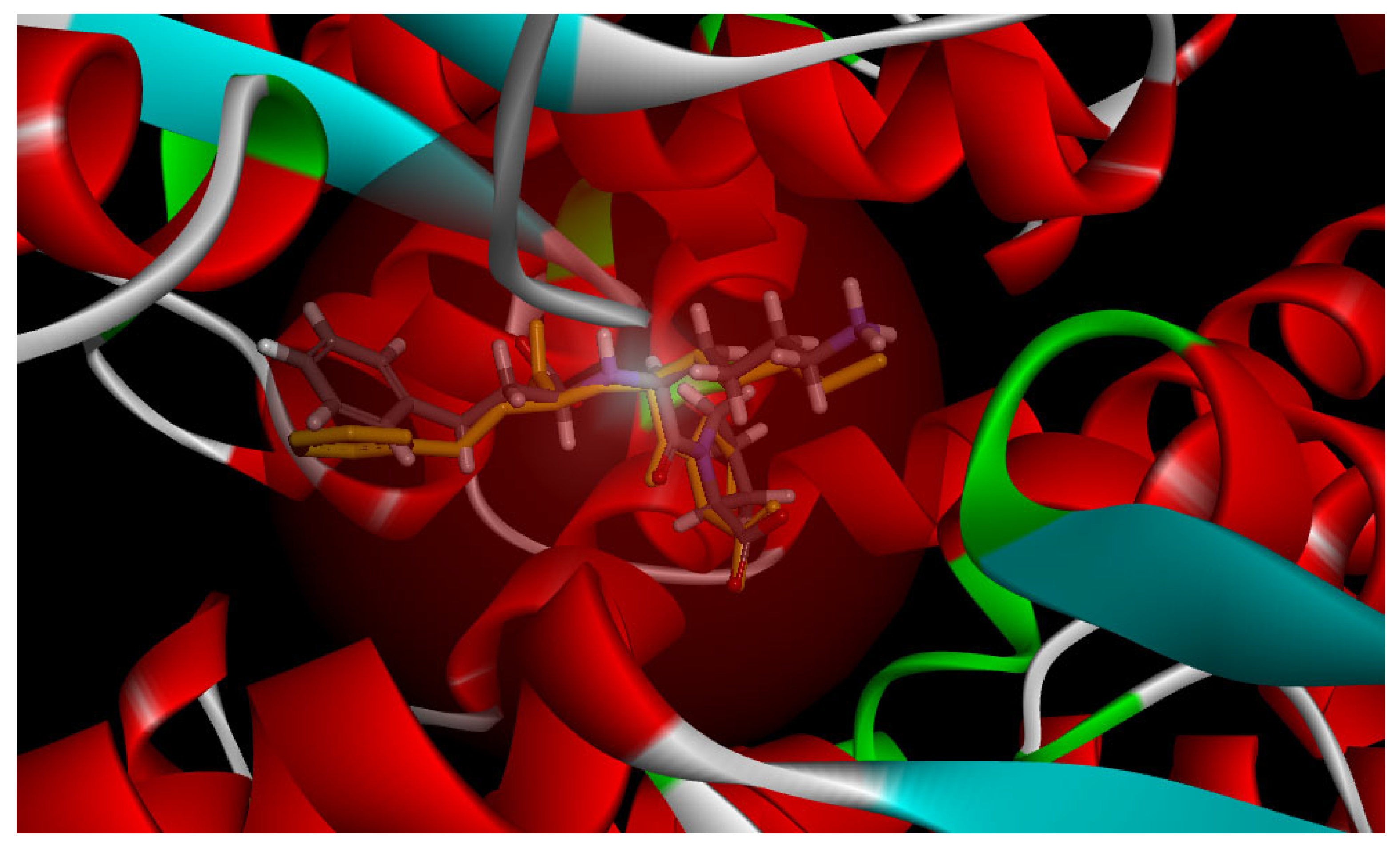
A docking simulation was performed to investigate the interactions between ACE inhibitory peptides (ligands) and ACE molecules (receptors) [7,41]. Calculations were performed using the CDocker energy value (known as interaction energies), the binding sites, and the information on formed interaction bonds. Lisinopril was selected to be docked first to tACE (PDB code: 1O86), before the processing of molecular docking of ACE inhibitory peptides to validate the precision of this current model. A significant positional similitude of lisinopril and the one obtained from the lisinopril–tACE complex (1O86.pdb) proved the accuracy of this model (Figure A6).
The docking simulation of human tACE (PDB code: 1O86) with potential peptides (WLQL and LPSW) was performed using Discovery Studio Visualized 3.0 software (Accelrys Software, Cambridge, UK). The main active sites of ACE conformation were named pocket S1 (Ala354, Glu384, and Tyr523 residues), S2 (Gln281, Tyr520, Lys511, His513, and His353 residues), and S1’ (Glu162 residue) [26,42,43]. Alternatively, Zn (II), which plays a critical role in the ACE activity, binds with ACE residues His383, His387, and Glu411 through coordination bonds to form the more stable zinc-binding motif HEXXH (tetrahedral structure) [26,44]. The stability of the zinc-binding motif HEXXH is a key factor in the binding affinity between ACE and inhibitors [26,44].
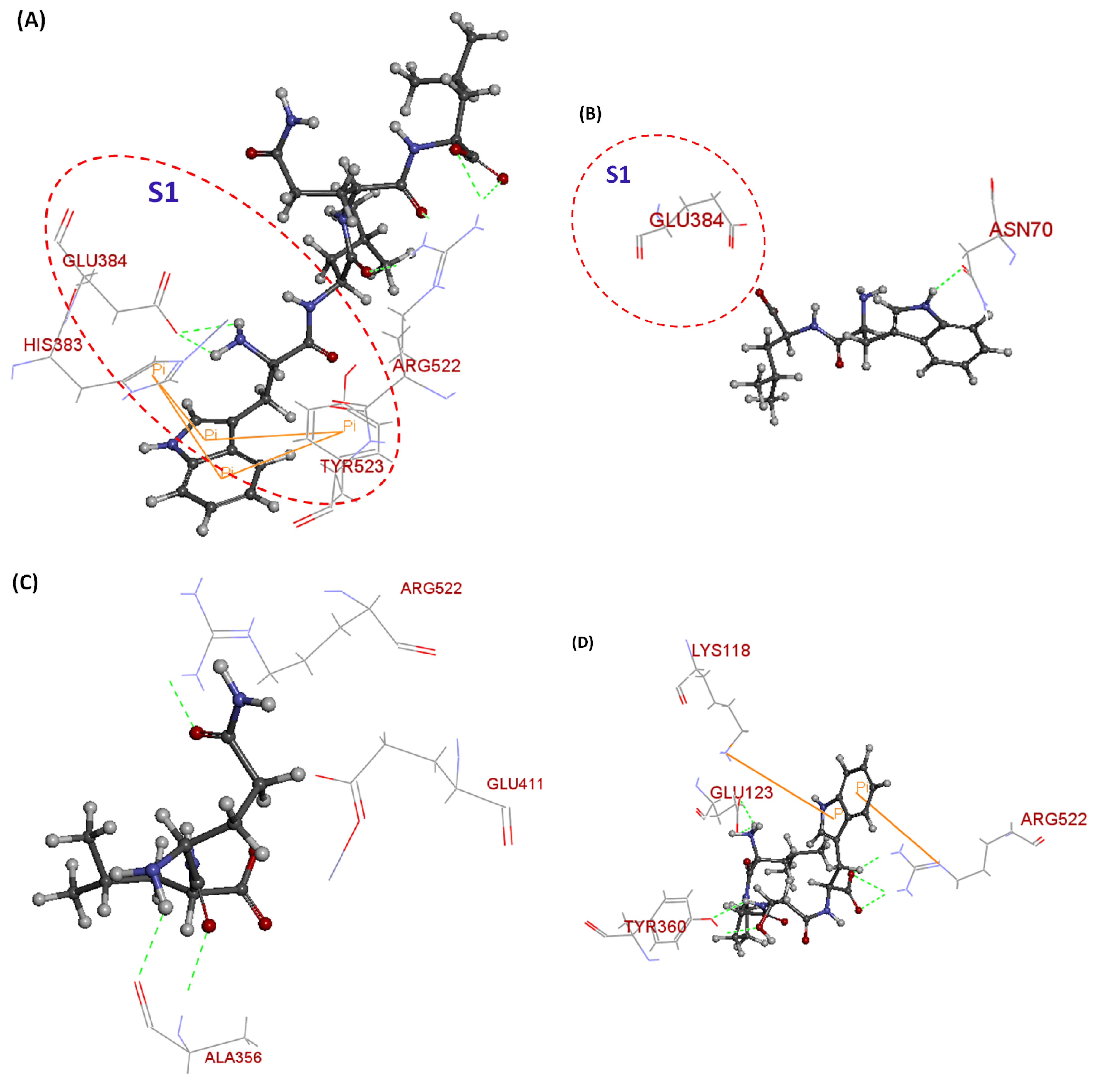
Moreover, the Arg522 residue that works as a notable ligand to the second Cl of the ACE, the binding site for lisinopril, and the substrate of ACE, may enhance ACE inhibitory activity [26,44]. These molecules can interact with the enzyme through hydrogen bonding (H-bond), hydrophobic interactions, electrostatic forces, and van der Waals forces. Among them, H-bonds are considered as contributing elements to the structural stability of the ligand–receptor complex [8,45]. Two tetrapeptides WLQL and LPSW were selected for the docking simulation with tACE based on their high ACE inhibitory activity compared to the remaining peptides. Small fragments (WL and QL from WLQL; LP and SW from LPSW) obtained from pre-incubation with ACE were also used for molecular docking to clearly understand their role in enhancing the ACE inhibitory activity. As shown in Figure 4, all the obtained peptides could interact with the ACE receptor through the S1, and S2 pockets, and the other amino acid residues.
Theoretically, the lower binding free energy of the peptide–ACE complexes can be interpreted as a thermodynamic property, resulting in a more stable complex, meaning a higher inhibitory effect of the peptides. According to their CDocker energies, the ACE inhibitory activity of the following peptides decreased: WLQL, QL, SW, WL, LPSW, and LP (Table 3). Via in vitro assay, the peptide WLQL was proven to possess a higher ACE inhibitory effect with an IC50 value of 16.87 µM, as compared to LPSW with an IC50 value of 20.80 µM.
The CDocker energy of WLQL was −80.4321 kJ/mol. The tetrapeptide WLQL formed five H-bonds with Arg522 (2.0 Å; 2.0 Å; 2.2 Å) (important ligand to the second Cl¯ of the ACE) and Glu384 (2.2 Å; 2.4 Å), and four π-π bonds with Tyr523 (5 Å; 6.3 Å), and His383 (4 Å; 4.3 Å), which was bound to a zinc ion. The amino group of tryptophan in WLQL also formed four H-bonds with Tyr523 (5 Å; 6.3 Å) and His383 (4 Å; 4.3 Å) residues, which were located at the S1 pockets in the active site, respectively (Figure 4A; Table 3, Table 4 and Table 5). The potent ACEI effect of WLQL could be rationalized through the above-mentioned interactions.
The CDocker energy of WL was 58.8494 kJ/mol. The dipeptide WL interacted with the ACE receptor via two H-bonds, including Glu384 (3.3 Å) at the S1 pocket and another residue, such as Asn70 (2.0 Å). Moreover, no π-π bonds were recognized in this case. The binding to the active site of ACE may allow the peptide WL to more easily react with the ACE, leading to an enhanced ACE inhibitory effect (Figure 4B; Table 3 and Table 5). As shown in Figure 4C and Table 3, the dipeptide QL bound to the second binding site instead of the active sites of the ACE receptor. Moreover, the interaction of QL and the ACE involved the formation of four H-bonds, including Ala356 (2.0 Å; 2.1 Å), Glu411 (2.9 Å), and Arg522 (2.4 Å), and no π-π binding was detected. However, Glu411 and Arg522 are well known for their ability to bind to zinc ions and are important ligands to the second Cl¯ of ACE, respectively. The appearance of these essential residues in the QL–ACE complex may explain the enhanced ACE inhibitory activity after the pre-incubation experiment. This peptide showed the CDocker energy of −62.3901 kJ/mol.
The tetrapeptide LPSW only interacted with the ACE receptor via the second binding sites that contained Arg522, Lys118, and Tyr360 Glu123. Binding bonds were formed in this LPSW–ACE complex, including two π-π interactions with Arg522 (6.8 Å); Lys118 (6.8 Å), and six hydrogen bonds with Tyr360 (2.2 Å; 2.2 Å); Glu123 (2.0 Å;2.7 Å) and Arg522 (1.9 Å; 2.1 Å). As is well known, Arg522 is considered as an important ligand to the second Cl¯ of the ACE. Therefore, the high ACE inhibitory effect of LPSW may be caused by Arg522 and interaction bonds, such as H-bonds and pi-pi interactions. The CDocker energy of LPSW was −55.1801 kJ/mol (Figure 4D; Table 3, Table 4 and Table 5).
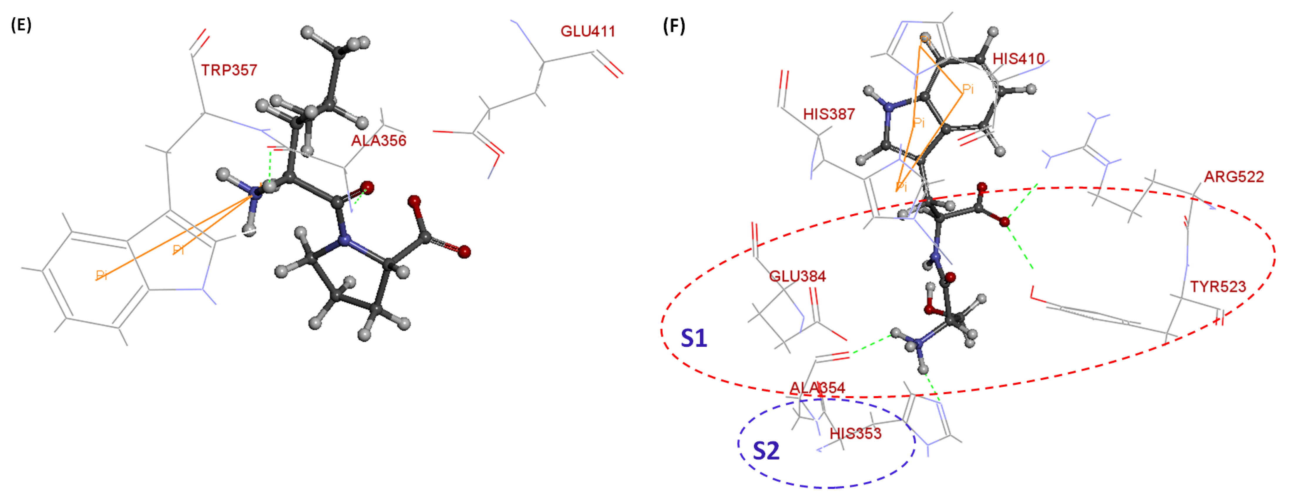
The CDocker energy of LP was 53.3389 kJ/mol. In this case, LP bound to the ACE receptor via the second binding position that contained Glu411, Ala356, and Trp357. Hydrogen and pi-pi bonds were identified in this LP–ACE complex, including three H-bonds with the residue Glu411 (3.0 Å), Ala356 (1.7 Å; 1.9 Å), and one π-π interaction with Trp357 (4.9 Å). The expression of Glu411 bound to a zinc ion may play a role in improving the ACE inhibitory activity (Figure 4E; Table 3, Table 4 and Table 5). The dipeptide SW can bind to the S1 pocket of the ACE receptor at Glu384, Ala354, and Tyr523 and the S2 pocket at His353. Furthermore, docking also occurred with different residues, including His387, Arg522, and His410. The interaction between SW and the ACE receptor involved the formation of four π-π bonds at His387 (4.4 Å; 5.4 Å) and His410 (4.2 Å; 4.7 Å) and six H-bonds, including Glu384 (3.7 Å), Ala354 (1.9 Å), Tyr523 (2.2Å), His353 (2.2 Å) and Arg522 (2.1 Å; 2.5 Å) (Figure 4F; Table 3, Table 4 and Table 5). The CDocker energy of SW was −61.9276 kJ/mol. The appearance of Arg522 (a notable ligand to the second Cl- of the ACE) and the active sites at both pockets 1 and 2 may significantly contribute to enhancing the ACE inhibitory activity of SW (Figure 4F; Table 3, Table 4 and Table 5). The CDocker energy of SW was −61.9276 kJ/mol.
According to our docking results (Table 3 and Table 5), the binding interaction of π-π may also play an important role in contributing to the structural stability of the ACE–peptide complex, leading to the strong inhibition of ACE by peptides. Liu et al. found similar results to ours and concluded that the high ACE inhibitory effect of hazelnut peptides (AVKVL, YLVR, and TLVGR) is not only determined by hydrogen and electrostatic bonds, but that the π-π interactions also offer a significant contribution [46].
Based on the kinetic and docking results, it can be concluded that WLQL acts as a competitive inhibitor and can interact with the active sites of ACE receptors. Interestingly, the peptide LPSW was determined as a non-competitive inhibitor via kinetic analysis. In this way, this peptide can only bind to non-active sites and this was confirmed through molecular docking. However, LPSW was cleavaged after being pre-incubated with ACE, and then small fragments (LP and SW) were also identified via LC-MS analysis. Therefore, the mechanism of LPSW must be studied more deeply to clearly understand this inconsistency.
Docking simulation is considered as an efficient method to predict the biological properties of peptides through the analysis of specific binding interactions formed between peptides and proteins. However, the determination of the characteristics of bioactive peptides in silico does not provide enough evidence to be conclusive; therefore, a corresponding in vitro assay must be performed for the predicted peptides [25,47].

4. Conclusions

The peptides derived from SSTY gastrointestinal hydrolysates have shown remarkable ACEI activity. Among these ACEI peptides, WLQL showed the highest IC50 value of 16.87 ± 0.54 µM. Molecular docking studies revealed that WLQL was docked to ACE through the S1 pockets of ACE, while LPSW interacted with ACE through the secondary binding sites. Moreover, the Lineweaver–Burk plot indicated that LPSW acted as a non-competitive agent against ACE. In contrast, the remaining peptides displayed patterns of competitive inhibition. Furthermore, ACE pre-incubation analysis revealed that LPSW, WLQL, and VPGLAL were pro-drug inhibitors against ACE, while LPLF was a substrate, and LVGLPL was a true inhibitor.

Author Contributions

N.T.P.N.: Writing—original draft, Data curation, Investigation, Formal analysis, Conceptualization, Investigation, Formal analysis and Writing—review and editing. C.C.Y.S.: Investigation, Formal analysis, Conceptualization, Investigation and Formal analysis. W.-T.H.: Investigation, Formal analysis, Conceptualization, Investigation and Formal analysis. J.-L.H.: Conceptualization, Investigation, Methodology, Software, Writing—review and editing, Supervision and Funding acquisition. P.-H.W.: Investigation, Methodology, Software, Writing—review and editing and Funding acquisition. All authors have read and agreed to the published version of the manuscript.

Funding

The budget for the current research was provided through the NPUST-KMU Joint Research Project (NPUST-KMU-111-P001), the Ministry of Science and Technology (MOST 111-2113-M-020-001), and Animal Biologics Research of the Featured Area Research Center within the framework of Higher Education Sprout Project supported by both the Ministry of Education and Ministry of Science and Technology (MOST 111-2634-F-020-001), Taiwan.

Institutional Review Board Statement

Not applicable.

Informed Consent Statement

Not applicable.

Data Availability Statement

Not applicable.

Acknowledgments

We appreciate the assistance with the valuable mass spectrometry analysis from the Precision Instrument Center of NPUST and Mithra Biotechnology Inc.

Conflicts of Interest

The authors declare no conflict of interest.

Appendix A

Figure A1. The ACE inhibitory effects of peptides present via the IC50 values with (A) WLQL, (B) LPSW, (C) LPLF, and (D) VPGLAL.
Applsci 12 12340 g0a1
Figure A2. The ACE inhibitory activity of peptides with pre-incubation assay. The error bars present the standard deviation (* indicated as significant difference between groups without and after pre-incubation with ACE when p < 0.05). The concentrations utilized for comparison are dependent on each peptide.
Applsci 12 12340 g0a2
Figure A3. The stability of LPLF against the ACE activity under the pre-incubation condition with ACE. (A) LC-MS profile of LPLF. (B) The MS spectrum of fragment LF (m/z 279.05).
Applsci 12 12340 g0a3
Figure A4. The stability of VPGLAL against the ACE activity under the pre-incubation condition with ACE. (A) LC-MS profile of VPGLAL. Two small fragments derived from VPGLAL after pre-incubation with ACE include: (B) the MS spectrum of fragment VPGL (m/z 385.17), and (C) the MS spectrum of fragment AL (m/z 202.92).
Applsci 12 12340 g0a4
Figure A5. The stability of LVGLPL against the ACE activity under the pre-incubation condition with ACE. (A) LC-MS analysis of LVGLPL. (B) The MS spectrum of fragment LVGLPL (m/z 611.37).
Applsci 12 12340 g0a5
Figure A6. The interaction of lisinopril and tACE (1O86.pdb). The ribbon demonstrates tACE. The substantial overlapping of lisinopril was achieved via docking (yellow color) and the enzyme derived from PBD (1O86) proved the accuracy of the current model. The docking energy score between lisinopril and tACE is −92.8494 kJ/mol (CDocker).
Applsci 12 12340 g0a6

References

  1. Majumder, K.; Wu, J. Molecular targets of antihypertensive peptides: Understanding the mechanisms of action based on the pathophysiology of hypertension. Int. J. Mol. Sci. 2015, 16, 256–283. [Google Scholar] [CrossRef] [PubMed] [Green Version]
  2. Ahmad, M.I.; Mendys, P.M.; Kelly, S.P.; Chen, L.Y.; Soliman, E.Z. Relationship between high blood pressure, atrial cardiopathy, and mortality in the general population. Am. J. Hypertens. 2022. [Google Scholar] [CrossRef]
  3. Zheng, Y.; Wang, X.; Zhuang, Y.; Li, Y.; Shi, P.; Tian, H.; Li, X.; Chen, X. Isolation of novel ACE-inhibitory peptide from naked oat globulin hydrolysates in silico approach: Molecular docking, in vivo antihypertension and effects on renin and intracellular endothelin-1. J. Food Sci. 2020, 85, 1328–1337. [Google Scholar] [CrossRef] [PubMed]
  4. Udenigwe, C.C.; Mohan, A. Mechanisms of food protein-derived antihypertensive peptides other than ACE inhibition. J. Funct. Foods 2014, 8, 45–52. [Google Scholar] [CrossRef]
  5. Li, S.; Tang, S.; He, Q.; Hu, J.; Zheng, J. In vitro antioxidant and angiotensin-converting enzyme inhibitory activity of fermented milk with different culture combinations. J. Dairy Sci. 2020, 103, 1120–1130. [Google Scholar] [CrossRef]
  6. Schiffrin, E.L.; Flack, J.M.; Ito, S.; Muntner, P.; Webb, R.C. Hypertension and COVID-19. Am. J. Hypertens. 2022, 33, 373–374. [Google Scholar] [CrossRef] [Green Version]
  7. Shih, Y.-H.; Chen, F.-A.; Wang, L.-F.; Hsu, J.-L. Discovery and study of novel antihypertensive peptides derived from Cassia obtusifolia seeds. J. Agric. Food Chem. 2019, 67, 7810–7820. [Google Scholar] [CrossRef]
  8. Tu, M.; Wang, C.; Chen, C.; Zhang, R.; Liu, H.; Lu, W.; Jiang, L.; Du, M. Identification of a novel ACE-inhibitory peptide from casein and evaluation of the inhibitory mechanisms. Food Chem. 2018, 256, 98–104. [Google Scholar] [CrossRef]
  9. Chamata, Y.; Watson, K.A.; Jauregi, P. Whey-derived peptides interactions with ACE by molecular docking as a potential predictive tool of natural ACE inhibitors. Int. J. Mol. Sci. 2020, 21, 864. [Google Scholar] [CrossRef] [Green Version]
  10. Salim, M.A.S.M.; Gan, C.-Y. Dual-function peptides derived from egg white ovalbumin: Bioinformatics identification with validation using in vitro assay. J. Funct. Foods 2020, 64, 103618. [Google Scholar] [CrossRef]
  11. Fan, H.; Wang, J.; Liao, W.; Jiang, X.; Wu, J. Identification and characterization of gastrointestinal-resistant angiotensin-converting enzyme inhibitory peptides from egg white proteins. J. Agric. Food Chem. 2019, 67, 7147–7156. [Google Scholar] [CrossRef] [PubMed]
  12. Liu, L.; Lu, B.; Gong, L.; Wu, X.; Zhang, Y. Studies on bioactive peptide from Chinese soft-shelled turtle (Pelodiscus sinensis) with functionalities of ACE inhibition and antioxidation. Afr. J. Biotechnol. 2012, 11, 6723–6729. [Google Scholar]
  13. Huanling, Y.; Yong, L.; Junbo, W.; Liping, Z.; Weixing, Y. Chinese soft-shelled turtle egg powder lowers serum cholesterol, increases faecal neutral steroids and bile acid excretion, and up-regulates liver cytochrome P450 mRNA level in rats. Br. J. Nutr. 2005, 94, 315–320. [Google Scholar] [CrossRef] [PubMed] [Green Version]
  14. Chiu, L.H.; Hsu, G.S.W.; Lu, Y.F. Antihypertensive capacity of defatted soft-shelled turtle powder after hydrolysis by gastrointestinal enzymes. J. Food Biochem. 2006, 30, 589–603. [Google Scholar] [CrossRef]
  15. Rawendra, R.D.; Chen, S.-H.; Chang, C.-I.; Shih, W.-L.; Huang, T.-C.; Liao, M.-H.; Hsu, J.-L. Isolation and characterization of a novel angiotensin-converting enzyme-inhibitory tripeptide from enzymatic hydrolysis of soft-shelled turtle (Pelodiscus sinensis) egg white: In vitro, in vivo, and in silico study. J. Agric. Food Chem. 2014, 62, 12178–12185. [Google Scholar] [CrossRef]
  16. Rawendra, R.D.; Chang, C.-I.; Chen, H.-H.; Huang, T.-C.; Hsu, J.-L. A novel angiotensin converting enzyme inhibitory peptide derived from proteolytic digest of Chinese soft-shelled turtle egg white proteins. J. Proteomics 2013, 94, 359–369. [Google Scholar] [CrossRef]
  17. Pujiastuti, D.Y.; Shih, Y.-H.; Chen, W.-L.; Hsu, J.-L. Screening of angiotensin-I converting enzyme inhibitory peptides derived from soft-shelled turtle yolk using two orthogonal bioassay-guided fractionations. J. Funct. Foods 2017, 28, 36–47. [Google Scholar] [CrossRef]
  18. Nong, N.T.P.; Chen, Y.-K.; Shih, W.-L.; Hsu, J.-L. Characterization of Novel Dipeptidyl Peptidase-IV Inhibitory Peptides from Soft-Shelled Turtle Yolk Hydrolysate Using Orthogonal Bioassay-Guided Fractionations Coupled with In Vitro and In Silico Study. Pharmaceuticals 2020, 13, 308. [Google Scholar] [CrossRef]
  19. Abouelkheir, M.; El-Metwally, T.H. Dipeptidyl peptidase-4 inhibitors can inhibit angiotensin converting enzyme. Eur. J. Pharmacol. 2019, 862, 172638. [Google Scholar] [CrossRef]
  20. Gomez, H.L.R.; Peralta, J.P.; Tejano, L.A.; Chang, Y.-W. In silico and in vitro assessment of portuguese oyster (Crassostrea angulata) proteins as precursor of bioactive peptides. Int. J. Mol. Sci. 2019, 20, 5191. [Google Scholar] [CrossRef] [Green Version]
  21. Sasaki, C.; Tamura, S.; Tohse, R.; Fujita, S.; Kikuchi, M.; Asada, C.; Nakamura, Y. Isolation and identification of an angiotensin I-converting enzyme inhibitory peptide from pearl oyster (Pinctada fucata) shell protein hydrolysate. Process Biochem. 2019, 77, 137–142. [Google Scholar] [CrossRef]
  22. Wang, R.; Lu, X.; Sun, Q.; Gao, J.; Ma, L.; Huang, J. Novel ACE inhibitory peptides derived from simulated gastrointestinal digestion in vitro of sesame (Sesamum indicum L.) protein and molecular docking study. Int. J. Mol. Sci. 2020, 21, 1059. [Google Scholar] [CrossRef] [PubMed] [Green Version]
  23. Cushman, D.W.; Cheung, H.; Sabo, E.; Ondetti, M. Design of potent competitive inhibitors of angiotensin-converting enzyme. Carboxyalkanoyl and mercaptoalkanoyl amino acids. Biochemistry 1977, 16, 5484–5491. [Google Scholar] [CrossRef] [PubMed]
  24. Joel, C.H.; Sutopo, C.C.; Prajitno, A.; Su, J.-H.; Hsu, J.-L. Screening of angiotensin-I converting enzyme inhibitory peptides derived from Caulerpa lentillifera. Molecules 2018, 23, 3005. [Google Scholar] [CrossRef] [PubMed] [Green Version]
  25. Sutopo, C.C.; Sutrisno, A.; Wang, L.-F.; Hsu, J.-L. Identification of a potent angiotensin-I converting enzyme inhibitory peptide from black cumin seed hydrolysate using orthogonal bioassay-guided fractionations coupled with in silico screening. Process Biochem. 2020, 95, 204–213. [Google Scholar] [CrossRef]
  26. Natesh, R.; Schwager, S.; Sturrock, E.; Acharya, K. Crystal Structure of Human Angiotensin Converting Enzyme in complex with lisinopril. Nature 2003, 421, 551–554. [Google Scholar] [CrossRef] [Green Version]
  27. Liu, P.; Lan, X.; Yaseen, M.; Wu, S.; Feng, X.; Zhou, L.; Sun, J.; Liao, A.; Liao, D.; Sun, L. Purification, characterization and evaluation of inhibitory mechanism of ACE inhibitory peptides from pearl oyster (Pinctada fucata martensii) meat protein hydrolysate. Mar. Drugs 2019, 17, 463. [Google Scholar] [CrossRef] [Green Version]
  28. Fan, H.; Wu, J. Purification and identification of novel ACE inhibitory and ACE2 upregulating peptides from spent hen muscle proteins. Food Chem. 2021, 345, 128867. [Google Scholar] [CrossRef]
  29. Sun, S.; Xu, X.; Sun, X.; Zhang, X.; Chen, X.; Xu, N. Preparation and identification of ACE inhibitory peptides from the marine macroalga Ulva intestinalis. Mar. Drugs 2019, 17, 179. [Google Scholar] [CrossRef] [Green Version]
  30. García-Mora, P.; Martín-Martínez, M.; Bonache, M.A.; González-Múniz, R.; Peñas, E.; Frias, J.; Martinez-Villaluenga, C. Identification, functional gastrointestinal stability and molecular docking studies of lentil peptides with dual antioxidant and angiotensin I converting enzyme inhibitory activities. Food Chem. 2017, 221, 464–472. [Google Scholar] [CrossRef] [Green Version]
  31. Cheung, B.M.; Li, C. Diabetes and hypertension: Is there a common metabolic pathway? Curr. Atheroscler. Rep. 2012, 14, 160–166. [Google Scholar] [CrossRef] [PubMed]
  32. Nong, N.T.P.; Hsu, J.-L. Characteristics of Food Protein-Derived Antidiabetic Bioactive Peptides: A Literature Update. Int. J. Mol. Sci. 2021, 22, 9508. [Google Scholar] [CrossRef]
  33. Chen, M.; Pan, D.; Zhou, T.; Gao, X.; Dang, Y. Novel Umami Peptide IPIPATKT with Dual Dipeptidyl Peptidase-IV and Angiotensin I-Converting Enzyme Inhibitory Activities. J. Agric. Food Chem. 2021, 69, 5463–5470. [Google Scholar] [CrossRef] [PubMed]
  34. Wu, J.; Xie, D.; Chen, X.; Tang, Y.-J.; Wang, L.; Xie, J.; Wei, D. Inhibitory mechanism of a substrate-type angiotensin I-converting enzyme inhibitory peptide. Process Biochem. 2019, 79, 97–104. [Google Scholar] [CrossRef]
  35. Lunow, D.; Kaiser, S.; Rückriemen, J.; Pohl, C.; Henle, T. Tryptophan-containing dipeptides are C-domain selective inhibitors of angiotensin converting enzyme. Food Chem. 2015, 166, 596–602. [Google Scholar] [CrossRef] [PubMed]
  36. Lunow, D.; Kaiser, S.; Brückner, S.; Gotsch, A.; Henle, T. Selective release of ACE-inhibiting tryptophan-containing dipeptides from food proteins by enzymatic hydrolysis. Eur. Food Res. Technol. 2013, 237, 27–37. [Google Scholar] [CrossRef]
  37. Rao, S.-Q.; Liu, S.; Ju, T.; Xu, W.-Q.; Mei, G.-M.; Xu, Y.-S.; Yang, Y.-J. Design of substrate-type ACE inhibitory pentapeptides with an antepenultimate C-terminal proline for efficient release of inhibitory activity. Biochem. Eng. J. 2012, 60, 50–55. [Google Scholar] [CrossRef]
  38. Fujita, H.; Yoshikawa, M. LKPNM: A prodrug-type ACE-inhibitory peptide derived from fish protein. Immunopharmacology 1999, 44, 123–127. [Google Scholar] [CrossRef]
  39. Li, J.; Su, J.; Chen, M.; Chen, J.; Ding, W.; Li, Y.; Yin, H. Two novel potent ACEI peptides isolated from Pinctada fucata meat hydrolysates using in silico analysis: Identification, screening and inhibitory mechanisms. RCS Adv. 2021, 11, 12172–12182. [Google Scholar] [CrossRef]
  40. Ruiz, J.Á.G.; Ramos, M.; Recio, I. Angiotensin converting enzyme-inhibitory activity of peptides isolated from Manchego cheese. Stability under simulated gastrointestinal digestion. Int. Dairy J. 2004, 14, 1075–1080. [Google Scholar] [CrossRef]
  41. Salmaso, V.; Moro, S. Bridging molecular docking to molecular dynamics in exploring ligand-protein recognition process: An overview. Front. Pharmacol. 2018, 9, 923. [Google Scholar] [CrossRef] [PubMed]
  42. Qian, B.; Tian, C.; Huo, J.; Ding, Z.; Xu, R.; Zhu, J.; Yu, L.; Villarreal, O.D. Design and evaluation of four novel tripeptides as potent angiotensin converting enzyme (ACE) inhibitors with anti-hypertension activity. Peptides 2019, 122, 170171. [Google Scholar] [CrossRef] [PubMed]
  43. Cao, S.; Wang, Y.; Hao, Y.; Zhang, W.; Zhou, G. Antihypertensive effects in vitro and in vivo of novel angiotensin-converting enzyme inhibitory peptides from bovine bone gelatin hydrolysate. J. Agric. Food Chem. 2019, 68, 759–768. [Google Scholar] [CrossRef] [PubMed]
  44. Liu, C.; Fang, L.; Min, W.; Liu, J.; Li, H. Exploration of the molecular interactions between angiotensin-I-converting enzyme (ACE) and the inhibitory peptides derived from hazelnut (Corylus heterophylla Fisch.). Food Chem. 2018, 245, 471–480. [Google Scholar] [CrossRef] [PubMed]
  45. Liu, C.; Liu, J.; Wang, M.; Zhang, B.; Wang, E.; Liu, B.; Zhang, T. Construction and application of membrane-bound angiotensin-I converting enzyme system: A new approach for the evaluation of angiotensin-I converting enzyme inhibitory peptides. J. Agric. Food Chem. 2020, 68, 5723–5731. [Google Scholar] [CrossRef]
  46. Khedr, S.; Martin, M.; Deussen, A. Inhibitory efficacy and biological variability of tryptophan containing dipeptides on human plasma angiotensin converting enzyme activity. J. Hypertens. 2015, 4. [Google Scholar] [CrossRef]
  47. Chen, Y.-C. Beware of docking! Trends Pharmacol. Sci. 2015, 36, 78–95. [Google Scholar] [CrossRef]
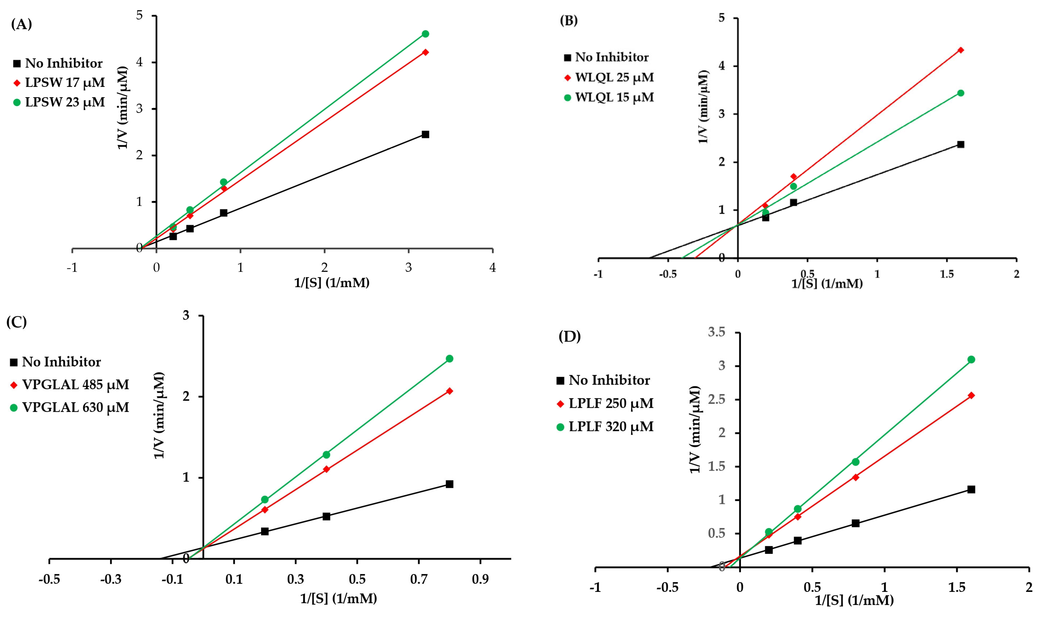
Figure 1. Lineweaver–Burk double reciprocal plots for ACE inhibition with (A) LPSW (non-competitive), (B) WLQL (competitive), (C) VPGLAL (competitive), and (D) LPLF (competitive) determined at concentrations close to their IC50 values.
Applsci 12 12340 g001
Figure 2. Liquid chromatography–mass spectrometry (LC-MS) profile of (A) LPSW full scan (m/z 502), (B) SW fragment (m/z 292.25), and (C) LP fragment (m/z 229.24) derived from LPSW preincubated with ACE for 3 h at 37 °C.
Applsci 12 12340 g002
Figure 3. Liquid chromatography–mass spectrometry (LC-MS) profile of (A) WLQL full scan (m/z 559.30), (B) WL fragment (m/z 318.02); (C) QL fragment (m/z 259.99) derived from WLQL pre-incubated with ACE for 3 h at 37 °C.
Applsci 12 12340 g003aApplsci 12 12340 g003b
Figure 4. The interaction model of inhibitory peptides and the ACE receptor. The best docking poses of peptides are (A) WLQL, (B) WL, (C) QL, (D) LPSW, (E) LP, and (F) SW at the ACE pockets. The amino acid residues that belong to pocket S1 and S2 are shown via a red circle and blue circle, respectively.
Applsci 12 12340 g004aApplsci 12 12340 g004b
Table 1. DPP-IV and ACE inhibitory activity of five peptides derived from SSTY protein hydrolysate.
Peptide Molecular Mass (Da)LengthDPP-IV IC50 (µM) [18]ACE IC50 (µM)Mode of ACE InhibitionStability of Peptide with ACE ACEI IC50 (µM) after Pre-Incubation with ACE
WLQL5594432.5 ± 11.8516.87 ± 0.54CompetitiveHydrolyzed8.5 ± 0.86
LPSW5024269.7 ± 15.9120.80 ± 0.79 Non-competitiveHydrolyzed13.39 ± 0.88
LPLF4894463.6 ± 5.52300.08 ± 17.30CompetitiveHydrolyzednd
VPGLAL5696289.2 ± 11.85573.00 ± 54.10CompetitiveHydrolyzednd
LVGLPL6116>2000>2000ndNot hydrolyzednd
Table 2. Kinetic constants of enzyme-catalyzed reactions at different peptide concentrations.
PeptideCatalytic Parameter *Concentration (µM)
LPSW 0 µM17 µM23 µM
Vmax6.994.653.76
Km5.065.845.13
WLQL 0 µM15 µM25 µM
Vmax1.471.441.42
Km1.572.483.24
VPGLAL 0 µM485 µM630 µM
Vmax7.198.067.17
Km7.0219.6420.83
LPLF 0 µM250 µM320 µM
Vmax7.526.157.59
Km4.839.1913.99
* Vmax is the maximum reaction velocity; Km is the Michaelis constant.
Table 3. Potential binding sites of ACE inhibitory peptides based on molecular docking analysis.
SequencesPotential Binding SiteTotal Binding SitesNumber of H-BondsNumber of Pi-Pi BondsCDOCKER Energy (kJ/mol)
WLQLTyr523, Arg522, Glu384, His38395480.4321
LPSWTyr360, Lys118, Glu123, Arg52286255.1801
WLGlu384, Asn7022058.8494
QLArg522, Ala356, Glu41144062.3901
LPTrp357, Ala356, Glu41143153.3389
SWGlu384, His353, Tyr523, Arg522, His387, His410, Ala354106461.9276
Table 4. Hydrogen bonds observed between the top-ranked docked poses of peptides and tACE a.
tACE Residues Involved in H-Bonding bNo. of H-Bonds and Corresponding Distance (Å)
WLQLLPSWWLQLLPSW
Arg522: O2; 2; 2.21.9; 2.12.42.1; 2.5
Glu384: H2.2; 2.4
Glu384: O3.3
Glu384: N3.7
Asn70: H2.0
Glu411: O2.93
Ala356: OH 2; 2.11.7; 1.9
Glu123: NH2; 2.7
Tyr360: H2.2; 2.2
Ala354: H1.9
His353: H2.2
Tyr523: O2.2
Total562436
a The residue number of tACE (PBD code: 108A). b H-bond position: donor or receptor atoms. Repeated residues indicate formation of two separate H-bonds by the same amino acid.
Table 5. Pi-pi bonds observed between the top-ranked docked poses of peptides and tACE a.
tACE ResiduesNo. of pi-pi Bonds and Corresponding Distance (Å)
WLQLLPSWWLQLLPSW
Tyr523: H5; 6.3
His383: H4; 4.3
Arg522: H6.8
Lys118: H6.8
Trp357: H4.9
His387: H4.4; 5.4
His410: H4.2; 4.7
Total420014
a The residue number of tACE (PBD code: 108A).
Publisher’s Note: MDPI stays neutral with regard to jurisdictional claims in published maps and institutional affiliations.