Article

Relationship between Reflectivity, Chemical Composition and Mechanical Behaviour of Orthodontic Bonding Nanofiller Resin Materials: A Proposal of an Alternative Method of Investigation

1
Department of Clinical Sciences and Translational Medicine, University of Rome “Tor Vergata”, Via Montpellier, 1, 00133 Rome, Italy
2
Institute for Microelectronics and Microsystems, National Research Council, Via del Fosso del Cavaliere, 100, 00133 Rome, Italy
3
Institute for Photonics and Nanotechnologies, National Research Council, Via del Fosso del Cavaliere, 100, 00133 Rome, Italy
4
Istituto di Struttura della Materia, Consiglio Nazionale delle Ricerche, Via del Fosso del Cavaliere, 100, 00133 Rome, Italy
5
Department of Life, Health & Environmental Sciences, University of L’Aquila, 67100 L’Aquila, Italy
*
Author to whom correspondence should be addressed.
Appl. Sci. 2022, 12(24), 12538; https://doi.org/10.3390/app122412538
Submission received: 14 November 2022 / Revised: 26 November 2022 / Accepted: 5 December 2022 / Published: 7 December 2022
(This article belongs to the Special Issue Application of Nanomaterials in Dentistry)

Abstract

:
Background: Relationships between reflectivity, hardness and chemical composition of the dispersed phase, included in orthodontic composites Transbond XTTM (Trans), Light-Cure Orthodontic Paste (Leone) and Bisco Ortho Bracket Paste LC (Bisco), were investigated in vitro to evaluate whether reflectivity results can be useful in internal material composition interpretation, thus obtaining information on mechanical behaviours. Methods: Light transmission through 36 resin discs was measured with a UV/Vis spectrophotometer, evaluating the spectral range from 190–1100 nm. To have a benchmark of material hardness and internal composition, Vickers measurements and Cross-Sectional Focus Ion Beam Scanning Electron Microscopy (FIB/SEM) analysis were provided. Results: Bisco has the highest reflectivity, Leone shows an absorption pattern in the UV region similar to Bisco and Transbond has the lowest reflectivity compared to the others. This trend is confirmed by FIB/SEM imaging, showing a more similar induced roughness and internal composition for Bisco and Leone, with respect to Transbond. Higher filler presence in the composition of Bisco and Leone justifies a higher hardness of these two materials, with respect to Transbond, as confirmed by Vickers measurements. Conclusions: Bisco and Leone show similar optical responses and similarities in mechanical performance. This statement is explained by the lower and similar filler content as confirmed also by FIB/SEM analysis. The inner composition of Bisco and Leone provides a higher value of microhardness, as demonstrated by Vickers measurements. Therefore, this study confirms that the UV-Vis analysis can also offer a significant overview on the internal material composition, thus indirectly providing information on the mechanical properties of orthodontic composites.

1. Introduction

At first, the real discriminant in the choice of composite resin for orthodontic bonding was the mechanical resistance to stress induced by orthodontic forces, especially in critical clinical cases [1,2,3,4]. An ideal orthodontic adhesive system, in fact, is one that is able both to resist the orthodontic-chewing forces during all phases of treatment and to preserve the integrity of the dental enamel following the removal of the bracket [5].
The bond strength of the bracket to the enamel is strictly influenced by each step of the application process, such as acid etching and drying time, the method of application of the adhesive and the curing time [6]. In recent years, optical properties (color, translucency, opalescence and fluorescence) and translucency in particular, have become fundamental properties to be evaluated for choosing the right composite resin, not only for bracket bonding procedures [7]. More recently, treatments with transparent aligners must involve additional auxiliaries, such as attachments in composite materials, which, acting as force transducers, make the orthodontic movement more effective, thus improving the biomechanics while respecting aesthetics [8]. Translucency, together to color, is the most easily observed optical property of materials that describes the partial passage of light through a certain structure [7]. The main factor that determines the optical properties of a material is its chemical composition: it is for this reason that the refractive index is different between dental biomaterials such as ceramics, glass ionomer cements and composite resins [9,10,11,12,13,14,15,16,17]. For composite resins, some of the factors responsible for the optical dispersion of light throughout the material are the overall composition and the filler content, both in terms of shape and size [18,19].
Most orthodontic adhesives consist of composite resin. Though, the orthodontic adhesive systems represent a variant, in terms of formulation, to the adhesives and composites used in conservative dentistry, differing from these mainly in the increased concentration of co-monomers found in their basic formulation [6]. The orthodontic composite resin is a complex material consisting of an organic matrix, (Bis-GMA, UDMA, TEGDMA), synthetic polymers (ceramic reinforcing fillers and silane coupling agents) and molecules that promote or modify the polymerization reaction. Resin adhesives consist of methacrylates (e.g., hydroxyethyl methacrylate or HEMA, and 4 methacryl-oxyethyl-trimellitic-anhydride, or 4-META), dimethacrylates (e.g., triethylen-glycol dimethacrylate, or TEGDMA, and Bis-GMA, consisting of a molecule of Bisphenol A and two molecules of glycidyl methacrylate), phosphonated pentacrylic esters, acrylic amides, aldehydes (for example glutaraldehyde) and organic acids [20]. Bis-GMA, together with UDMA (urethane dimethacrylate), is the main constituent of the resin matrix of composites; one of the most important characteristics of this dimethacrylate is its high viscosity, which makes it necessary to use a more fluid resin to dilute it. TEGDMA and HEMA have lower viscosity and can be considered diluting monomers. Resin adhesives also contain solvents, such as ethanol, acetone or water, which act as a vehicle for water between the collagen fibers affected by the adhesive bond, preventing their collapse. If not evaporated properly, however, these substances can negatively affect the quality of the adhesion [20]. In orthodontic resins, the inorganic fillers added to the organic matrix act by modifying the mechanical behaviour of these adhesive materials, conferring reinforcement and stiffness, decreasing dimensional variations and improving their versatility during clinical use [21]. In recent decades, resin composites have generally been classified on the basis of the size of the particles making up the filler, distinguishing themselves into: hybrids (8–30 μm), microhybrids (0.6–3.6 μm) and microfill (approximately 0.04 μm) [22,23]. Recently, a new class of nanofilled resinous composites, so-called nanocomposites, has been introduced. Some in vitro studies show that nanocomposites perform well towards dental restorations, but there are few studies currently in the literature on their light transmission capacity, and even less can be found in the orthodontic scientific research field [23]. Nowadays, research and innovations are also focusing on bioactive materials and their minimally invasive, regenerative and caries-preventing capabilities [22].
Knowing the variability in filler content present in the different adhesive resins used for bracket bonding can be useful to explain some mechanical behaviours and describe clinical performances on these adhesive materials.
In the present study, the relationship between optical reflectivity and chemical composition of the dispersed filler phase were investigated in three orthodontic adhesive resins employed for bracket bonding in order to evaluate for the first time whether the reflectivity results can be used to interpret the mechanical behaviours of orthodontic composites in addition to their optical qualities. To prove this statement, these data have been compared with the real mechanical and chemical properties of the three composites by using FIB/SEM analysis and Vickers measurements. This approach is totally new, and it aims to maximize the amount of information coming from a low-cost and non-destructive analysis such as the optical one to also compare the chemical and the mechanical properties of orthodontic bonding materials in one step.

2. Materials and Methods

Three light-cured orthodontic adhesive composites, Transbond XTTM Light Cure Adhesive (Trans), Light-Cure Orthodontic Paste (Light) and Bisco Ortho Bracket Paste LC (Bisco) have been selected for the in vitro test; their chemical compositions are shown in Table 1.

2.1. Specimen Preparation

Samples of each composite were made using a thermo-formed polyurethane mould, replicating the negative shape of a disc. The mould was filled with one uncured composite at a time and, using a transparent strip (Have Neos Dental, Bioggio, Switzerland) pressed on top of the mould, the surface was flattened and uniformed; in addition, microscope glass slides were pressed over and under the mould.
A light-curing unit (LCU) (LED starlight lamp) was employed, whose power density was measured by a curing radiometer (Model 100, Demetron Research Corp., Danbury, CT, USA, Serial no. 129540) and then set at 400 mW/cm2.
The orthodontic adhesive composites have been light-activated as suggested by the producers, applying the LCU at the top and bottom surfaces, with the light tip positioned in contact with the glass plate at a distance of 1.0 mm from the samples.
Twelve test discs (10.0 mm in diameter and 4.0 mm in thick) obtained from each orthodontic composite were produced for a total of 36 samples (Figure 1).
To replicate a condition as close as possible to the clinical situations, no finishing and polishing were carried out [21].

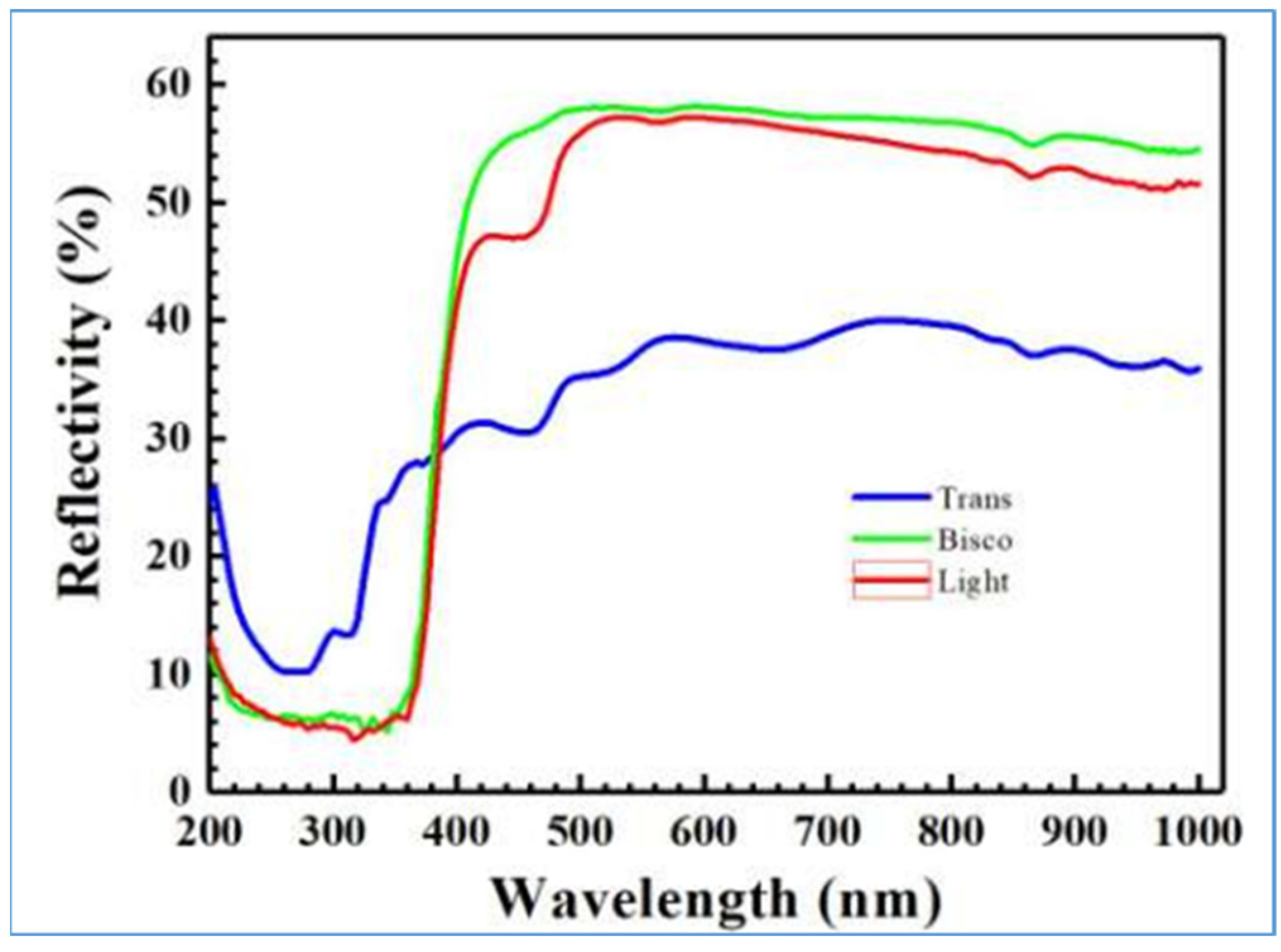
2.2. UV-Visible Spectrophotometry

Light transmission through the orthodontic composite resins was measured using a Lambda35 UV/Vis Spectrophotometer (PerkinElmer S/N 101N7060404) to evaluate the angle-integrated total reflectivity in the spectral range from 190–1100 nm. Due to the non-crystalline nature of the samples, reflected light was collected by an integrating sphere. Measurements were performed at room temperature or 25° (±1 °C) and at 30–50% of humidity. Specimens were stored in a holder without adhesive bonding in order to avoid contamination during the tests.
The transmittance values obtained at each wavelength were recorded by a computer connected to the spectrophotometer using specific software that displays the average values of transmittance in percentages.

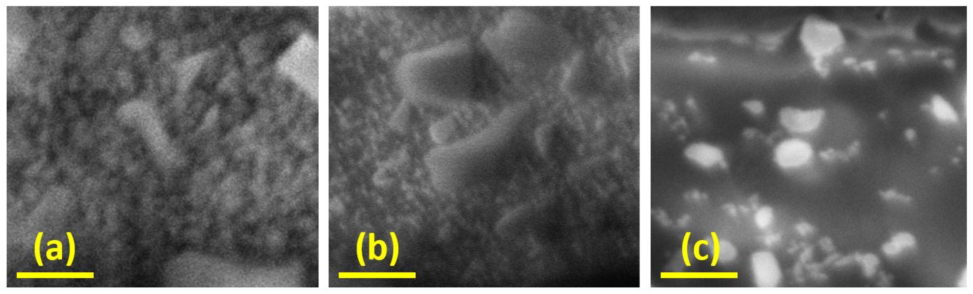
2.3. FIB/SEM Analysis

By using a FEI-Helios Nanolab 600 (FEI/Thermo Fisher Scientific Phillips, Research Center of Eindhoven, Eindhoven, The Netherlands) dual-beam equipment with a Ga+ ion source operated at 30 keV acceleration energy and 6.5 nA ion current, a controlled milling process was performed on the three different resins to observe their internal structure. To prepare the samples, the resin discs were metallized by using an e-gun evaporator, depositing a layer of chromium 20 nm thick to control the charging effect during SEM imaging. The entire procedure can be summarized as follows: A series of rectangular areas were created starting from the samples’ surface with a fixed dwell time and ion dose (respectively, 1 µs and 10 nC/µm2). The resins were treated at different tilted angles, measuring the depth of the excavation and collecting the cross-sectional images to observe the internal composition of the samples. All the tests were performed at room temperature and in a controlled vacuum at a working pressure of 1 × 10⁻⁶ mbar. In all of the milling experiments, the metal layer was removed by ions within the first 2 s of the process. This step did not affect the evaluation of the measurement of the depth of excavation since the time needed to eliminate the metal layer is negligible with respect to the time used milling the resin bulk.

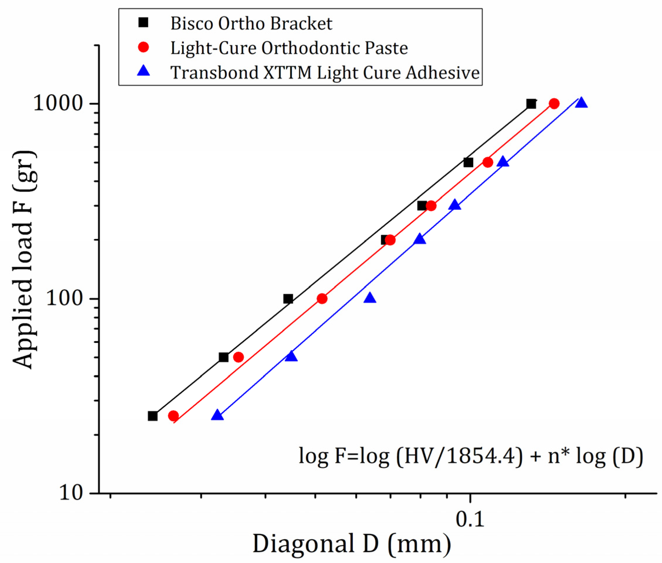
2.4. Indentation Strength Test

Hardness of the orthodontic adhesive resins was measured be means of the Leica VMHT apparatus (Leica GmbH, Wetzlar, Germany) equipped with the standard Vickers pyramidal indenter (diamond square-based pyramid with face angle of 136°). The loading speed was 5 × 10⁻⁶ m/s, and the time under each load was 20 s.
In order to provide reproducible and reliable Vickers hardness (HV) values, experimental parameters were set according to the ASTM E381-16 standard [24], with a dwell time of 20 s and a load of 500 g.
Additional measurements were performed at different loads in order to address the dependence of the HV in the investigated specimens on the applied load. Loads of 25, 50, 100, 200, 300, 500 and 1000 g were used for this purpose; 10 indentations were performed at each load and successively averaged to obtain a mean HV value and a standard deviation (SD) for each applied load.

3. Results

In Figure 2, the spectra relating to the reflectivity of each orthodontic adhesive resin reveals a lower reflectivity in the Trans XT compared to the other two materials, while for all the testing samples, the reflectivity drops down at 380 nm. Bisco is the resin that possesses the greatest reflectivity. Bisco suddenly reaches its maximum peak around 470 nm and remains constant beyond this wavelength, except for a slight drop between 800 and 900 nm, which is followed by a downward trend. This drop between 800 and 900 nm of wavelength is present for all three composite resins, but refers to different percentage values of reflectivity. Light has a curvilinear pattern very similar to Bisco.
The surface roughness and the internal composition of the samples have been investigated through the FIB/SEM analysis. A first comparison can be performed that directly evaluates the surface structure of the resins at SEM imaging: as can be seen in Figure 3, the Bisco and Light samples show a quite similar morphology with a homogeneous grainy shape. Conversely, Trans XT shows a smoother surface with the presence of small spikes and troughs. After the milling process, the internal composition of the resins can be evaluated: again Bisco and Light show similar characteristics that present a higher density of grains in the matrix with respect to Trans XT. As can been observed in Figure 4, the SEM analysis performed at a magnification of 60,000 Trans samples presents essentially two types of grains embedded in the matrix: a big one with an average size of tens of microns and a sub-micron class of grains less dense than the other two resins.
Vickers microhardness measurements were carried out for three orthodontic composite resins. As recommended by ASTM E381-16 ISO standard [24], to obtain HV hardness values for each sample, the indentations were made at 500 g of the applied load with a dwell time of 20 s. The diagonal length was measured after each indentation and used to calculate the HV value by applying the Crawford equation [25,26]:
HV = ( 1854.4   F ) D 2
where HV is Vickers hardness, F is applied load in grams and D is diagonal length in mm. Experimental values of diagonal lengths and relative calculated mean HV values are presented in Table 2:
The obtained HV values followed this order: Bisco > Light > Trans XT and, namely, 96, 83 and 69, respectively. In particular, the value relative to Trans XT resin is in agreement with the literature value [27,28].
Moreover, a further investigation of the three orthodontic composite resins was performed in order to establish a dependence of HV values on the applied load. Is it known that microhardness does not represent an absolute characteristic of a plastic material and is dependent on the applied load [25,29]. To this end, a complete set of indentations was carried out at 25, 50, 100, 200, 300, 500 and 1000 g of the applied load. In order to quantify the dependence of HV on the applied load, the applied load and the corresponding diagonal length values were plotted in a logarithmic graph (see Figure 5), as suggested by Crawford [25] and Wu [29].
In Figure 5, the sets of values of the applied load F and diagonal D for each sample: Bisco (black squares), Light (red circles) and Trans XT (blue triangles) is plotted. Logarithmic plots allowed us to linearize the F vs. D trends as a result of the reformulation of Equation (1) in the form of Equation (2):
log F = log ( HV 1854.4 ) + n   logD
where F is applied force in grams, HV is Vickers hardness value, n is an exponent of D and D is diagonal length. In an ideal system, HV should be constant and proportional to the inverse square value of the diagonal (1/D2, n = 2). Nevertheless, HV in real systems is dependent on the applied load, implying a deviation of the n value from the ideal one (n = 2). The extent of this deviation can provide a measure of the dependence of HV on the applied load. A higher deviation of n value from the ideal value indicates a higher dependence of the HV on the applied load.
Linear regression on each specimen’s dataset allowed us to extrapolate their relative n values. The obtained results are reported in Table 3:
The calculated n values indicate that the dependence of HV on the applied load follows this order: Trans XT > Bisco > Light.

4. Discussion

Translucency is referred to as the intermediate state between opacity and transparency. Unlike what happens in a transparent material, in a translucent one, the light still manages to pass through its entire structure but then undergoes dispersion; this prevents the bodies behind it from being clearly visible [30]. As composite resins, these materials are based on methacrylate compounds, made of an organic phase (matrix), a ceramic phase (filler) and photoinitiators and pro-adhesive agents (silanes). The organic matrix mainly consists of bisphenol A-glycidyl methacrylate (Bis-GMA), triethylene glycol dimethacrylate (TEGMA) and urethane dimethacrylate (UDMA). The inorganic phase, on the other hand, consists of admixtures of glassy particles of different shapes and sizes (macro, micro or nanofillers); these are mainly compounds of silicon, which determine the material’s properties [25]. According to the clinical requirements, fillers may make up 50% to 70% of the volume, and particle size may range from 20 nm to 5 μm [31,32,33,34,35,36].
In composite resins, despite the difference in composition of the different constituents, the resinous matrix and the inorganic fillers can show very similar refractive indices; this is what makes the composites themselves highly translucent materials. The resinous organic matrix, after polymerization, in fact presents refraction values very close to those of the fillers, between 1.47 and 1.52. Otherwise, a discrepancy in the refractive indices between the resin matrix and the filler can increase the opacity of the composite due to excessive refraction and reflection at the filler-matrix interfaces because, as the refractive indices vary, the direction of the light changes. Therefore, as the variation of the refractive index between matrix and filler decreases, the translucency increases and, consequently, the opacity of the composite decreases. All this, in essence, would affect the light scattering and, consequently, the color match in the composite material. However, in addition to the refractive index of the filler, the optical properties of the composite resins are also influenced by the volume fraction of the filler, the morphological characteristics (such as size, shape and distribution) of the particles and by the silane coupling. Several studies have focused on evaluating the effect of the load ratio, type, shape and size of the filler particles on the optical properties and, above all, on the appearance of resin composites. [23]. Indeed, there is a huge difference between orthodontic and dental restorative composite resins, essentially represented by the fact that the latter are accessible in multiple opacities, which are typically referred to as dentin, body or opaque and enamel or translucent, all to try to mimic the optical features of the natural dentin and enamel. It has been stated that composites that exhibit sufficient light diffusivity, i.e., translucency within their resinous structure, are capable of producing a chameleon effect which would lead to improved color matching. However, the chameleon effect of resin composites appears to be influenced not only by color variations but also by the initial size of the restoration. An in vitro study investigated, through spectrophotometric analysis, the possible effect of the thickness on the chromatic coordinates and translucency of different composites for dental restorations. In particular, it assessed whether color variation and transparency varied independently of the transparency of the composite. From the results, it was evident that the thickness is able to significantly influence both the color coordinates and the translucency of the material itself [30].
To investigate the relationship between the filler particles of composite resins and their mechanical properties, conventional studies have been conducted, nonetheless, no agreement has yet been reached on what is the optimal level of filler content for a biological area subjected to stress [37].
Previously conducted studies on composite resins for dental restorations have largely shown that factors such as the kind of monomer, the type and volume fraction of the filler, as well as the refractive index of the polymer matrix and the filler itself, are able to influence transmission of light in these resinous polymer blends [32,38]. UV-Visible Spectrophotometry analysis uses visible light to define the concentration of chemicals in an admixture. Atoms and molecules adsorb amounts of energy from light and undergo electronic transitions. Each compound will possess its own absorption or reflection profile based on its atomic composition. The specific profile enables for the quantitative determination of analytes in a sample. Therefore, the purpose of the UV-Visible Spectrophotometry analysis presented in this in vitro study was not only to establish the superior reflectivity of the Bisco resin compared to Light (although similar) and the lower reflectivity of Trans XT compared to the other composites: refraction isn’t of great interest in orthodontic adhesive resin. Rather, the objective of this work was to demonstrate for the first time in the orthodontic field how, using an alternative method of investigation such as a direct optical test, it is possible to provide additional information on the relationship between reflectivity, chemical composition and mechanical behaviour of orthodontic bonding nanofiller resin materials. In particular, it was possible to argue that the difference in the chemical composition of Trans XT and the constitution analogy existing between Bisco and Light resins is strongly influenced by the percentage of fillers constituting the inorganic phase of these polymers. In particular, among all the adhesive resins, Trans XT had the lowest content in fillers while Bisco and Light contained higher concentrations and, in any case, were similar to each other.
In particular, it has been observed that the bond strength of UDMA-based adhesives depends on the filler content: those reinforced with a higher percentage of fillers reveal greater strength than unfilled or lightly filled adhesives, which show a lower bond strength due to the reduced stiffness of the resin compared to the loaded adhesive, with detachment occurring within the adhesive resin (cohesive fracture) and not at the adhesive-bracket interface [18,39]. The high filler load in composite systems appears to be based on the concept of achieving high mechanical properties as established by conventional mechanical testing [40]. The quantity of the filler particles contained, together with the coupling characteristics between the filler and the matrix resin, also represent significant variables, i.e., capable of influencing the mechanical features of the composite resin. The compressive strength, hardness, flexural strength and modulus of elasticity grows whenever the filling volume fraction increases, while the shrinkage reduces [21]. In the literature, it has been reported that highly filled resin composites bond to metal brackets with better mechanical retention than lesser filled composites [13]. A greater mechanical retention of the Trans XT resin at the metal base of the bracket compared to the other two orthodontic adhesive resins reported in the literature can therefore be justified by its lower content of fillers in the inorganic phase [21]. It is also known that a resinous composite with less fillers turns out to be less rigid than one highly loaded in fillers. From this it is therefore conceivable that Bisco and Light, precisely because of their higher content in fillers, can clinically express a more rigid behaviour in transferring orthodontic forces to the tooth, as demonstrated in previous works by the type of bonding between the enamel and the bracket [21,41].
As observed in some previous studies [41,42,43,44,45,46,47,48], one in particular in which the Raman Spectroscopy analysis of the same orthodontic composites has revealed strong affinities between the spectra of Bisco and Light, while Trans XT exhibits a peculiar peak at 450 cm−1 associated to the high percentage of quartz in the chemical composition [Table 1] [39]. Similar results were obtained in the present in vitro study through the UV-Vis analysis: the higher content of fused quartz in Trans XT fillers resulting from UV-Vis Spectrometry analysis improves some important mechanical properties such as: the attainment of high compressive strength and stiffness, the abrasion resistance and the reduction in thermal dimensional change in the resin to a value matching that of tooth structure, effectively increasing adhesion to both the adhesive/enamel interface and the adhesive/bracket base interface through temperature changes often happening in the oral cavity.
All these speculations regarding the differences in the internal compositions of the three resins can be corroborated also by the FIB/SEM analysis. This technique is not only used for imaging but also for locally removing or patterning small areas through the ion bombardment of Ga+ ions. In this way it is possible to customize the surface of the samples and thin the samples while checking the results via SEM imaging. Even if historically this technique has been deeply utilized for the preparation of transmission electron microscopy (TEM) specimens [43], the process can provide valuable information regarding the hardness of a material and can shed light on its internal microscopic composition. Recently, we have proposed to use this technique to compare the hardness of different orthodontic materials by evaluating the deepness of the excavation on a surface for a fixed time of milling [21]. Even if several potential issues must be considered such as the sputtered material of the sample, the specific interaction between the sample and the ions, the heating of the sample surface, etc., we believe that such a technique can offer a deep insight into the material while providing information on the internal composition of samples that are usually composed of multiple components and on material consistency. In particular, in this case we observed that both the initial surface roughness and the cross-section views of Bisco and Light samples look very similar in terms of classes of grain dimensions dispersed in the matrix and homogeneity of the grain density. On the other hand, Transbond XTTM Light Cure Adhesive samples show a significant reduction both in grain homogeneity and grain dimensions. In particular, the grains at the sub-micro scale are the ones that show the largest difference, as can be seen in Figure 4. Indeed, their distribution is significantly reduced in the Trans XT sample, and the presence of the matrix (dark region in the image) is largely increased with respect to the other examined materials. As reported by Drummond [49] the presence of particles and grains in the filler contributes to an increase in the material hardness, so we can attribute greater hardness values to the two orthodontic adhesive resins Leone and Bisco with respect to Trans XT, as observed by the FIB/SEM analysis. Actually, this hypothesis is confirmed by the direct evaluation of the microhardness of the examined materials: indeed, based on the obtained HV values, it is possible to conclude that the Bisco Ortho Bracket Paste LC (Bisco) is characterized by HV 96 ± 16, being the hardest one, the Light-Cure Orthodontic Paste (Light) by HV 82 ± 8 and the Transbond XTTM Light Cure Adhesive (Trans) by HV 69 ± 3.

5. Conclusions

The UV-Vis analysis presented in this work was used as an alternative method of investigation to evaluate indirectly the compositional analogy that characterizes the inorganic phases of three orthodontic bonding nanofiller resins (Bisco, Light and Trans XT). Indeed, the goal of the paper was to evaluate and demonstrate the potentiality of UV-Vis Spectrophotometry as a tool useful for extrapolating not only optical but also compositional information. This could represent a novel and significant advantage in discriminating the properties of orthodontic resins without the use of more expensive techniques such as TEM or FIB/SEM imaging. From the analysis of the spectra, Bisco and Light adhesive resins showed a high and very similar filler content in percentage, which may explain some similarities in their mechanical performance. At the same time, the higher content of quartz observed in the inorganic phase of Trans XT and expressed by the lower reflectivity of the composite can confirm the improvement of some important mechanical properties. These results compared with FIB/SEM analysis obtained similar outcomes: analogous internal structures for Bisco and Light with a reduced presence of the matrix with respect to Trans XT and significant diversity in the distribution and the grain size of Trans XT, compared to both the Bisco and Light resins. Finally, a higher presence of filler in Bisco and Light contributes to a higher hardness of the composites, as also revealed by Vickers measurements, thus confirming the hypothesis that the materials with a high content in the ceramic phase present a more rigid mechanical behaviour. These findings confirm the potentiality of UV-Vis analysis in revealing multiple information regarding the optical and mechanical properties of orthodontic composite materials.

Author Contributions

Conceptualization: R.C.; Methodology: L.M. and A.C. (Annalisa Convertino); Validation: A.G.; Investigation: L.M., F.M., I.L., A.N., A.C. (Annalisa Convertino), J.V.R. and M.F.; Resources: G.M. (Giuseppe Marzo); Data Curation: A.C. (Alessandro Cioffi), P.P. and G.M. (Gianluca Mampieri); Writing—Original Draft Preparation: R.C.; Writing—Review & Editing: R.C. and L.M.; Supervision: R.C. and L.M. Project Administration: R.C. All authors have read and agreed to the published version of the manuscript.

Funding

This research received no external funding.

Conflicts of Interest

The authors declare no conflict of interest.

References

  1. Giancotti, A.; Mampieri, G.; Arcuri, C. Tunnel Traction Procedure for Deeply Impacted Canines and Resorbed Lateral Incisors. J. Clin. Orthod. JCO 2015, 49, 784–790. [Google Scholar] [PubMed]
  2. Mampieri, G.; Castroflorio, T.; Conigliaro, A.; Giancotti, A. Treatment of impacted canines with aligners: An alternative and viable option. Clin. Case Rep. 2021, 9, e04856. [Google Scholar] [CrossRef] [PubMed]
  3. Mampieri, G.; Condò, R.; Di Caccamo, G.; Pirelli, P.; Giancotti, A. Clear Aligner Treatments in Orthoperio Patients. Case Rep. Dent. 2022, 2022, 8932770. [Google Scholar] [CrossRef] [PubMed]
  4. Sas, B. Impact of self-ligating orthodontic brackets on dental biofilm and periodontal pathogens in adolescents. J. Biol. Regul. Homeost. Agents 2021, 35, 107–115. [Google Scholar] [CrossRef]
  5. Pick, B.; Rosa, V.; Azeredo, T.R.; Cruz Filho, E.A.; Miranda, W.G., Jr. Are flowable resin-based composites a reliable material for metal orthodontic bracket bonding? J. Contemp. Dent. Pract. 2010, 11, E017–E024. [Google Scholar]
  6. Samantha, C. Comparative Evaluation of Two Bis-GMA Based Orthodontic Bonding Adhesives–A Randomized Clinical Trial. J. Clin. Diagn. Res. 2017, 11, ZC40–ZC44. [Google Scholar] [CrossRef]
  7. Miyagawa, Y.; Powers, J.M.; O’Brien, W.J. Optical Properties of Direct Restorative Materials. J. Dent. Res. 1981, 60, 890–894. [Google Scholar] [CrossRef]
  8. Nucera, R.; Dolci, C.; Bellocchio, A.M.; Costa, S.; Barbera, S.; Rustico, L.; Farronato, M.; Militi, A.; Portelli, M. Effects of Composite Attachments on Orthodontic Clear Aligners Therapy: A Systematic Review. Materials 2022, 15, 533. [Google Scholar] [CrossRef]
  9. Masotti, A.S.; Onófrio, B.; Conceição, E.N.; Spohr, A.M. Uv–vis spectrophotometric direct transmittance analysis of composite resins. Dent. Mater. 2007, 23, 724–730. [Google Scholar] [CrossRef]
  10. Azzopardi, N.; Moharamzadeh, K.; Wood, D.J.; Martin, N.; Van Noort, R. Effect of resin matrix composition on the translucency of experimental dental composite resins. Dent. Mater. 2009, 25, 1564–1568. [Google Scholar] [CrossRef]
  11. Yeh, C.L.; Miyagawa, Y.; Powers, J.M. Optical Properties of Composites of Selected Shades. J. Dent. Res. 1982, 61, 797–801. [Google Scholar] [CrossRef]
  12. Johnston, W.; Ma, T.; Kienle, B.H. Translucency parameter of colorants for maxillofacial prostheses. Int. J. Prosthodont. 1995, 8, 79–86. [Google Scholar] [PubMed]
  13. Johnston, W.M.; Reisbick, M. Color and translucency changes during and after curing of esthetic restorative materials. Dent. Mater. 1997, 13, 89–97. [Google Scholar] [CrossRef]
  14. Taira, M.; Okazaki, M.; Takahashi, J. Studies on optical properties of two commercial visible-light-cured composite resins by diffuse reflectance measurements. J. Oral Rehabil. 1999, 26, 329–337. [Google Scholar] [CrossRef]
  15. Arikawa, H.; Fujii, K.; Kanie, T.; Inoue, K. Light transmittance characteristics of light-cured composite resins. Dent. Mater. 1998, 14, 405–411. [Google Scholar] [CrossRef]
  16. Lee, Y.-K. Influence of scattering/absorption characteristics on the color of resin composites. Dent. Mater. 2007, 23, 124–131. [Google Scholar] [CrossRef]
  17. Watts, D.; Cash, A. Analysis of optical transmission by 400–500 nm visible light into aesthetic dental biomaterials. J. Dent. 1994, 22, 112–117. [Google Scholar] [CrossRef]
  18. Campbell, P.; Johnston, W.; O’Brien, W. Light Scattering and Gloss of an Experimental Quartz-filled Composite. J. Dent. Res. 1986, 65, 892–894. [Google Scholar] [CrossRef] [Green Version]
  19. Faltermeier, A.; Rosentritt, M.; Faltermeier, R.; Reicheneder, C.; Mussig, D. Influence of filler level on the bond strength of orthodontic adhesives. Angle Orthod. 2007, 77, 494–498. [Google Scholar] [CrossRef] [PubMed] [Green Version]
  20. Van Meerbeek, B.; Inokoshi, S.; Braem, M.; Lambrechts, P.; Vanherle, G. Morphological Aspects of the Resin-Dentin Interdiffusion Zone with Different Dentin Adhesive Systems. J. Dent. Res. 1992, 71, 1530–1540. [Google Scholar] [CrossRef] [PubMed]
  21. Condò, R.; Mampieri, G.; Pasquantonio, G.; Giancotti, A.; Pirelli, P.; Cataldi, M.; La Rocca, S.; Leggeri, A.; Notargiacomo, A.; Maiolo, L.; et al. In Vitro Evaluation of Structural Factors Favouring Bacterial Adhesion on Orthodontic Adhesive Resins. Materials 2021, 14, 2485. [Google Scholar] [CrossRef]
  22. Lardani, L.; Derchi, G.; Marchio, V.; Carli, E. One-Year Clinical Performance of Activa™ Bioactive-Restorative Composite in Primary Molars. Children 2022, 9, 433. [Google Scholar] [CrossRef] [PubMed]
  23. Oivanen, M.; Keulemans, F.; Garoushi, S.; Vallittu, P.K.; Lassila, L. The effect of refractive index of fillers and polymer matrix on translucency and color matching of dental resin composite. Biomater. Investig. Dent. 2021, 8, 48–53. [Google Scholar] [CrossRef] [PubMed]
  24. ASTM E384-16; Standard Test Method for Microindentation Hardness of Materials. ASTM International: West Conshohocken, PA, USA, 2016.
  25. Crawford, R.J. Microhardness testing of Plastics. Polym. Test. 1982, 3, 37–54. [Google Scholar] [CrossRef]
  26. Lopez, J. Microhardness testing of Plastics: Literature Review. Polym. Test. 1993, 12, 437–458. [Google Scholar] [CrossRef]
  27. Sifakakis, I.; Zinelis, S.; Patcas, R.; Eliades, T. Mechanical properties of contemporary orthodontic adhesives used for lingual fixed retention. Biomed. Eng./Biomed. Tech. 2016, 62, 289–294. [Google Scholar] [CrossRef] [PubMed] [Green Version]
  28. Iijima, M.; Muguruma, T.; Brantley, W.A.; Yuasa, T.; Uechi, J.; Mizoguchi, I. Effect of mechanical properties of fillers on the grindability of composite resin adhesives. Am. J. Orthod. Dentofac. Orthop. 2010, 138, 420–426. [Google Scholar] [CrossRef]
  29. Wu, H.; Dave, F.; Mokhtari, M.; Ali, M.M.; Sherlock, R.; McIlhagger, A.; Tormey, D.; McFadden, S. On the Application of Vickers Micro Hardness Testing to Isotactic Polypropylene. Polymers 2022, 14, 1804. [Google Scholar] [CrossRef]
  30. Ardu, S.; Rossier, I.; di Bella, E.; Krejci, I.; Dietschi, D. Resin composite thickness’ influence on L*a*b* coordinates and translucency. Clin. Oral Investig. 2019, 23, 1583–1586. [Google Scholar] [CrossRef] [Green Version]
  31. Condo’, R.; Cerroni, L.; Pasquantonio, G.; Mancini, M.; Pecora, A.; Convertino, A.; Mussi, V.; Rinaldi, A.; Maiolo, L. A Deep Morphological Characterization and Comparison of Different Dental Restorative Materials. BioMed Res. Int. 2017, 2017, 7346317. [Google Scholar] [CrossRef] [Green Version]
  32. Szalewski, L.; Kamińska, A.; Wallner, E.; Batkowska, J.; Warda, T.; Wójcik, D.; Borowicz, J. Degradation of a Micro-Hybrid Dental Composite Reinforced with Polyaramide Fiber under the Influence of Cyclic Loads. Appl. Sci. 2020, 10, 7296. [Google Scholar] [CrossRef]
  33. Björkman, U.H.; Sundström, F.; Bosch, J.J.T. Fluorescence in dissolved fractions of human enamel. Acta Odontol. Scand. 1991, 49, 133–138. [Google Scholar] [CrossRef] [PubMed]
  34. Spitzer, D.; Ten, J.J.B. The total luminescence of bovine and human dental enamel. Calcif. Tissue Res. 1976, 20, 201–208. [Google Scholar] [CrossRef] [PubMed]
  35. McLaren, E.A. Luminescent Veneers. J. Esthet. Restor. Dent. 1997, 9, 3–12. [Google Scholar] [CrossRef] [PubMed]
  36. Lee, Y.-K.; Lu, H.; Powers, J.M. Changes in opalescence and fluorescence properties of resin composites after accelerated aging. Dent. Mater. 2006, 22, 653–660. [Google Scholar] [CrossRef]
  37. Papadogiannis, Y.; Lakes, R.S.; Palaghias, G.; Helvatjoglu-Antoniades, M.; Papadogiannis, D. Fatigue of packable dental composites. Dent. Mater. 2007, 23, 235–242. [Google Scholar] [CrossRef] [PubMed]
  38. dos Santos, G.; Alto, R.M.; Filho, H.S.; da Silva, E.; Fellows, C. Light transmission on dental resin composites. Dent. Mater. 2008, 24, 571–576. [Google Scholar] [CrossRef]
  39. Condò, R.; Mampieri, G.; Cioffi, A.; Cataldi, M.E.; Frustaci, I.; Giancotti, A.; Campanella, V.; Mussi, V.; Convertino, A.; Maiolo, L.; et al. Physical and chemical mechanisms involved in adhesion of orthodontic bonding composites: In vitro evaluations. BMC Oral Health 2021, 21, 350. [Google Scholar] [CrossRef]
  40. Zadeh, P.N.; Stawarczyk, B.; Hampe, R.; Liebermann, A.; Mayinger, F. Edge chipping resistance of veneering composite resins. J. Mech. Behav. Biomed. Mater. 2021, 116, 104349. [Google Scholar] [CrossRef]
  41. Cerroni, S.; Pasquantonio, G.; Condo’, R.; Cerroni, L. Orthodontic Fixed Appliance and Periodontal Status: An Updated Systematic Review. Open Dent. J. 2018, 12, 614–622. [Google Scholar] [CrossRef] [Green Version]
  42. Condo’, R.; Pazzini, L.; Cerroni, L.; Pasquantonio, G.; Lagana’, G.; Pecora, A.; Mussi, V.; Rinaldi, A.; Mecheri, B.; Licoccia, S.; et al. Mechanical properties of “two generations” of teeth aligners: Change analysis during oral permanence. Dent. Mater. J. 2018, 37, 835–842. [Google Scholar] [CrossRef]
  43. Daniele, V.; Macera, L.; Taglieri, G.; Di Giambattista, A.; Spagnoli, G.; Massaria, A.; Messori, M.; Quagliarini, E.; Chiappini, G.; Campanella, V.; et al. Thermoplastic Disks Used for Commercial Orthodontic Aligners: Complete Physicochemical and Mechanical Characterization. Materials 2020, 13, 2386. [Google Scholar] [CrossRef]
  44. Memè, L.; Notarstefano, V.; Sampalmieri, F.; Orilisi, G.; Quinzi, V. ATR-FTIR Analysis of Orthodontic Invisalign® Aligners Subjected to Various In Vitro Aging Treatments. Materials 2021, 14, 818. [Google Scholar] [CrossRef] [PubMed]
  45. Condò, R.; Mampieri, G.; Giancotti, A.; Cerroni, L.; Pasquantonio, G.; Divizia, A.; Convertino, A.; Mecheri, B.; Maiolo, L. SEM characterization and ageing analysis on two generation of invisible aligners. BMC Oral Health 2021, 21, 316. [Google Scholar] [CrossRef] [PubMed]
  46. Daniele, V.; Macera, L.; Taglieri, G.; Spera, L.; Marzo, G.; Quinzi, V. Color Stability, Chemico-Physical and Optical Features of the Most Common PETG and PU Based Orthodontic Aligners for Clear Aligner Therapy. Polymers 2021, 14, 14. [Google Scholar] [CrossRef]
  47. Valeri, C.; Aloisio, A.; Mummolo, S.; Quinzi, V. Performance of Rigid and Soft Transfer Templates Using Viscous and Fluid Resin-Based Composites in the Attachment Bonding Process of Clear Aligners. Int. J. Dent. 2022, 2022, 1637594. [Google Scholar] [CrossRef] [PubMed]
  48. Paglia, M.; Beretta, M.; Quinzi, V.; Colombo, S. PEEK polymer in orthodontics: A scoping review. Eur. J. Paediatr. Dent. 2022, 23, 137–139. [Google Scholar] [CrossRef]
  49. Drummond, J.L. Nanoindentation of dental composites. J. Biomed. Mater. Res. B Appl. Biomater. 2006, 78, 27–34. [Google Scholar] [CrossRef]
Figure 1. The test discs obtained from each orthodontic composite resin: (a) Light-Cure Orthodontic Paste (Light); (b) Transbond XTTM Light Cure Adhesive (Trans); (c) Bisco Ortho Bracket Paste LC (Bisco).
Applsci 12 12538 g001
Figure 2. The spectra relating to the reflectivity of each orthodontic adhesive resin.
Applsci 12 12538 g002
Figure 3. SEM images of the surfaces of (a) the Bisco Ortho Bracket Paste LC (Bisco), (b) Light-Cure Orthodontic Paste (Leone), and (c) Transbond XTTM Light Cure Adhesive (Trans) collected at a tilt angle of 52°. The scale bars indicate 10 μm.
Applsci 12 12538 g003
Figure 4. Cross-sectional analysis: SEM images collected on the sidewalls of the FIB milled areas on (a) the Bisco Ortho Bracket Paste LC (Bisco), (b) Light-Cure Orthodontic Paste (Leone), and (c) Transbond XTTM Light Cure Adhesive (Trans) after FIB milling. The images have been collected at a magnification of 60,000 via in-lens detector. The scale bars indicate 500 nm.
Applsci 12 12538 g004
Figure 5. Logarithmic plots of applied load (F) vs. diagonal length (D) for: Bisco Ortho Bracket (black squares), Light-Cure Orthodontic Paste (red circles) and Transbond XTTM Light Cure Adhesive (blue triangles). Straight lines are linear regressions of each dataset.
Applsci 12 12538 g005
Table 1. The chemical compositions of the three light-cured orthodontic adhesive composites considered for the study.
Orthodontic Composite ResinManufacturerCompositionAcronym
Bisco Ortho Bracket Paste LCBisco, Schaumburg, IL, USAUDMA (5–10%), TEGDMA (5–10%), molten silicon (50–75%). The substances contained in the remaining part of the adhesive are not specified by the supplierBisco
Light-Cure Orthodontic PasteLeone s.p.a., Sesto Fiorentino, FI, ItalyBis-GMA, UDMA, TEGDMA, Silica and other inert fillers, catalysts and stabilizers (unknown percentages since not provided by the manufacturer)Light
Transbond XTTM Light Cure Adhesive 3M Unitek, Monrovia, CA, USABis-GMA (5–10%), bis-EMA (10–20%), TEGDMA (5–10%), reaction products with quartz (70–80%), reaction products with dichlorodimethylsinane with silica (<2%)Trans XT
Table 2. Set of experimental diagonal lengths and calculated HV values obtained for applied load of 500 g.
Bisco Ortho Bracket Paste LC (Bisco)Light-Cure Orthodontic Paste (Light)Transbond XTTM Light Cure Adhesive (Trans)
RunD (μm)HVRunD (μm)HVRunD (μm)HV
1102.089.21101.689.91116.868.0
2110.675.62103.486.72113.572.1
3113.771.83106.981.03114.071.4
491.2111.54106.981.24111.374.9
589.8116.55100.891.35113.671.9
689.0117.26112.773.16117.267.6
796.898.97114.770.57118.366.3
898.695.58108.978.28116.168.8
9102.788.09111.874.29121.163.2
1097.897.010113.971.510114.970.3
Mean value + SD99.2 ± 8.396 ± 16Mean value + SD108.1 ± 6.482 ± 8Mean value + SD115.6 ± 2.869 ± 3
Table 3. Dependence of HV on the applied load for investigated orthodontic composite resins.
Bisco Ortho Bracket Paste LC (Bisco)Light-Cure Orthodontic Paste (Light)Transbond XTTM Light Cure Adhesive (Trans)
n value2.192.142.33
Publisher’s Note: MDPI stays neutral with regard to jurisdictional claims in published maps and institutional affiliations.

