Article

Design and Experimental Evaluation of a New RNA-FISH Probe to Detect and Identify Paenibacillus sp.

by
Sílvia Arantes
1,
Patrícia Branco
1 and
Ana Teresa Caldeira
1,2,3,*
1
Laboratório HERCULES, Instituto de Investigação e Formação Avançada, Universidade de Évora, 7000-809 Évora, Portugal
2
Departamento de Química e Bioquímica, Escola de Ciências e Tecnologia, Universidade de Évora, 7000-671 Évora, Portugal
3
City U Macau Chair in Sustainable Heritage, Instituto de Investigação e Formação Avançada, Universidade de Évora, 7000-809 Évora, Portugal
*
Author to whom correspondence should be addressed.
Appl. Sci. 2022, 12(5), 2348; https://doi.org/10.3390/app12052348
Submission received: 1 February 2022 / Revised: 21 February 2022 / Accepted: 22 February 2022 / Published: 24 February 2022

Abstract

:
Paenibacillus, rod-saped gram-positive endospores forming aerobic or facultative anaerobic bacteria, colonize diverse ecosystems and are involved in the biodegradation of cultural heritage assets. Biodeteriogenic microorganisms can be easily detected/identified by ribonucleic acid- fluorescent in situ hybridization RNA-FISH with specific probes. In this work, probes designed in silico were analyzed to calculate hybridization efficiency and specificity by varying the formamide concentration in the hybridization. The Pab489 probe showed excellent in silico performance with high theoretical maximum efficiency hybridization (99.99%) and specificity and was selected for experimental assays with target Paenibacillus sp. and non-target biodeteriogenic microorganisms. Results assessed by epifluorescence microscopy and flow cytometry revealed that, regardless of the formamide concentration, it was possible to observe that the Pab489-Cy3 probe had a similar signal intensity to the EUB338-Cy3 probe (positive control), so the presence of formamide, a highly toxic and carcinogenic compound used to aid the hybridization process, is not necessary. The designed probe used in FISH assays allows specific in situ identification of Paenibacillus spp. in microbial communities in a culture-independent way. This approach can be employed for screening Paenibacillus spp., showing great potential for future application in biodeterioration of heritage assets, in the search for Paenibacillus strains that produce compounds with biotechnological or medical potential.

1. Introduction

The genus Paenibacillus (Paenibacillaceae family), defined as belonging to the genus Bacillus, was reclassified in 1993 by Ash et al. [1], based on the sequence comparison of the 16S RNA gene, and is characterized as a Gram-positive, rod-shaped endospore-forming aerobic or facultative anaerobic bacteria [2,3,4]. The genus Paenibacillus contains 211 recognized species, isolated from different environments, such as soil, water and salt water, sewage, sediments, compost, rhizosphere, food, plants, insect larvae and clinics [2,5].
The species of the genus Paenibacillus colonize different environments [2,5,6,7,8,9,10,11,12,13,14,15] and most of them are found in the soil, often associated with plant roots, in a symbiotic relationship (promote plant growth), and can be exploited for use in agriculture. Some studies also suggest their biotechnological potential for producing antimicrobial compounds that are useful in medicine or as pesticides, and enzymes with application in bioremediation or to produce valuable chemicals [3,9]. However, although some species of Paenibacillus are harmless or beneficial, others have shown the ability to promote infections in humans.
Infections by Paenibacillus sp. are generally opportunistic and tend to infect the elderly and immunocompromised people. Diseases associated with Paenibacillus sp. infections include chronic kidney disease, sickle cell disease, hydrocephalus, skin cancer, chronic interstitial nephropathy, acute lymphoblastic leukaemia, and premature births [2,9,10], so their rapid detection will influence the onset and development of the disease. Its rapid detection may contribute to the use of an appropriate antimicrobial and thus mitigate the harmful effect of this type of infection. Additionally, many species of Paenibacillus produce antimicrobial compounds that are useful in medicine, and some have shown biodeteriogenic potential to cultural heritage (CH) assets (i.e., biodeterioration of murals) [5,8]. Due to its ability to colonize different environments, the genus Paenibacillus, often in association with Arthrobacter and Bacillus species, has been associated with the biodeterioration of wall paintings, with a variety of hygroscopic salts, including carbonates, chlorides, nitrates and sulfates, which form efflorescences on their surfaces [8,16,17]. Microbial biodeterioration is a growing problem in the preservation of indoor and outdoor patrimonial assets in CH [18]. Thus, detection and identification of CH biodeteriogenic microorganisms, such as Paenibacillus spp., is crucial for the CH objects safeguarding [19,20].
The fluorescent in situ hybridization (FISH) technique has been demonstrated to be a powerful tool for in situ analysis [21]. However, its real potential for the research of biodeteriogenic agents of cultural property has not yet been investigated in depth. The FISH technique is widely used in the detection, visualization and counting of viable target cells present in a sample, without the need for isolation in culture. Due to its speed and sensitivity, this technique is considered an important tool for microbiological studies in several research areas such as the clinical, environmental and food industry, as it allows the identification of species integrated into consortiums of microorganisms that are difficult to cultivate and maintain [22,23]. Fluorescence microscopy allows the visualization of target microorganisms by shining a beam of UV light, which will excite the fluorochromes attached to the FISH probes allowing their detection [24]. On the other hand, the flow-FISH technique (FISH associated with detection by flow cytometry) allows the measurement of physicochemical properties of cells/particles in a flow, by the incidence of laser light through a capillary—the cells scatter the light and emit fluorescence, which is filtered, processed and digitized to be analysed in a computer [21,25].
This study aimed to design in silico a DNA-FISH probe with high specificity to the target microorganism Paenibacillus sp. and experimentally evaluate its performance to detect/identify these bacteria.

2. Materials and Methods

2.1. Design and In Silico Evaluation of a DNA-FISH Probe for Detection and Identification of Paenibacillus spp.

The DNA-FISH probe for Paenibacillus sp. was designed using the method described by Branco et al. [26]. For this purpose, several sequences of ribosomal RNA (rRNA) 16S from the genus Paenibacillus were searched in the National Center for Biotechnology Information database (NCBI; http://www.ncbi.nlm.nih.gov/; accessed on 4 June 2021; Altschul et al. [27]). Subsequently, we proceeded to its alignment with the BioEdit program (www.mbio.ncsu.edu/BioEdit/bioedit.html accessed on 4 June 2021) and then submitted it to the DECIPHER probe design tool (http://www2.decipher.codes/DesignProbes.html accessed on 4 June 2021; Wright [28]). Several species-specific probes to Paenibacillus sp. were obtained. For in silico analysis, 10 probes were selected with the best specificity (score close to zero) and efficiencies given by the program (Table 1). The in silico analysis was performed using the followed tools: Nucleotide BLAST (http://blast.ncbi.nlm.nih.gov/Blast.cgi accessed on 4 June 2021) to confirm the specificity of the probe for the target microorganism; Oligo Calc: Oligonucleotide Properties Calculator [29] to evaluate the probe properties, including length, molecular weight, GC contents, melting temperature, formation of “hairpin”, loop formation; and mathFISH software (http://mathfish.cee.wisc.edu/probeaff.html accessed on 4 June 2021; Yilmaz et al. [30]) to simulate the performance and efficiency of FISH probes and their hybridization.

2.2. Experimental Assessment of the Efficiency and Specificity of the Designed Probe

2.2.1. Microorganisms and Growth Conditions

For this study, a Paenibacillus sp. strain was used as target bacteria and six non-target bacterial bacteriogenic strains of equity goods were selected: two strains of Arthrobacter sp., two strains of Bacillus sp., and two strains of Rubrobacter radiotolerans (1 and 2). The microorganisms used in this study were previously isolated from colonized heritage assets and deposited in the culture collection of the HERCULES Laboratory (HERCULES Biotech Laboratory, Évora University, Évora, Portugal). All bacteria were maintained in Nutrient Agar medium (NA) and cultured in nutrient broth medium (NB) for 7 days at 30 °C, with agitation at 120 rpm.

2.2.2. RNA-FISH Procedure

To proceed with the experimental analysis of the Pab489-Cy3 probe, the seven bacterial strains selected were cultured in nutrient broth medium (NB) and submitted to the FISH protocol [26,31]: fixation/permeabilization, hybridization and washing.
After 7 days of incubation, cultured cells were recovered by centrifugation (13,000 rpm, 15 min, 4 °C) and washed with 10 mL PBS 10× (130.0 mm NaCl, 8.0 mm NaH2PO4, 2.7 mm KCL, 1.5 mm KH2PO4, pH 7.2). After discarding the supernatant, cells were fixed/permeabilized by adding 5 mL of absolute ethanol (EtOH), with incubation at room temperature (1 h). The fixed cells were preserved in EtOH: PBS 10× (50:50, v/v) at −20 °C.
For hybridization, microtubes were prepared with identical aliquots containing 106 cells fixed previously. After centrifugation (13,000 rpm, 15 min, 4 °C), 80 μL of hybridization buffer—HB (0.9 M NaCl, 20 mM Tris-HCl, 0.1% SDS aqueous solution, pH 7.2)—was added. The volume (1.0 µL) of the corresponding RNA-FISH probe stock solution (120 ng/µL) was then added to each FISH assay. The samples were stirred and incubated in a water bath at 46 °C for 2 h. Negative controls were used: (i) a specific probe for eukaryotes EUK516-CY3 (5′-ACCAGACTTGCCCTCC-3′ [32]), and (ii) the corresponding blanks (without the addition of the probe); and as a positive control, a specific probe for eubacteria EUB338-CY3 (5′-GCTGCCTCCCGTAGGAGT-3′ [33]). Tests were also carried out in the presence of formamide 0% (v/v) to 55% (v/v).
After hybridization, cells were washed with HB (100 μL) and incubated in a water bath (46 °C, 30 min). Finally, after centrifugation (13,000 rpm, 15 min, 4 °C), cells were resuspended in 400 μL of PBS 10×.
Samples were analyzed by epifluorescence microscopy (EM) and flow cytometry (FC) according to the procedure described by Branco et al. [26]. In the flow cytometric analysis, the percentage of hybridized cells was calculated according to the following formula: ((Σnumber of cells hybridized with Pab489-Cy3 probe)—(Σnumber of cells hybridized with EUK516-Cy3 probe)) × 100/1000.

3. Results and Discussion

3.1. Design and In Silico Evaluation of a Paenibacillus sp. Specific Probe

To detect/identify Paenibacillus sp. in a reliable way is essential to design a DNA-FISH probe with a good performance, for it is required that the probe holds a low probability of hairpin and self-dimer formation and a high affinity and level of specificity and hybridization efficiency [34,35]. The affinity of the DNA-FISH is the propensity of the probe to bind to its target under given hybridization conditions (melting temperature, GC content and probe length [36]). The DNA-FISH probe specificity is its suitability to bind only to its target.
To design a DNA-FISH probe for Paenibacillus sp., the DECIPHER program was used and the ten best candidates in terms of score and specificity given by the DECIPHER program (data not shown) were selected for their in silico evaluation (Table 1).
Taking into consideration all the criteria described above for obtaining a DNA-FISH probe with a good performance, the Pab489 probe proved to be the best candidate to identify Paenibacillius sp., i.e., its GC content was shown to be less than 60% (58.8%), no possibility of hairpin and self-dimer formation was predicted, and it presented the highest theoretical hybridization efficiency (99.99%), combined with a high number of matches for Paenibacillius sp., 444 matches out of 500 (Table 1).

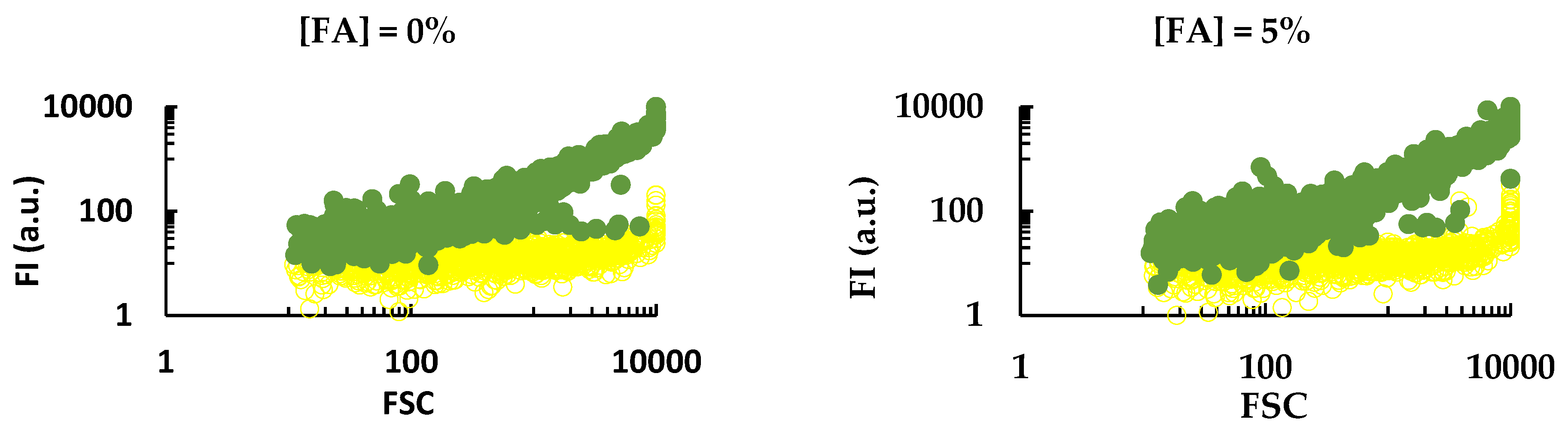
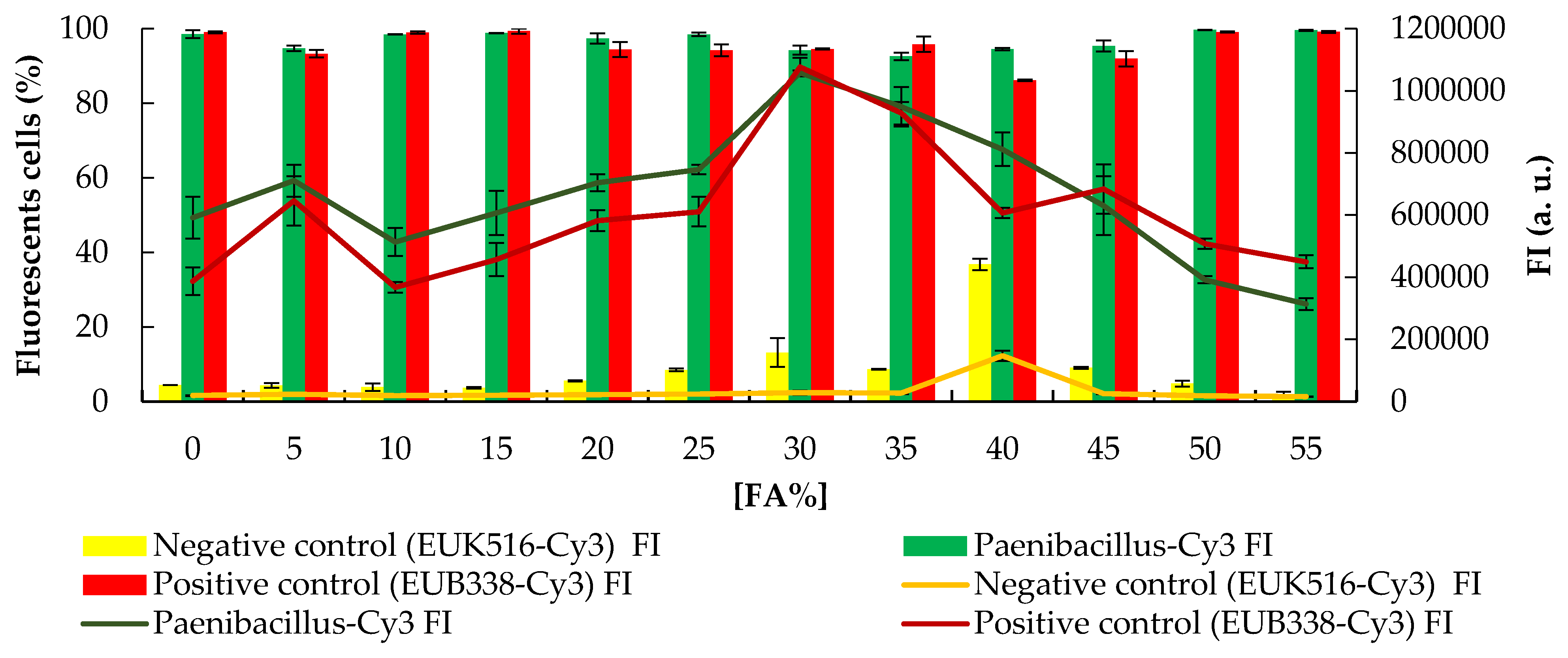
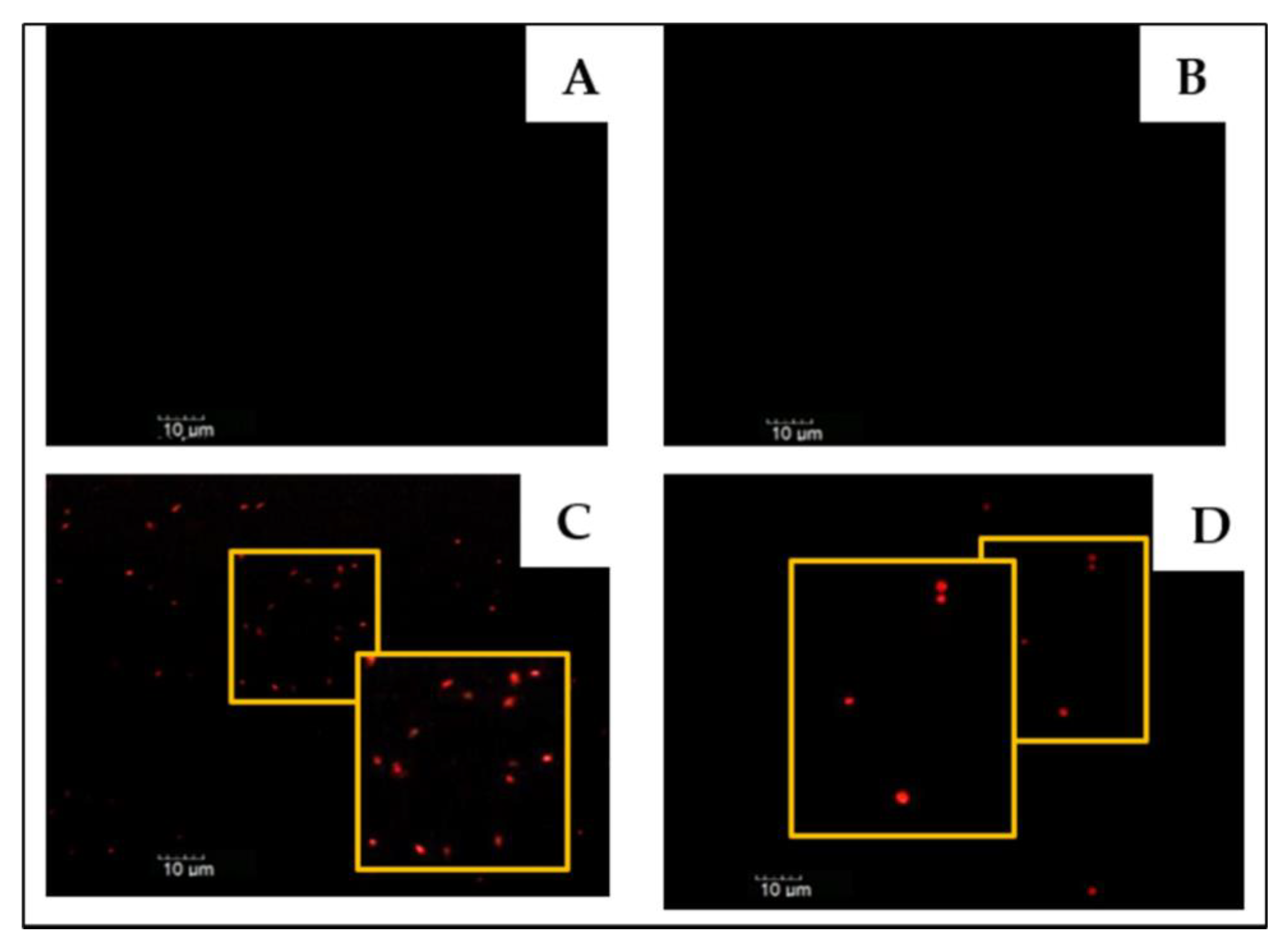
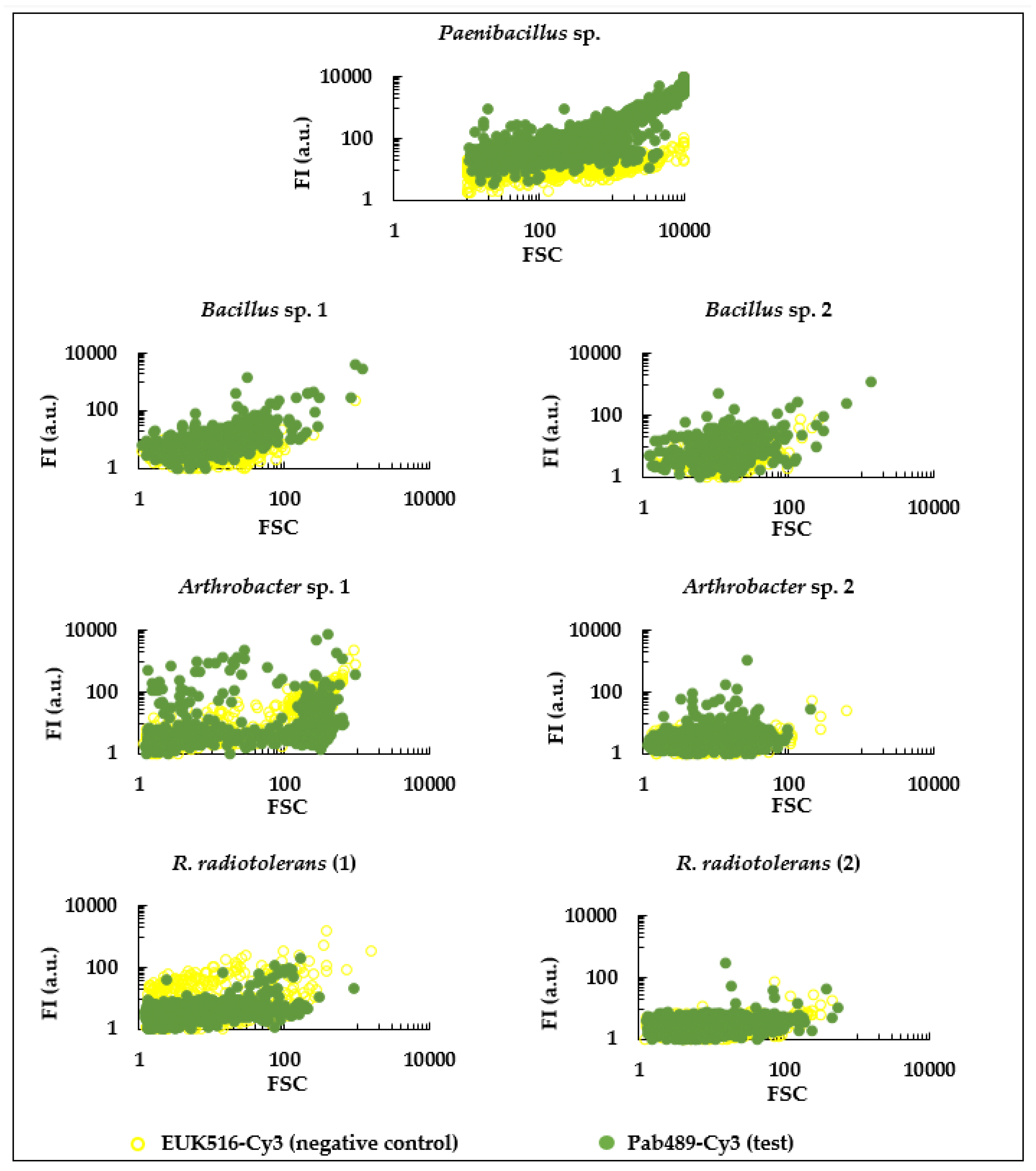
3.2. Experimental Evaluation of a Paenibacillus sp. Specific Probe

Normally, when designing a probe, several experimental conditions are tested for the evaluation and optimization of the probes, however, excellent in silico results do not always translate into excellent experimental results [37]. Therefore, after the design of the probes, it is necessary to proceed to the experimental evaluation, as well as to tests to optimize the stringency conditions, in which the hybridization reaction occurs between the probe and its complementary target (generally located in the rRNA), as well as how to test the probe against non-target organisms from the same ecosystem as the target organism. The stringency of the reaction is adjusted by varying the incubation temperature or the concentration of formamide in the hybridization buffer in concentration steps of 5% (v/v) [38,39,40].
In the experimental evaluation of the hybridization efficiency of the FISH probe, it is still of vital importance to carry out, under the appropriate experimental conditions, different assays: blank (to exclude autofluorescence of the microorganism), positive and negative controls, and the test probe [21,41].
The results of the experimental evaluation and stringency of the Pab489-Cy3 probe are shown in Figure 1 (flow cytometry (FC) results) and Figure 2 (percentage of fluorescent cells and fluorescence intensity). Comparing the flow cytometry results (Figure 1) of the negative control (EUK516-Cy3) and the Pab489-Cy3 probe, we observed that the probe was able to hybridize with Paenibacillus sp. (target microorganism).
It was also possible to observe that, regardless of the formamide concentration (stringency assay), the Pab489-Cy3 probe is always able to hybridize with Paenibacillus sp. cells (green) when compared to the negative control EUK516-Cy3 (yellow).
After processing the results, it was possible to calculate the percentage of fluorescent cells and the fluorescence intensity (FI). As we can see in Figure 2, the Pab489-Cy3 probe, regardless of the formamide concentration, hybridizes with the target cells, showing values close to those observed for the positive control (EUB338-Cy3). The Pab489-Cy3 probe shows fluorescence intensity values > 500,000 a.u. for formamide concentrations below 30%, where they reach the maximum percentage of hybridized cells (FI > 1,000,000 a.u.). The positive control probe EUB338-Cy3 has a similar profile to the test probe Pab489-Cy3, with maximum FI when [FA] = 30%. The negative control EUK516-Cy3 showed FI values less than 200,000 a.u. for all formamide concentrations, showing a slightly higher FI value for [FA] = 40%. Regarding the percentage of hybridized cells, we observed that for the probes Pab489-Cy3 and EUB338-Cy3, the values were always above 90%, while for the negative control (EUK516-Cy3), we observed values of percentage of fluorescent cells below 10%, except for [FA] = 40% which showed about 40% fluorescent cells. These results confirmed that, for [FA] < 40%, hybridization of the Pab489-Cy3 probe occurs in the target cells, with no false positives observed.
The analysis by epifluorescence microscopy showed results in agreement with those obtained by flow cytometry, with fluorescent cells being observed for the probes Pab489-Cy3 and EUB338-Cy3, while for the blank (without probe) and negative control samples (EUK516-Cy3) there were no fluorescent cells, regardless of the formamide concentration. In Figure 3, a microphotography of each sample in the absence of formamide is shown, by way of example.
Knowing that formamide is toxic and a carcinogenic compound to humans [42,43,44], and that in its absence the Pab489-Cy3 probe presents good hybridization results, the hybridization assays of the Pab489-Cy3 probe with non-target microorganisms (Figure 4) were carried out in the absence of formamide. Experimental tests performed with the Pab489-Cy3 probe in the presence of non-target microorganisms suggests the specificity of this probe for the target microorganism since the populations of cells hybridized with the Pab489-Cy3 probe are like the populations of Bacillus spp., Arthrobacter spp. and R. radiotolerans cells hybridized with the EUK516-Cy3 probe (negative control). It should be noted that RNA-FISH was not tested on the spores and therefore the presence of Paenibacillus sp. in the samples studied could be underestimated.
By the technique of epifluorescence microscopy, no signal was observed in the cells of non-target microorganisms hybridized with the probe Pab489-Cy3, thus confirming the results observed by flow cytometry.
This new probe Pab489 represents a considerable advance in the detection/identification of strains of the genus Paenibacillus, especially in comparison with the other methodologies. On the one hand, the classic microbiology methods for the detection/identification of microorganisms imply isolation from the original matrix and allow characterization of only those microorganisms capable of growing, multiplying, and forming colonies in the selected growth medium and conditions [45,46]. In addition to being conditioned by the use of the appropriate culture medium, culture conditions can affect microbial communities, affecting the complete observation of the ecosystem, since it is estimated that only 1% of microorganisms that exist in nature can be cultivated in the laboratory [22,46,47,48]. On the other hand, molecular biology techniques, based on DNA amplification, which are more specific, more sensitive, and faster than traditional methods, do not allow collection of information about spatial distribution, identity, and morphology, nor in situ analysis [21,24,47].
Using the FISH methodology, the analysis of microbial communities is fast and culture-independent, having as the main advantage the specific in situ identification of individual microbial cells [47,49,50], presenting a high potential for the study of the biodeterioration of the cultural heritage [51,52].

4. Conclusions

Excellent performance was observed with high maximal theoretical in silico hybridization efficiency (99.99%) and specificity (444 matches of the target organism in 500 sequences).
The experimental results revealed that, in the absence of formamide, it was possible to observe that the Pab489-Cy3 probe had a signal intensity slightly higher than that of the positive control (EUB338-Cy3), with a percentage of fluorescent cells greater than 90%. Furthermore, the presence of formamide, a highly toxic and carcinogenic compound used to aid the hybridization process, does not contribute to a considerable increase in the signal of the Pab489-Cy3 probe—in the absence of formamide, the probe has a high signal intensity and percentage of hybridized cells. Under the experimental tested conditions, the probe Pab489 showed high efficiency and specificity to detect Paenibacillus sp.
These results contribute to the future implementation of a simple and fast methodology for the detection of Paenibacillus spp. using specific RNA-FISH probes, with possible application to the study of the biodeterioration of heritage assets, in the search for Paenibacillus strains that produce compounds with biotechnological or medical potential.

Author Contributions

Conceptualization, A.T.C.; methodology, S.A., P.B. and A.T.C.; formal analysis, S.A., P.B. and A.T.C.; investigation, S.A., P.B. and A.T.C.; resources, A.T.C.; writing—original draft preparation, S.A.; writing—review and editing, S.A., P.B. and A.T.C.; supervision, A.T.C.; project administration, A.T.C.; funding acquisition, A.T.C. All authors have read and agreed to the published version of the manuscript.

Funding

This research received no external funding.

Institutional Review Board Statement

Not applicable.

Informed Consent Statement

Not applicable.

Data Availability Statement

Not applicable.

Acknowledgments

The authors acknowledge financial support for the PROBIOMA project (0483_PROBIOMA_5_E, EP), co-financed by the FEDER, through the program INTERREG VA España—Portugal (POCTEP); SCREAM Project (ALT20-03-0145-FEDER-031577) and the City University of Macau endowment to the Sustainable Heritage Chair.

Conflicts of Interest

The authors declare no conflict of interest.

References

  1. Ash, C.; Priest, F.G.; Collins, M.D. Molecular identification of rRNA group 3 bacilli (Ash, Farrow, Wallbanks and Collins) using a PCR probe test. Proposal for the creation of a new genus Paenibacillus. Antonie Leeuwenhoek 1993, 64, 253–260. [Google Scholar] [CrossRef] [PubMed]
  2. Saez-Nieto, J.A.; Medina-Pascual, M.J.; Carrasco, G.; Garrido, N.; Fernandez-Torres, M.A.; Villalon, P.; Valdezate, S. Paenibacillus spp. isolated from human and environmental samples in Spain: Detection of 11 new species. New Microbes New Infect. 2017, 19, 19–27. [Google Scholar] [CrossRef] [PubMed]
  3. Tiwari, S.; Prasad, V.; Lata, C. Bacillus: Plant Growth Promoting Bacteria for Sustainable Agriculture and Environment. In New and Future Developments in Microbial Biotechnology and Bioengineering; Elsevier: Amsterdam, The Netherlands, 2019; pp. 43–55. [Google Scholar]
  4. Rafigh, S.M.; Yazdi, A.V.; Vossoughi, M.; Safekordi, A.A.; Ardjmand, M. Optimization of culture medium and modeling of curdlan production from Paenibacillus polymyxa by RSM and ANN. Int. J. Biol. Macromol. 2014, 70, 463–473. [Google Scholar] [CrossRef]
  5. Son, J.S.; Kang, H.U.; Ghim, S.Y. Paenibacillus dongdonensis sp. nov., isolated from rhizospheric soil of Elymus tsukushiensis. Int. J. Syst. Evol. Microbiol. 2014, 64, 2865–2870. [Google Scholar] [CrossRef] [PubMed] [Green Version]
  6. Shin, S.K.; Kim, E.; Yi, H. Paenibacillus crassostreae sp. nov., isolated from the Pacific oyster Crassostrea gigas. Int. J. Syst. Evol. Microbiol. 2018, 68, 58–63. [Google Scholar] [CrossRef]
  7. Kong, B.H.; Liu, Q.F.; Liu, M.; Liu, Y.; Liu, L.; Li, C.L.; Yu, R.; Li, Y.H. Paenibacillus typhae sp. nov., isolated from roots of Typha angustifolia L. Int. J. Syst. Evol. Microbiol. 2013, 63, 1037–1044. [Google Scholar] [CrossRef] [Green Version]
  8. Smerda, J.; Sedlacek, I.; Pacova, Z.; Krejci, E.; Havel, L. Paenibacillus sepulcri sp. nov., isolated from biodeteriorated mural paintings in the Servilia tomb. Int. J. Syst. Evol. Microbiol. 2006, 56, 2341–2344. [Google Scholar] [CrossRef] [Green Version]
  9. Grady, E.N.; MacDonald, J.; Liu, L.; Richman, A.; Yuan, Z.C. Current knowledge and perspectives of Paenibacillus: A review. Microb. Cell Fact. 2016, 15, 203. [Google Scholar] [CrossRef] [Green Version]
  10. Roux, V.; Fenner, L.; Raoult, D. Paenibacillus provencensis sp. nov., isolated from human cerebrospinal fluid, and Paenibacillus urinalis sp. nov., isolated from human urine. Int. J. Syst. Evol. Microbiol. 2008, 58, 682–687. [Google Scholar] [CrossRef] [Green Version]
  11. Nakamura, L.K. Paenibacillus apiarius sp. nov. Int. J. Syst. Bacteriol. 1996, 46, 688–693. [Google Scholar] [CrossRef] [Green Version]
  12. Chung, Y.R.; Kim, C.H.; Hwang, I.; Chun, J. Paenibacillus koreensis sp. nov., a new species that produces an iturin-like antifungal compound. Int. J. Syst. Evol. Microbiol. 2000, 50, 1495–1500. [Google Scholar] [CrossRef] [PubMed]
  13. Lee, J.S.; Lee, K.C.; Chang, Y.H.; Hong, S.G.; Oh, H.W.; Pyun, Y.R.; Bae, K.S. Paenibacillus daejeonensis sp. nov., a novel alkaliphilic bacterium from soil. Int. J. Syst. Evol. Microbiol. 2002, 52, 2107–2111. [Google Scholar] [CrossRef] [PubMed] [Green Version]
  14. Takeda, M.; Kamagata, Y.; Shinmaru, S.; Nishiyama, T.; Koizumi, J. Paenibacillus koleovorans sp. nov., able to grow on the sheath of Sphaerotilus natans. Int. J. Syst. Evol. Microbiol. 2002, 52, 1597–1601. [Google Scholar] [CrossRef] [PubMed] [Green Version]
  15. Logan, N.A.; De Clerck, E.; Lebbe, L.; Verhelst, A.; Goris, J.; Forsyth, G.; Rodriguez-Diaz, M.; Heyndrickx, M.; De Vos, P. Paenibacillus cineris sp. nov. and Paenibacillus cookii sp. nov., from Antarctic volcanic soils and a gelatin-processing plant. Int. J. Syst. Evol. Microbiol. 2004, 54, 1071–1076. [Google Scholar] [CrossRef] [PubMed] [Green Version]
  16. Huang, Z.; Zhao, F.; Li, Y.H. Isolation of Paenibacillus tumbae sp. nov., from the tomb of the emperor Yang of the Sui dynasty, and emended description of the genus Paenibacillus. Antonie Leeuwenhoek 2017, 110, 357–364. [Google Scholar] [CrossRef] [PubMed]
  17. Jurado, V.; Gonzalez-Pimentel, J.L.; Hermosin, B.; Saiz-Jimenez, C. Biodeterioration of Salón de Reinos, Museo Nacional del Prado, Madrid, Spain. Appl. Sci. 2021, 11, 8858. [Google Scholar] [CrossRef]
  18. Rojas, T.I.; Aira, M.J.; Batista, A.; Cruz, I.L.; González, S. Fungal biodeterioration in historic buildings of Havana (Cuba). Grana 2012, 51, 44–51. [Google Scholar] [CrossRef]
  19. Sterflinger, K. Fungi: Their role in deterioration of cultural heritage. Fungal Biol. Rev. 2010, 24, 47–55. [Google Scholar] [CrossRef]
  20. Rosado, T.; Gil, M.; Mirão, J.; Candeias, A.; Caldeira, A.T. Oxalate biofilm formation in mural paintings due to microorganisms –A comprehensive study. Int. Biodeterior. Biodegrad. 2013, 85, 1–7. [Google Scholar] [CrossRef] [Green Version]
  21. Amann, R.; Fuchs, B.M.; Behrens, S. The identification of microorganisms by fluorescence in situ hybridisation. Curr. Opin. Biotechnol. 2001, 12, 231–236. [Google Scholar] [CrossRef]
  22. Bottari, B.; Ercolini, D.; Gatti, M.; Neviani, E. Application of FISH technology for microbiological analysis: Current state and prospects. Appl. Microbiol. Biotechnol. 2006, 73, 485–494. [Google Scholar] [CrossRef] [PubMed]
  23. Souza, J.V.B.; Junior, R.M.D.S.; Koshikene, D.; Silva, E.S. Applications of fluorescent in situ hybridization (FISH) in environmental microbiology. J. Food Agric. Environ. 2007, 5, 408–411. [Google Scholar]
  24. Franco-Duarte, R.; Cernakova, L.; Kadam, S.; Kaushik, K.S.; Salehi, B.; Bevilacqua, A.; Corbo, M.R.; Antolak, H.; Dybka-Stepien, K.; Leszczewicz, M.; et al. Advances in Chemical and Biological Methods to Identify Microorganisms-From Past to Present. Microorganisms 2019, 7, 130. [Google Scholar] [CrossRef] [PubMed] [Green Version]
  25. Kempf, V.A.; Mandle, T.; Schumacher, U.; Schafer, A.; Autenrieth, I.B. Rapid detection and identification of pathogens in blood cultures by fluorescence in situ hybridization and flow cytometry. Int. J. Med. Microbiol. 2005, 295, 47–55. [Google Scholar] [CrossRef]
  26. Branco, P.; Candeias, A.; Caldeira, A.T.; Gonzalez-Perez, M. A simple procedure for detecting Dekkera bruxellensis in wine environment by RNA-FISH using a novel probe. Int. J. Food Microbiol. 2020, 314, 108415. [Google Scholar] [CrossRef]
  27. Altschul, S.F.; Gish, W.; Miller, W.; Myers, E.W.; Lipman, D.J. Basic local alignment search tool. J. Mol. Biol. 1990, 215, 403–410. [Google Scholar] [CrossRef]
  28. Wright, E. Using DECIPHER v2.0 to Analyze Big Biological Sequence Data in R. R J. 2016, 8, 352–359. [Google Scholar] [CrossRef] [Green Version]
  29. Kibbe, W.A. OligoCalc: An online oligonucleotide properties calculator. Nucleic Acids Res. 2007, 35, W43–W46. [Google Scholar] [CrossRef]
  30. Yilmaz, L.S.; Parnerkar, S.; Noguera, D.R. MathFISH, a web tool that uses thermodynamics-based mathematical models for in silico evaluation of oligonucleotide probes for fluorescence in situ hybridization. Appl. Environ. Microbiol. 2011, 77, 1118–1122. [Google Scholar] [CrossRef] [Green Version]
  31. González-Pérez, M.; Brinco, C.; Vieira, R.; Rosado, T.; Mauran, G.; Pereira, A.; Candeias, A.; Caldeira, A.T. Dual phylogenetic staining protocol for simultaneous analysis of yeast and bacteria in artworks. Appl. Phys. A 2017, 123, 142. [Google Scholar] [CrossRef]
  32. Amann, R.; Fuchs, B.M. Single-cell identification in microbial communities by improved fluorescence in situ hybridization techniques. Nat. Rev. Microbiol. 2008, 6, 339–348. [Google Scholar] [CrossRef] [PubMed]
  33. Bouvier, T.; Del Giorgio, P.A. Factors influencing the detection of bacterial cells using fluorescence in situ hybridization (FISH): A quantitative review of published reports. FEMS Microbiol. Ecol. 2003, 44, 3–15. [Google Scholar] [CrossRef] [Green Version]
  34. Amann, R.I.; Ludwig, W.; Schleifer, K.H. Phylogenetic identification and in situ detection of individual microbial cells without cultivation. Microbiol. Rev. 1995, 59, 143–169. [Google Scholar] [CrossRef] [PubMed]
  35. Keller, G.H.; Cumming, C.U.; Huang, D.P.; Manak, M.M.; Ting, R. A chemical method for introducing haptens onto DNA probes. Anal. Biochem. 1988, 170, 441–450. [Google Scholar] [CrossRef]
  36. Owczarzy, R.; Vallone, P.M.; Gallo, F.J.; Paner, T.M.; Lane, M.J.; Benight, A.S. Predicting sequence-dependent melting stability of short duplex DNA oligomers. Biopolymers 1997, 44, 217–239. [Google Scholar] [CrossRef]
  37. Henriques, A.; Cereija, T.; Machado, A.; Cerca, N. In silico vs. in vitro analysis of primer specificity for the detection of Gardnerella vaginalis, Atopobium vaginae and Lactobacillus spp. BMC Res. Notes 2012, 5, 637. [Google Scholar] [CrossRef] [Green Version]
  38. Yilmaz, L.S.; Loy, A.; Wright, E.S.; Wagner, M.; Noguera, D.R. Modeling formamide denaturation of probe-target hybrids for improved microarray probe design in microbial diagnostics. PLoS ONE 2012, 7, e43862. [Google Scholar] [CrossRef]
  39. Icgen, B.; Yilmaz, F. Design a cadA-targeted DNA probe for screening of potential bacterial cadmium biosorbents. Environ. Sci. Pollut. Res. Int. 2016, 23, 5743–5752. [Google Scholar] [CrossRef]
  40. Daims, H.; Stoecker, K.; Wagner, M. Fluorescence In Situ Hybridization for the Detection of Prokaryotes. In Advanced Methods in Molecular Microbial Ecology; Osborn, A.M., Smith, C.J., Eds.; Bios-Garland: Abingdon, UK, 2005; pp. 213–239. [Google Scholar]
  41. Fuchs, B.M.; Glockner, F.O.; Wulf, J.; Amann, R. Unlabeled helper oligonucleotides increase the in situ accessibility to 16S rRNA of fluorescently labeled oligonucleotide probes. Appl. Environ. Microbiol. 2000, 66, 3603–3607. [Google Scholar] [CrossRef] [Green Version]
  42. Program, N.T. Toxicology and Carcinogenesis Studies of Formamide (Cas No. 75-12-7) in F344/N Rats and B6C3F1 Mice (Gavage Studies); 0888-8051; National Toxicology Program Technical Report Series; National Toxicology Program: Springfield, IL, USA, 2008; pp. 1–192.
  43. George, J.D.; Price, C.J.; Marr, M.C.; Myers, C.B.; Jahnke, G.D. Evaluation of the developmental toxicity of formamide in Sprague-Dawley (CD) rats. Toxicol. Sci. 2000, 57, 284–291. [Google Scholar] [CrossRef] [Green Version]
  44. IARC. Formaldehyde, 2-Butoxyethanol and 1-tert-butoxypropan-2-ol, 2007/03/21 ed.; IARC: Lyon, France, 2006; Volume 88, pp. 1–478.
  45. Wei, S.; Jiang, Z.; Liu, H.; Zhou, D.; Sanchez-Silva, M. Microbiologically induced deterioration of concrete—A review. Braz. J. Microbiol. 2013, 44, 1001–1007. [Google Scholar] [CrossRef]
  46. Urzi, C.; La Cono, V.; Stackebrandt, E. Design and application of two oligonucleotide probes for the identification of Geodermatophilaceae strains using fluorescence in situ hybridization (FISH). Environ. Microbiol. 2004, 6, 678–685. [Google Scholar] [CrossRef] [PubMed]
  47. Moter, A.; Gobel, U.B. Fluorescence in situ hybridization (FISH) for direct visualization of microorganisms. J. Microbiol. Methods 2000, 41, 85–112. [Google Scholar] [CrossRef]
  48. Palkova, Z. Multicellular microorganisms: Laboratory versus nature. EMBO Rep. 2004, 5, 470–476. [Google Scholar] [CrossRef] [PubMed]
  49. Lukumbuzya, M.; Schmid, M.; Pjevac, P.; Daims, H. A Multicolor Fluorescence in situ Hybridization Approach Using an Extended Set of Fluorophores to Visualize Microorganisms. Front. Microbiol. 2019, 10, 1383. [Google Scholar] [CrossRef] [PubMed]
  50. Shakoori, A.R. Fluorescence In Situ Hybridization (FISH) and Its Applications. In Chromosome Structure and Aberrations; Springer: Berlin/Heidelberg, Germany, 2017; pp. 343–367. [Google Scholar]
  51. Urzì, C.; La Cono, V.; De Leo, F.; Donato, P. Fluorescent In Situ Hybridization (FISH) to Study Biodeterioration in Cultural Heritage. In Molecular Biology and Cultural Heritage; Routledge: London, UK, 2017; pp. 55–60. [Google Scholar]
  52. González, M.; Vieira, R.; Nunes, P.; Rosado, T.; Martins, S.; Candeias, A.; Pereira, A.; Caldeira, A.T. Fluorescence In Situ Hybridization: A Potentially Useful Technique for Detection of Microorganisms on Mortars. e-conservation J. 2014, 2, 45–52. [Google Scholar] [CrossRef]
Figure 1. Flow cytometry (FC) results (fluorescence intensity (FI)/forward scattering (FSC)) of the Pab489-Cy3 probe for the target microorganism (Paenibacillus sp.) at different concentrations of formamide (FA) from 0 to 55%.
Applsci 12 02348 g001aApplsci 12 02348 g001b
Figure 2. Percentage of Paenibacillus sp. fluorescent cells and fluorescence intensity (FI) obtained by flow cytometry after RNA-FISH treatment with Pab489-Cy3 probe (test), EUB338-Cy3 probe (positive control) and EUK516-Cy3 probe (negative control) at different concentrations of formamide (FA, 0 to 55%). The percentage of hybridized cells was calculated according to the following formula: ((Σ number of cells hybridized with Pab489-Cy3 probe) − (Σ number of cells hybridized with EUK516-Cy3 probe)) × 100/1000.
Applsci 12 02348 g002
Figure 3. Microphotography captured under the epifluorescence microscope in objective amplification of 100 × ([FA] = 0%). (A) Blank sample (without probe); (B) negative control (EUK516-Cy3); (C) test probe (Pab489-Cy3); and (D) positive control (EUB338-Cy3).
Applsci 12 02348 g003
Figure 4. Flow cytometry (FC) results (fluorescence intensity (FI)/forward scattering (FSC)) of Pab489-Cy3 (test) and EUK516-Cy3 (negative control) in the presence of target (Paenibacillus sp.) and non-target microorganisms, i.e., two strains of Bacillus sp., (Bacillus sp., 1 and 2), two strains of Arthrobacter sp. (Arthrobacter sp., 1 and 2) and two strains of Rubrobacter radiotolerans (R. radiotolerans 1 and 2).
Applsci 12 02348 g004
Table 1. In silico analysis of the 10 best DNA-FISH probes specific for Paenibacillius sp. generated by the DECIPHER program.
NameSequence (5′–3′)Score aEfficiency bGC (%) LengthHybridization
Efficiency c
(%)
HairpinSelf-DimerTarget
Microorganisms d
Non-Target
Microorganisms d
Pab489CCGGGGCTTTCTTCTCA00.71758.801799.99nonenone4440
Pab482GCTTTCTTCTCAGGTACCG00.73152.601999.99nonenone3941
Pab516TACCGCGGCTGCTGGC00.77275.001699.99nonenone20
Pab492AGCCGGGGCTTTCTTC00.64262.501699.99nonenone1680
Pab533CCCTACGTATTACCGCGG070.8161.001899.86nonenone1710
Pab435GCAACAGAGCTTTACGATCC00.58450.002098.71nonenone4260
Pab433GCATTCTTCCCTGGCAAC00.71355.601898.06nonenone2381
Pab525GCGGCTGCTGGCACGT00.79280.001588.12nonenone133
Pab564GCGCGCTTTACGCCCA00.77968.801656.26nonenone21
Pab428CCCTGGCAACAGAGCTTTA00.71752.601954.91nonenone3961
a Score of probes calculated by DECIPHER program (probe specificity). The specificity is greater the closer the score is to 0. b Efficiency of the probes calculated by the DECIPHER program. c Hybridization efficiency without formamide, predicted by the mathFISH program (Yilmaz, et al. [30]). d Number of sequences with 100% match identity found in 500 sequences in BLAST (Altschul et al. [27]).
Publisher’s Note: MDPI stays neutral with regard to jurisdictional claims in published maps and institutional affiliations.

