Review

Diamonds for Life: Developments in Sensors for Biomolecules

by
Nádia E. Santos
1,2,
Flávio Figueira
3,
Miguel Neto
3,
Filipe A. Almeida Paz
3,
Susana Santos Braga
1,* and
Joana C. Mendes
2,*
1
LAQV-REQUIMTE, Department of Chemistry, University of Aveiro, 3810-193 Aveiro, Portugal
2
Departamento de Eletrónica, Telecomunicações e Informática, Instituto de Telecomunicações, Universidade de Aveiro, 3810-193 Aveiro, Portugal
3
Department of Chemistry, CICECO—Aveiro Institute of Materials, University of Aveiro, 3810-193 Aveiro, Portugal
*
Authors to whom correspondence should be addressed.
Appl. Sci. 2022, 12(6), 3000; https://doi.org/10.3390/app12063000
Submission received: 23 February 2022 / Revised: 8 March 2022 / Accepted: 11 March 2022 / Published: 15 March 2022
(This article belongs to the Special Issue Feature Papers in Surface Sciences and Technology Section)

Abstract

:
Diamond-based electrodes and biosensors are interesting in analytics because of their particular set of properties, namely: large potential window, chemical inertness, low baseline current, stability, and transparency. Diamond-based electrodes and biosensors were shown to detect biological molecules such as neurotransmitters and proteins, respectively. In this review, we summarise the different types of diamond electrodes and biosensors based on their type of detection (electrochemical or optical), functionalisation, and target analyte. The last section presents a discussion on the different analytical responses obtained with electrodes or biosensors, according to the type of analyte. Electrodes work quite well for detecting small molecules with redox properties, whereas biosensors are more suited for detecting molecules with a high molecular weight, such as DNA and proteins.

1. Introduction

A biosensor is, generally speaking, a device composed of a receptor layer (RL) and a transducer, as schematically depicted in Figure 1. Upon contact with a solution of the target analyte (e.g., DNA, proteins, lipids, saccharides), it binds to the RL, and the transducer typically translates the event into an output signal, such as an optical, thermal, piezoelectric, electrochemical, or electrochemiluminescent signal, to name a few.
Diamond gathers a unique set of chemical and electronic properties that make it a quite a promising material for the composition of the transducer layer of a biosensor [1]. Chemical inertness makes diamond thin films safe to use in biological applications [2,3] while the inherent hardness and mechanical resistance of diamond allow diamond-based sensors to be very durable and to operate under aggressive conditions such as high or low pH media [4].
Natural diamond is an insulator with a very wide bandgap (≈5.5 eV) [5] and an exceedingly high electrical resistivity (1016 Ω·cm), hindering its application in electronics [6]. In turn, synthetic diamond deposited by chemical vapour deposition (CVD) in hydrogen-saturated conditions presents a much lower surface resistivity, with a value around 106 Ω·cm [7]. The conductivity of diamond can be further tailored by including lattice-selected impurities, such as boron, nitrogen, and phosphorous, during the CVD process.
The target-specific receptor layer depicted in Figure 1 can be achieved by the functionalisation of hydrogen-terminated CVD diamond films with amino or carboxylic pending moieties capable of linking a biomolecule. Photochemical methods (e.g., alkenes) or electrochemical modifications (via reduction of diazonium salts) are the most common methods to attach these pending groups to the diamond surface [8]. This is followed by a second reactional step, where a complementary biomolecule (to the target analyte) is attached to these linkers, thus conveying high sensitivity and specificity to the diamond layer.
This review describes the main methods of fabricating diamond-based biosensors and is organised as follows: Section 2 describes the characteristics of diamond films deposited by CVD and the functionalisation of their surface using plasma treatments; Section 3 presents an overview of the photochemical, electrochemical, and green chemistry methods used in the surface modification of diamond films; Section 4 and Section 5 describe the practical applications of diamond-based electrochemical sensors and of biosensors with a diamond receptor layer, respectively. We note that the sensing devices are listed according to the type of analyte, with electrodes finding applications in the analysis of small, redox-active biomolecules such as histamine, dopamine and other neurotransmitters, and biosensors with a larger surface area being used on the recognition of biomolecules such as DNA and proteins. At the end of the review, a comparison weighing the benefits and drawbacks of these various types of detectors is presented.

2. Chemical Vapour Deposited Diamond Thin Films

Diamond thin films can be deposited by CVD on a variety of non-diamond substrates in a carbon-containing atmosphere saturated with hydrogen. The latter plays a pivotal role: on the one hand, hydrogen atoms generate radicals on both the hydrocarbon source and the diamond substrate; on the other, they etch sp2-hybridised carbon (graphite) away at a much higher rate than sp3-hybridised carbon (diamond), which results in the growth of high-quality diamond films. Hydrogen atoms further stabilise the dangling bonds at the diamond surface [9], being at the genesis of some of the most unique properties of diamond, such as high chemical inertness, large potential window, hydrophobicity, and negative electron affinity (NEA).
Natural diamond is an insulator. When films are deposited by CVD in a hydrogen-rich environment, they exhibit a property deemed “surface conductivity” [10]. This happens because carbon is slightly more electronegative than hydrogen, resulting in the formation of a dense surface dipole layer with slightly negative charged carbon atoms (C) and slightly positive charged hydrogen atoms (H+), which causes an electrostatic potential step ΔV perpendicular to the surface over a distance of the order of the C–H bond length, i.e., 1.1 Å. Energy variation over this dipole is in the range of 1.6 eV [1,11].
The conductivity of diamond can be further tailored by including charge-carrier elements, such as boron, nitrogen, and phosphorous, into its lattice. Doping diamond with boron makes it become a p-type semiconductor, with an activation energy (Ea) of 0.37 eV, while n-type behaviour can be obtained by doping diamond with nitrogen, Ea = 1.7 eV, or phosphorous, Ea = 0.56 eV [12].
The surface of a diamond thin film is typically polycrystalline, with grain sizes within the micrometric range. The versatility of the CVD technique allows for the preparation of multi-layered materials, where doped and undoped layers can be deposited one on top of each other simply by changing the composition of the gas environment, allowing the fabrication of films with tailored properties. An as example, the electrical properties of the material stack can be improved by growing a thin layer of non-doped nanocrystalline diamond on top of a boron-doped polycrystalline film (Figure 2).
When the undoped nanolayer is deposited over a base layer of conducting polycrystalline diamond, the combined material can be employed as an electrode with improved selectivity for small biomolecules. A literature example reports the deposition of nanocrystalline diamond over boron-doped diamond using two hours of deposition at 20 Torr, 50 sccm CH4, 200 sccm H2, 2 sccm C2H2, a substrate bias of −200 V, and a substrate temperature of 800 °C) [13].
More recently, Song et al. showed that it is possible to produce boron-doped diamond (BDD) starting from non-doped diamond and using a secondary deposition to perform the boron doping. As expected, BDD samples presented electrochemical properties that the diamond film did not have. As the thickness of the doped layer increased (by increasing the time of doping), there was an increase in the potential window, but there was no change in electrochemical activation, because this is a surface phenomenon [14].

Plasma Treatment of Diamond Thin Films

The surface of a diamond thin film typically bears hydrogen atoms (H-diamond). It is possible, nevertheless, to produce diamond films with other terminal groups, influencing, in this way, properties such as electron affinity, surface conductivity and wettability and ultimately changing its ability to adsorb and attach molecules, cells, and bacteria [15]. Oxygen and nitrogen are two of the most common terminal groups of diamond surfaces. This termination can be achieved by exposing the diamond to selected gas atmospheres under conditions that allow them to react with the diamond surface.
The replacement of H atoms with O ones can be achieved by exposure to a plasma of oxygen [16], or to atomic oxygen generated from a radiofrequency (RF) plasma [17], or by thermal oxidation in oxygen atmosphere [18,19]. Unlike the original H-diamond, the surface of diamond, being rich in oxygen, is hydrophilic, and has a positive electron affinity (PEA). Its electrochemical properties (that is, electron transfer kinetics and potential window) are different from those of the surface of H-diamond.
While the existence of oxygen terminal atoms guarantees a hydrophilic surface, the presence of nitrogen ones brings the additional advantage of allowing the covalent binding of biomolecules such as DNA and peptides [20,21]. In a similar fashion to the oxidation process, the inclusion of nitrogen atoms onto the diamond surface can be achieved with an RF plasma treatment in an atmosphere of He/NH3 [22] or in N2 [23]. The latter process can generate multiple bonding configurations, such as C≡N, C=N, and C–NH2.

3. Surface Modification of Diamond Thin Films

In addition to the plasma treatment, specific molecules can be attached to the surface of the diamond films using a variety of techniques, such as photochemical and electrochemical functionalisation. ”Greener” chemical methods have been further reported in the literature.

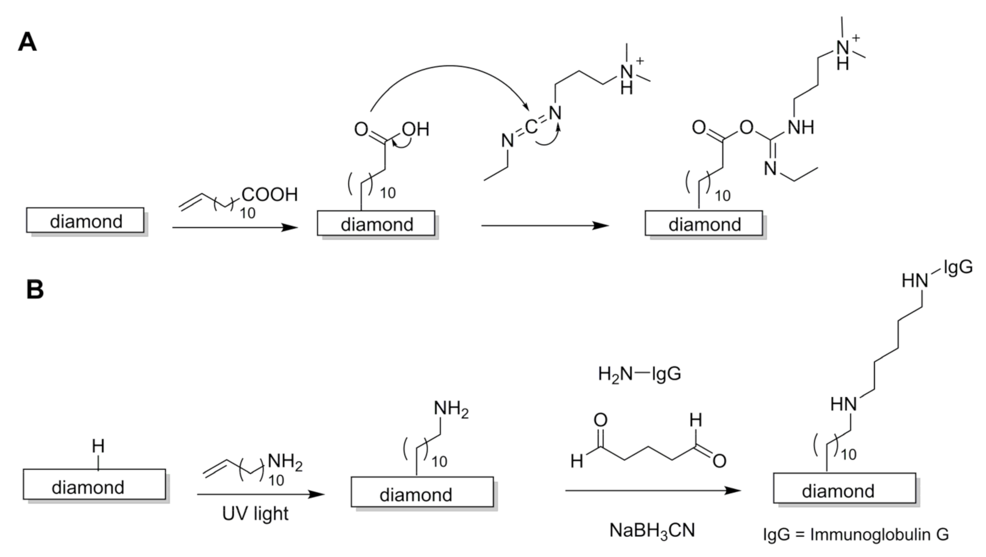
3.1. Photochemical Reactions

One of the first methods for the inclusion of different functional groups onto the surface of H-diamond resorted to processes initiated by photochemistry. In these, UV light is employed to attach simple NH2 groups or bridging molecules, such as acid or amine-terminated alkenes, to the surface of the diamond film (Figure 2). The bridging molecules create a spacer arm that can subsequently form a covalent bond with a target-specific molecule, such as a DNA oligonucleotide chain or a protein. Moreover, surface modification by photochemical methods has advantages over wet-chemical modification because it allows a facile patterning of the substrate with functional groups. As a setback, the presence of two different functional groups in the amino alkenes, e.g., a double bond at one end and an amine at the other end, requires the use of protecting groups to ensure that the photochemical reaction occurs with the double bond site and to avoid reaction of the amine headgroups, which may cause etching of the substrate [24].
The simplest form of functionalisation involves the generation of NH2-terminated surfaces in diamond. These can be obtained by the exposure of H-diamond to chlorine gas under irradiation with a 450 W Hg arc lamp (254 nm), with subsequent exposure of the chlorine-terminated to ammonia gas, also under irradiation. Both reactions are conducted at low pressure (c.a., 10−7 atm) [25].
Methods using spacer arms are preferred for a better spatial accommodation of large molecules such as DNA. The first report on the functionalisation of nanocrystalline diamond films with a DNA oligonucleotide sequence dates back to 2002 and employed 10-aminodec-1-ene (with the amine group being protected by trifluoroacetamide) to form a covalent bond with diamond carbons by irradiation with UV light (0.35 mW cm−2, 254 nm) [26]. After deprotection, the primary amine was allowed to react with a heterobifunctional crosslinker, sulfosuccinimidyl-4-(N-maleimidomethyl)cyclohexane-1-carboxylate (SSMCC), to form a group that subsequently reacted with a thiol-modified DNA oligonucleotide chain.
Alternatively, DNA oligonucleotide chains can be linked to H-diamond by an alkene bridge terminated with a carboxylic acid group at the other end. A commonly employed linker is 10-undecenoic acid, which reacts by activation with UV irradiation (20 h, 2.5 mW cm−2, 254 nm) to form the bridging layer; subsequently, the free carboxylate groups are treated with 1-ethyl-3-[3-dimethylaminopropyl]-carbodiimide (EDC), generating an active intermediate species that readily reacts with the amine groups of the desired DNA oligonucleotides (Figure 3A) [27,28]. Activation of the carboxylate groups with EDC permits a much faster, high-yield reaction because the active intermediate is more reactive than the native COOH. Further development of this method compared the effect of reacting 10-undecenoic acid in the unprotected and protected forms (trifluoroethyl undecenoate), having shown that the use of protecting groups leads to a lower density of carboxylate groups on the surface of diamond [24]. This is likely due to the bulkier nature of the protected molecules that create a steric hindrance to the functionalisation reactions, reducing the density of the attached molecules.
The inclusion of an antibody to the surface of H-diamond followed a process similar to that already described for DNA, in which trifluoroacetamide-protected 10-aminodec-1-ene was employed in the first reaction step (Figure 3B). In a second step, the substrate was treated with a solution of glutaraldehyde, a protein cross-linking agent able to react with amines to form Schiff bases [29]. This method required, however, an extra treatment step (addition of 0.1 M solution of glycine in sodium cyanoborohydride coupling buffer) to remove unreacted aldehyde groups that could affect the stability of the surface of diamond [29].

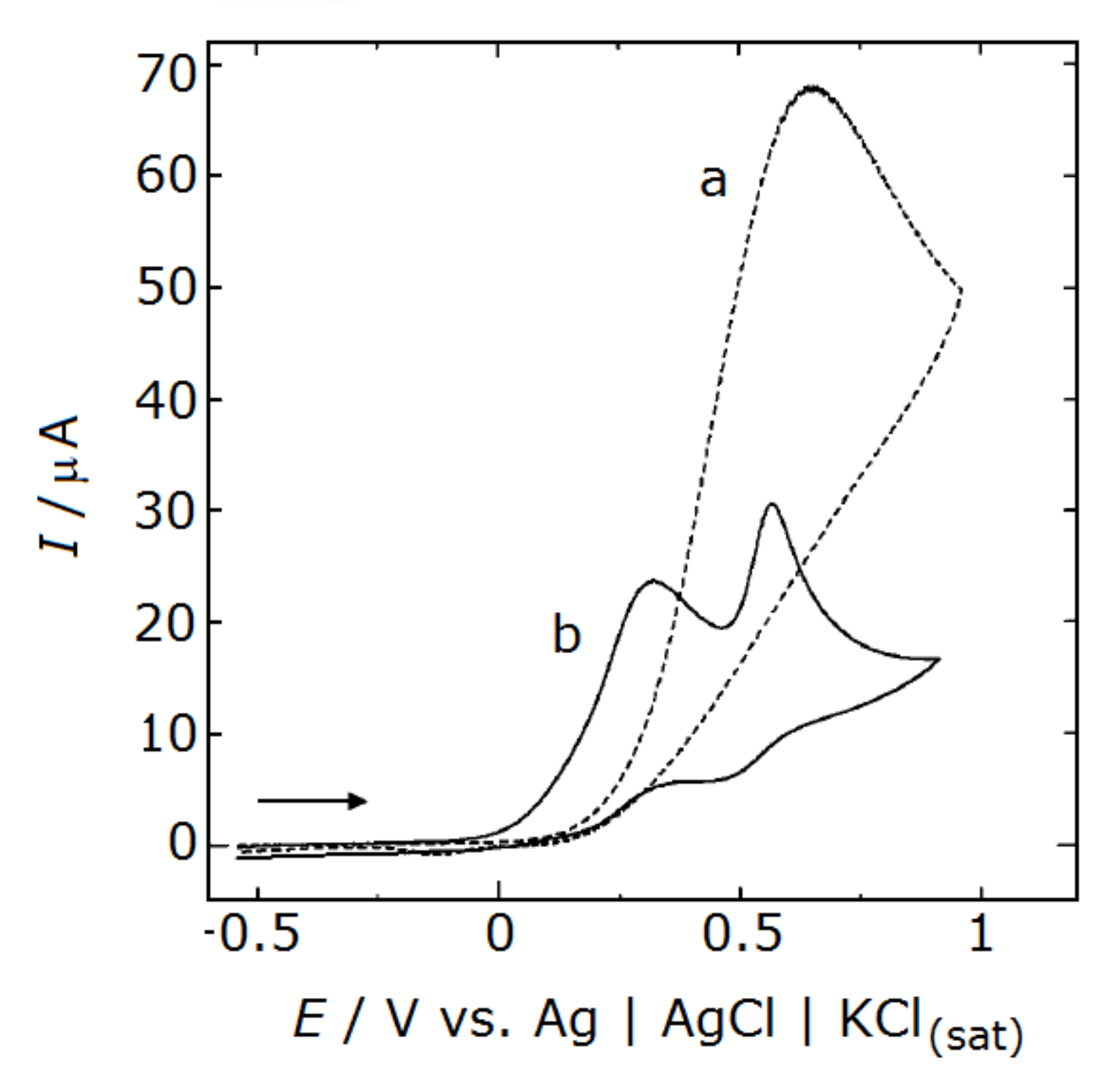
3.2. Electrochemical Reactions

There procedures are based on the reaction of charged molecules with the surface of H-diamond through the use of an electric current.

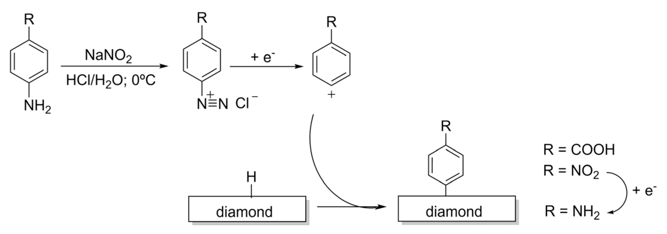
3.2.1. Reactions Involving Diazonium Salts

The use of phenyl diazonium salts was first reported by Kuo et al. in 1999 [30] and quickly became an extensively employed method for the functionalisation of diamond surfaces with linker arms that serve as anchors for the covalent bonding of biomolecules such as antibodies [31] or DNA oligonucleotide chains [32]. The process starts with the synthesis of a diazonium salt, e.g., 4-nitrobenzenediazonium tetrafluoroborate (Figure 4) [33], which is then used to treat H-terminated diamond thin films (40 mM) in the presence of a solution of 1% SDS (sodium dodecyl sulfate) to produce a surface terminated with 4-nitro-aryl groups. Subsequent application of the current reduces the nitro groups to primary amines.
Primary amines formed on the surface of diamond by the phenyl diazonium salt method can be used as anchoring points for a variety of molecules. An example of this is the reaction with thiol-modified DNA oligonucleotides via SSMCC cross-linking [32] (details of the process for cross linking DNA with NH2-terminated diamond are described in Section 3.1).
A drawback of this method is when the phenyl radical species react not only with the surface of the diamond film, but with the phenyl ring grafted on the surface, ultimately forming undesirable multilayers [34].

3.2.2. In Situ Electrochemical Polymerisation

Electrochemical reactions can be fine-tuned to achieve in situ polymer deposition. An example of this reported the use of pyrrole, a molecule with a positive charge, to form a polypyrrole film: the diamond surface was immersed in a solution of 50 mM pyrrole in 0.2 M KCl and cycled twice from 0 to 1.1 V (sweep rate of 500 mV s⁻1) [35]. Subsequent oxidation (by adding 0.5 M NaOH and applying ten electrical cycles), promoted the formation of COO⁻ groups, resulting in a polymer coating with multiple negative charges. This method was reported to improve the detection of small biologic cationic molecules such as dopamine [35,36].

3.3. Reactions Promoted by Ionic Liquids

Most of the methods reported so far rely on the use of an external energy source (RF/thermal/electrical energy of UV light) to provide the required activation energy to establish the bonds between the atoms on the diamond surface and the different chemical entities. A new perspective for the use of diazonium salts in the chemical modification of the surface of diamond films bearing hydrogen atoms has been proposed and uses ionic liquids [37]. The process can be conducted without the use of any source of activation, that is, without thermal, electrical, or photochemical activation. In their report, Szunerits et al. used only 4-nitroazobenzene dissolved in ionic liquids, taking advantage of their low vapour pressure: droplets of an ionic liquid solution of the diazonium salt are deposited locally on carbon materials by using simple spotting techniques [37], opening the way for new opportunities in the chemical functionalisation of diamond surfaces.

4. Diamond-Based Electrodes

Boron-doped diamond (BDD) electrodes are increasingly employed in bioanalytical applications because of their unique electrochemical properties, such as a low and stable baseline current [38] and wide potential window of water decomposition [39,40]. Chemical stability is a key factor: while most of the commonly employed electrodes cannot be used in corrosive, acidic, or basic media, the chemical inertness of diamond electrodes allows for their use in every type of media [41]. Another benefit is the ‘anti-fouling’ behaviour, i.e., their poor adsorption of most types of organic molecules [42], which ensures a very good stability against the degradation of the electrochemical signal over time due to surface passive layer build-up [43,44].
Diamond-based electrodes are reported to detect a variety of biogenic amines, including neurotransmitters such as dopamine [13,34,35,36,45,46,47], adrenalin [46], noradrenalin [46], serotonin [48], and histamine [48]. Detection of this class of molecules is typically based on their oxidation reactions, in which the alcohol moieties of the catechol are transformed into ketones. Electrodes described in this section are typically not functionalised for target-specific analysis, and their sensitivity is rather attained by finding the oxidation (or reduction) peak voltage of the desired analyte and then operating in amperometric mode; that is, fixing the voltage at the previously determined optimal value. Increased sensitivity can be achieved with simple coating procedures, such as using an oxidised polypyrrole polymer to repel anionic contaminants [35], or by reshaping the electrode surface with nano-grains to narrow the redox peak of a target analyte [13]. Specificity can be increased by coupling the electrode with a separation method such as capillary electrophoresis [46,47] or flow injection analysis [45].

4.1. Histamine Detection

The electrochemical response of a polycrystalline, BDD thin-film electrode to histamine in aqueous media at a pH of 7.2 was investigated with cyclic voltammetry and hydrodynamic voltammetry [47]. The cyclic voltammograms of histamine were well defined, peaking at 1.40 V, having a detection limit of 1 μM and a linear response range for histamine concentrations between 0.5 and 100 μM.

4.2. Serotonin Detection

The same BDD thin films described in the previous subsection were applied to the detection of serotonin [47]. Cyclic voltammograms showed well-defined sweeps, with peaks indicating no adsorption of the oxidation products of serotonin on the surface of the electrode. Consistently, no fouling or deactivation of the electrode was observed within the entire length of the experiment (several hours). The linear dynamic range was within the 0.01 to 100 μM interval with a remarkably low limit of detection around 10 nM.

4.3. Dopamine Detection

Voltammetric detection of dopamine was achieved with a micro-electrode prepared from a BDD film and modified by a secondary deposition process (see details in Section 2) to exhibit an external layer of nano-sized undoped crystalline diamond. This strategy helped to narrow the oxidation peak of dopamine, making it discernible from the signals of two common interferents, ascorbic acid and uric acid [13]. Note that, when the diamond electrode was not treated (with the second deposition) to feature the nanocrystals, the peaks of these two interferents overlapped with that of dopamine.
An alternative strategy to detect dopamine in solutions with other contaminants resorted to a BDD electrode deposited on a microfiber and coated with a polymeric layer of oxidised polypyrrole (see manufacturing details in Section 3.2.2) [35,36]. The presence of the oxidised polypyrrole coating helped to reduce the interference originated from ascorbic acid and 3,4-dihydroxyphenylacetic acid (DOPAC), a common metabolite of dopamine. This behaviour is associated with the abundance of negatively charged carboxylate groups (from the oxidised polypyrrole coating) on the electrode surface that attracts dopamine cations while repelling the negatively charged ascorbate anions. Even when an excess of ascorbic acid (0.2 mM) was present in regard to the target analyte, dopamine (0.05 mM), voltammograms showed two separate peaks, one for dopamine at 0.5 V and another for ascorbate at around 0.3 V (Figure 5) [35]. The limit of detection for dopamine was 0.1 nM at S/N = 3 (S/N is the signal-to-noise ratio), and the linear dynamic range laid between 0.5 nM and 100 μM. The amperometric response for 0.5 nM dopamine showed high stability (relative standard deviation of 5.4% for n = 5) and good reproducibility (within ± 6.2% for n = 10, with a solution of 1 nM of dopamine). Furthermore, the response remained the same during measurements conducted over a period of seven days, with no variation in the efficiency of rejection of the interferents (ascorbic acid and DOPAC).
Sensitivity to dopamine in matrices containing complex backgrounds can be further increased using coupled systems. In one of such report, amperometric detection of dopamine using a BDD microelectrode was coupled with a pretreatment using flow injection analysis [45]. The system detected dopamine with good reliability and reproducibility at concentrations as low as 6.0 μM (the lowest tested concentration) even when the analyte was dissolved in complex matrices such as river water and human serum. In another report, electrochemical detection of dopamine using a BDD microelectrode was coupled with capillary electrophoresis [48]. The system allowed the separation and analysis of dopamine in a mixture containing dopamine, catechol, and ascorbic acid (all at concentrations of 10 mM in phosphate buffer at pH = 6). The system performance was evaluated at different applied potentials, showing that the background current (~100 pA) was very stable with time and good reproducibility of the times of elution and detection (peak current or area) for each of the components of the mixture. The response had a precision better than 4.1%, a linear response between 0.1 and 1 mM, and a limit of detection of 1.7 fM.

4.4. Multi-Component Analysis: Dopamine, Adrenaline and Noradrenaline

A similar setting to that previously described, that is, comprised of a boron-doped microelectrode coupled with capillary electrophoresis, was reported to simultaneously detect three main neurotransmitters: dopamine, adrenaline, and noradrenaline [46]. The system exhibited high separation efficiency for these three catecholamines and a good detection performance, with high reproducibility for the current response over ten repetitive injections of mixtures of the catecholamines, at 50 μM each, and detection limits as low as 20 nM.

4.5. Towards In Vivo Applications: Noradrenaline and Adenosine Detection in Excised Tissue

4.5.1. Noradrenaline

In an attempt to bring sensors closer to real-life applications in living organisms, a diamond-coated carbon fibre microsensor was developed and tested regarding noradrenalin detection in an in vitro setting with excised rat mesenteric tissue. Following electrical stimulation of the mesenterium (60 pulses at 20 Hz for a total time of 3 s, repeating the stimulation after 10 min), noradrenaline was released from sympathetic nerves innervating the mesenteric artery and was measured as an electric response, presenting a peak of c.a. 10 pA. The response exhibited good reproducibility and recyclability over time [49,50].

4.5.2. Adenosine

Adenosine, besides being a base component of DNA, is present in the brain at low concentrations (around 50 to 200 nM), participating in neurotransmission modulation, particularly in the control of respiratory function. In a pilot study, Xie et al. focused on the PreBötzinger complex in rodent brains, a site that is rich in adenosine-releasing neurons [51]. Excised sections of this brain region were maintained in artificial cerebrospinal fluid (with 8 mM K+). Measurements resorted to a BDD thin film grown on the tip of a tungsten microelectrode (with an approximate diameter of 30 μm) that was shown to detect a concentration of 10 nM of adenosine and had a signal-to-noise ratio of 20 with a limit of detection of 2 nM (S/N = 3). When placed over the excised brain tissue slice stimulated with a synthetic drug to release adenosine, the microelectrode was able to detect the adenosine release in concentrations oscillating between 2 and 5 μM.

4.6. Detection of NADH, a Dinucleotide of Adenine and Nicotinamide

Nicotinamide adenine dinucleotide (NADH) is composed of two nucleotides linked through their phosphate groups. This essential biomolecule for cell metabolism is involved in a variety of cellular redox reactions, including the citric acid cycle and the oxidative phosphorylation chain, two essential processes for energy production in living cells. Detection of NADH was achieved with a BDD thin film electrode, with well-defined voltammograms even at low NADH concentrations (0.2 mM), with a good amperometric performance and a detection limit of 10 nM (S/N ratio of 7). The electrode response was found to be stable for a storage period of up to 3 months [43].

4.7. A Glutathione Electrode Operating In Vivo

Glutathione, a redox-active tripeptide with the sequence L-γ-glutamyl-L-cysteinyl-glycine, is the main biomolecule responsible for the removal of oxidising species and free radicals that can cause damage to living tissue. A BDD was reported to detect glutathione both in chimico and in vivo in a mouse model [52]. Amperometric measurements conducted in solutions of glutathione in diluted phosphate buffer saline (PBS) showed a limit of detection of 0.3 mM for glutathione at S/N = 3. In vivo measurements were conducted subcutaneously in immunodeficient mice with xenograft tumours derived from human cancer cells. Glutathione is typically overexpressed in tumour cells due to their augmented metabolic rate. The electrode was shown to detect the difference in concentrations of glutathione between cancerous and healthy tissues with high reproducibility.

5. Diamond-Based Electric Biosensors

The possibility of functionalising the surface of the diamond films with a specific detector layer opens the door to the detection of larger and specific molecules such as DNA or even viruses. The operating principle of these biosensors is based on the occurrence of intermolecular interactions between the target molecules and the receptor layer, which bring them improved selectivity. The use of more than one type of receptor molecule on the surface allows for the production of sensors capable of detecting multiple analytes. In addition, when molecules of the analyte are recognized by their counterparts at the receptor layer, the supramolecular interaction results in a change in the electrical properties of the surface of the biosensor that allows for other interrogation techniques in addition to cyclic voltammetry, such as impedance measurements. By specifically probing the changes in the impedance of the surface, these measurements may provide a more accurate tool to detect the presence of the molecules of the analyte.

5.1. Biosensors for DNA

Yang et al. observed the recognition of DNA oligonucleotides at the surface of diamond thin films by frequency-dependent impedance measurements [53]. Measurements as a function of potential show that the impedance at 106 Hz was dominated by the space-charge region of the diamond film and that the hybridisation of a complementary DNA oligonucleotide was able to induce a field effect in the diamond space-charge layer. This effect altered the impedance of the diamond film, thus allowing for a real-time hybridisation-dependent response.
Zhong et al. tested the electrical detection of DNA using two types of diamond thin films, one composed of non-doped diamond and another of BDD with dopant concentrations above 1019 cm⁻1. Both films were functionalised with undecenoic acid with subsequent attachment of a DNA probe chain [24]. Interestingly, monitoring DNA recognition by impedance measurements demonstrated that there was an insignificant change in impedance following hybridisation of DNA at the BDD-based biosensor, while the DNA sensor built from non-doped diamond exhibited a clear reduction in the impedance response. This was explained as the result of ‘reduced charge-transfer resistance of the molecular layers following the π–π stacking of the duplex DNA, as well as a reduction in the subsurface depletion due to an accumulation of holes’.
A DNA impedimetric biosensor built from undecenoic acid/DNA-functionalised, non-doped nanocrystalline diamond films was reported by Vermeeren et al. [54]. This device operated in a similar way to that constructed by Yang et al., that is, it showed a decrease in impedance when the complementary target DNA was added. Further tests included challenging the biosensor with a solution containing a DNA sequence that was not complementary to that of the receptor layer (with a single nucleotide mismatch, or 1-mismatch) to investigate if it could be differentiated from the target analyte. Results showed that differentiation between 1-mismatch and complementary target DNA was possible at frequencies between 100 and 1500 Hz. The biosensor still showed, however, some limitations for a wide range of applications, because its performance suffered negatively from the low ionic content of the sample media. Hybridisation in 1× PBS buffer was unsuccessful, while the separation between complementary and 1-mismatch hybridisation curves was very clear and reproducible when using the concentrated buffer, 10× PCR buffer (at 30 °C).

5.2. Biosensors for Immunoglobulins

Detection of antigen–antibody recognition using a biosensor built from a diamond thin film was first reported in 2007, resorting to electric impedance measurements of the immunoglobulin G/anti-immunoglobulin G antigen–antibody system [29].
A biosensor for immunoglobulin E (IgE) was subsequently reported by Tran et al. [55]. Instead of using the native anti-IgE antibody, an alternative approach was employed, which resorted to a smaller, synthetic biomolecule called an aptamer for the receptor layer (bridged via undecenoic acid). Aptamers are oligonucleotide or peptide chains designed to recognise DNA or proteins. The aptamer was a synthetic chain of oligonucleotides enriched with a terminal sequence of twenty thymidine bases for maximum flexibility in binding to the IgE protein. The biosensor offered a low detection limit of 0.03 µg/mL (at S/N = 3) and a linear dynamic range between 0.03 and 42.8 μg/mL. Reusability was tested for six cycles, with relative standard deviations (after the sixth cycle) rounding to 1%. The practical utility of the biosensor was tested in human serum samples, with results showing a linear dose–response for concentrations of IgE up to 5.8 μg/mL and an excellent correlation coefficient of 0.99 between quantification data provided by the biosensor and the reference method, enzyme-linked immunosorbent assay (ELISA).

5.3. Biosensors for Influenza

5.3.1. Detecting M1 Protein as a Universal Influenza Biomarker

Influenza A virus matrix protein M1 is a widely abundant membrane-associated protein that lies under the envelope of the virus particles, helping to maintain structural integrity and participating in several essential processes of the viral life cycle. When released into the serum, M1 proteins can agglomerate and help the various components of the virus assemble to form a new viral particle [56]. M1 can be monitored as a biomarker of influenza infection with an important advantage over other antigen proteins because it is transversal to all serotypes of influenza virus.
A biosensor for the M1 protein of influenza was built from a nanocrystalline BDD diamond thin film functionalised with polyclonal antibodies via diazonium salts electrodeposition [31,33]. The biosensor was able to detect the M1 protein in the H1N1 influenza strain by impedance measurements in PBS supplemented with 0.5% Triton X, a non-ionic surfactant. Under these conditions, the sensor response stabilised within 5 min, presenting a linear range of 10 to 100 fg/mL and a limit of detection as low as 0.7 fg/mL [31], which corresponds to approximately 40 viral particles [33]. The biosensor was also able to detect the H3N2 strain under the same conditions with almost no change in the impedimetric response [31].

5.3.2. Detecting Whole Influenza Viral Particles via Hemagglutinin (HA) Recognition

A biosensor for whole viral particles of influenza was built from a BDD thin film functionalised by the electrochemical deposition of a diazonium salt with the subsequent attachment of a sialic acid mimic dimeric peptide [57]. This peptide was designed for the recognition of hemagglutinin (HA), one of the surface antigens of the influenza virus. The first sequence of tests demonstrated that the biosensor does recognize pure HA in a PBS solution when operating in the mode of electric impedance spectroscopy. The biosensor was tested with whole viral particles, having proven successful in responding to both H1N1 and H3N2 strains in the concentration range of 400–8000 pfu/mL (where pfu is a plaque-forming unit).

6. Discussion and Future Outlook

The electrical detection of biomolecules using diamond-based devices can be done either with simple electrodes or with target-specific biosensors, as reviewed in the present work.
Electrodes built from BDD (Section 4) can serve as practical, resilient, and reusable instruments in bioanalytical applications because of their unique electrochemical properties, chemical stability, and ‘anti-fouling’ behaviour. Unlike the biosensors described in Section 5, these electrodes are not functionalised for target-specific analysis, their optimisation being attained by finding the oxidation (or reduction) peak voltage of the desired analyte and operating under that voltage. This raises the obvious question of selectivity, particularly for the electrodes described in Section 4.1, Section 4.2, Section 4.3 to Section 4.4, which were only evaluated in chimico, that is, in buffered solutions containing either just the target analyte or mixtures of the analyte with one or two common interferents. Their ability to operate in complex biological matrices, such as biological fluids or even in a living tissue, remains to be demonstrated, being an important target of investigation for those aiming to pursue further studies in this field. A second important issue is their sensitivity (LOD and/or linearity range), because biologic amines exist in very low quantities in the human body. Plasma levels of dopamine range between 0.05 and 0.3 nM [58], while plasma noradrenaline is within 0.9 to 8 nM [59]. These values are below, or within close proximity of, the linear response range of the electrodes, thus meaning that their applicability to real-case scenarios with plasma samples will require a pretreatment step to increase the concentration of the analyte, or the improvement of their performance. The approach employed by Roy et al. is a possible strategy for curbing these issues: by coating the electrode surface with an anionic polymer, the LOD was successfully lowered to the fentomolar range; however, the linear response range was still quite high, within the micromolar range [36], warranting further improvement. A recent strategy to overcome reduced sensitivity was recently presented by de Brosse et al. [60]. Using an oil-membrane composite protection chamber, interferences from the biofluid of hydrophilic solutes and solvents were mitigated. This oil-membrane protection allowed lowering the LOD of two standard inorganic compounds, hexacyanoferrate (II/III) and hexaammineruthenium (II/III), by over 100-fold compared to traditional electrodes (that is, those that were not covered with the membrane/oil chamber) [60]. While this concept was only demonstrated for the two inorganic standards, it is an innovative and promising approach for detecting biomolecules in biofluids with fewer performance-diminishing interferent effects; moreover, it does not require the change of the biosensor system itself, relying, instead, on the use of a careful selection of the semi-permeable membrane and oil composition for each specific application.
Electrodes described in Section 4.5, Section 4.6 through Section 4.7 constitute interesting proofs-of-concept on the application of diamond-based devices for sensing biological transmitters. The adenosine electrode, tested by Xie et al. on excised rat brain tissue and shown to operate within the nanomolar range [51], may also find use in monitoring plasma adenosine levels, typically around 0.6–1.1 mM [61]. The NADH sensor (Section 4.6) also operates in the nanomolar range. Further emphasis should be given to the electrode reposted by Fierro et al. (Section 4.7), already demonstrated to operate in vivo with promising results in cancer detection [52].
In the case of diamond-based biosensors, specificity of response is implemented by design, because the response relies on a specific interaction between the analyte and a complementary biomolecule on the organic receptor layer. The supramolecular recognition occurring between these two biomolecules, the covalently bonded one at the RL and the target analyte, creates a high degree of selectivity, which cannot be matched by electrodes. An additional advantage of biosensors is that they can operate with very small amounts of sample: a droplet of the solution can be simply placed on the receptor layer of the diamond-based biosensor. Furthermore, carbon-made biosensors with electrochemical detection have already been used to detect highly proliferative cells, such as human breast adenocarcinoma (MDA-MB-231), breast cancer isolated cell line (MCF-7), and hepatocellular carcinoma isolated cell line (HepG2), as portrayed and summarized by Suhito et al. [62], showing a great potential of carbon-based materials not only to detect biomolecules, but also the ability to provide a label-free, non-invasive, and non-destructive method for detecting early-stage cancer. This could be an interesting approach for boron-doped diamond in the future.
In conclusion, when weighing diamond-based electrodes against biosensors in the detection of biomolecules, there are, in one hand, the practical advantages of the easy-to-produce and easy-to use diamond-based electrodes and, on the other hand, the outstanding ability of biosensors to detect biological molecules of high molecular weight with a lower amount of analyte and very high selectivity. The manufacturing of biosensors requires, however, a sequence of chemical functionalisation steps, which may pose as a disadvantage because of the increased time, cost, and complexity of fabrication. For these reasons, the choice between a diamond-based electrode and a biosensor depends on several issues: the size, charge, and solution mobility of the target molecule; the amount of analyte; the intended type of detection; the intended selectivity and the time and money for fabrication.

Author Contributions

This review was written through the equal contribution of all authors.

Funding

We acknowledge University of Aveiro and FCT/MCTES (Fundação para a Ciência e a Tecnologia, Ministério da Ciência, da Tecnologia e do Ensino Superior) for financial support to LAQV-REQUIMTE (Ref. UIDB/50006/2020), and to CICECO—Aveiro Institute of Materials (Refs. UIDB/50011/2020, UIDP/50011/2020 & LA/P/0006/2020) through national founds (PIDDAC) and, where applicable, co-financed by the European Regional Development Fund (FEDER), within the PT2020 Partnership Agreement. The work of Instituto de Telecomunicações is funded by FCT/MCTES through national funds and when applicable co-funded EU funds under the project UIDB/50008/2020-UIDP/50008/2020. The research contract of FF (REF-168-89-ARH/2018) is funded by national funds (OE), through FCT, in the scope of the framework contract foreseen in Nos. 4, 5 and 6 of article 23 of the Decree-Law 57/2016, of 29 August, changed by Law 57/2017, of 19 July.

Conflicts of Interest

The authors declare no conflict of interest.

References

  1. Nebel, C.E.; Rezek, B.; Shin, D.; Uetsuka, H.; Yang, N. Diamond for bio-sensor applications. J. Phys. D Appl. Phys. 2007, 40, 6443–6466. [Google Scholar] [CrossRef]
  2. Tang, L.; Tsai, C.; Gerberich, C.C.; Kruckeberg, L.; Kania, D.R. Biocompatibility of chemical-vapour-deposited diamond. Biomaterials 1995, 16, 483–488. [Google Scholar] [CrossRef]
  3. Xue, Y.; Feng, X.; Roberts, S.C.; Chen, X. Diamond and carbon nanostructures for biomedical applications. Funct. Diam. 2021, 1, 221–242. [Google Scholar] [CrossRef]
  4. Szunerits, S.; Nebel, C.E.; Hamers, R.J. Surface functionalization and biological applications of CVD diamond. MRS Bull. 2014, 39, 517–524. [Google Scholar] [CrossRef]
  5. Field, J.E. The Properties of Natural and Synthetic Diamond, 1st ed.; Academic Press: London, UK, 1992. [Google Scholar]
  6. Luong, J.H.T.; Male, K.B.; Glennon, J.D. Boron-doped diamond electrode: Synthesis, characterization, functionalization and analytical applications. Analyst 2009, 134, 1965–1979. [Google Scholar] [CrossRef] [PubMed] [Green Version]
  7. Landstrass, M.I.; Ravi, K.V. Resistivity of chemical vapor-deposited diamond films. Appl. Phys. Lett. 1989, 55, 975–977. [Google Scholar] [CrossRef]
  8. Wenmackers, S.; Vermerren, V.; vandeVen, M.; Ameloot, M.; Bijnes, N.; Haenen, K.; Michiels, L.; Wagner, P. Diamond-based DNA sensors: Surface functionalization and read-out strategies. Phys. Status Solidi Appl. Mater. Sci. 2009, 206, 391–408. [Google Scholar] [CrossRef] [Green Version]
  9. Raymakers, J.; Haenen, K.; Maes, W. Diamond surface functionalization: From gemstone to photoelectrochemical applications. J. Mater. Chem. C 2019, 7, 10134–10165. [Google Scholar] [CrossRef]
  10. Gracio, J.J.; Fan, Q.H.; Madaleno, J.C. Diamond growth by chemical vapour deposition. J. Phys. D Appl. Phys. 2010, 43, 43. [Google Scholar] [CrossRef]
  11. Sque, S.J.; Jones, R.; Briddon, P.R. Structure, electronics, and interaction of hydrogen and oxygen on diamond surfaces. Phys. Rev. B 2006, 73, 085313. [Google Scholar] [CrossRef]
  12. Eaton, S.C.; Anderson, A.B.; Angus, J.C.; Evstefeeva, Y.E.; Pleskov, Y.V. Co-doping of diamond with boron and sulfur. Electrochem. Solid State Lett. 2002, 5, G65–G68. [Google Scholar] [CrossRef]
  13. Siew, P.S.; Loh, K.P.; Poh, W.C.; Zhang, H. Biosensing properties of nanocrystalline diamond film grown on polycrystalline diamond electrodes. Diam. Relat. Mater. 2005, 14, 426–431. [Google Scholar] [CrossRef]
  14. Song, C.W.; Cho, D.S.; Lee, J.M.; Song, P.K. Effect of boron doping on diamond film and electrochemical properties of BDD according to thickness and morphology. Coatings 2020, 10, 331. [Google Scholar] [CrossRef] [Green Version]
  15. Klauser, F.; Ghodban, S.; Boukherroub, R.; Sznurits, S.; Steinmüller-Nethl, D.; Bertel, E.; Memmel, N. Comparison of different oxidation techniques on single-crystal and nanocrystalline diamond surfaces. Diam. Relat. Mater. 2010, 19, 474–478. [Google Scholar] [CrossRef]
  16. O’Donnell, K.M.; Edmonds, M.T.; Ristein, J.; Tadich, A.; Thomsen, L.; Wu, Q.; Pakes, C.I.; Ley, L. Diamond surfaces with air-stable negative electron affinity and giant electron yield enhancement. Adv. Funct. Mater. 2013, 23, 5608–5614. [Google Scholar] [CrossRef]
  17. Loh, K.P.; Xie, X.N.; Yang, S.W.; Zheng, J.C. Oxygen adsorption on (111)-oriented diamond: A study with ultraviolet photoelectron spectroscopy, temperature-programmed desorption, and periodic density functional theory. J. Phys. Chem. B 2002, 106, 5230–5240. [Google Scholar] [CrossRef]
  18. Nakamura, J.; Ito, T. Oxidization process of CVD diamond (100): H 2 × 1 surfaces. Appl. Surf. Sci. 2005, 244, 301–304. [Google Scholar] [CrossRef]
  19. Jiang, M.; Yu, H.; Li, X.; Lu, S.; Hu, X. Thermal oxidation induced high electrochemical activity of boron-doped nanocrystalline diamond electrodes. Electrochim. Acta 2017, 258, 61–70. [Google Scholar] [CrossRef]
  20. Kuga, S.; Yang, J.H.; Takahashi, H.; Hirama, K.; Iwasaki, T.; Kawarada, H. Detection of mismatched DNA on partially negatively charged diamond surfaces by optical and potentiometric methods. J. Am. Chem. Soc. 2008, 130, 13251–13263. [Google Scholar] [CrossRef]
  21. Coffinie, Y.; Szuerits, S.; Jama, C.; Desmet, R.; Melnyk, O.; Marcus, B.; Gengembre, L.; Payen, E.; Delabouglise, D.; Boukherroub, R. Peptide immobilization on amine-terminated boron-doped diamond surfaces. Langmuir 2007, 3, 4494–4497. [Google Scholar] [CrossRef]
  22. Szunerits, S.; Jama, C.; Coffinier, Y.; Marcus, B.; Delabouglise, D.; Boukherroub, R. Direct amination of hydrogen-terminated boron doped diamond surfaces. Electrochem. Commun. 2006, 8, 1185–1190. [Google Scholar] [CrossRef]
  23. Chandran, M.; Shasha, M.; Michaelson, S.; Hoffman, A. Nitrogen termination of single crystal (100) diamond surface by radio frequency N2 plasma process: An in-situ x-ray photoemission spectroscopy and secondary electron emission studies. Appl. Phys. Lett. 2015, 107, 111602. [Google Scholar] [CrossRef]
  24. Zhong, Y.L.; Chong, K.F.; May, P.W.; Chen, Z.K.; Loh, K.P. Optimizing biosensing properties on undecylenic acid-functionalized diamond. Langmuir 2007, 23, 5824–5830. [Google Scholar] [CrossRef] [PubMed]
  25. Miller, J.B.; Brown, D.W. Properties of photochemically modified diamond films. Diam. Rel. Mater. 1995, 4, 435–440. [Google Scholar] [CrossRef]
  26. Yang, W.; Auciello, O.; Butler, J.E.; Cai, W.; Carlisle, J.A.; Gerbi, J.E.; Gruen, D.M.; Knickerbocker, T.; Lasseter, T.L.; Russel, J.N., Jr.; et al. DNA-modified nanocrystalline diamond thin-films as stable, biologically active substrates. Nature 2003, 1, 253–258. [Google Scholar] [CrossRef] [PubMed]
  27. Christiaens, P.; Vermeeren, V.; Wenmackers, S.; Daenen, M.; Haenen, K.; Nesládek, M.; VanDeVen, M.; Ameloot, M.; Michiels, L.; Wagner, P. EDC-mediated DNA attachment to nanocrystalline CVD diamond films. Biosens. Bioelectron. 2006, 22, 170–177. [Google Scholar] [CrossRef]
  28. Wenmackers, S.; Christiaens, P.; Daenen, M.; Haenen, K.; Nesládek, M.; van deVen, M.; Vermeeren, V.; Michiels, L.; Ameloot, M.; Wagner, P. DNA attachment to nanocrystalline diamond films. Phys. Status Solidi A 2005, 202, 2212–2216. [Google Scholar] [CrossRef]
  29. Yang, W.; Butler, J.E.; Russell, J.N.; Hamers, R.J. Direct electrical detection of antigen-antibody binding on diamond and silicon substrates using electrical impedance spectroscopy. Analyst 2007, 132, 296–306. [Google Scholar] [CrossRef]
  30. Kuo, T.C.; McCreery, R.L.; Swain, G.M. Electrochemical modification of boron-doped chemical vapor deposited diamond surfaces with covalently bonded monolayers. Electrochem. Solid-State Lett. 1999, 2, 288–290. [Google Scholar] [CrossRef]
  31. Nidzworski, D.; Siuzdak, K.; Niedziałkowski, P.; Bogdanowicz, R.; Sobaszek, M.; Ryl, J.; Weiher, P.; Sawczak, M.; Wnuk, E.; Goddard, W.A.; et al. A rapid-response ultrasensitive biosensor for influenza virus detection using antibody modified boron-doped diamond. Sci. Rep. 2017, 7, 1–10. [Google Scholar] [CrossRef]
  32. Yang, W.; Baker, S.E.; Butler, J.E.; Lee, C.S.; Russell, J.N.; Shang, L.; Sun, B.; Hamers, R.J. Electrically addressable biomolecular functionalization of conductive nanocrystalline diamond thin films. Chem. Mater. 2005, 17, 938–940. [Google Scholar] [CrossRef]
  33. Siuzdak, K.; Niedziałkowski, P.; Sobaszek, M.; Łęga, T.; Sawczak, M.; Czaczyk, E.; Dziąbowska, K.; Ossowski, T.; Nidzworski, D.; Bogdanowicz, R. Biomolecular influenza virus detection based on the electrochemical impedance spectroscopy using the nanocrystalline boron-doped diamond electrodes with covalently bound antibodies. Sens. Actuators B Chem. 2019, 280, 263–271. [Google Scholar] [CrossRef]
  34. Pinson, J.; Podvorica, F. Attachment of organic layers to conductive or semiconductive surfaces by reduction of diazonium salts. Chem. Soc. Rev. 2005, 34, 429–439. [Google Scholar] [CrossRef] [PubMed]
  35. Olivia, H.; Sarada, B.V.; Shin, D.; Rao, T.N.; Fujishima, A. Selective amperometric detection of dopamine using OPPy-modified diamond microsensor system. Analyst 2002, 127, 1572–1575. [Google Scholar] [CrossRef] [PubMed]
  36. Roy, P.R.; Saha, M.S.; Okajima, T.; Park, S.-G.; Fujishima, A.; Ohsaka, T. Selective detection of dopamine and its metabolite, DOPAC, in the presence of ascorbic acid using diamond electrode modified by the polymer film. Electroanalysis 2004, 16, 1777–1784. [Google Scholar] [CrossRef]
  37. Shul, G.; Actis, P.; Marcus, B.; Opallo, M.; Boukherroub, R.; Szunerits, S. Solvent-free chemical functionalization of hydrogen-terminated boron-doped diamond electrodes with diazonium salts in ionic liquids. Diam. Relat. Mater. 2008, 17, 1394–1398. [Google Scholar] [CrossRef]
  38. Yano, T.; Tryk, D.A.; Hashimoto, K.; Fujishima, A. Electrochemical behavior of highly conductive boron-doped diamond electrodes for oxygen reduction in alkaline solution. J. Electrochem. Soc. 1998, 145, 1870–1876. [Google Scholar] [CrossRef]
  39. Strojek, J.W.; Granger, M.C.; Swain, G.M.; Dallas, T.; Holtz, M.W. Enhanced signal-to-background ratios in voltammetric measurements made at diamond thin-film electrochemical interfaces. Anal. Chem. 1996, 68, 2031–2037. [Google Scholar] [CrossRef] [PubMed]
  40. Vinokur, N.; Miller, B.; Avyigal, Y.; Kalish, R. Electrochemical behavior of boron-doped diamond electrodes. J. Electrochem. Soc. 1996, 143, L238–L240. [Google Scholar] [CrossRef]
  41. Hébert, C.; Ruffinatto, S.; Bergonzo, P. Diamond Biosensors. In Carbon for Sensing Devices, 1st ed.; Demarchi, D., Tagliaferro, A., Eds.; Springer International Publishing: New York, NY, USA, 2015; pp. 227–264. [Google Scholar] [CrossRef]
  42. Xu, J.; Swain, G.M. Oxidation of azide anion at boron-doped diamond thin-film electrodes. Anal. Chem. 1998, 70, 1502–1510. [Google Scholar] [CrossRef]
  43. Rao, T.N.; Yagi, I.; Miwa, T.; Tryk, D.A.; Fujishima, A. Electrochemical oxidation of NADH at highly boron-doped diamond electrodes. Anal. Chem. 1999, 71, 2506–2511. [Google Scholar] [CrossRef] [PubMed]
  44. Vanhove, E.; de Sanoit, J.; Arnault, J.C.; Saada, C.; Mer, C.; Mailley, P.; Bergonzo, P.; Nesladek, M. Stability of H-terminated BDD electrodes: An insight into the influence of the surface preparation. Phys. Status Solidi 2007, 204, 2931–2939. [Google Scholar] [CrossRef]
  45. Wong, A.; Santos, A.M.; Fatibello-Filho, O. Simultaneous determination of dopamine and cysteamine by flow injection with multiple pulse amperometric detection using a boron-doped diamond electrode. Diam. Relat. Mater. 2018, 85, 68–73. [Google Scholar] [CrossRef]
  46. Shin, D.; Sarada, B.V.; Tryk, D.A.; Fujishima, A.; Wang, J. Application of diamond microelectrodes for end-column electrochemical detection in capillary electrophoresis. Anal. Chem. 2003, 75, 530–534. [Google Scholar] [CrossRef]
  47. Cvačka, J.; Quaiserová, V.; Park, J.W.; Show, Y.; Muck, A.; Swain, G.M. Boron-doped diamond microelectrodes for use in capillary electrophoresis with electrochemical detection. Anal. Chem. 2003, 75, 2678–2687. [Google Scholar] [CrossRef]
  48. Sarada, B.V.; Rao, T.N.; Tryk, D.A.; Fujishima, A. Electrochemical oxidation of histamine and serotonin at highly boron-doped diamond electrodes. Anal. Chem. 2000, 72, 1632–1638. [Google Scholar] [CrossRef]
  49. Park, J.; Quaiserová-Mocko, V.; Pecková, K.; Galligan, J.J.; Fink, G.D.; Swain, G.M. Fabrication, characterization, and application of a diamond microelectrode for electrochemical measurement of norepinephrine release from the sympathetic nervous system. Diam. Relat. Mater. 2006, 15, 761–772. [Google Scholar] [CrossRef]
  50. Park, J.; Show, Y.; Quaiserova, V.; Galligan, J.J.; Fink, G.D.; Swain, G.M. Diamond microelectrodes for use in biological environments. J. Electroanal. Chem. 2005, 583, 56–68. [Google Scholar] [CrossRef]
  51. Xie, S.; Shafer, G.; Wilson, C.G.; Martin, H.B. In vitro adenosine detection with a diamond-based sensor. Diam. Relat. Mater. 2006, 15, 225–228. [Google Scholar] [CrossRef]
  52. Fierro, S.; Yoshikawa, M.; Nagano, O.; Yoshimi, K.; Saya, H.; Einaga, Y. In vivo assessment of cancerous tumors using boron doped diamond microelectrode. Sci. Rep. 2012, 2, srep00901. [Google Scholar] [CrossRef]
  53. Yang, W.; Butler, J.E.; Russell, J.N.; Hamers, R.J. Interfacial electrical properties of DNA-modified diamond thin films: Intrinsic response and hybridization-induced field effects. Langmuir 2004, 20, 6778–6787. [Google Scholar] [CrossRef] [PubMed]
  54. Vermeeren, V.; Bijnens, N.; Wenmackers, S.; Daenen, M.; Haenen, K.; Williams, O.A.; Ameloot, M.; van de Ven, M.; Wagner, P.; Michiels, L. Towards a real-time, label-free, diamond-based DNA sensor. Langmuir 2007, 23, 13193–13202. [Google Scholar] [CrossRef]
  55. Tran, D.T.; Vermeeren, V.; Grieten, L.; Wenmackers, S.; Wagner, P.; Pollet, J.; Janssen, K.P.F.; Michiels, L.; Lammertyn, J. Nanocrystalline diamond impedimetric aptasensor for the label-free detection of human IgE. Biosens. Bioelectron. 2011, 26, 2987–2993. [Google Scholar] [CrossRef] [PubMed]
  56. Hilsch, M.; Goldenbogen, B.; Sieben, C.; Höfer, C.T.; Rabe, J.P.; Klip, E.; Herrmann, A.; Chiantia, S. Influenza A matrix protein m1 multimerizes upon binding to lipid membranes. Biophys. J. 2014, 107, 912–923. [Google Scholar] [CrossRef] [PubMed] [Green Version]
  57. Matsubara, T.; Ujie, M.; Yamamoto, T.; Akahori, M.; Einaga, Y.; Sato, T. Highly sensitive detection of influenza virus by boron-doped diamond electrode terminated with sialic acid-mimic peptide. Proc. Natl. Acad. Sci. USA 2016, 113, 8981–8984. [Google Scholar] [CrossRef] [Green Version]
  58. Goldstein, D.S.; Swoboda, K.J.; Miles, J.M.; Coppack, S.W.; Aneman, A.; Holmes, C.; Lamensdorf, I.; Eisenhofer, G. Sources and physiological significance of plasma dopamine sulphate. J. Clin. Endocrinol. Metab. 1999, 84, 2523–2531. [Google Scholar] [CrossRef] [PubMed]
  59. Pillet, L.E.; Taccola, C.; Cotoni, J.; Thiriez, H.; André, K.; Verpillot, R. Correlation between cognition and plasma noradrenaline level in Alzheimer’s disease: A potential new blood marker of disease evolution. Transl. Psychiatry 2020, 10, 1–10. [Google Scholar] [CrossRef]
  60. DeBrosse, M.; Yuan, Y.; Brothers, M.; Karajic, A.; van Duren, J.; Kim, S.; Hussain, S.; Heikenfeld, J. A dual approach of an oil–membrane composite and boron-doped diamond electrode to mitigate biofluid interferences. Sensors 2021, 21, 8063. [Google Scholar] [CrossRef]
  61. Simard, T.; Jung, R.; Labinaz, A.; Faraz, M.A.; Ramirez, F.D.; Di Santo, P.; Perry-Nguyen, D.; Pitcher, I.; Motazedian, P.; Gaudet, C.; et al. Evaluation of plasma adenosine as a marker of cardiovascular risk: Analytical and biological considerations. J. Am. Heart Assoc. 2019, 8, e012228. [Google Scholar] [CrossRef] [PubMed] [Green Version]
  62. Suhito, I.R.; Koo, K.-M.; Kim, T.-H. Recent advances in electrochemical sensors for the detection of biomolecules and whole cells. Biomedicines 2021, 9, 15. [Google Scholar] [CrossRef]
Figure 1. Schematic representation of a biosensor.
Applsci 12 03000 g001
Figure 2. Electron microscopy image of: (left) polycrystalline boron-doped-diamond film; (right) the same film after a secondary deposition with non-doped diamond, generating nano-sized crystalline grains (magnification was increased to better show the very small grains). Reprinted with permission from Siew et al. [13]. Copyright 2004 Elsevier BV.
Applsci 12 03000 g002
Figure 3. Examples of surface modification of H-diamond through photochemical grafting of two different kinds of linker arms: (A) an unsaturated carboxylic or (B) an unsaturated amine.
Applsci 12 03000 g003
Figure 4. Surface modification of H-diamond through electrochemical reduction of phenyl diazonium salts.
Applsci 12 03000 g004
Figure 5. Cyclic voltammograms, at a potential scan rate of 100 mV·s⁻1, of a mixed solution containing 0.2 mM of ascorbic acid and 0.05 mM dopamine in 0.2 M phosphate buffer (pH 7): (a) bare BDD electrode and (b) oxidised polypyrrole film-coated BDD electrode. Reprinted with permission from Roy et al. [36]. Copyright 2004 Wiley-VCH Verlag Gmbh & Co. KGaA, Weinhelm.
Applsci 12 03000 g005
Publisher’s Note: MDPI stays neutral with regard to jurisdictional claims in published maps and institutional affiliations.

