Materials Separation via the Matrix Method Employing Energy-Discriminating X-ray Detection
Abstract
:1. Introduction
2. Materials and Methods
2.1. The PiXirad Detector
2.2. Matrix Equation for Materials Separation Using the PiXirad Detector
2.3. Simulation Study
3. Experimental Results
4. Conclusions
Supplementary Materials
Author Contributions
Funding
Institutional Review Board Statement
Informed Consent Statement
Data Availability Statement
Acknowledgments
Conflicts of Interest
References
- Sakellariou, A.; Arns, C.H.; Sheppard, A.P.; Sok, R.M.; Averdunk, H.; Limaye, A.; Jones, A.C.; Senden, T.J.; Knackstedt, M.A. Developing a virtual materials laboratory. Mater. Today 2007, 10, 44–51. [Google Scholar] [CrossRef]
- Arhatari, B.D.; Gureyev, T.E.; Abbey, B. Elemental Contrast X-ray Tomography Using Ross Filter Pairs with a Polychromatic Laboratory Source. Sci. Rep. 2017, 7, 218. [Google Scholar] [CrossRef] [Green Version]
- O’Connell, D.W.; Morgan, K.S.; Ruben, G.; Schaff, F.; Croton, L.C.; Buckley, G.A.; Paganin, D.M.; Uesugi, K.; Kitchen, M.J. Photon-counting, energy-resolving and super-resolution phase contrast X-ray imaging using an integrating detector. Opt. Express 2020, 28, 7080–7094. [Google Scholar] [CrossRef]
- Chen, P.; Han, Y.; Li, Y. X-ray Multispectrum CT Imaging by Projection Sequences Blind Separation Based on Basis-Effect Decomposition. IEEE Trans. Instrum. Meas. 2021, 70, 4502208. [Google Scholar] [CrossRef]
- Dehlinger, A.; Blechschmidt, A.; Grötzsch, D.; Jung, R.; Kanngießer, B.; Seim, C.; Stiel, H. 3D nanoscale imaging of biological samples with laboratory-based soft X-ray sources. In Proceedings of the X-ray Lasers and Coherent X-ray Sources: Development and Applications XI, San Diego, CA, USA, 9–13 August 2015; pp. 82–90. [Google Scholar]
- Dudak, J.; Zemlicka, J.; Mrzilkova, J.; Zach, P.; Holcova, K. Applicability of large-area single-photon counting detectors Timepix for high-resolution and high-contrast X-ray imaging of biological samples. IEEE Trans. Nucl. Sci. 2022. Early Access. [Google Scholar] [CrossRef]
- Arhatari, B.D.; Stevenson, A.W.; Abbey, B.; Nesterets, Y.I.; Maksimenko, A.; Hall, C.J.; Thompson, D.; Mayo, S.C.; Fiala, T.; Quiney, H.M.; et al. X-ray Phase-Contrast Computed Tomography for Soft Tissue Imaging at the Imaging and Medical Beamline (IMBL) of the Australian Synchrotron. Appl. Sci. 2021, 11, 4120. [Google Scholar] [CrossRef]
- Gureyev, T.E.; Nesterets, Y.I.; Baran, P.M.; Taba, S.T.; Mayo, S.C.; Thompson, D.; Arhatari, B.; Mihocic, A.; Abbey, B.; Lockie, D.; et al. Propagation-based x-ray phase-contrast tomography of mastectomy samples using synchrotron radiation. Med. Phys. 2019, 46, 5478–5487. [Google Scholar] [CrossRef]
- Peterson, I.; Abbey, B.; Putkunz, C.T.; Vine, D.J.; van Riessen, G.A.; Cadenazzi, G.A.; Balaur, E.; Ryan, R.; Quiney, H.M.; McNulty, I.; et al. Nanoscale Fresnel coherent diffraction imaging tomography using ptychography. Opt. Express 2012, 20, 24678–24685. [Google Scholar] [CrossRef]
- Bravin, A.; Coan, P.; Suortti, P. X-ray phase-contrast imaging: From pre-clinical applications towards clinics. Phys. Med. Biol. 2012, 58, R1–R35. [Google Scholar] [CrossRef]
- Wang, X.; Meier, D.; Taguchi, K.; Wagenaar, D.J.; Patt, B.E.; Frey, E.C. Material separation in X-ray CT with energy resolved photon-counting detectors. Med. Phys. 2011, 38, 1534–1546. [Google Scholar] [CrossRef] [Green Version]
- Taguchi, K. Energy-sensitive photon counting detector-based X-ray computed tomography. Radiol. Phys. Technol. 2017, 10, 8–22. [Google Scholar] [CrossRef]
- Yokhana, V.S.; Arhatari, B.D.; Gureyev, T.E.; Abbey, B. Elemental contrast imaging with a polychromatic laboratory X-ray source using energy-discriminating detectors. In Proceedings of the SPIE BioPhotonics Australasia, Adelaide, Australia, 17–19 October 2016; Volume 10013, p. 1001339. [Google Scholar]
- Delogu, P.; Oliva, P.; Bellazzini, R.; Brez, A.; De Ruvo, P.; Minuti, M.; Pinchera, M.; Spandre, G.; Vincenzi, A. Characterization of Pixirad-1 photon counting detector for X-ray imaging. J. Instrum. 2016, 11, P01015. [Google Scholar] [CrossRef]
- O’Flynn, D.; Bellazzini, R.; Minuti, M.; Brez, A.; Pinchera, M.; Spandre, G.; Moss, R.; Speller, R. Energy-windowed, pixellated X-ray diffraction using the Pixirad CdTe detector. J. Instrum. 2017, 12, P01004. [Google Scholar] [CrossRef] [Green Version]
- Vincenzi, A.; De Ruvo, P.; Delogu, P.; Bellazzini, R.; Brez, A.; Minuti, M.; Pinchera, M.; Spandre, G. Energy characterization of Pixirad-1 photon counting detector system. J. Instrum. 2015, 10, C04010. [Google Scholar] [CrossRef] [Green Version]
- Matsuyama, S.; Shimura, M.; Fujii, M.; Maeshima, K.; Yumoto, H.; Mimura, H.; Sano, Y.; Yabashi, M.; Nishino, Y.; Tamasaku, K. Elemental mapping of frozen-hydrated cells with cryo-scanning X-ray fluorescence microscopy. X-ray Spectrom. 2010, 39, 260–266. [Google Scholar] [CrossRef]
- Jones, M.W.; de Jonge, M.D.; James, S.A.; Burke, R. Elemental mapping of the entire intact Drosophila gastrointestinal tract. J. Biol. Inorg. Chem. 2015, 20, 979–987. [Google Scholar] [CrossRef]
- De Jonge, M.D.; Vogt, S. Hard X-ray fluorescence tomography—An emerging tool for structural visualization. Curr. Opin. Struct. Biol. 2010, 20, 606–614. [Google Scholar] [CrossRef]
- Tillman, C.; Mercer, I.; Svanberg, S.; Herrlin, K. Elemental biological imaging by differential absorption with a laser-produced X-ray source. JOSA B 1996, 13, 209–215. [Google Scholar] [CrossRef]
- Luu, M.B.; Tran, C.Q.; Arhatari, B.; Balaur, E.; Kirby, N.; Mudie, S.; Pham, B.T.; Vo, N.T.; Putkunz, C.T.; De Carlo, F. Multi-wavelength elemental contrast absorption imaging. Opt. Express 2011, 19, 25969–25980. [Google Scholar] [CrossRef]
- Fornaro, J.; Leschka, S.; Hibbeln, D.; Butler, A.; Anderson, N.; Pache, G.; Scheffel, H.; Wildermuth, S.; Alkadhi, H.; Stolzmann, P. Dual-and multi-energy CT: Approach to functional imaging. Insights Imaging 2011, 2, 149–159. [Google Scholar] [CrossRef] [Green Version]
- Handschuh, S.; Beisser, C.J.; Ruthensteiner, B.; Metscher, B.D. Microscopic dual-energy CT (microDECT): A flexible tool for multichannel ex vivo 3D imaging of biological specimens. J. Microsc. 2017, 267, 3–26. [Google Scholar] [CrossRef] [PubMed]
- Yokhana, V.S.K.; Arhatari, B.D.; Gureyev, T.E.; Abbey, B. Soft-tissue differentiation and bone densitometry via energy-discriminating X-ray microCT. Opt. Express 2017, 25, 29328–29341. [Google Scholar] [CrossRef]
- Lehmann, L.; Alvarez, R.; Macovski, A.; Brody, W.; Pelc, N.; Riederer, S.J.; Hall, A. Generalized image combinations in dual KVP digital radiography. Med. Phys. 1981, 8, 659–667. [Google Scholar] [CrossRef] [PubMed]
- McCollough, C.H.; Leng, S.; Yu, L.; Fletcher, J.G. Dual-and multi-energy CT: Principles, technical approaches, and clinical applications. Radiology 2015, 276, 637–653. [Google Scholar] [CrossRef] [PubMed]
- Hidas, G.; Eliahou, R.; Duvdevani, M.; Coulon, P.; Lemaitre, L.; Gofrit, O.N.; Pode, D.; Sosna, J. Determination of renal stone composition with dual-energy CT: In vivo analysis and comparison with x-ray diffraction. Radiology 2010, 257, 394–401. [Google Scholar] [CrossRef]
- Alvarez, R.E.; Macovski, A. Energy-selective reconstructions in x-ray computerised tomography. Phys. Med. Biol. 1976, 21, 733. [Google Scholar] [CrossRef]
- Goo, H.W.; Goo, J.M. Dual-Energy CT: New Horizon in Medical Imaging. Korean J. Radiol. 2017, 18, 555–569. [Google Scholar] [CrossRef] [Green Version]
- Cheong, S.-K.; Jones, B.L.; Siddiqi, A.K.; Liu, F.; Manohar, N.; Cho, S.H. X-ray fluorescence computed tomography (XFCT) imaging of gold nanoparticle-loaded objects using 110 kVp X-rays. Phys. Med. Biol. 2010, 55, 647–662. [Google Scholar] [CrossRef]
- Romero, I.O.; Fang, Y.; Lun, M.; Li, C. X-ray fluorescence computed tomography (XFCT) imaging with a superfine pencil beam x-ray source. Photonics 2021, 8, 236. [Google Scholar] [CrossRef]
- Hampai, D.; Cherepennikov, Y.M.; Liedl, A.; Cappuccio, G.; Capitolo, E.; Iannarelli, M.; Azzutti, C.; Gladkikh, Y.P.; Marcelli, A.; Dabagov, S. Polycapillary based μXRF station for 3D colour tomography. J. Instrum. 2018, 13, C04024. [Google Scholar] [CrossRef] [Green Version]
- Hampai, D.; Liedl, A.; Cappuccio, G.; Capitolo, E.; Iannarelli, M.; Massussi, M.; Tucci, S.; Sardella, R.; Sciancalepore, A.; Polese, C. 2D-3D μXRF elemental mapping of archeological samples. Nucl. Instrum. Methods Phys. Res. Sect. B Beam Interact. Mater. At. 2017, 402, 274–277. [Google Scholar] [CrossRef]
- Veale, M.C.; Seller, P.; Wilson, M.; Liotti, E. HEXITEC: A High-Energy X-ray Spectroscopic Imaging Detector for Synchrotron Applications. Synchrotron Radiat. News 2018, 31, 28–32. [Google Scholar] [CrossRef]
- Vazquez, I.; Fredette, N.R.; Das, M. Quantitative phase retrieval of heterogeneous samples from spectral X-ray measurements. In Proceedings of the Medical Imaging 2019: Physics of Medical Imaging, San Diego, CA, USA, 16–21 February 2019; p. 109484R. [Google Scholar]
- Kurdzesau, F. Energy-dispersive Laue experiments with X-ray tube and PILATUS detector: Precise determination of lattice constants. J. Appl. Crystallogr. 2019, 52, 72–93. [Google Scholar] [CrossRef]
- Alle, P.; Wenger, E.; Dahaoui, S.; Schaniel, D.; Lecomte, C. Comparison of CCD, CMOS and hybrid pixel x-ray detectors: Detection principle and data quality. Phys. Scr. 2016, 91, 063001. [Google Scholar] [CrossRef]
- Jayarathna, S.; Ahmed, M.F.; O’ryan, L.; Moktan, H.; Cui, Y.; Cho, S.H. Characterization of a pixelated cadmium telluride detector system using a polychromatic X-ray source and gold nanoparticle-loaded phantoms for benchtop X-ray fluorescence imaging. IEEE Access 2021, 9, 49912–49919. [Google Scholar] [CrossRef]
- Ordavo, I.; Ihle, S.; Arkadiev, V.; Scharf, O.; Soltau, H.; Bjeoumikhov, A.; Bjeoumikhova, S.; Buzanich, G.; Gubzhokov, R.; Günther, A.; et al. A new pnCCD-based color X-ray camera for fast spatial and energy-resolved measurements. Nucl. Instrum. Methods Phys. Res. Sect. A Accel. Spectrometers Detect. Assoc. Equip. 2011, 654, 250–257. [Google Scholar] [CrossRef]
- Batey, D.J.; Cipiccia, S.; Van Assche, F.; Vanheule, S.; Vanmechelen, J.; Boone, M.N.; Rau, C. Spectroscopic imaging with single acquisition ptychography and a hyperspectral detector. Sci. Rep. 2019, 9, 12278. [Google Scholar] [CrossRef]
- Taguchi, K.; Iwanczyk, J.S. Vision 20/20: Single photon counting X-ray detectors in medical imaging. Med. Phys. 2013, 40, 100901. [Google Scholar] [CrossRef]
- Cajipe, V.B.; Calderwood, R.F.; Clajus, M.; Hayakawa, S.; Jayaraman, R.; Tumer, T.O.; Grattan, B.; Yossifor, O. Multi-energy X-ray imaging with linear CZT pixel arrays and integrated electronics. In Proceedings of the IEEE Symposium Conference Record Nuclear Science 2004, Rome, Italy, 16–22 October 2004; Volume 4547, pp. 4548–4551. [Google Scholar]
- Iwanczyk, J.S.; Nygård, E.; Meirav, O.; Arenson, J.; Barber, W.C.; Hartsough, N.E.; Malakhov, N.; Wessel, J.C. Photon Counting Energy Dispersive Detector Arrays for X-ray Imaging. IEEE Trans. Nucl. Sci. 2009, 56, 535–542. [Google Scholar] [CrossRef] [Green Version]
- Uher, J.; Harvey, G.; Jakubek, J. X-ray fluorescence imaging with the Medipix2 single-photon counting detector. IEEE Trans. Nucl. Sci. 2010, 59, 54–61. [Google Scholar] [CrossRef] [Green Version]
- Shikhaliev, P.M. Computed tomography with energy-resolved detection: A feasibility study. Phys. Med. Biol. 2008, 53, 1475–1495. [Google Scholar] [CrossRef] [PubMed]
- Gogolev, A.; Kazaryan, M.; Filatov, N.; Obkhodsky, A.; Popov, A.; Chistyakov, S.; Kiziridi, A. Tomography Imaging Taking into Account Spectral Information. Bull. Lebedev Phys. Inst. 2018, 45, 176–181. [Google Scholar] [CrossRef]
- Rajendran, K.; Walsh, M.; De Ruiter, N.; Chernoglazov, A.; Panta, R.; Butler, A.; Butler, P.; Bell, S.; Anderson, N.; Woodfield, T. Reducing beam hardening effects and metal artefacts in spectral CT using Medipix3RX. J. Instrum. 2014, 9, P03015. [Google Scholar] [CrossRef] [Green Version]
- Bellazzini, R.; Spandre, G.; Brez, A.; Minuti, M.; Pinchera, M.; Mozzo, P. Chromatic X-ray imaging with a fine pitch CdTe sensor coupled to a large area photon counting pixel ASIC. J. Instrum. 2013, 8, C02028. [Google Scholar] [CrossRef] [Green Version]
- Del Sordo, S.; Abbene, L.; Caroli, E.; Mancini, A.M.; Zappettini, A.; Ubertini, P. Progress in the Development of CdTe and CdZnTe Semiconductor Radiation Detectors for Astrophysical and Medical Applications. Sensors 2009, 9, 3491–3526. [Google Scholar] [CrossRef] [PubMed]
- NIST (National Institute of Standards and Technology). Physical Measurement Laboratory. Available online: http://physics.nist.gov/PhysRefData/FFast/html/form.html (accessed on 9 September 2020).
- QRM. Quality Assurance in Radiology and Medicine GmbH. Available online: https://www.qrm.de/en/ (accessed on 10 January 2018).
- Lacey, D.; Hickey, P.; Arhatari, B.D.; O’Reilly, L.A.; Rohrbeck, L.; Kiriazis, H.; Du, X.J.; Bouillet, P. Spontaneous retrotransposon insertion into TNF 3’UTR causes heart valve disease and chronic polyarthritis. Proc. Natl. Acad. Sci. USA 2015, 112, 9698–9703. [Google Scholar] [CrossRef] [Green Version]
- Boutaleb, S.; Pouget, J.-P.; Hindorf, C.; Pèlegrin, A.; Barbet, J.; Kotzki, P.-O.; Bardiès, M. Impact of mouse model on preclinical dosimetry in targeted radionuclide therapy. Proc. IEEE 2009, 97, 2076–2085. [Google Scholar] [CrossRef] [Green Version]
- Mousa, A.; Kusminarto, K.; Suparta, G.B. A New Simple Method to Measure the X-ray Linear Attenuation Coefficients of Materials using Micro-Digital Radiography Machine. Int. J. Appl. Eng. Res. 2017, 12, 10589–10594. [Google Scholar]
- Paganin, D.; Barty, A.; McMahon, P.J.; Nugent, K.A. Quantitative phase-amplitude microscopy. III. The effects of noise. J. Microsc. 2004, 214, 51–61. [Google Scholar] [CrossRef]
- Mair, B.A. Tikhonov Regularization for Finitely and Infinitely Smoothing Operators. SIAM J. Math. Anal. 1994, 25, 135–147. [Google Scholar] [CrossRef]

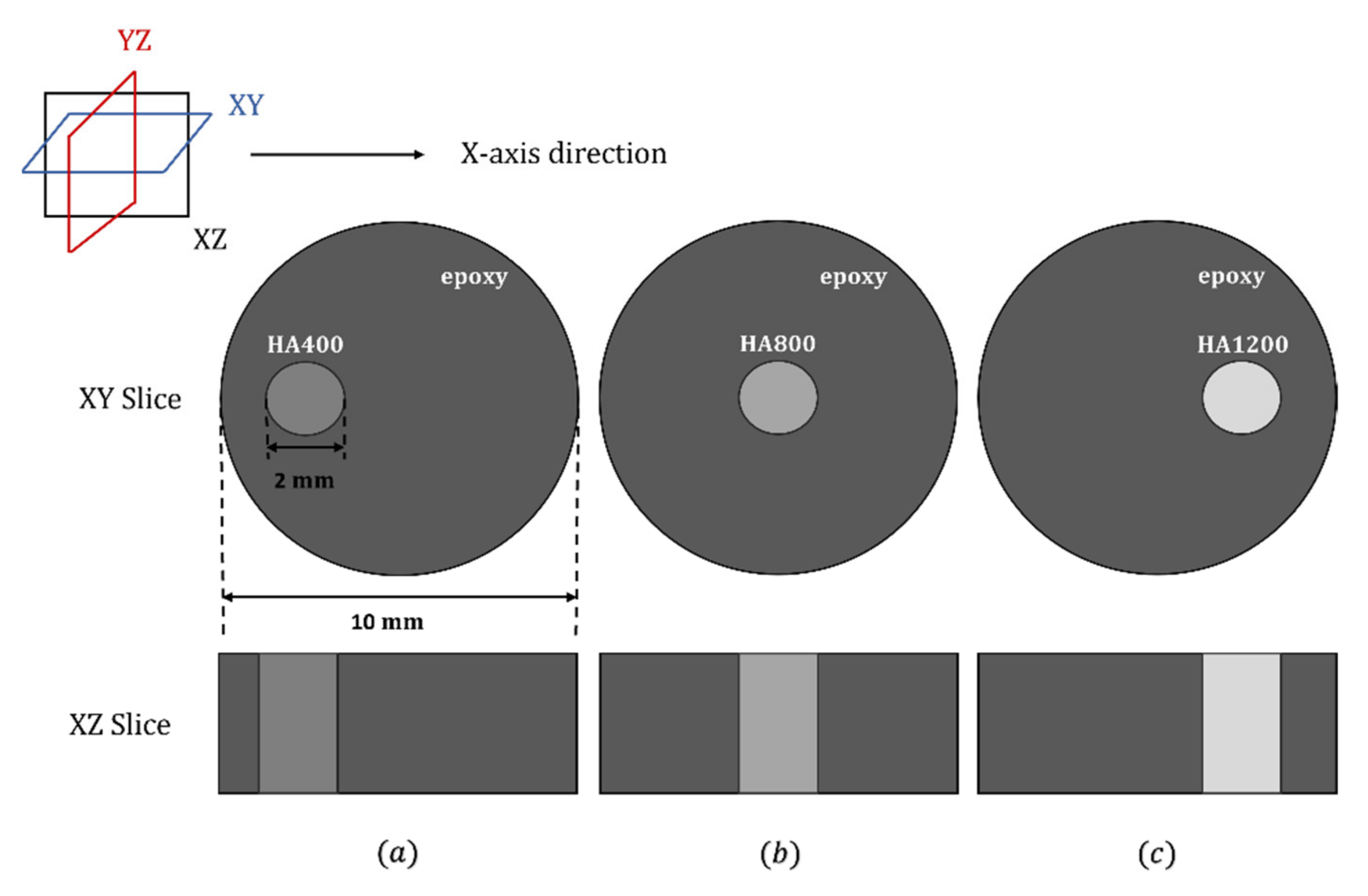

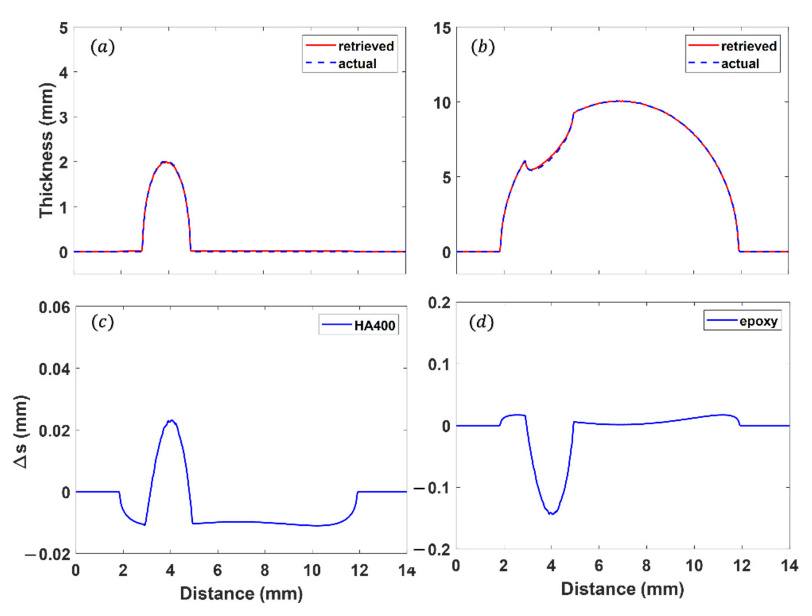
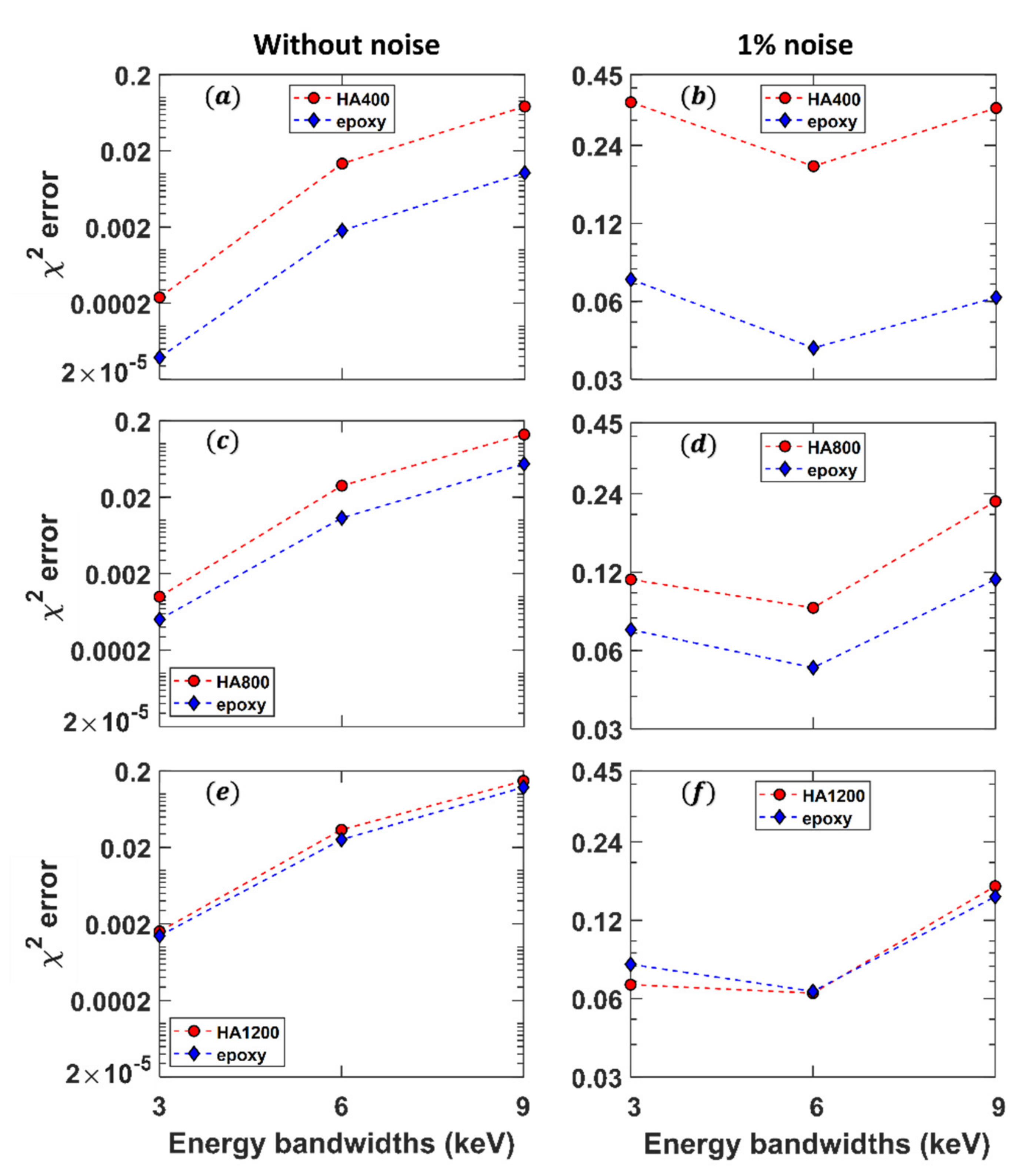
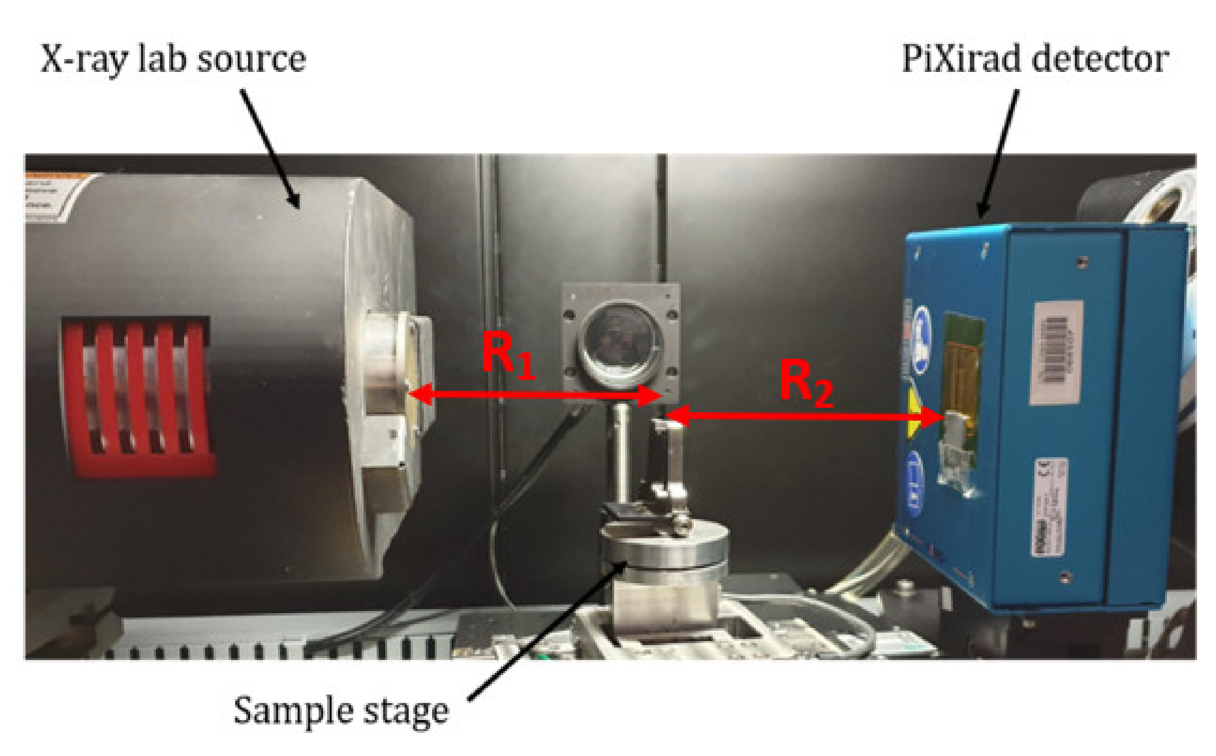


| Material | Density ± 0.02 (g/cm3) | Base (Epoxy Resin) (%) | HA Content (%) |
|---|---|---|---|
| epoxy | 1.13 | 100 | 0 |
| HA400 | 1.39 | 71.31 | 28.87 |
| HA800 | 1.65 | 51.2 | 48.8 |
| HA1200 | 1.9 | 36.51 | 63.49 |
| Threshold Position (keV) | Bandwidth (keV) | ||
|---|---|---|---|
| = 3 | = 6 | = 9 | |
| 16 | 16 | 16 | |
| 19 | 22 | 25 | |
| 27 | 24 | 21 | |
| 30 | 30 | 30 | |
Publisher’s Note: MDPI stays neutral with regard to jurisdictional claims in published maps and institutional affiliations. |
© 2022 by the authors. Licensee MDPI, Basel, Switzerland. This article is an open access article distributed under the terms and conditions of the Creative Commons Attribution (CC BY) license (https://creativecommons.org/licenses/by/4.0/).
Share and Cite
Yokhana, V.S.K.; Arhatari, B.D.; Abbey, B. Materials Separation via the Matrix Method Employing Energy-Discriminating X-ray Detection. Appl. Sci. 2022, 12, 3198. https://doi.org/10.3390/app12063198
Yokhana VSK, Arhatari BD, Abbey B. Materials Separation via the Matrix Method Employing Energy-Discriminating X-ray Detection. Applied Sciences. 2022; 12(6):3198. https://doi.org/10.3390/app12063198
Chicago/Turabian StyleYokhana, Viona S. K., Benedicta D. Arhatari, and Brian Abbey. 2022. "Materials Separation via the Matrix Method Employing Energy-Discriminating X-ray Detection" Applied Sciences 12, no. 6: 3198. https://doi.org/10.3390/app12063198
APA StyleYokhana, V. S. K., Arhatari, B. D., & Abbey, B. (2022). Materials Separation via the Matrix Method Employing Energy-Discriminating X-ray Detection. Applied Sciences, 12(6), 3198. https://doi.org/10.3390/app12063198


