Article

Comparison of the Biological Properties of Plasma-Treated Solution and Solution of Chemical Reagents

by
Tatyana Ivanovna Pavlik
*,
Namik Guseynaga-ogly Gusein-zade
,
Leonid Viktorovich Kolik
and
Nikolay L’vovich Shimanovskii
Pirogov Russian National Research Medical University, 117997 Moscow, Russia
*
Author to whom correspondence should be addressed.
Appl. Sci. 2022, 12(8), 3704; https://doi.org/10.3390/app12083704
Submission received: 14 March 2022 / Revised: 30 March 2022 / Accepted: 1 April 2022 / Published: 7 April 2022
(This article belongs to the Special Issue Low-Temperature Plasma and Its Applications)

Abstract

:
Presently, plasma-treated solutions (PTS) are widely introduced into medicine. Plasma-activated solutions contain various reactive forms of oxygen and nitrogen which provide the desired biological effects. Yet it remains unclear exactly which components of the treated solution are the most important and what the difference is between the plasma-treated solution and a chemically prepared solution composed of the same components. In this work, we show that the chemically prepared mixture of nitrites, nitrates, and hydrogen peroxide with concentrations similar to the plasma-treated solution exerts a fundamentally different effect on a cell culture. The chemically prepared solution has higher cyto- and genotoxicity and causes necrosis, while under the action of the plasma-treated solution, apoptotic processes develop in the cells slowly.

1. Introduction

By the beginning of the 21st century, a new field, plasma medicine, emerged, whose main applications include the sterilization of wounds, coagulation of blood, recovery of destroyed tissues, selective programmed destruction of cancerous cells (apoptosis), diagnostics of skin diseases, cosmetology, etc. [1,2,3,4,5,6,7,8].
In recent years, at the same time as biological objects were treated by CAP, it also became common to study the effect of plasma-treated solutions (PTS) on the vital activity of living organisms. PTS retains the main biological effects of CAP, and it also contains the long-lived reactive nitrogen species (RONS), such as the nitrite (NO2) and nitrate (NO3), as well as hydrogen peroxide (H2O2) and short-lived species of oxygen, hydrogen, and nitrogen generated as a result of their interaction [9,10,11,12]. It was shown that PTS demonstrate anti-tumor activity with regards to several types of cancer, including ovarian cancer, pancreatic cancer, colorectal cancer, and stomach cancer [13,14,15,16].
The advantages of using PTS for treating oncological diseases compared to the direct CAP processing is that they can serve to treat deep tissues as well as surface tumors and tumor beds after their surgical extraction.
Today, a relatively detailed picture of the chemical reactions connected to plasma discharges both in air in the boundary area of the discharge, and in the surface layer of the treated solution has been formed. During the interaction with surrounding gases and surfaces, such discharges bring their molecules into a high-energy active state and help their breaking and creation of radicals [9,17,18]. The radicals created in water and in air interact with each other and with the nonactivated molecules, as a result of which different reactive oxygen and nitrogen species are created. When cold plasma is generated in pure gases (argon, helium, oxygen), oxygen-reactive forms are dominant, while nitrogen compounds almost do not emerge [9,10,11,12,13]. Due to their high chemical activity, in water, the reactive nitrogen and oxygen forms participate in another type of interaction and create stable compounds that exist in the solution during a therapeutically significant time. Mainly, these are nitrites, nitrates, and oxygen peroxide. The nitrites and the nitrates are in kinetic equilibrium with nitrogen oxides of corresponding valence. The long-lived biologically active compounds can decompose into short-lived species during catalysis of the components of the nutritional mediums or biological liquids [12].
In [13,19,20], G. Bauer showed that the hydrogen peroxide and nitrite ions exhibit biological action in cold-plasma-treated solutions. Cancerous cells are protected from the action of hydrogen peroxide by the catalase ferment that is found on their cellular membrane and neutralizes the peroxide. During the chemical interaction of hydrogen peroxide with nitrites, peroxinitrite (ONOO) is created, which becomes a source of singlet oxygen (1O2). The singlet oxygen inhibits the catalase and allows hydrogen peroxide, peroxinitrite, and other short-lived species to penetrate the cell. Normally, the cancerous cells themselves also generate hydrogen peroxide and peroxinitrite, but they are inactivated. Due to the inhibition of catalase, the cell begins to release the oxygen and nitrogen reactive species into the surrounding medium and initiates similar processes in other tumor cells. The creation of HOCl from chlorine and peroxide leads to the peroxidation of lipids and triggers apoptosis. In this process, it is the reactive oxygen and nitrogen species released by the cells themselves that play a major role in the destruction of cancerous cells. The cold-plasma-treated solution only starts the self-destruction of the cells by activating the catalase. Healthy cells are less affected than the tumor ones because they contain other ferments on their membranes and do not generate the reactive oxygen and nitrogen species.
Based on the works of Bauer, we make an assumption in this work that a chemically prepared solution of hydrogen peroxide, nitrites, and nitrates will act on the cells similarly to the cold-plasma-treated solution under the condition that it has similar concentrations of hydrogen peroxide, nitrites, and nitrates.
A similar work was performed in 2016 by Kurake et al. [21], the cold plasma being used to treat the nutritious medium for the cell cultures of the lacteous gland and glioblastoma. Kurake showed that adding nitrites to the nutritious medium increases the survivability of the cells; adding hydrogen peroxide decreases their survivability; adding both nitrites and hydrogen peroxide decreases the survivability more than pure hydrogen peroxide; and adding the cold-plasma-treated nutritious medium decreases their survivability even further.
Turini’s studies showed that T-lymphoblastoid leukemia cells begin to die by apoptosis one day after exposure to cold plasma or the addition of hydrogen peroxide to the nutrient medium. At the same time, the level of toxicity for different modes of cold plasma and hydrogen peroxide did not differ [22].
It has been shown that direct and indirect effects of cold plasma on blood cells do not change the parameters of hemostasis, but they do cause the hemolysis of erythrocytes [23].
The objective of this work is to compare the effect of the physiological solution treated by the direct piezodischarge and a similar solution of nitrites, nitrates, and hydrogen peroxide on the survivability and DNA damage of biological objects: the cell cultures of tumor cells (K562, the myeloblastosis) and cells of conventionally healthy donors (mononuclear leucocytes).

2. Materials and Methods

2.1. Reagents

Physiological solution 0.9% NaCl (SOLOFARM, St Petersburg, Russia, RU-LP-002619), sodium nitrate (Moskhimtorg, Moscow, Russia, GOST 4168-79), sodium nitrite (OOO PraimKhemikalsGrup, Moscow region, Mytishchi, Russia), hydrogen peroxide 34% (Sigma-Aldrich, Saint Louis, MO, USA, 18304-1Lf), Griess reagent (Lenkreativ, St Petersburg, Russia 373131L), luminol (Aldaich Chemistry, Saint Louis, MO, USA, 1002521482), hemoglobin (SERVA, Wichita Falls, TX, USA, 24510), RPMI nutritional medium (OOO NPP PanEko, Moscow, Russia cat. no. C350), amphotericin (OOO NPP PanEko, cat. no. A006), gentamicin (OOO NPP PanEko, cat. no. A011), fetal bovine serum (OOO NPP PanEko, cat. no. FB-1001), PBS (Sigma-Aldrich, P4417-100TAB), Ficoll solution (OOO NPP PanEko, cat. no. R053p), tris-hydroxymethylaminomethane (OOO NPP PanEko, cat. no. X270), hydrochloric acid (Lenkreativ, 170266), sodium chloride (OOO NPP PanEko, cat. no. F085), EDTA (ReaKhim, Izhevsk, Russia, 26040, lot 64), sodium carbonate (Lenkreativ, 130274), dimethyl sulphoxide (DMSO) (Tatkhimpreparaty, Republic of Tatarstan, Kazan, Russia LSR-003126/08), Triton X-100 (VWR (Amresco), Solon, OH, USA, Am-O694-0.1), high-melting agarose (Diaem, Moscow, Russia, D00214), low-melting agarose (Diaem, Moscow, Russia, 55140006), sodium hydroxide (Lachema, Brno, Czech Republic 501891190), SYBR Green I (Sintol, Singapore, 160919), ethanol 96% (PAO Bryntsalov-A, LSR-009126/10), MTT (Thyazolil Blue Tetrazolium Bromide) (Diaem, 38745*), trypan blue dye (OOO NPP PanEko, cat. no. O130), and acridine orange (OOO NPP PanEko, cat. no. O050).

2.2. Method of Preparation of Samples of Cold-Plasma-Treated Solutions

In this work, cold plasma was generated by direct piezodischarge in atmospheric air [24,25]. The cold plasma generator consists of a power source of a single-layer piezoelectric transformer TP-R1, which is a resonant amplifier operating at a frequency of 21 ± 0.3 kHz [26]. The high-voltage (5 ± 1 kV) is generated at the output of the piezotransformer. When the high-voltage contact of the piezotransformer approaches the surface of the solution to the distance of about 0.5 cm, a direct piezodischarge appeared. The average gas temperature during the processing did not exceed 40 °C, and the electron energy could reach several eVs. The average source power is about 8 W [27].
The processing of solutions by direct piezodischarge was conducted over 15 to 180 s. To treat the solution that acted on cells, we used times equal to 30, 60, and 90 s. The treated physiological solution (5 mL) was placed in a 6-cm-diameter Petri dish or a plate well of the same shape. This size of the well showed the best results for the production of ROS [14,28]. The solutions were treated at the temperature of the surrounding medium equal to 20 ± 2 °C and air humidity 70 ± 3%.

2.3. Measurement of the Concentration of Reactive Oxygen and Nitrogen Forms in the Solution

The concentrations of nitrite ions were measured by the photometric method with Griess reagent [29]. The nitrate concentration in the solution was measured using the ALICE 121 nitrate-selective electrode. The hydrogen peroxide concentration was measured by the chemoluminescent method and using the luminol–methemoglobin–hydrogen peroxide system. When this system was applied, the analyzed solution and hydrogen peroxide used for calibration were added to the reagent mixture from luminol, methemoglobin, and a phosphate buffer, after which the chemoluniniscence of the combination was measured.
In addition to these components, the content of ClO in the plasma-treated solution was also analyzed by spectral photometry. The ClO concentration is proportional to the absorption of the solution at wavelength of 292 nm [30].
In addition, pH was measured with the application of the pH-electrode HI1131B.

2.4. Method of Cell Cultivation

The mononuclear leucocytes were derived from the blood of conventionally healthy donors (without diseases of the hemic system nor immunodeficiencies). The comparison was made with the cell line of myeloblastosis K562. All cells were kept in the RPMI nutritional medium with the addition of 10% fetal bovine serum, amphotericin, and gentamycin in a thermostat with increased CO2 concentration (5%) at temperature of 37 °C. In the plate, the analyzed solution was added to all wells with cells at 20% of the volume of nutritional medium, and 24 h later, cell analysis was carried out (The processing time of the solution before it was added to the cell was of 30 min).

2.5. Analysis of Viability and Damage of Cell DNA

The viability of the cells is their ability to maintain a state necessary to carry out specific functions, such as the ability of cells capable of division to realize their mitotic potential. Analysis of the viability of the cells was carried out parallel to the MTT test (analysis of the metabolic activity of the cells) [31] by dyeing them with the trypan blue dye [32].
The type of cell death (apoptosis or necrosis) was defined using the acridine orange dye that details the internal structure of the cell and allows one to estimate the damage to its chromatin and cell membrane [33] (Examples are given in Figure 1).
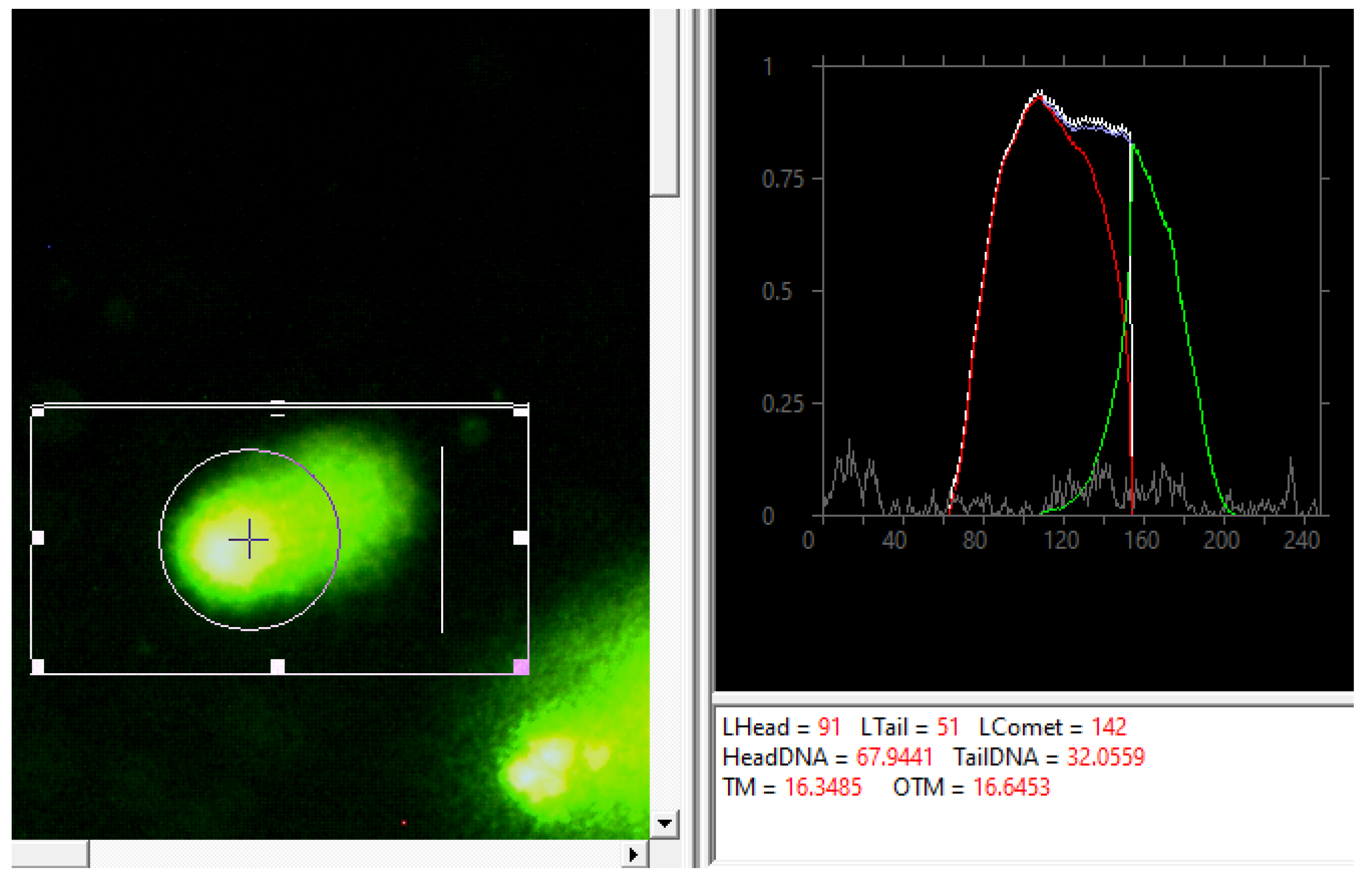
To analyze the DNA damage, the DNA comet method was applied, similarly to [34]. The Casplab_1.2.3b2 software was used to calculate the percentage of the DNA in the “comet tail” (Figure 2).
During electrophoresis, DNA fragments torn away from the main molecule leave the nucleus and create the “comet tail”. Therefore, the percentage of the DNA in the tail was considered proportional to the damage to the DNA.

2.6. Statistical Analysis

For each measurement, an average value and average deviation of results were calculated. To compare the results for the tumor and healthy cells, the nonparametric Mann–Whitney test was applied. Taking into account the correction, the threshold p value that we used in multiple comparison was of 0.05.

3. Results and Their Discussion

As a result of our studies of the processing of physiological solution by the cold plasma of the piezodischarge, we obtained the following dependencies of the increase in concentrations of the long-lived reactive oxygen and nitrogen forms (NO2, NO3, and H2O2) on processing time (Figure 3, Figure 4 and Figure 5). The concentration of nitrites increases in short of cold-plasma treatment times and at times of ~90 s, it reaches saturation, which is caused by their oxidation into nitrates.
The concentration of HOCl in the studied solutions weakly depends on the processing time and does not exceed 5 µM.
Neutral pH decreases to 4.53 already after 30 s of treatment and then remains at this value (Figure 6). A nonlinear decrease in pH indicates that the production of new compounds in the solution improves its buffer properties.
In the studied time range for the processing of the physiological solution by direct piezodischarge, the vitality of myeloblastosis cells decreased, at maximum, to 88% (Figure 7), which not a clinically significant result, and it did not exhibit direct correlation with the processing time. The vitality of mononuclear leucocytes decreased proportionally to the processing time to 63%, at processing time of 90 s.
Apoptosis was recorded as the unique type of cell death in this experiment. At the same time, the results of the MTT test, which estimates the physiological activity of the cells, did not show a dependable difference of all points from the control. To construct the dependence shown in Figure 7, we only used the data obtained by the trypan blue dye, which shows cell vitality directly.
Figure 8 shows the level of damage to the DNA at different processing times by the direct peiozodischarge of the physiological solution added to the nutritional medium. The selectivity of DNA damage of mononuclear leucocytes and mieloblastosis cells is seen when the processing time is 30 s. The maximum level of DNA damage of the mononuclear leucocytes is 51% at processing time of 90 s, and the maximum damage of mieloblastosis cells is 76% at a processing time of 30 s.
Figure 9 shows that the chemically prepared solution of nitrites, nitrates, and hydrogen peroxide has a similar effect on the mononuclear leucocytes and a stronger cytotoxic effect on the mieloblastosis cells than the physiological solution treated by the direct piezodischarge. In this case, there was a correlation between the MTT test and the trypan blue dye.
Although nitrites (NO2) themselves do not have a damaging effect on the cells, and they even increase the vitality of mieloblastosis cells compared to the control, together with hydrogen peroxide (H2O2), they have a stronger cytotoxic effect than hydrogen peroxide alone. The addition of nitrates (NO3) increases the cytotoxic effect further (by 30–50%). This result confirms Bauer’s studies [19,20], which talk about the combined action of the reactive forms of nitrogen and oxygen.
The damage to the DNA of both types of cells under the action of the chemically prepared solution of nitrites, nitrates, and hydrogen peroxide shown in Figure 10 is also more pronounced than under the action of the physiological solution treated by direct piezodischarge; it reaches 100%. Same as the vitality, the level of DNA damage after nitrites are added is the same as in the control and is even lower for some concentrations. When both nitrites and hydrogen peroxide are added, the DNA damage is more severe than one only hydrogen peroxide is added. The addition of the combination of nitrites, nitrates and hydrogen peroxide produces approximately the same result as the addition of nitrites and hydrogen peroxide.
Figure 11 shows that necrosis is prevalent among the types of cell death during the action on the cells of the chemically prepared solution of nitrites, nitrates, and hydrogen peroxide. This can explain the high level of cell death and DNA damage by the rougher action of the chemically prepared solution compared to the solution treated by the piezodischarge.
Thus, both the solution treated by cold plasma and the chemically prepared solution of nitrites, nitrates, and hydrogen peroxide have cyto- and genotoxic effects on cells. However, in the first case, cells die due to apoptosis and in the second, due to necrosis, which demonstrates that the chemically prepared solution is more toxic to the studied cells than the cold-plasma-treated solution. In the therapy of oncological diseases, the ability to cause the selectively programmed death of tumor cells that does not affect healthy cells is a priority.

4. Conclusions

The results of the studies of treating the physiological solution by the direct piezodischarge showed a certain dynamic of the change in the chemical composition of the solution. At the same time, the action in vitro on the cells of the physiological solution treated by direct piezodischarge differs from the action of a similar chemically prepared solution of nitrites, nitrates, and hydrogen peroxide. To initiate the programmed cell death in tumor cells, it is preferable to use the plasma-treated solution, since apoptosis, which is prevalent in this case, is a process regulated by the body, and it selectively kills the tumor cells without damaging the healthy ones. In case of treatment with the chemically prepared solution, necrosis is prevalent, which is an uncontrolled cell death that leads to damaging the surrounding healthy cells and forms an inflammation site.

Author Contributions

Conceptualization, N.G.-o.G.-z. and N.L.S.; methodology, T.I.P. and L.V.K.; software, T.I.P.; validation, T.I.P., L.V.K. and N.G.-o.G.-z.; formal analysis, T.I.P.; investigation, T.I.P.; resources, N.L.S.; data curation, N.L.S.; writing—original draft preparation, T.I.P., L.V.K. and N.G.-o.G.-z.; writing—review and editing, N.G.-o.G.-z. and N.L.S.; visualization, T.I.P. and L.V.K.; supervision, N.G.-o.G.-z. and N.L.S.; project administration, N.L.S. All authors have read and agreed to the published version of the manuscript.

Funding

This work was carried out with the financial support of the Russian Foundation for Basic Research within the framework of scientific project No. 19-02-00378.

Institutional Review Board Statement

Not applicable.

Informed Consent Statement

Not applicable.

Data Availability Statement

Not applicable.

Conflicts of Interest

The authors declare no conflict of interest.

References

  1. Metelmann, H.-R.; Woedtke, T.; Weltmann, K.-D. Comprehensive Clinical Plasma Medicine: Cold Physical Plasma for Medical Application; Springer: Berlin/Heidelberg, Germany, 2018. [Google Scholar]
  2. Cha, S.; Park, Y.-S. Plasma in dentistry. Clin. Plasma Med. 2014, 2, 4–10. [Google Scholar] [CrossRef] [PubMed] [Green Version]
  3. Kim, J.-H.; Lee, M.-A.; Han, G.-J.; Cho, B.-H. Plasma in dentistry: A review of basic concepts and applications in dentistry. Acta Odontol. Scand. 2013, 72, 1–12. [Google Scholar] [CrossRef] [PubMed]
  4. Heinlin, J.; Morfill, G.; Landthaler, M.; Stolz, W.; Isbary, G.; Zimmermann, J.L.; Shimizu, T.; Karrer, S. Plasma medicine: Possible applications in dermatology. JDDG J. der Dtsch. Dermatol. Ges. 2010, 8, 968–976. [Google Scholar] [CrossRef] [PubMed]
  5. Bernhardt, T.; Semmler, M.L.; Schäfer, M.; Bekeschus, S.; Emmert, S.; Boeckmann, L. Plasma Medicine: Applications of Cold Atmospheric Pressure Plasma in Dermatology. Oxid. Med. Cell. Longev. 2019, 2019, 1–10. [Google Scholar] [CrossRef] [PubMed] [Green Version]
  6. Griffin, M.; Palgrave, R.; Baldovino-Medrano, V.G.; Butler, P.E.; Kalaskar, D.M. Argon plasma improves the tissue integration and angiogenesis of subcutaneous implants by modifying surface chemistry and topography. Int. J. Nanomed. 2018, 13, 6123–6141. [Google Scholar] [CrossRef] [Green Version]
  7. Nguyen, L.; Lu, P.; Boehm, D.; Bourke, P.; Gilmore, B.F.; Hickok, N.J.; Freeman, T.A. Cold atmospheric plasma is a viable solution for treating orthopedic infection: A review. Biol. Chem. 2018, 400, 77–86. [Google Scholar] [CrossRef] [PubMed]
  8. Al Dybiat, I.; Baitukha, A.; Pimpie, C.; Kaci, R.; Pocard, M.; Arefi Khonsari, F.; Mirshahi, M. Multi-nanolayer drug delivery using radiofrequency plasma technology. BMC Cancer 2020, 20, 565. [Google Scholar] [CrossRef] [PubMed]
  9. Joslin, J.M.; McCall, J.R.; Bzdek, J.P.; Johnson, D.C.; Hybertson, B.M. Aqueous Plasma Pharmacy: Preparation Methods, Chemistry, and Therapeutic Applications. Plasma Med. 2016, 6, 135–177. [Google Scholar] [CrossRef] [PubMed] [Green Version]
  10. Lindsay, A.; Anderson, C.; Slikboer, E.; Shannon, S.; Graves, D. Momentum, heat, and neutral mass transport in convective atmospheric pressure plasma-liquid systems and implications for aqueous targets. J. Phys. D Appl. Phys. 2015, 48, 424007. [Google Scholar] [CrossRef]
  11. Kovačević, V.V.; Dojčinović, B.P.; Jović, M.; Roglić, G.M.; Obradović, B.M.; Kuraica, M.M. Measurement of reactive species generated by dielectric barrier discharge in direct contact with water in different atmospheres. J. Phys. D Appl. Phys. 2017, 50, 155205. [Google Scholar] [CrossRef]
  12. Liu, D.; Liu, Z.; Chen, C.; Yang, A.; Li, D.; Rong, M.Z.; Chen, H.L.; Kong, M.G. Aqueous reactive species induced by a surface air discharge: Heterogeneous mass transfer and liquid chemistry pathways. Sci. Rep. 2016, 6, 23737. [Google Scholar] [CrossRef] [PubMed]
  13. Bauer, G.; Sersenová, D.; Graves, D.B.; Machala, Z. Dynamics of Singlet Oxygen-Triggered, RONS-Based Apoptosis Induction after Treatment of Tumor Cells with Cold Atmospheric Plasma or Plasma-Activated Medium. Sci. Rep. 2019, 9, 1–34. [Google Scholar] [CrossRef] [Green Version]
  14. Tanaka, H.; Bekeschus, S.; Yan, D.; Hori, M.; Keidar, M.; Laroussi, M. Plasma-Treated Solutions (PTS) in Cancer Therapy. Cancers 2021, 13, 1737. [Google Scholar] [CrossRef] [PubMed]
  15. Solé-Martí, X.; Espona-Noguera, A.; Ginebra, M.-P.; Canal, C. Plasma-Conditioned Liquids as Anticancer Therapies In Vivo: Current State and Future Directions. Cancers 2021, 13, 452. [Google Scholar] [CrossRef] [PubMed]
  16. Azzariti, A.; Iacobazzi, R.M.; Di Fonte, R.; Porcelli, L.; Gristina, R.; Favia, P.; Fracassi, F.; Trizio, I.; Silvestris, N.; Guida, G.; et al. Plasma-activated medium triggers cell death and the presentation of immune activating danger signals in melanoma and pancreatic cancer cells. Sci. Rep. 2019, 9, 4099. [Google Scholar] [CrossRef] [PubMed]
  17. Lu, X.; Naidis, G.V.; Laroussi, M.; Reuter, S.; Graves, D.B.; Ostrikov, K. Reactive species in non-equilibrium atmospheric-pressure plasmas: Generation, transport, and biological effects. Phys. Rep. 2016, 630, 1–84. [Google Scholar] [CrossRef] [Green Version]
  18. Saadati, F.; Mahdikia, H.; Abbaszadeh, H.-A.; Abdollahifar, M.-A.; Khoramgah, M.S.; Shokri, B. Comparison of Direct and Indirect cold atmospheric-pressure plasma methods in the B16F10 melanoma cancer cells treatment. Sci. Rep. 2018, 8, 1–15. [Google Scholar] [CrossRef] [PubMed] [Green Version]
  19. Bauer, G. Intercellular singlet oxygen-mediated bystander signaling triggered by long-lived species of cold atmospheric plasma and plasma-activated medium. Redox Biol. 2019, 26, 101301. [Google Scholar] [CrossRef]
  20. Bauer, G.; Sersenová, D.; Graves, D.B.; Machala, Z. Cold Atmospheric Plasma and Plasma-Activated Medium Trigger RONS-Based Tumor Cell Apoptosis. Sci. Rep. 2019, 9, 1–28. [Google Scholar] [CrossRef]
  21. Kurake, N.; Tanaka, H.; Ishikawa, K.; Kondo, T.; Sekine, M.; Nakamura, K.; Kajiyama, H.; Kikkawa, F.; Mizuno, M.; Hori, M. Cell survival of glioblastoma grown in medium containing hydrogen peroxide and/or nitrite, or in plasma-activated medium. Arch. Biochem. Biophys. 2016, 605, 102–108. [Google Scholar] [CrossRef]
  22. Turrini, E.; Laurita, R.; Stancampiano, A.; Catanzaro, E.; Calcabrini, C.; Maffei, F.; Gherardi, M.; Colombo, V.; Fimognari, C. Cold Atmospheric Plasma Induces Apoptosis and Oxidative Stress Pathway Regulation in T-Lymphoblastoid Leukemia Cells. Oxid. Med. Cell. Longev. 2017, 2017, 1–13. [Google Scholar] [CrossRef] [PubMed]
  23. Golpour, M.; Alimohammadi, M.; Mohseni, A.; Zaboli, E.; Sohbatzadeh, F.; Bekeschus, S.; Rafiei, A. Lack of Adverse Effects of Cold Physical Plasma-Treated Blood from Leukemia Patients: A Proof-of-Concept Study. Appl. Sci. 2021, 12, 128. [Google Scholar] [CrossRef]
  24. Artem’Ev, K.V.; Bogachev, N.N.; Gusein-Zade, N.G.; Dolmatov, T.V.; Kolik, L.V.; Konchekov, E.M.; Andreev, S.E. Study of Characteristics of the Cold Atmospheric Plasma Source Based on a Piezo Transformer. Sov. Phys. J. 2020, 62, 2073–2080. [Google Scholar] [CrossRef]
  25. Korzec, D.; Hoppenthaler, F.; Nettesheim, S. Piezoelectric Direct Discharge: Devices and Applications. Plasma 2020, 4, 1–41. [Google Scholar] [CrossRef]
  26. Kolik, L.V.; Kharchev, N.K.; Borzosekov, V.D.; Malakhov, D.V.; Koncnekov, E.M.; Stepakhin, V.D.; Gusein-zade, N.G.; Bogachev, N.N. Low Temperature Plasma Generator. Patent RU181459U1, 9 April 2018. [Google Scholar]
  27. Artem’Ev, K.; Kolik, L.; Podkovyrov, I.; Sevostyanov, S.; Kosolapov, V.; Meshalkin, V.; Diuldin, M. Generation of plasma-activated water using a direct piezo-discharge: Physicochemical aspects. IOP Conf. Series Earth Environ. Sci. 2019, 390, 012039. [Google Scholar] [CrossRef]
  28. Rowe, W.; Cheng, X.; Ly, L.; Zhuang, T.; Basadonna, G.; Trink, B.; Keidar, M.; Canady, J. The Canady Helios Cold Plasma Scalpel Significantly Decreases Viability in Malignant Solid Tumor Cells in a Dose-Dependent Manner. Plasma 2018, 1, 177–188. [Google Scholar] [CrossRef] [Green Version]
  29. Tsikas, D. Analysis of nitrite and nitrate in biological fluids by assays based on the Griess reaction: Appraisal of the Griess reaction in the l-arginine/nitric oxide area of research. J. Chromatogr. B Analyt Technol. Biomed Life Sci. 2007, 851, 51–70. [Google Scholar] [CrossRef]
  30. Wang, L.; Bassiri, M.; Najafi, R.; Najafi, K.; Yang, J.; Khosrovi, B.; Hwong, W.; Barati, E.; Belisle, B.; Celeri, C.; et al. Hypochlorous acid as a potential wound care agent: Part I. Stabilized hypochlorous acid: A component of the inorganic armamentarium of innate immunity. J. Burns Wounds 2007, 6, e5. [Google Scholar]
  31. Mosmann, T. Rapid colorimetric assay for cellular growth and survival: Application to proliferation and cytotoxicity assays. J. Immunol. Methods 1983, 65, 55–63. [Google Scholar] [CrossRef]
  32. Stoddart, M.J. Cell viability assays: Introduction. Methods Mol. Biol. 2011, 740, 1–6. [Google Scholar]
  33. Thomé, M.P.; Filippi-Chiela, E.C.; Villodre, E.S.; Migliavaca, C.B.; Onzi, G.R.; Felipe, K.B.; Lenz, G. Ratiometric analysis of acridine orange staining in the study of acidic organelles and autophagy. J. Cell Sci. 2016, 129, 4622–4632. [Google Scholar] [CrossRef] [PubMed] [Green Version]
  34. Collins, A.R. The Comet Assay for DNA Damage and Repair. Mol. Biotechnol. 2004, 26, 249–261. [Google Scholar] [CrossRef]
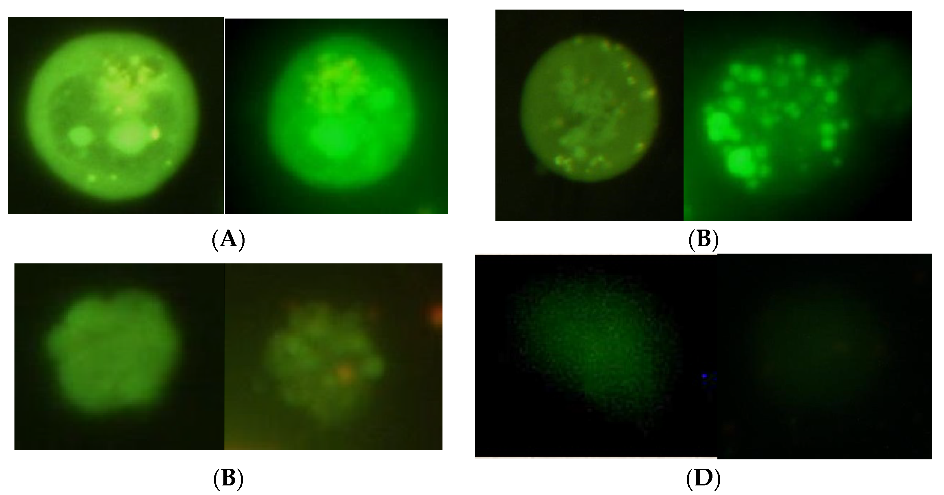
Figure 1. External view of live and dead cells in microslides dyed by acridine orange ((A)-living cell, (B)-apoptosis (early stage), (C)-apoptosis (late stage), (D)-necrosis).
Applsci 12 03704 g001
Figure 2. “DNA-comet tail” length calculation using the Casplab_1.2.3b2 software.
Applsci 12 03704 g002
Figure 3. The dependence of nitrite concentration in the physiological solution on the time of processing by direct piezodischarge. The distance between the piezotransformer and the solution surface was 0.5 mm. The temperature of the surrounding medium was 20 ± 2 °C, and the air humidity was 70 ± 3%. In detail, the experimental conditions are described in the Section 2.
Applsci 12 03704 g003
Figure 4. Dependence of oxygen peroxide concentration in the physiological solution on the processing time by direct piezodischarge. The distance between the piezotransformer and the solution surface was 0.5 mm. The temperature of the surrounding medium was 20 ± 2 °C, and the air humidity was 70 ± 3%. In detail, the experimental conditions are described in the Section 2.
Applsci 12 03704 g004
Figure 5. Dependence of nitrate concentration in the physiological solution on the time of processing by direct piezodischarge. The distance between the piezotransformer and the solution surface was 0.5 mm. The temperature of the surrounding medium was 20 ± 2 °C, and the air humidity was 70 ± 3%. In detail, the experimental conditions are described in the Section 2.
Applsci 12 03704 g005
Figure 6. Changes in the pH of the physiological saline after exposure to piezoelectric direct discharge of variable duration. An asterisk * indicates a statistically significant difference (p < 0.05) from the control.
Applsci 12 03704 g006
Figure 7. Dependence of the level of vitality of the healthy (blue diamonds) and tumor (red squares) cells on the time of processing by the direct piezodischarge of the physiological solution added to the nutritional medium. The cells were kept in the RPMI nutritional medium with the addition of 10% fetal bovine serum, amphotericin, and gentamycin, in a thermostat with increased CO2 concentration (5%) at temperature of 37 °C. In detail, the experimental conditions are described in the Section 2.
Applsci 12 03704 g007
Figure 8. Dependence of the level of damage to the DNA cells on the time of processing by the direct piezodischarge of the physiological solution added to the nutritional medium. The cells were kept in the RPMI nutritional medium with the addition of 10% fetal bovine serum, amphotericin, and gentamycin, in a thermostat with increased CO2 concentration (5%) at temperature of 37 °C. In detail, the experimental conditions are described in the Section 2.
Applsci 12 03704 g008
Figure 9. Dependence of the level of vitality of (A) conventionally healthy and (B) tumor (mieloblastosis) cells on the concentration of nitrites, nitrates, and hydrogen peroxide added to the nutritional medium. Concentrations 1–3 correspond to those in the physiological solution treated by the direct piezodischarge during 30, 60, and 90 s, respectively (1: H2O2—2.08 µM, NO2—0.058 mM, NO3—0.286 mM; 2: H2O2—6.91 µM, NO2—0.091 mM, NO3—0.378 mM; 3: H2O2—10.08 µM; NO2—0.103 mM, NO3—0.725 mM). An asterisk * indicates a statistically significant difference (p < 0.05) from the control, an asterisk above the frame indicates a statistically significant difference (p < 0.05) between different experimental samples. The experimental conditions are described in the Section 2.
Applsci 12 03704 g009aApplsci 12 03704 g009b
Figure 10. Dependence of the level of DNA damage of (A) conventionally healthy and (B) tumor (mieloblastosis) cells on the concentration of nitrites, nitrates, and hydrogen peroxide added to the nutritional medium. Concentrations 1–3 correspond to those in the physiological solution treated by the direct piezodischarge during 30, 60, and 90 s, respectively (1: H2O2—2.08 µM, NO2—0.058 mM, NO3—0.286 mM; 2: H2O2—6.91 µM, NO2—0.091 mM, NO3—0.378 mM; 3: H2O2–10.08 µM; NO2—0.103 mM, NO3—0.725 mM). An asterisk * indicates a statistically significant difference (p < 0.05) from the control, an asterisk above the frame indicates a statistically significant difference (p < 0.05) between different experimental samples. The experimental conditions are described in the Section 2.
Applsci 12 03704 g010
Figure 11. Distribution of types of death of (A) conventionally healthy and (B) tumor (mieloblastosis) cells at different concentrations of nitrites, nitrates, and hydrogen peroxide added to the nutritional medium. Concentrations 1–3 correspond to those in the physiological solution treated by the direct piezodischarge during 30, 60, and 90 s, respectively (1: H2O2—2.08 µM, NO2—0.058 mM, NO3—0.286 mM; 2: H2O2—6.91 µM, NO2—0.091 mM, NO3—0.378 mM; 3: H2O2–10.08 µM; NO2—0.103 mM, NO3—0.725 mM). An asterisk * indicates a statistically significant difference (p < 0.05) from the control, an asterisk above the frame indicates a statistically significant difference (p < 0.05) between different experimental samples. The experimental conditions are described in the Section 2.
Applsci 12 03704 g011
Publisher’s Note: MDPI stays neutral with regard to jurisdictional claims in published maps and institutional affiliations.