Article

Exogenous Selenium Treatment Promotes Glucosinolate and Glucoraphanin Accumulation in Broccoli by Activating Their Biosynthesis and Transport Pathways

1
College of Horticulture, Hunan Agricultural University, Changsha 410128, China
2
Engineering Research Center for Horticultural Crop Germplasm Creation and New Variety Breeding, Ministry of Education, Changsha 410128, China
3
Key Laboratory for Vegetable Biology of Hunan Province, Changsha 410128, China
*
Author to whom correspondence should be addressed.
Appl. Sci. 2022, 12(9), 4101; https://doi.org/10.3390/app12094101
Submission received: 2 April 2022 / Revised: 10 April 2022 / Accepted: 14 April 2022 / Published: 19 April 2022
(This article belongs to the Special Issue Genetics and Breeding of Broccoli)

Abstract

:
Supplementation using selenium (Se) on plants is an effective and widely used approach. It can not only be converted to more Se rich compounds but promote the accumulation of glucosinolates (GSLs) with anti-carcinogenic properties. However, the molecular mechanism of Se in regulating GSLs synthesis remains unclear. In the present study, we analyzed the effects of Se treatment (50 μM sodium selenite) on GSLs, glucoraphanin (4MSOB), and sulforaphane compounds in broccoli tissues. The transcript levels of genes involved in sulfur absorption and transport, GSLs biosynthesis, translocation, and degradation pathways were also evaluated. The study showed that Se treatment remarkably promoted the accumulation of total sulfur and total Se contents and increased Trp-derived GSLs levels in roots by 2 times. The 4MSOB concentration and sulforaphane content in fresh leaves was increased by 67% and 30% after Se treatment, respectively. For genes expressions, some genes involved in sulfate uptake and transporters, GSLs biosynthesis, and transporters were induced strongly upon Se exposure. Results revealed that exogenous Se treatment promotes the overaccumulation of GSLs and 4MSOB content in broccoli by activating the transcript levels of genes involved in sulfur absorption, GSLs biosynthesis, and translocation pathways.

1. Introduction

Broccoli (Brassica oleracea var. italica Plenck) is rich in a variety of nutrients, including minerals, vitamins, and dietary fiber. It is the main source of anti-carcinogenic phytochemicals, such as polyphenols, sulfur- and nitrogen-rich compounds; GSLs [1,2]. Isothiocyanates (ITCs) are formed in Brassica vegetables after tissue disruption because their precursors, GSLs, come into contact with endogenous enzymes known as myrosinases (MYRs). This disruption-caused GSLs degradation is known as “autolysis” [3]. Two main groups of GSLs are found in broccoli, including Met-derived GSLs and Trp-derived GSLs. One Met-derived GSL is 4-(methylsulfinyl)butyl GSL (4MSOB); the corresponding ITC 4-(methylsulfinyl)butyl ITC is commonly known as sulforaphane and has been particularly studied in nutritional science [4]. Among known effects are effective detoxification and anti-cancer properties [5,6]. The most simple Trp-derived GSL is indol-3-ylmethyl GSL (I3M). Chemically, the Trp-derived GSLs do not form stable ITCs but various other products. Indeed, the two groups of GSLs show some differentiation in biological properties. For example, sulforaphane confers pathogen resistance to plants by inhibiting bacterial virulence [7]. Likewise, Trp-derived GSLs play roles in defending against herbivore attack and pathogen invasion [8]. Concerning biosynthesis, the groups not only have different amino acid precursors but also differ in the actual biosynthetic enzymes and their regulation [4,9,10] (Figure S1). Hence, in physiological studies including this, the two groups are generally discussed separately. Some transcription factors, including MYB28, MYB29, and MYB76, are involved in regulating Met-derived GSLs biosynthesis [11,12]. Other transcription factors, such as MYB34, MYB51, and MYB122 genes, are responsible for accumulating Trp-derived GSLs [13]. As specialized metabolites, GSLs contents and degradation products are susceptible to external stimuli [14,15,16]. Among them, supplementation using inorganic Se on broccoli is considered to be an effective and widely used approach because it can not only affect GSLs accumulation but produce more Se-rich compounds with anti-carcinogenic properties [17,18].
Se is an essential micronutrient for many living organisms, which plays a crucial role in enhancing human immunity, reducing cancer risks, mitigating heavy metals toxicity, and alleviating abiotic stress [19,20,21,22]. The Recommended Dietary Allowance (RDA) of Se consumption for adult males and females is 55–75 μg/day. When the daily consumed level is low, 40 μg or over 400 μg, people will be suffered from corresponding dietary deficiency and toxicity symptoms, respectively [23]. Se uptake in humans primarily depends on the ingestion of Se-enriched fruits, vegetables, and crops [24,25]. Se biofortification at an appropriate concentration on plants is an effective and convenient strategy for producing more Se-containing edible foods [26,27]. Besides, Se supplementation can boost the accumulation of plants’ secondary metabolites with benefits for human health [28], such as capsaicin in peppers [29], tocopherol and total phenols in mustards [30], phenolic acids and anthocyanins in lettuces [31], flavonoids and chalcone in tomatoes [32].
Se biofertilization on plants may also affect the production of GLSs, a class of sulfur-containing specialized metabolites [33,34,35]. Due to the similarities in chemical properties and physical structures between Se and sulfur, Se competes with the same sulfate transporters (SULTRs) and sulfur assimilation genes with sulfur in plants [36,37]. Previous studies have concluded that Se application leads to a reduction in GSLs contents, especially Met-derived GSLs, including ITCs and sulforaphane levels [38]. Similar effects of Se treatment on primary metabolites and total GSLs contents were reported in broccoli sprouts [39]. Conversely, recent evidence suggested that Se supplementation can increase Met-derived GSLs; 4MSOB and Trp-derived GSLs; I3M concentrations of radish roots by eliciting the expression abundances of sulfate transport (SULTRs) and sulfur assimilation genes (APSKs) [40,41]. The same conclusion was shown in cabbages [42].
Previous studies have demonstrated that Se treatment boosts the enrichment of selenocompounds and the differential accumulation of GSLs in broccoli tissues [43,44]. However, the mechanism of Se in regulating GSLs production has not been well elucidated. In this study, the effects of Se treatment on the total sulfur and Se contents, GSLs, 4MSOB, and sulforaphane levels in different tissues of broccoli, especially the effects on GSLs biosynthesis and translocation pathways, were investigated. The transcription levels of genes involved in the sulfate transport, GSLs biosynthesis, translocation, and degradation pathways were examined to preliminarily figure out the molecular mechanism of the response of broccoli to Se stimuli. These results will provide a useful guideline for the cultivation of broccoli with more nutritional value.

2. Materials and Methods

2.1. Plant Materials and Treatments

The broccoli (Brassica oleracea var. italica Plenck) cultivars ‘XiangLv No. 3′ were cultivated in greenhouse conditions (a 16-h-day/8-h-night cycle, 22 °C temperature, 80% relative humidity) at Hunan Agricultural University, Changsha, Hunan province, China (28°18′ N, 113°03′ E). The seedlings with four true leaves were transplanted into 30 L hydroponic tanks containing 1/2 Hoagland’s nutrient solution until the plants had eight true leaves. The treatment was applied 4 weeks after transplantation, which consisted of two experiments: plants treated with 50 μM sodium selenite (final concentration in the hydroponic tank) and the control group treated with water. Adding treatment was repeated once every 5 days for 2 weeks. All plants were harvested after 15 days of treatment. Senescent leaves (rosette leaves closest to the roots), fresh leaves (fully expanded 6th to 8th functional leaves), and roots were immediately separated. The roots were washed with distilled water twice and dried with filter papers. A portion of fresh samples was used to analyze the MYRs activity. The remaining samples were immediately frozen in liquid nitrogen. A few samples were stored at −80 °C for quantitative real-time polymerase chain reaction (RT-qPCR) analysis. The rest of the samples were lyophilized and ground into powder to measure total sulfur and Se contents, GSLs contents, and sulforaphane level after autolysis.

2.2. Determination of Total Sulfur and Se Contents

Approximately 2.0 g of freeze-dried samples were placed in glass digestion tubes with 10 mL of an acid mixture (HNO3:HClO4, 4:1) for acid digestion at 120 °C, then transferred to colorimetric tubes and diluted to a volume of 25 mL. Total sulfur and Se contents were measured with a Varian ICP-OES Analyzer (model Vista-MPX, Varian, PaloAlto, CA, USA) at 182 nm and 196 nm, respectively. The unit of total sulfur and Se content is expressed as g/kg DW and μg/g DW, respectively.

2.3. Determination of GSLs Composition and Content

Total GSLs were extracted and quantified using described methods with minor modifications [45,46]. Overall, 0.5 g broccoli samples were mixed with 0.1 mL 5 mM standards; Sinigrin (PREN, Sigma Aldrich, ≥99% purity, Merck, Shanghai, China). The mixtures were extracted with 7 mL 70% methanol at 75 °C for 20 min. After cooling, 2 mL barium acetate was added and centrifuged at 8000 r/min to collect the supernatants. The precipitations were reextracted with 4 mL of 70% methanol and 0.5 mL of barium acetate. The supernatants collected twice were fixed to 10 mL with 70% methanol and isolated by anion-exchange chromatography using a DEAE Sephadex A-25 column (Solarbio, Beijing, China). The column was incubated at 37 °C for 16 h to allow desulfation, then eluted with 2 mL of deionized water, and collected into 2 mL sample bottles through a 0.22 μm filter membrane.
DesulfoGSLs were separated using a UHPLC-MS system (Waters Acquity TQD, Waters, Shanghai, China), which were analyzed by both UV-detection at 229 nm (for quantification) and MS-detection (for identity confirmation). The detailed procedures were as described by previous studies [47,48]. The chemical name, precursors, and GSLs abbreviations are listed in Table 1. The molecular weight, retention time, and correction coefficient of each DesulfoGSL are shown in Table S2. The sinigrin was used as the reference standard to calculate the GSLs contents per ISO 9167-12. The recovery rate of the internal standard is shown in Table S3. The calculation formula of GSLs quantification is shown in Table S4, and the unit is expressed as μmol/g DW.

2.4. Determination of MYR Activity

The method of MYR extraction was based on Lim et al. with some modifications [49]. Fresh samples (0.1 g) were weighed and homogenized with 1 mL of extraction buffers (10 mM potassium phosphate containing 3 mM Dithiothreitol, 1 mM Ethylenediaminetetraacetic acid, and 5% glycerol, pH 7.2) (Coolaber, Beijing, China). The homogenization buffers were centrifuged to collect the supernatants.
Glucose Assay Kit (Abbkine, Wuhan, China) was used to detect the glucose production from PREN catalyzed by the MYR. 100 μL of crude extracts were reacted with 200 μL of potassium phosphate buffers (33 mM, pH 6.5, with 2 mM PREN). Another 100 μL of crude extracts were blended with 200 μL buffers without PREN. All samples were incubated at 37 °C for 15 min, the reaction was stopped at 95 °C for 10 min, and then the reaction was performed according to the manufacturer’s protocol. The absorbance of the reaction solutions was measured by a Microplate Reader at 630 nm. The myrosinase activity is expressed as μmol glucose formed per g fresh sample per minute (μmol/g min FW).

2.5. Determination of Sulforaphane Level

The sulforaphane extraction and detection methods were as described by a previous study [50]. Approximately 0.5 g of freeze-dried samples were added with 6 mL of distilled water, hydrolyzed at 25 °C for 4 h, then added to 30 mL of dichloromethane for extraction. The mixtures were filtrated with a funnel and dried using rotary evaporation, eluted with 2 mL of acetonitrile, then collected into a sample bottle through a 0.45 μm filter membrane.
High-performance liquid chromatography (HPLC; Agilent 1100 Series, Agilent, Beijing, China) using a C18 reversed-phase column was used to measure the sulforaphane level. The mobile phase contained the water phase and acetonitrile phase, and the procedures were as follows: the column temperature was set as 30 ℃, the sample load was 10 μL, the flow rate was 1 mL/min, and the wavelength was 240 nm. The standard curve was obtained by purchased sulforaphane solution (Sigma Aldrich, USA, ≥99% purity) and was used to quantify the sulforaphane content of samples. The unit is expressed as μmol/g DW.

2.6. RNA Extraction and RT-qPCR Analysis

Total RNA was extracted using a Polysaccharides and Polyphenolic-rich RNAprep Pure Plant Kit (Tiangen, Beijing, China) following the manufacturer’s instructions. Then, 1.5 μg of total RNA was synthesized into cDNA using the All-In-One 5X RT Master Mix with gDNA remover (Abm, Zhenjiang, China). We performed the RT-qPCR analysis using the SYBR Green Premix Pro Taq HS qPCR kit (AG, Wuhan, China). All primers used for RT-qPCR analysis were designed by the Primer-BLAST online website (https://www.ncbi.nlm.nih.gov/tools/primer-blast/, accessed on 8 June 2021), and the Actin2 broccoli gene was used as an internal reference gene for normalization (Table S1). The relative expression level was calculated by a 2−ΔΔCT method as described in the BioRad Real-time PCR Application Guide. The expression abundance in broccoli roots from the control treatment was defined as “1”.

2.7. Statistical Analysis

Experimental data were processed with GraphPad Prism 5. All data represented the mean ± standard deviation (SD) of four biological replicates. Data were statistically analyzed by one-way ANOVA with Duncan’s multiple comparison test using the SPSS 20.0 software (SPSS Inc., Chicago, IL, USA). Differences at p < 0.05 were regarded as significant.

3. Results

3.1. Effect of Se on Total Sulfur and Se Contents

In the absence of Se supply, the total sulfur content in fresh leaves was higher than in roots and senescent leaves. In comparison with the normal conditions, total sulfur content considerably increased by 28%, up to 21 g/kg DW in roots with 50 μM Na2SeO3 treatment via the hydroponic method. However, the Se treatment remarkably decreased total sulfur concentration in senescent leaves and fresh leaves by 30% and 17%, respectively, compared to the control (Figure 1a). Total Se content in broccoli tissues was not detected under the control conditions, while it was dramatically increased in all tissues of broccoli by Se supplementation. When broccoli roots are exposed to Se conditions, the total Se content in broccoli roots is higher than in leaves (Figure 1b).

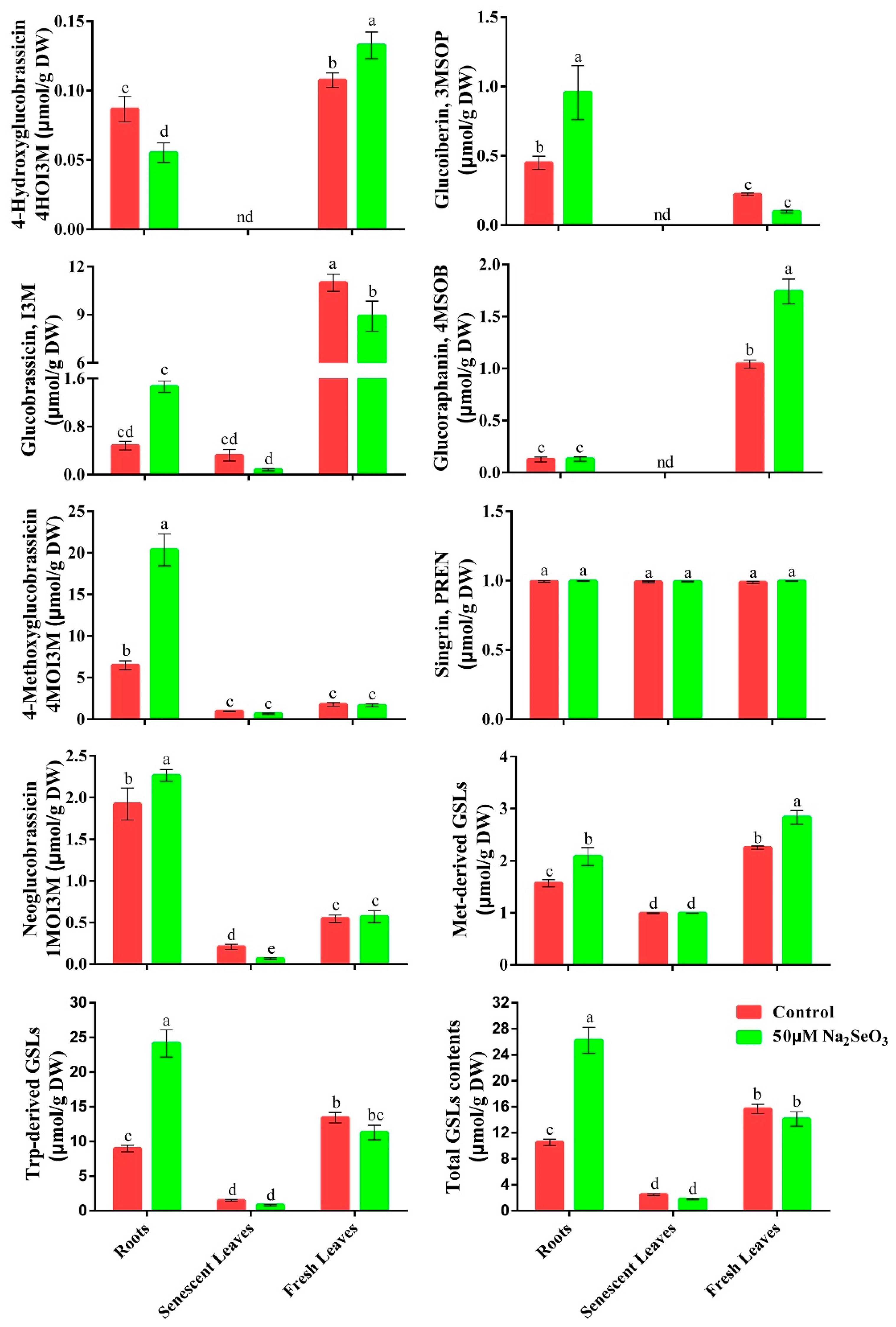
3.2. Effect of Se on GSLs Contents

We detected three Met-derived GSLs and four Trp-derived GSLs in fresh leaves and roots of young broccoli plants (Table 1). Only three Trp-derived GSLs (excluding 4-Hydroxyindol-3-ylmethyl GSL, 4HOI3M) were identified in senescent leaves. Under the normal conditions, GSLs content was the highest in fresh leaves, followed by those in roots and senescent leaves. Indol-3-ylmethyl GSL (I3M) was the main GSL in fresh leaves, accounting for 71% of total GSLs content. 4-Methoxyindol-3-ylmethyl GSL (4MOI3M) was the predominant GSL in broccoli roots, comprising about 62% of total GSLs level. Total GSLs contents in the aerial tissues were not affected by Se treatment but increased in roots by 3 folds. Se treatment significantly enhanced Met-derived GSLs content in fresh leaves and roots, particularly the 4MSOB and 3-(Methylsulfinyl)propyl GSL (3MSOP) contents, increased by 21% and 33%, respectively. 4MOI3M content was strongly accumulated in broccoli roots and increased from 6 μmol/g DW to 20 μmol/g DW after Se treatment. Trp-derived GSLs content in leaves was not affected by Se supplementation, except for the reduction of I3M content in fresh leaves (Figure 2).

3.3. Effects of Se on MYR Activity and Sulforaphane Level after Autolysis

The differential partitioning of MYR activity among the broccoli tissues upon Se exposure is shown in Figure 3a. Compared with the control treatment, MYR activity in roots remarkably increased by 96%, whereas those in fresh leaves and senescent leaves had no remarkable difference after the 50 μM Na2SeO3 treatment. As shown in Figure 3b, the sulforaphane level after autolysis in fresh leaves was increased by 13% when the broccoli roots were exposed to Se treatment. However, sulforaphane level was barely detectable in roots and senescent leaves (Figure 2 and Figure 3a).

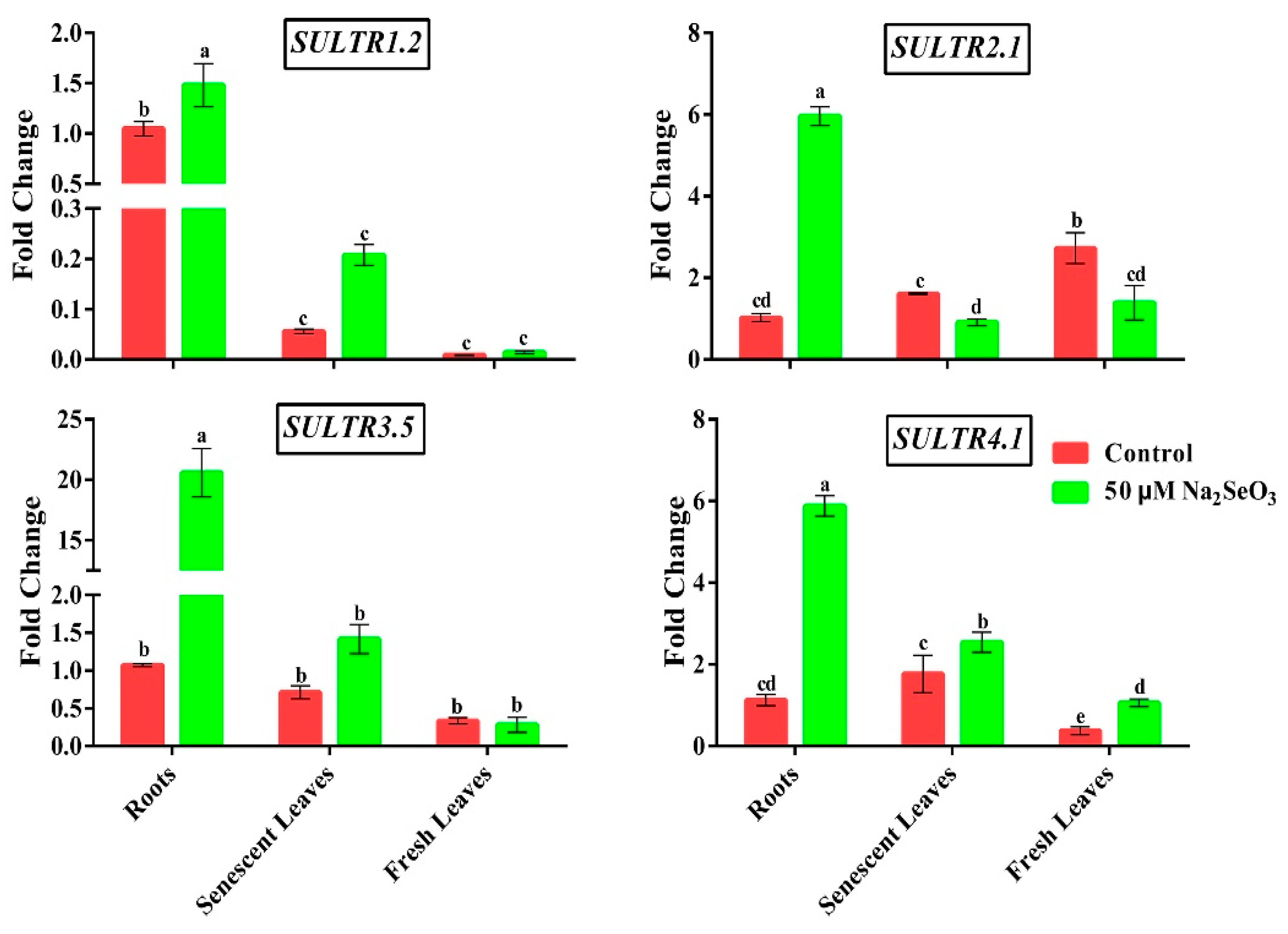
3.4. Effect of Se on the Transcript Levels of Genes Related to Sulfate Uptake and Transport

Compared with the control treatment, the transcript levels of SULTR1.2, SULTR2.1, SULTR3.5, and SULTR4.1 genes in roots increased by almost 1, 6, 19, and 5 folds, respectively, under the Se treatment. Furthermore, the transcript level of the SULTR4.1 gene was up-regulated strongly in all tissues when broccoli seedlings were exposed to Se stimuli. On the contrary, the expression abundance of the SULTR2.1 gene in fresh leaves and senescent leaves was decreased by 49% and 43%, respectively, after Se treatment (Figure 4).

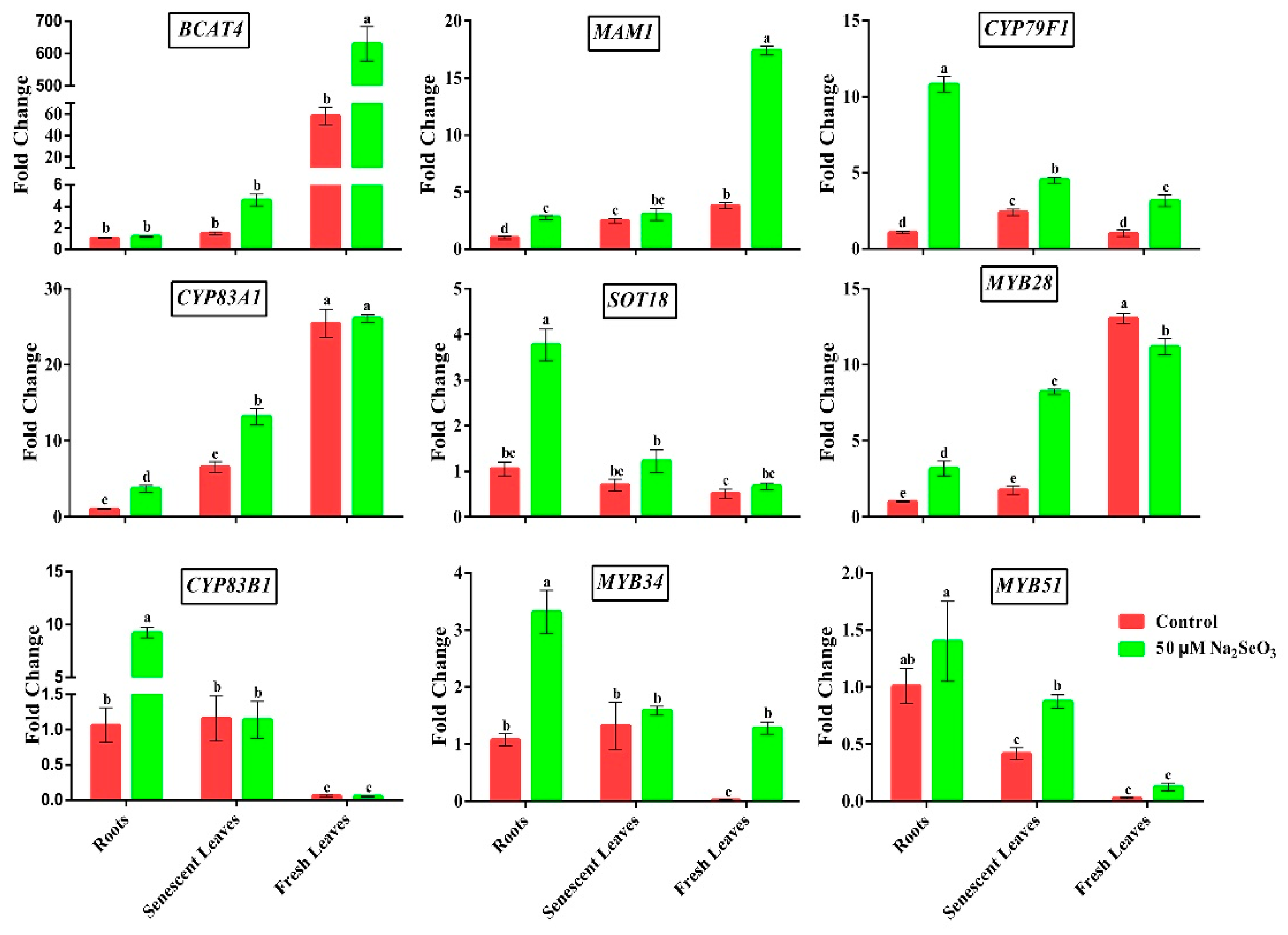
3.5. Effect of Se on the Transcript Levels of Genes Related to GSLs Biosynthesis

The expression patterns of GSLs biosynthesis genes were tissue-specific. Most genes involved in Met-derived GSLs biosynthesis, including BCAT4, MAM1, CYP83A1, and MYB28 genes were primarily expressed in fresh leaves, while others (SOT18 gene) were mainly expressed in broccoli roots. The transcript levels of BCAT4, MAM1, and CYP79F1 genes in fresh leaves were strongly induced by Se treatment, while the expression of the MYB28 gene was suppressed. In addition, Se-induced up-regulation of MAM1, CYP79F1, CYP83A1, SOT18, and MYB28 expression abundance occurred in broccoli roots (Figure 5).
The expression abundance of Trp-derived GSLs biosynthesis genes (CYP83B1 and MYB34) in broccoli roots increased significantly after 50 μM Na2SeO3 treatment. The transcript level of MYB51 in senescent leaves was substantially activated by Se treatment. Furthermore, the expression abundances of these genes (CYP83B1, MYB34, and MYB51) in roots were higher than those in broccoli leaves (Figure 5).

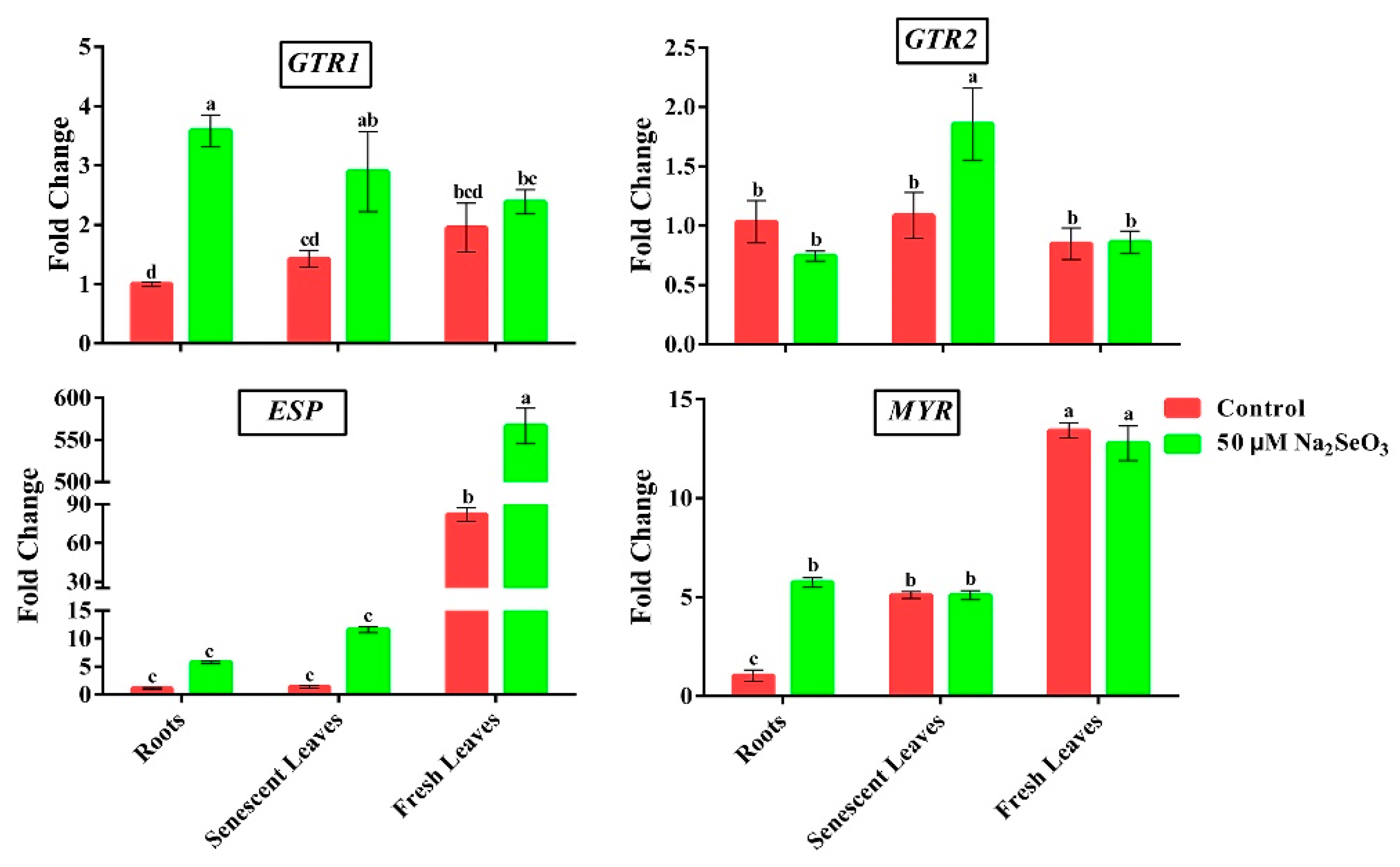
3.6. Effect of Se on the Transcript Levels of GSLs Transporters and 4MSOB Degradation Genes

As shown in Figure 6, the expression abundance of the GTR1 gene in broccoli roots and senescent leaves was significantly stimulated by Se treatment. Besides, the transcript level of the GTR2 gene was only induced in senescent leaves after Se treatment.
The ESP and MYR genes were mostly expressed in fresh leaves. Compared with control conditions, the expression abundance of the ESP gene in fresh leaves was increased by 7 folds with Se supplementation. However, Se-induced up-regulation of MYR expression abundance only occurred in broccoli roots (Figure 6).

4. Discussion

4.1. Se-Induced Total Sulfur and GSLs Accumulation in Broccoli Roots

GSLs are a diverse group of nitrogen- and sulfur-rich specialized metabolites whose contents represent up to 30% of the total sulfur content of plant tissues [51]. Thus, GSLs concentrations may depend on the sulfur status of the entire plant [44]. In the study, the sulfur content in fresh leaves was significantly higher than in broccoli roots and senescent leaves under the control treatment. This result is in accordance with a previous study, which showed that the young growing tissues are the main sulfur sinks, and their sulfur content exhibits a downward trend after full expansion [52]. Furthermore, GSLs contents in fresh leaves were the highest, followed by that in broccoli roots and senescent leaves under the normal conditions. The same results were shown in Chinese cabbage [53]. Broccoli is a typical Selenium (Se) and Sulfur (S) accumulator, while both Se and S belong to chalcogen elements with similar chemical characteristics [54]. Presumably, Se supplementation could influence sulfur uptake and GSLs metabolism in plants. Based on results, exogenous Se treatment considerably increases the sulfur and GSLs contents in broccoli roots. This result is similar to Se addition in boosting the accumulation of total sulfur and GSLs contents in Brassica oleracea [38] and radish roots [41]. Accumulation of the GSLs contents is mainly dependent on the sulfur status of plants and transcriptional activity of GSLs biosynthesis genes [17,34]. The study showed that the sulfur content in broccoli roots was greatly induced through activating SULTRs expressions by Se treatment. However, the sulfur content of broccoli leaves was decreased under Se treatment. This result may be due to the substantial repression of SULTR2.1 expression in fresh leaves and senescent leaves by Se treatment. The role of SULTR2.1 is mainly responsible for the translocation of the sulfur from the roots to aboveground tissues [55,56]. Consequently, exogenous Se treatment promotes the overaccumulation of sulfur and GSLs contents in broccoli roots, while probably inhibiting the partitioning and translocation of the sulfur from broccoli roots to the shoots. On the other hand, the activation of gene expression, such as MAM1, BCAT4, and CYP79F1, was the consequence of an increase in GSLs accumulation in the Se-treated cabbage [42]. Se treatment could promote GSLs production in broccoli florets by stimulating the transcriptional activity of ST5b (SOT18) gene, which was involved in the Met-derived GSLs biosynthesis pathway [47]. In addition, the expression of CYP79B2 and CYP83B1 genes were induced by Se treatment. This led to an increase of Trp-derived GSLs contents in radish seedlings, especially at 7 and 14 days after germination (DAG) [40]. Results revealed that the Se-induced increase of GSLs contents in broccoli roots was largely attributed to the up-regulation expressions of the GSLs biosynthesis genes.

4.2. Induction of 4MSOB Accumulation in Young Leaves by Se through the GSLs Transporters

Within-leaf distribution of defense compounds, such as GSLs and 4MSOB, is plants’ strategy to defend against insect herbivory, whose accumulation in the tissue resulted from in-situ synthesis and long-distance transportation [57]. The study showed that exogenous Se treatment significantly enhances the Met-derived GSLs contents in fresh leaves of broccoli, especially the 4MSOB concentration, but has no remarkable changes in senescent leaves. The results corresponded to the previously described study, broccoli with Se addition via foliar spraying remarkably enhanced the nitriles and ITCs concentrations in the florets and leaves, but slightly reduced those in broccoli roots [58]. Results revealed that the increase of the 4MSOB content in young broccoli leaves after Se treatment was largely attributed to the up-regulated expressions of the GSLs biosynthesis genes, such as BCAT4, MAM1, and CYP79F1 genes. However, the MYB28 gene expression in fresh leaves was suppressed by Se treatment, probably because the response of MYB28 gene was time-dependent and earlier than that of the structural genes [59]. Se-induced upregulation of the structural genes involved in the GSLs biosynthesis could make up for the function [47]. In addition, the effects of exogenous Se treatment on the GSLs and 4MSOB accumulation could have tissue-specificity, and the 4MSOB accumulation may be preferentially distributed to fresh leaves of broccoli. Previous studies indicated that the GSLs transporters NPF2.10/GTR1 and NPF2.11/GTR2 play an important role in the transport and allocation of GSLs among the different plant tissues [60,61]. In Arabidopsis thaliana, GTR1 and GTR2 genes are responsible for the long-distance movement of GSLs compounds, especially Met-derived GSLs between the rosettes and roots [62,63]. Likewise, the functions of GTR proteins in regulating the GSLs transport and accumulation in different tissues of Brassica crops were also demonstrated [64,65]. However, the regulatory mechanism of Se in the GSLs and 4MSOB translocation has not been well elucidated. The study showed that Se treatment promotes the transport of Met-derived GSLs from senescent leaves and roots to young broccoli leaves, yet more studies are needed to confirm this hypothesis.

4.3. Effect of Se Supplementation on Sulforaphane Content in Broccoli

Sulforaphane level after autolysis is largely dependent on the precursors of 4MSOB content and MYR activity [66]. The sulforaphane content was undetected in roots, largely attributed to the extremely low activity of MYRs in broccoli roots [67]. The study showed that sulforaphane content, MYR activity, and MYR transcript level in fresh broccoli leaves were not affected by Se treatment. However, the opposite results occurred in other studies, which showed that Se supplementation via foliar spraying activates the MYR activity, ultimately increasing sulforaphane levels in broccoli sprouts and florets [68,69]. This discrepancy could be caused by the different effects of spraying versus hydroponic conditions, growth periods, and Se forms or concentrations. Interestingly, the study showed that Se addition via hydroponics significantly enhances the MYR activity and the MYR transcript level in broccoli roots. On the other hand, some studies reported that the ESP enzyme could catalyze the 4MSOB into sulforaphane nitrile, reducing sulforaphane levels. Unlike sulforaphane, sulforaphane nitrile does not possess anti-cancer prosperities [70]. Results revealed that sulforaphane content in young leaves was not substantially changed under the Se treatment, probably due to the comparatively higher ESP expression level than the control conditions. A similar trend was observed in broccoli sprouts under Ca2+ channel blocker stress [48]. The conversion efficiency from the precursor compound 4MSOB to sulforaphane was low, and the sulforaphane was easily degraded during the extraction process [71], which probably resulted in an upward trend of sulforaphane level that does not look as large as the 4MSOB content. For humans ingestion, consuming broccoli with more of the 4MSOB content will produce more sulforaphane with anti-cancer activity because humans can completely hydrolyze the 4MSOB compound to sulforaphane in the body [72].
Based on these results, we propose a simple working model of Se-induced regulation of GSLs and 4MSOB accumulations in broccoli tissues, a better understanding of the molecular mechanism of Se in regulating GSL, and 4MSOB biosynthesis (Figure 7). When broccoli roots were exposed to Se conditions, the GSLs contents were remarkably increased in broccoli roots, possibly due to the increase of total sulfur content and the up-regulated expression of GSLs biosynthesis genes. The 4MSOB concentration in fresh broccoli leaves was enhanced significantly, increased by 67% with Se supplementation because of the comparatively higher transcript levels of Met-derived GSLs biosynthesis genes than the control conditions. Another possibility was that exogenous Se-induced up-regulated expressions of GTR1/2 genes in broccoli roots and senescent leaves promote the translocation of the 4MSOB from other tissues to young leaves. The sulforaphane level in fresh leaves of broccoli was not significantly increased by Se treatment, possibly because Se-induced up-regulated expression of ESP gene in fresh leaves undermined the conversion efficiency from the precursor compound 4MSOB to sulforaphane.

5. Conclusions

In the present study, the study showed that exogenous Se treatment exerts a significant effect on the GSLs and 4MSOB accumulation in young broccoli plants, and this effect has tissue-specificity. The GSLs contents in broccoli roots increased 2 times after Se treatment. The 4MSOB concentration and sulforaphane level in fresh leaves were increased by 67% and 30% with Se supplementation, respectively. Furthermore, some key genes involved in sulfate assimilation and transport, GSLs biosynthesis, and transport were induced strongly upon Se exposure. In conclusion, the exogenous supplementation of Se via root application mediates the biosynthesis and translocation of the GSLs and 4MSOB compounds, and has different effects among the broccoli tissues.

Supplementary Materials

The following supporting information can be downloaded at: https://www.mdpi.com/article/10.3390/app12094101/s1. Table S1: List of primer sequences of genes used for RT-qPCR analysis; Table S2: The desulfoGSLs identified in the broccoli plants using UPLC-MS analysis; Table S3: The recovery rate of Sinigrin (internal standard) in this study; Table S4: The calculation formula of the GSLs quantification; Figure S1: Simplified scheme of Met-derived Glucosinolates (GSLs) biosynthesis, Glucoraphanin hydrolysis and Trp-derived GSLs biosynthesis pathways. (A) represents the main biosynthetic pathway of Met-derived GSLs and glucoraphanin degradation pathways; (B) represents the main synthetic pathway of Trp-derived GSLs. Figure S2: Effect of incubation time on myrosinase activity in different tissues of broccoli. (A) represents the myrosinase activity after 30 min incubation time; (B) represents the myrosinase activity after 15 min incubation time.

Author Contributions

Q.W. (Qi Wu) planned and executed the hydroponic experiments and performed with MYR activity measurement, HPLC, and RT-qPCR analysis with H.H. and S.M.; Q.W. (Qi Wu), J.W., Q.W. (Qiuyun Wu) and K.H. analyzed these results; Q.W. (Qi Wu), wrote the manuscript using the contributions of all authors. All authors have read and agreed to the published version of the manuscript.

Funding

This research was funded by the Natural Science Foundation of China (Nos. 32072585, 32072568, 31772325, and 31902023), the Natural Science Foundation of Hunan province (Nos. 2021JJ30324, 2021JJ30345), the Natural Science Foundation of Changsha city (No. kq2007016), the Postgraduate Scientific Research Innovation Project of Hunan Province (CX20190503), and the Outstanding Youth Project of Educational Department of Hunan Province (20B275).

Institutional Review Board Statement

Not applicable.

Informed Consent Statement

Not applicable.

Data Availability Statement

The raw data supporting the conclusions of this study are presented in the article/Supplementary data. Further inquiries can be directed to the corresponding authors.

Conflicts of Interest

The authors declare no conflict of interest.

References

  1. Li, H.; Xia, Y.; Liu, H.Y.; Gan, R.Y. Nutritional values, beneficial effects, and food applications of broccoli (Brassica oleracea var. italica Plenck). Trends Food Sci. Technol. 2022, 119, 288–308. [Google Scholar] [CrossRef]
  2. Thomas, M.; Badr, A.; Desjardins, Y.; Gosselin, A.; Angers, P. Characterization of industrial broccoli discards (Brassica oleracea var. italica) for their glucosinolate, polyphenol and flavonoid contents using UPLC MS/MS and spectrophotometric methods. Food. Chem. 2018, 245, 1204–1211. [Google Scholar] [CrossRef]
  3. Winde, I.; Wittstock, U. Insect herbivore counteradaptations to the plant glucosinolate-myrosinase system. Phytochemistry 2011, 72, 1566–1575. [Google Scholar] [CrossRef]
  4. Wei, L.Y.; Liu, C.H.; Zheng, H.H.; Zheng, L. Melatonin treatment affects the glucoraphanin-sulforaphane system in postharvest fresh-cut broccoli (Brassica oleracea L.). Food Chem. 2020, 307, 125562. [Google Scholar] [CrossRef]
  5. Sangkret, S.; Pongmalai, P.; Devahastin, S.; Chiewchan, N. Enhanced production of sulforaphane by exogenous glucoraphanin hydrolysis catalyzed by myrosinase extracted from Chinese flowering cabbage (Brassica rapa var. parachinensis). Sci. Rep. 2019, 9, 9882. [Google Scholar] [CrossRef]
  6. Santín-Márquez, R.; Alarcón-Aguilar, A.; López-Diazguerrero, N.E.; Chondrogianni, N.; Königsberg, M. Sulforaphane role in aging and neurodegeneration. GeroScience 2019, 41, 655–670. [Google Scholar] [CrossRef]
  7. Wang, W.; Yang, J.; Zhang, J.; Liu, Y.X.; Tian, C.P.; Zhou, J.M. An Arabidopsis Secondary Metabolite Directly Targets Expression of the Bacterial Type III Secretion System to Inhibit Bacterial Virulence. Cell Host Microbe 2020, 27, 601–613. [Google Scholar] [CrossRef]
  8. Stahl, E.; Bellwon, P.; Huber, S.; Schlaeppi, K.; Bernsdorff, F.; Zeier, J. Regulatory and Functional Aspects of Indolic Metabolism in Plant Systemic Acquired Resistance. Mol. Plant 2016, 9, 662–681. [Google Scholar] [CrossRef] [Green Version]
  9. Sønderby, I.E.; Geu-Flores, F.; Halkier, B.A. Biosynthesis of glucosinolates-gene discovery and beyond. Trends Plant Sci. 2010, 15, 283–290. [Google Scholar] [CrossRef]
  10. Mitreiter, S.; Gigolashvili, T. Regulation of glucosinolate biosynthesis. J. Exp. Bot. 2021, 72, 70–91. [Google Scholar] [CrossRef]
  11. Yang, J.; Wang, J.; Li, Z.; Li, X.; Zhang, M. Genomic signatures of vegetable and oilseed allopolyploid Brassica juncea and genetic loci controlling the accumulation of glucosinolates. Plant Biotechnol. J. 2021, 19, 2619–2628. [Google Scholar] [CrossRef]
  12. Zuluaga, D.L.; Graham, N.S.; Klinder, A.; Marcotrigiano, A.R.; Aarts, M.G.M. Overexpression of the MYB29 transcription factor affects aliphatic glucosinolate synthesis in Brassica oleracea. Plant Mol. Biol. 2019, 101, 65–79. [Google Scholar] [CrossRef] [Green Version]
  13. Frerigmann, H.; Gigolashvili, T. MYB34, MYB51, and MYB122 Distinctly Regulate Indolic Glucosinolate Biosynthesis in Arabidopsis thaliana. Mol. Plant 2014, 7, 814–828. [Google Scholar] [CrossRef] [Green Version]
  14. Huseby, S.; Koprivova, A.; Lee, B.R.; Saha, S.; Mithen, R.; Wold, A.B. Diurnal and light regulation of sulphur assimilation and glucosinolate biosynthesis in Arabidopsis. J. Exp. Bot. 2013, 64, 1039–1048. [Google Scholar] [CrossRef] [Green Version]
  15. Esfandiari, A.; Saei, A.; McKenzie, M.J.; Matich, A.J.; Babalar, M.; Hunter, D.A. Preferentially enhancing anti-cancer isothiocyanates over glucosinolates in broccoli sprouts: How NaCl and salicylic acid affect their formation. Plant Physiol. Biochem. 2017, 115, 343–353. [Google Scholar] [CrossRef]
  16. Tian, M.; Yang, Y.; Avila, F.W.; Fish, T.; Yuan, Y.; Hui, M.X. Effects of selenium supplementation on glucosinolate biosynthesis in broccoli. J. Agric. Food Chem. 2018, 66, 8036–8044. [Google Scholar] [CrossRef]
  17. Ramos, S.J.; Yuan, Y.; Faquin, V.; Guilherme, L.R.; Li, L. Evaluation of Genotypic Variation of Broccoli (Brassica oleracea var. italic) in Response to Selenium Treatment. J. Agric. Food Chem. 2011, 5, 3657–3665. [Google Scholar] [CrossRef]
  18. Ávila, F.W.; Faquin, V.; Yang, Y.; Ramos, S.J.; Guilherme, L.R.G.; Thannhauser, T.W. Assessment of the Anticancer Compounds Se-Methylselenocysteine and Glucosinolates in Se-Biofortified Broccoli (Brassica oleracea L. var. italica) Sprouts and Florets. J. Agric. Food Chem. 2013, 61, 6216–6223. [Google Scholar] [CrossRef]
  19. Tsai, C.F.; Ou, B.R.; Liang, Y.C.; Yeh, J.Y. Growth inhibition and antioxidative status induced by selenium-enriched broccoli extract and selenocompounds in DNA mismatch repair-deficient human colon cancer cells. Food Chem. 2013, 139, 267–273. [Google Scholar] [CrossRef]
  20. Ulhassan, Z.; Gill, R.A.; Huang, H.; Ali, S.; Mwamba, T.M.; Zhou, W. Selenium mitigates the chromium toxicity in Brassica napus L. by ameliorating nutrients uptake, amino acids metabolism and antioxidant defense system. Plant Physiol. Biochem. 2019, 145, 142–152. [Google Scholar] [CrossRef]
  21. Silva, V.M.; Tavanti, R.F.; Gratão, P.L.; Alcock, T.D.; Reis, A.R.D. Selenate and selenite affect photosynthetic pigments and ROS scavenging through distinct mechanisms in cowpea (Vigna unguiculate L. walp) plants. Ecotoxicol. Environ. Saf. 2020, 201, 110777. [Google Scholar] [CrossRef]
  22. Han, D.; Tu, S.; Dai, Z.; Huang, W.; Jia, W.; Shao, H. Comparison of selenite and selenate in alleviation of drought stress in Nicotiana tabacum L. Chemosphere 2022, 287, 132136. [Google Scholar] [CrossRef]
  23. Johnson, L.J.; Meacham, S.L.; Kruskall, L.J. The antioxidants-vitamin C, vitamin E, selenium, and carotenoids. J. Agromed. 2003, 9, 65–82. [Google Scholar] [CrossRef]
  24. Wiesner-Reinhold, M.; Schreiner, M.; Baldermann, S.; Schwarz, D.; Hanschen, F.S.; McKenzie, M.J. Mechanisms of Selenium Enrichment and Measurement in Brassicaceous Vegetables, and Their Application to Human Health. Front. Plant Sci. 2017, 8, 1365. [Google Scholar] [CrossRef] [Green Version]
  25. Hossain, A.; Skalicky, M.; Brestic, M.; Maitra, S.; Sarkar, S.; Laing, A.M. Selenium Biofortification: Roles, Mechanisms, Responses and Prospects. Molecules 2021, 4, 881. [Google Scholar] [CrossRef]
  26. Ren, G.X.; Ran, X.L.; Zeng, R.Y.; Chen, J.W.; Wang, Y.B.; Yang, G.H. Effects of sodium selenite spray on apple production, quality, and sucrose metabolism-related enzyme activity. Food Chem. 2020, 339, 127883. [Google Scholar] [CrossRef]
  27. Alfthan, G.; Eurola, M.; Ekholm, P.; Venäläinen, E.R.; Root, T.; Aro, A. Selenium Working Group. Effects of nationwide addition of selenium to fertilizers on foods, and animal and human health in Finland: From deficiency to optimal selenium status of the population. J. Trace Elem. Med. Biol. 2015, 31, 142–147. [Google Scholar] [CrossRef]
  28. D’Amato, R.; Regni, L.; Falcinelli, B.; Mattioli, S.; Benincasa, P.; Businelli, D. Current Knowledge on Selenium Biofortification to Improve the Nutraceutical Profile of Food: A Comprehensive Review. J. Agric. Food Chem. 2020, 14, 4075–4097. [Google Scholar] [CrossRef]
  29. Li, Z.; Zheng, S.; Liu, Y.; Fang, Z.; Yang, L.; Xu, D. Characterization of glucosinolates in 80 broccoli genotypes and different organs using UHPLC-Triple-TOF-MS method. Food Chem. 2021, 334, 127519. [Google Scholar] [CrossRef]
  30. Ahmad, P.; Allah, E.F.; Hashem, A. Exogenous Application of Selenium Mitigates Cadmium Toxicity in Brassica juncea L. (Czern & Cross) by Up-Regulating Antioxidative System and Secondary Metabolites. J. Plant Growth Regul. 2016, 35, 936–950. [Google Scholar] [CrossRef]
  31. Pannico, A.; El-Nakhel, C.; Kyriacou, M.C.; Giordano, M.; Stazi, S.R.; Rouphael, Y. Combating Micronutrient Deficiency and Enhancing Food Functional Quality Through Selenium Fortification of Select Lettuce Genotypes Grown in a Closed Soilless System. Front. Plant Sci. 2019, 10, 1495. [Google Scholar] [CrossRef] [Green Version]
  32. Schiavon, M.; Dall’Acqua, S.; Mietto, A.; Pilon-Smits, E.A.H.; Sambo, P.; Malagoli, M. Selenium fertilization alters the chemical composition and antioxidant constituents of tomato (Solanum lycopersicon L.). J. Agric. Food Chem. 2013, 44, 10542–10554. [Google Scholar] [CrossRef]
  33. Ávila, F.W.; Yang, Y.; Faquin, V.; Ramos, S.J.; Guilherme, L.R.G.; Thannhauser, W.T. Impact of selenium supply on Se-methylselenocysteine and glucosinolate accumulation in selenium-biofortified Brassica sprouts. Food Chem. 2014, 165, 578–586. [Google Scholar] [CrossRef]
  34. Hsu, F.C.; Wirtz, M.; Heppel, S.C.; Bogs, J.; Krämer, U.; Khan, M.S. Generation of Se-fortified broccoli as functional food: Impact of Se fertilization on S metabolism. Plant Cell Environ. 2011, 34, 192–207. [Google Scholar] [CrossRef]
  35. Barickman, T.C.; Kopsell, D.A.; Sams, C.E. Selenium Influences Glucosinolate and Isothiocyanates and Increases Sulfur Uptake in Arabidopsis thaliana and Rapid-Cycling Brassica oleracea. J. Agric. Food Chem. 2013, 61, 202–209. [Google Scholar] [CrossRef]
  36. Lyi, S.M.; Heller, L.I.; Rutzke, M.; Welch, R.M.; Kochian, L.V.; Li, L. Molecular and Biochemical Characterization of the Selenocysteine Se-Methyltransferase Gene and Se-Methylselenocysteine Synthesis in Broccoli. Plant Physiol. 2005, 138, 409–420. [Google Scholar] [CrossRef] [Green Version]
  37. Malagoli, M.; Schiavon, M.; dall’Acqua, S.; Pilon-Smits, E.A. Effects of selenium biofortification on crop nutritional quality. Front. Plant Sci. 2015, 6, 280. [Google Scholar] [CrossRef] [Green Version]
  38. Charron, C.S.; Kopsell, D.A.; Randle, W.M.; Sams, C.E. Sodium selenate fertilization increases selenium accumulation and decreases glucosinolate concentration in rapid-cycling Brassica oleracea. J. Sci. Food Agric. 2011, 81, 962–966. [Google Scholar] [CrossRef]
  39. Tian, M.; Xu, X.Y.; Liu, F.X.; Fan, X.; Pan, S.Y. Untargeted metabolomics reveals predominant alterations in primary metabolites of broccoli sprouts in response to pre-harvest selenium treatment. Food Res. Int. 2018, 111, 205–211. [Google Scholar] [CrossRef]
  40. McKenzie, M.; Matich, A.; Hunter, D.; Esfandiari, A.; Trolove, S.; Lill, R. Selenium Application During Radish (Raphanus sativus) Plant Development Alters Glucosinolate Metabolic Gene Expression and Results in the Production of 4-(methylseleno)but-3-enyl glucosinolate. Plants 2019, 10, 427. [Google Scholar] [CrossRef] [Green Version]
  41. Schiavon, M.; Berto, C.; Malagoli, M.; Trentin, A.; Sambo, P.; Pilon-Smits, E.A. Selenium Biofortification in Radish Enhances Nutritional Quality via Accumulation of Methyl-Selenocysteine and Promotion of Transcripts and Metabolites Related to Glucosinolates, Phenolics, and Amino Acids. Front. Plant Sci. 2016, 7, 1371. [Google Scholar] [CrossRef] [Green Version]
  42. Wang, J.W.; Mao, S.X.; Xu, H.R.; Wu, Q.; Liang, M.T.; Wu, Q.Y. Effects of Sulfur and Selenium on Glucosinolate Biosynthesis in Cabbage. Plant Mol. Biol. Rep. 2019, 38, 62–74. [Google Scholar] [CrossRef]
  43. Gui, J.Y.; Rao, S.; Gou, Y.; Xu, F.; Cheng, S. Comparative study of the effects of selenium yeast and sodium selenite on selenium content and nutrient quality in broccoli florets (Brassica oleracea L. var. italica). J. Sci. Food Agric. 2022, 4, 1707–1718. [Google Scholar] [CrossRef]
  44. Aghajanzadeh, T.A.; Hawkesford, M.; De Kok, L.J. The significance of glucosinolates for sulfur storage in Brassicaceae seedlings. Front. Plant. Sci. 2014, 5, 704. [Google Scholar] [CrossRef] [Green Version]
  45. Shi, H.; Zhao, Y.; Sun, J.; Yu, L.; Chen, P. Chemical profiling of glucosinolates in cruciferous vegetables-based dietary supplements using ultra-high performance liquid chromatography coupled to tandem high resolution mass spectrometry. J. Food Compos. Anal. 2017, 61, 67–72. [Google Scholar] [CrossRef]
  46. Wang, J.W.; Mao, S.X.; Wu, Q.; Yuan, Y.M.; Liang, M.T.; Wu, Q.Y. Effects of LED illumination spectra on glucosinolate and sulforaphane accumulation in broccoli seedlings. Food Chem. 2021, 356, 129550. [Google Scholar] [CrossRef]
  47. Mao, S.X.; Wang, J.Y.; Wu, Q.; Liang, M.T.; Yuan, Y.M.; Huang, K. Effect of selenium-sulfur interaction on the anabolism of sulforaphane in broccoli. Phytochemistry 2020, 179, 112499. [Google Scholar] [CrossRef]
  48. Zhuang, L.; Xua, K.X.; Zhua, Y.L.; Wang, F.W.; Xiao, J.X.; Guo, L.P. Calcium affects glucoraphanin metabolism in broccoli sprouts under ZnSO4 stress. Food Chem. 2021, 334, 127520. [Google Scholar] [CrossRef]
  49. Lim, S.; Lee, E.J.; Kim, J. Decreased sulforaphane concentration and reduced myrosinase activity of radish (Raphanus sativus L.) root during cold storage. Postharvest Biol. Technol. 2015, 100, 219–225. [Google Scholar] [CrossRef]
  50. Huang, K.; Lin, J.C.; Wu, Q.Y.; Yan, J.Y.; Liu, M.Y.; Zhang, S.; Xiao, W.J. Changes in Sulforaphane and Selenocysteine Methyltransferase Transcript Levels in Broccoli Treated with Sodium Selenite. Plant Mol. Biol. Rep. 2016, 34, 807–814. [Google Scholar] [CrossRef]
  51. Agerbirk, N.; Olsen, C.E. Glucosinolate structures in evolution. Phytochemistry 2012, 77, 16–45. [Google Scholar] [CrossRef]
  52. Honsel, A.; Kojima, M.; Haas, R. Sulphur limitation and early sulphur deficiency responses in poplar: Significance of gene expression, metabolites, and plant hormones. J. Exp. Bot. 2012, 63, 1873–1893. [Google Scholar] [CrossRef]
  53. Kim, Y.B.; Li, X.H.; Kim, S.J.; Kim, H.H.; Lee, J.; Kim, H.R. MYB Transcription Factors Regulate Glucosinolate Biosynthesis in Different Organs of Chinese Cabbage (Brassica rapa ssp. pekinensis). Molecules 2013, 18, 8682–8695. [Google Scholar] [CrossRef] [Green Version]
  54. White, P.J. Selenium metabolism in plants. Biochim. Biophys. Acta Gen. Subj. 2018, 11, 2333–2342. [Google Scholar] [CrossRef]
  55. Takahashi, H. Sulfate transport systems in plants: Functional diversity and molecular mechanisms underlying regulatory coordination. J. Exp. Bot. 2019, 19, 4075–4087. [Google Scholar] [CrossRef]
  56. Maruyama-Nakashita, A.; Watanabe-Takahashi, A.; Inoue, E.; Yamaya, T.; Saito, K.; Takahashib, H. Sulfur-Responsive Elements in the 3′-Nontranscribed Intergenic Region Are Essential for the Induction of SULFATE TRANSPORTER 2;1 Gene Expression in Arabidopsis Roots under Sulfur Deficiency. Plant Cell 2015, 27, 1279–1296. [Google Scholar] [CrossRef] [Green Version]
  57. Shroff, R.; Vergara, F.; Muck, A.; Svatos, A.; Gershenzon, J. Nonuniform distribution of glucosinolates in Arabidopsis thaliana leaves has important consequences for plant defense. Proc. Natl. Acad. Sci. USA 2008, 105, 6196–6201. [Google Scholar] [CrossRef] [Green Version]
  58. Matich, A.J.; McKenzie, M.J.; Lill, R.E.; McGhie, T.K.; Chen, R.K.-Y.; Rowan, D.D. Distribution of selenoglucosinolates and their metabolites in Brassica treated with sodium selenite. J. Agric. Food Chem. 2015, 63, 1896–1905. [Google Scholar] [CrossRef]
  59. Shanks, C.M.; Hecker, A.; Cheng, C.Y.; Brand, L.; Collani, S.; Kieber, J.J. Role of BASIC PENTACYSTEINE transcription factors in a subset of cytokinin signaling responses. Plant J. 2018, 95, 458–473. [Google Scholar] [CrossRef] [Green Version]
  60. Nour-Eldin, H.H.; Andersen, T.H.; Burow, M.; Madsen, S.R.; Jørgensen, M.E.; Olsen, C.E. NRT/PTR transporters are essential for translocation of glucosinolate defense compounds to seeds. Nature 2013, 488, 531–534. [Google Scholar] [CrossRef]
  61. Madsen, S.R.; Olsen, C.E.; Nour-Eldin, H.H.; Halkier, B.A. Elucidating the role of transport processes in leaf glucosinolate distribution. Plant Physiol. 2014, 166, 1450–1462. [Google Scholar] [CrossRef]
  62. Andersen, T.G.; Nour-Eldin, H.H.; Fuller, V.L.; Olsen, C.E.; Burow, M.; Halkier, B.A. Integration of Biosynthesis and Long-Distance Transport Establish Organ-Specific Glucosinolate Profiles in Vegetative Arabidopsis. Plant Cell 2013, 25, 3133–3145. [Google Scholar] [CrossRef] [Green Version]
  63. Xu, D.Y.; Hunziker, P.; Koroleva, O.; Blennow, A.; Crocoll, C.; Halkier, B.A. GTR-Mediated Radial Import Directs Accumulation of Defensive Glucosinolates to Sulfur-Rich Cells in the Phloem Cap of Arabidopsis Inflorescence Stem. Mol. Plant 2019, 12, 1474–1484. [Google Scholar] [CrossRef]
  64. Nour-Eldin, H.H.; Madsen, S.R.; Engelen, S.; Jørgensen, M.E.; Olsen, C.E.; Andersen, J.S. Reduction of antinutritional glucosinolates in Brassica oilseeds by mutation of genes encoding transporters. Nat. Biotechnol. 2017, 35, 377–382. [Google Scholar] [CrossRef]
  65. Jiang, D.; Lei, J.; Cao, B.; Wu, S.; Chen, G.; Chen, C. Molecular Cloning and Characterization of Three Glucosinolate Transporter (GTR) Genes from Chinese Kale. Genes 2019, 10, 202. [Google Scholar] [CrossRef] [Green Version]
  66. Wu, Y.F.; Lv, C.Z.; Zou, L.G.; Sun, J.; Song, X.J.; Mao, J.W. Approaches for enhancing the stability and formation of sulforaphane. Food Chem. 2021, 345, 128771. [Google Scholar] [CrossRef]
  67. Dosz, E.B.; Jeffery, E.H. Commercially produced frozen broccoli lacks the ability to form sulforaphane. J. Funct. Foods 2013, 5, 987–990. [Google Scholar] [CrossRef]
  68. Tian, M.; Xu, X.Y.; Liu, Y.L.; Xie, L.; Pan, S.Y. Effect of Se treatment on glucosinolate metabolism and health-promoting compounds in the broccoli sprouts of three cultivars. Food Chem. 2016, 190, 374–380. [Google Scholar] [CrossRef]
  69. Kim, S.Y.; Park, J.E.; Kim, E.O.; Lim, S.J.; Nam, E.J.; Yun, J.H. Exposure of kale root to NaCl and Na2SeO3 increases isothiocyanate levels and Nrf2 signaling without reducing plant root growth. Sci. Rep. 2018, 8, 3999. [Google Scholar] [CrossRef] [Green Version]
  70. Daniel, E.; Carolina, E.; Pablo, V.; Jorge, P. Glucosinolates as an effective tool in plant-parasitic nematodes control: Exploiting natural plant defenses. Appl. Soil Ecol. 2022, 176, 104497. [Google Scholar] [CrossRef]
  71. Bello, C.; Maldini, M.; Baima, S.; Scaccini, C.; Natella, F. Glucoraphanin and sulforaphane evolution during juice preparation from broccoli sprouts. Food Chem. 2018, 268, 249–256. [Google Scholar] [CrossRef]
  72. Vanduchova, A.; Tomankova, V.; Anzenbacher, P.; Anzenbacherova, E. Influence of Sulforaphane Metabolites on Activities of Human Drug-Metabolizing Cytochrome P450 and Determination of Sulforaphane in Human Liver Cells. J. Med. Food 2016, 19, 1141–1146. [Google Scholar] [CrossRef]
Figure 1. Effect of Se on total sulfur and total Se contents. (a) Total sulfur content in different tissues of young broccoli plants under control and 50 μM Na2SeO3 treatments. (b) Total Se content in different tissues of broccoli under control and 50 μM Na2SeO3 treatments. Each value represents mean ± SD (the same below). Data with different lower-case are significantly different at p < 0.05.
Applsci 12 04101 g001
Figure 2. Effect of Se on the GSLs contents. Abbreviations of individual GSLs following a comprehensive system are explained in Table 1. Data with different lower-case are significantly different at p < 0.05. nd means not detected.
Applsci 12 04101 g002
Figure 3. Effects of Se on MYR activity and sulforaphane level after autolysis. (a) MYR activity in different tissues of broccoli under control and 50 μM Na2SeO3 treatments. (b) Sulforaphane level after autolysis in broccoli tissues under control and 50 μM Na2SeO3 treatments. Data with different lower-case are significantly different at p < 0.05. ns means no significant difference at p > 0.05 by t testing.
Applsci 12 04101 g003
Figure 4. Effect of Se on the transcript levels of genes involved in sulfate uptake and transport. The data were analyzed using the 2−ΔΔCt method, and the gene expression abundance in broccoli roots from the control group was defined as 1 (the same below). Different letters indicate significant differences compared to roots under control.
Applsci 12 04101 g004
Figure 5. Effect of Se on the transcript levels of these genes involved in GSLs biosynthesis. Different letters indicate significant differences compared to roots under control.
Applsci 12 04101 g005
Figure 6. Effect of Se on the genes expressions related to GSLs transport and 4MSOB degradation. Different letters indicate significant differences compared to roots under control.
Applsci 12 04101 g006
Figure 7. Model of Se-induced regulation of GSLs and 4MSOB contents in broccoli roots and leaves. Se, 50 μM Na2SeO3 treatment; S, total sulfur; AGS, Met-derived GSLs; IGS, Trp-derived GSLs; 4MSOB, glucoraphanin; SF, sulforaphane; MYR, myrosinase activity. (We suggested the reader refer to the web version of this article to interpret the colors used in the figure legend.
Applsci 12 04101 g007
Table 1. Nomenclature of the GSLs identified in broccoli leaves and roots.
No.Trivial NamePrecursorAbbreviationChemical NameCorrection Coefficient
1GlucoiberinMet3MSOP3-(Methylsulfinyl)propyl GSL1
2SinigrinMetPREN2-Propenyl GSL1
3GlucoraphaninMet4MSOB4-(Methylsulfinyl)butyl GSL1
44-HydroxyglucobrassicinTrp4HOI3M4-Hydroxyindol-3-ylmethyl GSL0.28
5GlucobrassicinTrpI3MIndol-3-ylmethyl GSL0.29
64-MethoxyglucobrassicinTrp4MOI3M4-Methoxyindol-3-ylmethyl GSL0.25
7NeoglucobrassicinTrp1MOI3M1-Methoxyindol-3-ylmethyl GSL0.2
Publisher’s Note: MDPI stays neutral with regard to jurisdictional claims in published maps and institutional affiliations.

