Article

Bacterial Diversity and Dominant Spoilage Microorganisms in Fresh-Cut Broccoli

1
Key Laboratory of Molecular Medicine and Biotherapy, School of Life Science, Beijing Institute of Technology, Beijing 100081, China
2
Institute of Agri-Food Processing and Nutrition, Beijing Academy of Agriculture and Forestry Sciences, Beijing 100097, China
*
Authors to whom correspondence should be addressed.
Appl. Sci. 2022, 12(7), 3370; https://doi.org/10.3390/app12073370
Submission received: 1 March 2022 / Revised: 20 March 2022 / Accepted: 21 March 2022 / Published: 25 March 2022
(This article belongs to the Special Issue Genetics and Breeding of Broccoli)

Abstract

:
Different cutting types not only affect the quality and bioactive substances of broccoli but may produce wounding stress that influences the quantity and diversity of microorganisms and finally leads to safety concerns. In this study, three different fresh-cutting types (floret (F), quarterly cut floret (QF) and shredded floret (FS)) and four storage stages (0, 1, 2 and 3 day(s)) were designed to detect the bacterial diversity in broccoli. Results showed that the quantity and growth of microorganisms in fresh-cut broccoli were positively correlated with the intensity of fresh-cut damage, and the 97 strains isolated using 16S rRNA gene sequences were mainly classified into four phyla: Proteobacteria, Actinobacteria, Bacteroidetes and Firmicutes. The predominant phylum was Proteobacteria, dominated by Pseudomonas spp. and Acinetobacter spp. Four representative strains of different species were selected to analyze their spoilage potential on fresh-cut broccoli. Pseudomonas spp. had great impacts on the corruption of broccoli, which affected fresh-cut broccoli by metabolizing to produce nitrite and consuming nutrition. Moreover, Comamonas spp. and Acinetobacter spp. also affected the quality of broccoli, but received little attention previously. This study provides a reference for precise bacteriostasis and preservation of fresh-cut vegetables such as broccoli.

1. Introduction

Broccoli is a health-promoting vegetable known for its rich content of bioactive components, such as vitamins, antioxidant substances, and anticarcinogenic precursors. It is cultivated worldwide due to high consumer popularity [1]. Studies show that glucosinolates have recently attracted great research interest because of their potential health benefits in cardiovascular diseases such as hypertension, atherosclerosis and cholesterol reduction [2,3]. Fresh-cut vegetables refer to those processed by washing, sorting, peeling, and slicing, and the most popular ones among them are cruciferous vegetables such as broccoli, cauliflower and cabbage [4]. Fresh-cut fruits and vegetables, which originated in the United States in the 1950s, have received much attention due to their nutrition, convenience and flavor [5]. While fresh-cut vegetables are rich in essential nutrients, they are susceptible to external biological, chemical, and physical processes, which may create a variety of safety issues. During the process of fresh-cut produce, cutting is an essential procedure that unavoidably produces wounding stress, which may cause fresh-cut broccoli to be infected by microorganisms [6]. Moreover, the mechanical damage caused by primary processing, such as peeling, cutting, and slicing, leads to the release of intracellular products in broken cells and tissues, enhancing the risk of bacterial contamination compared with vegetables without processing. Although fresh-cut fruits and vegetables will be contaminated by a variety of microorganisms during the process of picking and transportation, it is not easy to determine the quality of vegetables solely on the total bacterial count at the end of storage, because not all bacteria are capable of proliferating on vegetables behaving as spoilage bacteria.
In addition to enzymolysis of nutrients, spoilage of fruits and vegetables can be caused by the activity of a wide variety of microorganisms. Thus, a great number of studies have focused on identifying the bacteria responsible for vegetable spoilage. Most of the microorganisms causing fruits and vegetables to spoil are aerobic bacteria or molds, while a few are anaerobic microorganisms [7]. Multiple bacteria have been identified as spoilage microorganisms in fruits and vegetables, including Pseudomonas [8], Bacillus [9], and Erwinia [10]. Because vegetables are easily infected by bacteria in soil, the number of microbial colonies is high even in the primary stage and the common ones belonged to Pseudomonas and Bacillus, as reported in [11]. The total number of colonies in some vegetables can even reach up to a high level, which may be caused by mechanical damage and environmental pollution in the process of picking and processing [12].
To investigate the microorganisms in the process of broccoli becoming putrid, the culture-dependent method, relying on separation and identification methods, is considered to be a wide and conventional isolation technique, which lays a solid foundation for the study of microorganisms in foods. In addition, culture-independent molecular techniques can also be used to fully estimate microbial communities [13,14,15,16].
The objective of this study was to evaluate the effect of cutting types and storage on bacterial diversity of fresh-cut broccoli with culture-dependent methods and to conduct the spoilage potential analyses of dominant spoilage microorganisms. Many studies have investigated the contamination processes of specific bacteria, but the overall microbial diversity of rotten vegetables is still unclear. In order to keep vegetables fresh and prolong their shelf life, it is important to determine which kinds of microorganisms will actually deteriorate vegetables. The findings could provide information to food scientists and food industries in controlling microbial contamination and extending the shelf life of foods.

2. Materials and Methods

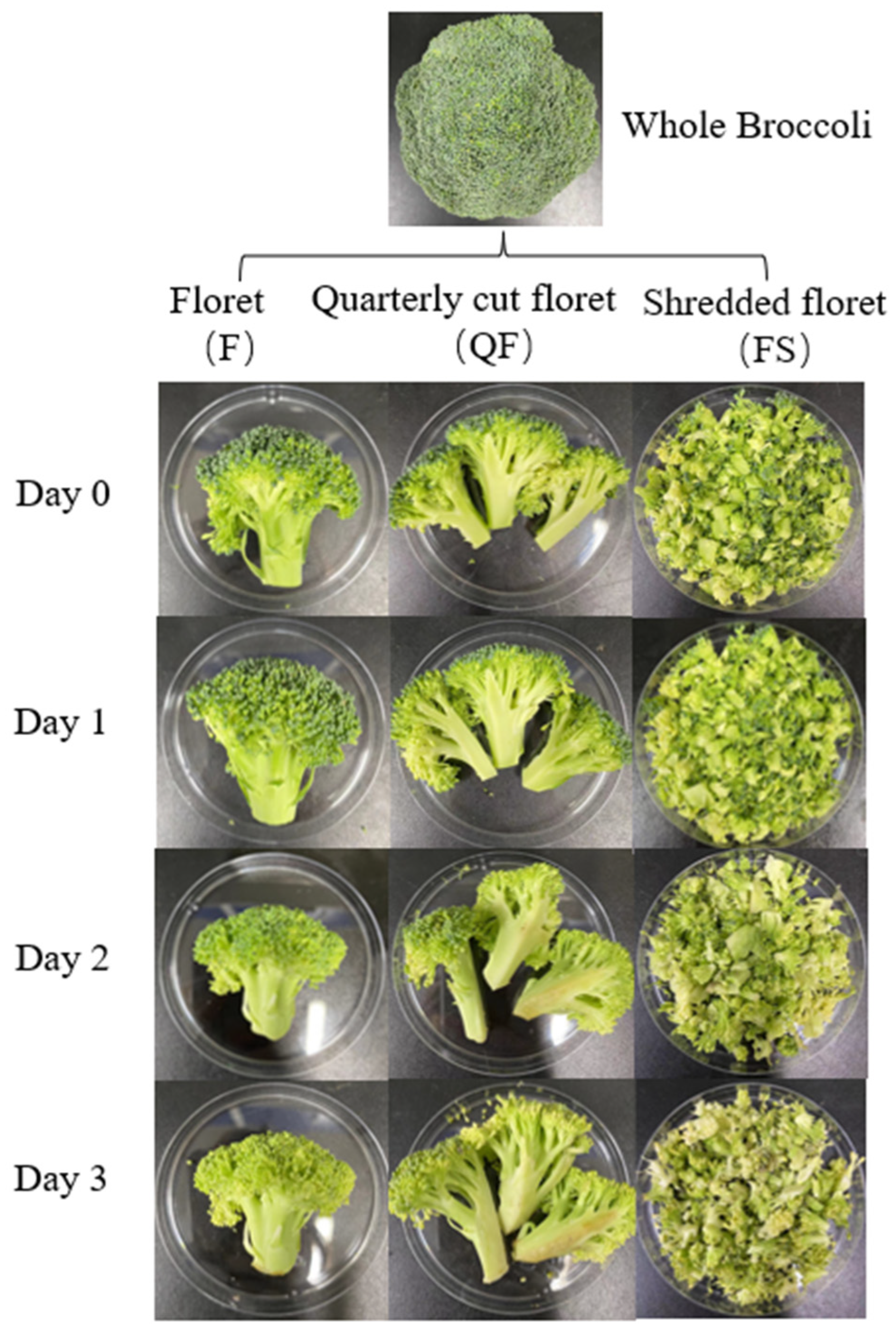
2.1. Sample Collection and Preparation

The raw material broccoli was harvested in April at Beijing National Vegetable Research Center (Beijing, China). Healthy broccoli with the same maturity and size were selected and transported to the lab with ice and stored at 20 °C for further analysis. The experiments were performed with three randomly selected biological replicates.

2.2. Pretreatment of Sample

The selected samples were washed with distilled water and then placed in a ventilator to dry naturally. The broccoli was subjected to three different cutting types using a sterile scalpel: (1) the broccoli was cut into single floret (floret, marked as F); (2) the broccoli floret was cut into four parts quarterly (quarterly cut floret, marked as QF); (3) the broccoli was shredded into pieces (shredded floret, marked as FS). All broccoli samples were packaged in a polyethylene plastic bag and then stored at 20 °C to study the change of microorganisms on it. During storage, broccoli was subsequentially sampled for further analyses at 0, 1, 2, 3 day(s). Three biological replicates were used for each sample. The characteristics of samples are shown in Figure 1.

2.3. Isolation of Bacteria

During 3 days of storage at 20 °C, the isolation and purification of bacteria were performed as Li et al. [17]. The total number of bacteria were determined by the standard plate count method using Plate Count Agar (PCA) medium. Briefly, 25 g broccoli samples were mixed with 225 mL sterilized 0.85% NaCl solution, placed in a sterilized homogenous bag, and homogenized at the speed of 10,000 r/min for 2 min to prepare a 1:10 sample homogenate. The sample suspension was serially diluted with sterilized 0.85% NaCl solution, 1 mL of which was spread, in triplicates, on PCA medium to enumerate bacteria. The PCA plates were incubated at 37 °C for 2 days and the bacterial counts were expressed as lg CFU/g with 3 replicates. In addition, different single colonies were picked out according to the morphology, color and size, and grown continuously on the plate until pure single colonies were obtained.

2.4. DNA Extraction

Strains were harvested for DNA extraction at 37 °C. Genomic DNA was extracted from cell suspension cultures using TIANamp Bacteria Genomic DNA kits (TIANGEN BIOTECH) according to the manufacturer’s instructions. DNA concentration and quality were determined spectrophotometrically at 260 and 280 nm.

2.5. PCR Amplification

After the extraction of genomic DNA, PCR amplification and sequencing of the 16S rRNA gene were performed according to Li et al. [18]. Amplification of the 16S rRNA gene was conducted using a pair of universal primers, forward primer 27F (5′-AGAGTTTGATCCTGGCTCAG-3′) and reverse primer 1525R (5′-AGAAAGGAGGTGATCCAGCC-3′). The PCR amplification were carried out under the following conditions: initial denaturation at 94 °C for 4 min followed by 35 cycles of denaturation at 94 °C for 1 min, annealing at 55 °C for 1 min, extension at 72 °C for 2 min and final extension at 72 °C for 10 min. The products above were purified and sequenced by BGI (The Beijing Genomics Institute). The 16S rRNA gene sequences of the isolates were sequenced with the Sanger sequencing method.

2.6. 16S rRNA Gene Phylogeny

After sequencing, the 16S rRNA gene sequences of different strains in broccoli were obtained and similarity searches were performed using the EzBioCloud server (http://www.ezbiocloud.net) [19]. The 16S rRNA sequencing data of all the isolates were aligned using Molecular Evolutionary Genetics Analysis 11 (MEGA 11) software and analyzed by Basic Local Alignment Search Tool (BLAST) with those of the closest strains retrieved from the National Center for Biotechnology Information (NCBI) database. The phylogenetic tree was constructed using the neighbor-joining method [20] in MEGA 11 software with representative type strains to determine the evolutionary relationship among the strains. Kimura’s two-parameter model was used to calculate the evolutionary distances [21]. Bootstrap values were determined based on 1000 replications [22].
Based on the results of identification, four representative strains of different species were selected as dominant spoilage bacteria to study their impacts on the spoilage of broccoli.

2.7. Spoilage Characteristics of Representative Strains in Spoilage of Broccoli

The broccoli samples were washed with sterile water three times, soaked in 75% alcohol for 10 s, then picked up and dried naturally. After sterilization, they were washed with sterile water and dried. The total number of bacterial colonies of sterile broccoli should be less than 2 log10 (CFU/g).
The dominant spoilage bacteria were inoculated on broccoli again, and the effects of different dominant spoilage bacteria on the physicochemical properties and nutritional quality of fresh-cut broccoli were measured.

2.7.1. Chlorophyll Content Determination

The content of total chlorophyll was determined by spectrophotometry according to Lichtenthaler [23], and the content of chlorophyll a, chlorophyll b was measured as described by Fang et al. [24] with slight modifications. An amount of 0.1 g plant tissues was homogenized in 80% acetone and extracted until the plant tissue completely became white and then centrifuged at 8000 rpm for 15 min. The optical density of the supernatant was recorded at 645 nm and 663 nm.

2.7.2. Nitrite Content Determination

The total content of nitrite was measured according to the naphthyl ethylenediamine hydrochloric acid spectrophotometric method by Nerdy et al. [25] with minor modifications. Approximately 5.0 g broccoli was crushed, deproteinated and defatted by precipitation followed by filtration. 40 mL of the filtrate was transferred into a 50 mL colorimetric tube with stopper, added 2 mL of p-Aminobenzenesulfonic Acid solution and 1 mL of N-(1-Naphthyl) Ethylenediamine Dihydrochloride solution, shaken until homogeneously mixed, diluted with distilled water to the marked line, shaken until well mixed. The mixtures were then kept at room temperature for 15 min. OD of the colored mixtures was read at 538 nm against the reagent blank as described by An et al. [26]. The experiments were carried out in triplicates and the results were calculated as the average obtained from them.

2.7.3. MDA Content Assay

Malondialdehyde (MDA) content was measured by the thiobarbituric acid (TBA) chromogenic method as described by Liu et al. [27]. Extraction was carried out by homogenizing 1.0 g of broccoli samples with 5 mL of 10% trichloroacetic acid (TCA), and then the homogenate was centrifuged at 8000 rpm at 4 °C for 20 min. The supernatant was collected and 2 mL of extract was mixed with 2 mL of 0.67% TBA solution to react in a boiling water bath for 20 min. The solution was cooled and centrifuged again. The extraction of supernatant was determined in absorbance at 450 nm, 532 nm, and 600 nm, respectively. The amount of MDA was expressed as nmol per gram (nmol/g fresh weight) of broccoli.

2.7.4. Soluble Sugar Content Analysis

The total soluble sugar content in each broccoli sample was measured by the anthrone colorimetric method [28] with slight modifications. Broccoli samples weighing 0.2 g were taken in test tubes. Then, to each test tube was added 1 mL of distilled water to disperse it and the samples were grinded into homogenate. After placing the test tubes in boiling water for 10 min, the extracts were collected and the volumes of the extracts were adjusted to 10 mL using distilled water. The absorbances of the extracts were determined at 620 nm wavelength after adding sulfuric acid and anthrone. The results were presented as milligram per gram (mg/g fresh weight) of broccoli, and the experiments were repeated three times.

2.7.5. Statistical Analysis for Spoilage Characteristic of Microorganisms in Broccoli

All data were analyzed statistically using a one-way analysis of variance (ANOVA) with SPSS software 23.0. Statistical differences between samples (* p < 0.05 was considered to be significant; ** p < 0.01 was considered to be extremely significant) were determined by Duncan’s multiple range test. All data were recorded as the mean ± standard errors (SE) of the three independent biological replicates.

3. Results

3.1. Changes in Bacterial Counts and Isolates Separated from Broccoli

Fresh-cut broccoli was easily infected by bacteria during storage. At the harvest period, the bacterial counts of broccoli were up to 5.12 lg (CFU/g), which might be contaminated with a large number of microorganisms during sample collection and transportation (Figure 2). After 3 days, the bacterial counts in broccoli which was cut into a single floret reached up to 8.54 lg (CFU/g), and different kinds of bacteria were observed. In addition, the number of microorganisms in QF (quarterly cut floret) and FS (shredded floret) reached up to 8.66 lg (CFU/g) and 9.60 lg (CFU/g), respectively. It can be seen from Figure 2 that mechanical damage had a great impact on the amounts of microbial colonies. The total number and growth rate of microorganisms on severely damaged broccoli, referring to the broccoli cut into shredded pieces, were much higher than that of lightly damaged broccoli, referring to the broccoli floret cut quarterly into four parts. Therefore, in the postharvest treatment, mechanical damage to broccoli should be lessened as much as possible, or supplementary strategies (such as cold storage) should be applied to prolong its shelf life.
The microorganisms could be separated from the broccoli during storage (Table 1). The analysis of the growth features and corruption characteristics of the spoilage microorganisms were, therefore, of great significance in assisting the control of quality and safety of fresh-cut broccoli.

3.2. Identification of Bacterial Isolation

To identify key microbes that accounted for the observed microbiota separation, community composition was analyzed. According to the different colonial morphologies, a total of ninety-seven bacterial colonies that appeared on culture medium were selected for obtaining pure cultures to be used in characterization for further research. The screened strains were sequenced, and the phylogenetic tree of the spoilage strains is shown in Figure 3. According to the 16S rRNA gene sequencing, the bacterial isolates were grouped into 21 different bacterial genera (Table 2). And the culturable bacterial composition of fresh-cut broccoli at the phylum and genus levels is shown in Figure 4. A similarities research based on near-complete 16S rRNA sequences of the isolates showed that there was a high similarity (>97%) between the test isolates and the representative strains of Pseudomonas, Stenotrophomonas, Acinetobacter, Alcaligenes, Enterobacter, Delftia, Comamonas, Agrobacterium, Sphingobacterium, Brevundimonas, Lysinibacillus, Brevibacterium, Glutamicibacter, Lelliottia, Myroides, Pectobacterium, Proteus, Microbacterium, Leclercia, Escherichia and Rothia. The majority of bacteria belonged to three phyla: Proteobacteria, Actinobacteria and Bacteroidota, accounting for more than 90% of the total. Furthermore, Proteobacteria was the predominant phylum, and the majority were Pseudomonas spp. and Acinetobacter spp. A few genera with the least number of isolates were also obtained, such as Lysinibacillus, a genus of phylum Firmicutes. Isolates belonging to the phyla Actinobacteria and Bacteroidetes were found in a low number. A diversity of microorganism distribution was observed in bacterial isolates.

3.3. Effects of Spoilage Microorganisms on the Quality of Broccoli

Regardless of the cutting types, the diversity of spoilage bacteria isolated from broccoli was similar. In the present study, we collected more than ninety colonies of bacteria from broccoli and selected four major isolates based on colony phenotype grown on PCA medium. The phylogenetic trees of the four strains are shown in Figure 5 and their impacts are shown in Table 3. The results showed that each of the strains had different effects on the quality of the broccoli, Pseudomonas spp. particularly.
Chlorophyll is an important indicator of plant vital signs in the photosynthesis of plant cells, which can absorb light energy to synthesize carbohydrates, carbon dioxide, and water. Thus, the change of chlorophyll content was considered to be an important factor affecting the quality of broccoli during the preservation. As shown in Table 3, the content of total chlorophyll in broccoli decreased during the storage. After 72 h, the total chlorophyll content in fresh-cut broccoli inoculated with Pseudomonas sp. BRO-C11 and Comamonas sp. BRO-C5 decreased by 51.9% and 48.1%, respectively, compared with the control. At the end of storage, the total chlorophyll content was decreased by 30.8–66.7% compared with the initial value, but Acinetobacter sp. BRO-F1 showed little effect on the change of chlorophyll content compared with the control.
Furthermore, the accumulation of nitrite is another common problem in the process of broccoli becoming putrid. Nitrite can react with amines or amides to form carcinogenic, mutagenic, and teratogenic N-nitroso compounds. High nitrate dietary intake was found to be associated with gastric cancer [29]. In addition, nitrosamine products, once accumulated to a certain amount, likely cause teratogenicity, carcinogenesis and mutagenicity. Therefore, it is of great importance to detect the content of nitrite in vegetables. It can be seen from Table 3 that with the time of storage, the content of nitrite had the law that first it increased, next decreased, then increased again to reach a relatively steady state in most samples. The content of nitrite in newly harvested broccoli was relatively low. During storage, the physiological activities of bacteria might be an important factor leading to the change of nitrite content. Among them, the nitrite in the group inoculated with Pseudomonas sp. BRO-C11 reached up to a maximum, 1.61 mg/kg fresh weight, indicating that the Pseudomonas spp. could promote the accumulation of nitrite. After 24 h, the nitrite content in broccoli inoculated with Pseudomonas sp. BRO-C11, Pseudomonas sp. BRO-G12 and Acinetobacter sp. BRO-F1 showed downward trends, which may be caused by the activity of microorganisms. Under the metabolism of microorganisms in broccoli, nitrate was greatly reduced to nitrite, and the content of nitrite increased. With the consumption of oxygen, the growth of aerobic bacteria was inhibited, resulting in the activity of nitrate reductase being inhibited, the effect of nitrate reduction to be reduced, and large amounts of nitrite to be decomposed. The nitrite content in the group inoculated with Pseudomonas sp. BRO-C11 increased at the end of storage, with significant differences among the other groups (p < 0.05). In addition, Acinetobacter sp. BRO-F1 and Comamonas sp. BRO-C5 had little effects on the content of nitrite.
The effects of spoilage bacteria on the content of MDA in fresh-cut broccoli is shown in Table 3. In all samples, the content of MDA increased first then decreased, and with storage, the content of MDA showed a downward trend, and the final contents were 2.47–4.79 nmol/g fresh weight. The MDA content in all samples reached a maximum at 48 h. In general, Pseudomonas spp. accelerated the accumulation of MDA in fresh-cut broccoli, which might be due to that the bacteria gradually infected and destroyed the membrane structure, resulting in the formation of malondialdehyde (MDA).
The effects of bacterial infection on the content of soluble sugar in fresh-cut broccoli are shown in Table 3. The soluble sugar content in fresh-cut broccoli was 2.66 mg/g fresh weight in the initial value. At 48 h of storage, the soluble sugar in broccoli inoculated with Pseudomonas sp. BRO-C11 and Pseudomonas sp. BRO-G12 increased by 33.08% and 19.17% compared with the initial value. However, there was no significant difference in the content of soluble sugar between the group inoculated with Acinetobacter sp. BRO-F1 and the control group. The soluble sugar content in broccoli inoculated with Pseudomonas sp. BRO-C11 was significantly increased under the bacterial infection for 48 h, and it displayed an increase of 25.09% compared with control.

4. Discussion

The cutting types had great impacts on the amounts of microorganisms in broccoli but had little impact on the species of microorganisms. The amounts of microorganisms gradually increased and conformed to the S-shaped curve of microbial growth. During storage, the total number of colonies of broccoli treated with different cutting types had the same trend, but the average count of bacteria in broccoli subjected to different cutting types showed a significant difference. The processed fruits and vegetables were more perishable than the raw products from which they were prepared. Cutting of fruits and vegetables might cause mechanical damage, outflow of nutrients, and provide favorable living conditions for microorganisms to grow and multiply. In addition, it increased the chance of contamination of fruits and vegetables and resulted in the microbial spoilage of fruits through the transfer of microflora on the outer surfaces to the interior tissue, where microorganisms had access to nutrient-laden juice. On the other hand, fresh-cut fruits and vegetables lost the barrier and protection of the epidermis, and the surface exposed to the air increased, resulting in the disorder of the original physiological function and the destruction of the self-protection system, so that the resistance of the body decreased, resulting in more microorganisms invading the internal tissues of fruits and vegetables from the wound. The larger the specific surface area formed by cutting treatment, the more serious the microbial infection was, and the worse the storage of fresh-cut products. The resistance of fresh-cut fruits and vegetables to microbial infection in the environment gradually decreased. However, even in the early storage, the amounts of microorganisms can also be large. There were three main ways fresh-cut fruits and vegetables were shown to be infected by microorganisms before and during storage. Firstly, fresh-cut fruits and vegetables were easily infected by microorganisms living in manure, fertilizer, soil and irrigation water sources during planting or harvest. The microorganisms contained were usually bacteria, molds, yeasts, and actinomycetes [30]. Secondly, in the process of cutting, the main pollution sources of fresh-cut fruits and vegetables were usually from cutting equipment or the environment [31]. Moreover, the cross contamination of microbial communities between fruits and vegetables could accelerate their decay and deterioration. Thirdly, fresh-cut fruits and vegetables were easily infected by microorganisms during storage. The resistance of harvested fruits and vegetables to microbial infection in the environment gradually decreased, and the number of microorganisms on the surface of fruits and vegetables increased.
From this study, the phylogenetic relationships of the 97 bacterial isolates were determined in comparison to type strains with the neighbor-joining method. Among them, Pseudomonas spp. were the most predominant and might have the greatest impact on food quality. The physiological properties of Pseudomonas spp. were adapted to the environment of vegetables. At the end of storage, the relative abundance of other flora decreased gradually. The main reason was that the growth of dominant bacteria was too fast, which inhibited the growth of other microorganisms. Pseudomonas spp. are aerobic, Gram-negative bacteria, and are widely distributed in nature. Different from enzymatic browning of vegetables, Pseudomonas spp. mainly cause the tissue of fresh-cut vegetables to become rotten and proved to result in a rapid browning reaction on the surface of fresh-cut vegetables [32]. Pseudomonas spp. can utilize organic compounds, oxidize glucose and maltose to produce acid, resulting in the decline of food quality. In addition, Pseudomonas spp. can also produce catalase and oxidase, and hydrolyze or lipolyze frozen food, so as to make it corrupt. Moreover, they produced pectolytic enzymes to hydrolyze the cell walls of fruits and vegetables, resulting in rot and deterioration of fruits and vegetables, such as pectolytic enzymes produced in Pseudomonas viridiflava and Pseudomonas chlororaphis, which caused organoleptic properties to decline as well as spoilage in fresh-cut vegetables [33].
The color of vegetables is considered to be an extremely crucial factor for consumer acceptance, especially the unique green color in vegetables, and the dominant reason for de-greening of broccoli was chlorophyll deterioration. Compared with the control group, the chlorophyll of broccoli infected by bacteria decreased in varying degrees, especially the group inoculated with Pseudomonas sp. BRO-C11, Pseudomonas sp. BRO-G12 and Comamonas sp. BRO-C5, which also indicated that the infected group was more prone to yellowing in the experiment, explaining that bacterial infection promoted the yellowing of broccoli and destroyed the appearance of broccoli. This result may explain the yellowing of fresh-cut broccoli after longer storage periods. In order to maintain the pleasant appearance of broccoli, measures to avoid broccoli being polluted by bacteria, Pseudomonas spp. particularly, should be taken.
Agricultural standards clearly stipulate the limit standards of nitrate and nitrite in vegetables. Our research investigated potential correlations between bacterial infection and the formation or depletion of nitrite during storage, and microorganisms in fresh-cut fruits and vegetables were the main cause of nitrite accumulation. With storage, the concentration of nitrite reached a maximum, the nitrite peak, and then began to decrease. Among the microorganisms, Pseudomonas sp. BRO-C11 had the most important effects on the content of nitrite in broccoli. In the harvest period, Pseudomonas sp. BRO-C11 with nitrate reductase proliferated in large numbers, resulting in the nitrate in fresh-cut broccoli to be reduced to nitrite. However, the growth of aerobic Pseudomonas sp. BRO-C11 was inhibited when the oxygen was consumed, resulting in the activity of nitrate reductase to be inhibited, promoting large amounts of nitrite to be decomposed. In general, controlling the content of nitrite and nitrate in vegetables was the key to scientific production and edible vegetables. In the follow-up study, the spoilage bacteria that had important impacts on nitrite content should be strictly controlled.
Malondialdehyde (MDA), as the main product of membrane lipid peroxidation, indicates the integrity of cell membranes. The content of malondialdehyde in plant is positively correlated with the degree of cell membrane damage. In our study, the content of MDA in all samples exhibited approximately the same first increased then decreased trend, and the group inoculated with Pseudomonas sp. BRO-C11 and Pseudomonas sp. BRO-G12 increased at a higher rate than the control group at 48 h of storage. The results showed that after being infected by bacteria, the permeability of the cell membrane increased, which might be due to the damage by bacteria to plant cells by secreting hydrolases and toxins, resulting in the loss of contained logistics in plant cells. The substance in cells, which was conducive for the growth and reproduction of microorganisms, flowed out due to the rupture of the cell membrane, and further aggravated the damage to the cell membrane, leading to the increase of MDA content. Overall, the content of MDA increased, and the senescence process of broccoli was enhanced after being infected.
Soluble sugar has the function to maintain the turgidity of cells, and it is able to protect the membranes of cells from damage caused by the abiotic stress. In this study, under infection, the soluble sugar content of broccoli inoculated with Pseudomonas sp. BRO-C11, Pseudomonas sp. BRO-G12 and Comamonas sp. BRO-C5 increased at 48 h, which meant that the broccoli could adapt to bacterial infection in the short term. Moreover, the soluble sugar had the function of acting as an osmotic agent, which was expressed directly by the content in plants. In a word, plants could adapt to the bacterial infection by regulating the content of soluble sugar.
The chlorophyll of the experimental group inoculated with bacteria was decreased, which affected the sensory quality of fresh-cut broccoli. In addition, the content of MDA in broccoli infected by Pseudomonas sp. BRO-C11 and Pseudomonas sp. BRO-G12 increased rapidly, and broccoli inoculated with Pseudomonas sp. BRO-C11 produced more nitrite, indicating that the Pseudomonas had great impacts on the quality and safety of vegetables and led to the production of harmful substances in fresh-cut broccoli, which should be paid more attention for food safety.

5. Conclusions

The results showed that cutting types had significant effects on the quantity of microorganisms in fresh-cut broccoli, and the number and growth rate of colonies of broccoli with severe mechanical damage were much higher than those with mild mechanical damage. The ninety-seven strains of spoilage microorganisms in broccoli samples were obtained by isolation. Through 16S rRNA gene sequencing, the main microorganisms in the samples were classified into four phyla: Proteobacteria, Actinobacteria, Bacteroidetes and Firmicutes. Furthermore, Proteobacteria was the predominant phylum, and the majority were the Pseudomonas spp. and Acinetobacter spp. Pseudomonas spp. was not only dominant in quantity, but also had great impacts on the quality of broccoli. Comamonas spp. and Acinetobacter spp. also affected the quality of broccoli to varying degrees, but received little attention in previous studies. The corruption phenomena included the production of harmful substances, such as nitrite, the consumption of nutrients and active components, and inhibition of key enzyme activities, causing spoilage characteristics such as malodor, softness, and browning. This study provided a foundation for targeted bacteriostasis, delaying the corruption and deterioration of fresh-cut vegetables and prolonging the edible period of broccoli. Therefore, necessary measures, such as ensuring a clean processing environment and storage environment, the addition of antibacterial preservative, and less mechanical damage need to be taken into account to reduce contamination from corrupt microorganisms and ensure the safety and flavor of broccoli.

Author Contributions

Conceptualization, S.C., J.Z. and G.L.; methodology, S.C. and X.Y.; software, S.C.; validation, S.C. and X.Y.; formal analysis, S.C.; resources, S.C. and X.Y.; writing—original draft preparation, S.C.; writing—review and editing, X.Y. and Y.C.; project administration, J.Z. and G.L. All authors have read and agreed to the published version of the manuscript.

Funding

This study is funded by the Collaborative Innovation Center of the Beijing Academy of Agriculture and Forestry Sciences, grant number KJCX201915, and the Innovation and Capacity-building Project of the Beijing Academy of Agriculture and Forestry Sciences, grant number KJCX20200213.

Institutional Review Board Statement

Not applicable.

Informed Consent Statement

Not applicable.

Data Availability Statement

The data that support the findings of this study are available from the corresponding author upon reasonable request.

Acknowledgments

We thank Hongju He for supporting the broccoli sample preparation. We thank Xuezhi Zhao for sample preparation.

Conflicts of Interest

The authors declare no conflict of interest.

References

  1. Fernández-León, M.F.; Fernández-León, A.M.; Lozano, M.; Ayuso, M.C.; González-Gómez, D. Different postharvest strategies to preserve broccoli quality during storage and shelf life: Controlled atmosphere and 1-MCP. Food Chem. 2013, 138, 564–573. [Google Scholar] [CrossRef] [PubMed]
  2. Li, Y.; Zhang, T.; Korkaya, H.; Liu, S.; Lee, H.F.; Newman, B.; Yu, Y.; Clouthier, S.G.; Schwartz, S.J.; Wicha, M.S.; et al. Sulforaphane, a dietary component of broccoli/broccoli sprouts, inhibits breast cancer stem cells. Clin. Cancer Res. 2010, 16, 2580–2590. [Google Scholar] [CrossRef] [PubMed] [Green Version]
  3. Wang, H.; Wang, L.; Cao, L.; Zhang, Q.; Song, Q.; Meng, Z.; Wu, X.; Xu, K. Inhibition of autophagy potentiates the anti-metastasis effect of phenethyl isothiocyanate through JAK2/STAT3 pathway in lung cancer cells. Mol. Carcinog. 2018, 57, 522–535. [Google Scholar] [CrossRef]
  4. Alvarez, M.V.; Moreira, M.; Roura, S.I.; Ayala-Zavala, J.F.; González Aguilar, G.A. Using natural antimicrobials to enhance the safety and quality of fresh and processed fruits and vegetables: Types of antimicrobials—ScienceDirect. In Handbook of Natural Antimicrobials for Food Safety and Quality; Woodhead Publishing: Cambridge, UK, 2015; pp. 287–313. [Google Scholar]
  5. Barrett, D.M.; Beaulieu, J.C.; Shewfelt, R. Color, flavor, texture, and nutritional quality of fresh-cut fruits and vegetables: Desirable levels, instrumental and sensory measurement, and the effects of processing. Crit. Rev. Food Sci. Nutr. 2010, 50, 369–389. [Google Scholar] [CrossRef]
  6. Oliveira, M.; Abadias, M.; Usall, J.; Torres, R.; Teixid, N. Application of Modified Atmosphere Packaging as a safety approach to fresh-cut fruits and vegetables—A review. Trends Food Sci. Tech. 2015, 46, 13–26. [Google Scholar] [CrossRef]
  7. Wang, C.W.; Wang, Y. First report of postharvest fruit rot caused by Fusarium equiseti on stored cerasus Pseudocerasus in China. Plant. Dis. 2017, 101, 1451. [Google Scholar] [CrossRef]
  8. Dakwa, V.; Powell, S.; Eyles, A.; Gracie, A.; Tamplin, M.; Ross, S. Effect of peroxyacetic acid treatment and bruising on the bacterial community and shelf-life of baby spinach. Int. J. Food Microbiol. 2021, 343, 109086. [Google Scholar] [CrossRef]
  9. Bah, A.; Ferjani, R.; Fhoula, I.; Gharbi, Y.; Najjari, A.; Boudabous, A.; Ouzari, H.I. Microbial community dynamic in tomato fruit during spontaneous fermentation and biotechnological characterization of indigenous lactic acid bacteria. Ann. Microbiol. 2019, 69, 41–49. [Google Scholar] [CrossRef]
  10. Voidarou, C.; Alexopoulos, A.; Tsinas, A.; Rozos, G.; Tzora, A.; Skoufos, I.; Varzakas, T.; Bezirtzoglou, E. Effectiveness of bacteriocin-producing lactic acid bacteria and bifidobacterium isolated from honeycombs against spoilage microorganisms and pathogens isolated from fruits and vegetables. Appl. Sci. 2020, 10, 7309. [Google Scholar] [CrossRef]
  11. Maffei, D.F.; Batalha, E.Y.; Landgraf, M.; Schaffner, D.W.; Franco, B.D. Microbiology of organic and conventionally grown fresh produce. Braz. J. Microbiol. 2016, 47, 99–105. [Google Scholar] [CrossRef] [Green Version]
  12. Barbosa, A.A.T.; Mantovani, H.C.; Jain, S. Bacteriocins from lactic acid bacteria and their potential in the preservation of fruit products. Crit. Rev. Biotechnol. 2017, 37, 852–864. [Google Scholar] [CrossRef] [PubMed]
  13. Stoll, D.A.; Müller, A.; Meinhardt, A.K.; Dötsch, A.; Greiner, R.; Kulling, S.E.; Huch, M. Influence of salt concentration and iodized table salt on the microbiota of fermented cucumbers. Food Microbiol. 2020, 92, 103552. [Google Scholar] [CrossRef] [PubMed]
  14. Nahidul-Islam, S.M.; Takashi, K.; Hajime, T.; Bon, K. Bacterial and fungal microbiota in traditional Bangladeshi fermented milk products analysed by culture-dependent and culture-independent methods. Food Res. Int. 2018, 111, 431–437. [Google Scholar] [CrossRef]
  15. Rocha, K.E.M.; Milani, C.; Ventura, M.; Valencia, F.E.; Madiedo, R.; Rusa-Madiedo, P.; Delgado, S. Bacterial diversity of the Colombian fermented milk “Suero Costeño” assessed by culturing and high-throughput sequencing and DGGE analysis of 16S rRNA gene amplicons. Food Microbiol. 2017, 68, 129–136. [Google Scholar]
  16. Liang, H.; Yin, L.; Zhang, Y.; Chang, C.; Zhang, W. Dynamics and diversity of a microbial community during the fermentation of industrialized Qingcai paocai, a traditional Chinese fermented vegetable food, as assessed by Illumina MiSeq sequencing, DGGE and qPCR assay. Ann. Microbiol. 2018, 68, 111–122. [Google Scholar] [CrossRef]
  17. Li, R.; Cai, L.; Gao, T.; Li, C.; Ye, K. Comparing the quality characteristics and bacterial communities in meatballs with or without blown pack spoilage. LWT Food Sci. Technol. 2020, 130, 109529. [Google Scholar] [CrossRef]
  18. Li, W.J.; Xu, P.; Schumann, P.; Zhang, Y.Q.; Pukall, R.; Xu, L.H.; Stackebrandt, E.; Jiang, C.L. Georgenia ruanii sp. nov. a novel actinobacterium isolated from forest soil in Yunnan (China), and emended description of the genus Georgenia. Int. J. Syst. Evol. Microbiol. 2007, 57, 1424–1428. [Google Scholar] [CrossRef]
  19. Yoon, S.H.; Ha, S.M.; Kwon, S.; Lim, J.; Chun, J. Introducing EzBioCloud: A taxonomically united database of 16S rRNA gene sequences and whole-genome assemblies. Int. J. Syst. Evol. Microbiol. 2017, 67, 1613–1617. [Google Scholar] [CrossRef]
  20. Saitou, N.; Nei, M. The neighbor-joining method: A new method for reconstructing phylogenetic trees. Mol. Biol. Evol. 1987, 4, 406–425. [Google Scholar]
  21. Kimura, M. A simple method for estimating evolutionary rates of base substitutions through comparative studies of nucleotide sequences. J. Mol. Evol. 1980, 16, 111–120. [Google Scholar] [CrossRef]
  22. Felsenstein, J. Confidence limits on phylogenies: An approach using the bootstrap. Evolution 1985, 38, 14–26. [Google Scholar]
  23. Lichtenthaler, H.K. Chlorophylls and carotenoids: Pigments of photosynthetic biomembranes. Methods Enzymol. 1987, 148, 350–382. [Google Scholar]
  24. Fang, H.; Luo, F.; Li, P.; Zhou, Q.; Zhou, X.; Wei, B.; Cheng, S.; Zhou, H.; Ji, S. Potential of jasmonic acid (JA) in accelerating postharvest yellowing of broccoli by promoting its chlorophyll degradation. Food Chem. 2020, 309, 125737. [Google Scholar] [CrossRef] [PubMed]
  25. Nerdy, N.; Putra, E. Spectrophotometric method for determination of nitrite and nitrate levels in broccoli and cauliflower with different fertilization treatment. Orient J Chem. 2018, 34, 2983–2991. [Google Scholar] [CrossRef]
  26. An, F.; Sun, H.; Wu, J.; Zhao, C.; Li, T.; Huang, H.; Fang, Q.; Mu, E.; Wu, R. Investigating the core microbiota and its influencing factors in traditional Chinese pickles. Food Res. Int. 2021, 147, 110543. [Google Scholar] [CrossRef]
  27. Liu, H.; Jiang, W.; Cao, J.; Ma, L. A combination of 1-methylcyclopropene treatment and intermittent warming alleviates chilling injury and affects phenolics and antioxidant activity of peach fruit during storage. Sci. Hortic. 2018, 229, 175–181. [Google Scholar] [CrossRef]
  28. Fairbairn, N.J. A modified anthrone reagent. Chem. and Ind. 1953, 4, 86. [Google Scholar]
  29. Rezaei, M.; Fani, A.; Moini, A.L.; Mirzajani, P.; Malekirad, A.A.; Rafiei, M. Determining Nitrate and Nitrite content in beverages, fruits, vegetables, and stews marketed in Arak, Iran. Int. Sch. Res. Not. 2014, 2014, 1–5. [Google Scholar] [CrossRef]
  30. Devleesschauwer, B.; Marvasi, M.; Giurcanu, M.C.; Hochmuth, G.J.; Speybroeck, N.; Havelaar, A.H.; Teplitski, M. High relative humidity pre-harvest reduces post-harvest proliferation of Salmonella in tomatoes. Food Microbiol. 2017, 66, 55–63. [Google Scholar] [CrossRef] [Green Version]
  31. Shen, C.; Rao, J.; Wu, Q.; Wu, D.; Chen, K. The effect of indirect plasma-processed air pretreatment on the microbial loads, decay, and metabolites of Chinese bayberries. LWT Food Sci. Technol. 2021, 150, 111998. [Google Scholar] [CrossRef]
  32. Pinto, L.; Ippolito, A.; Baruzzi, F. Control of spoiler Pseudomonas spp. on fresh cut vegetables by neutral electrolyzed water. Food Microbiol. 2015, 50, 102–108. [Google Scholar] [CrossRef] [PubMed]
  33. Dong, H.L.; Kim, J.B.; Kim, M.; Roh, E.; Jung, K.; Choi, M.; Changsik, O.H.; Choi, J.; Yun, J.; Heu, S. Microbiota on spoiled vegetables and their characterization. J. Food Prot. 2013, 76, 1350–1358. [Google Scholar]
Figure 1. Plant materials and sampling design. Broccoli was harvested, prepared and cut with three different types, floret, quarterly cut floret and shredded floret. Then they were stored at 20 °C and subsequentially collected at 0, 1, 2, 3 days for further analyses.
Applsci 12 03370 g001
Figure 2. Effects of different cutting types on the growth of total bacterial counts in fresh-cut broccoli during storage.
Applsci 12 03370 g002
Figure 3. The neighbor-joining phylogenetic tree of the representative 97 bacterial isolates and their closest relatives based on 16S rRNA gene sequences.
Applsci 12 03370 g003
Figure 4. The culturable bacterial composition of fresh-cut broccoli at the phylum (A) and genus (B) levels.
Applsci 12 03370 g004
Figure 5. Phylogenetic trees of the four selected spoilage microorganisms BRO-C11 (a), BRO-G12 (b), BRO-F1 (c) and BRO-C5 (d).
Applsci 12 03370 g005aApplsci 12 03370 g005b
Table 1. The descriptions of the sampled broccoli and the isolates.
SampleStrain NumberCutting TypeColor of
Broccoli
Peculiar SmellStorage
Time (d)
ABRO-A1~BRO-A20FloretGreenNo0
BBRO-B1~BRO-B21FloretGreenNo1
CBRO-C1~BRO-C12FloretLight greenMinor2
DBRO-D1~BRO-D13FloretLight yellowMinor3
EBRO-E1~BRO-E9Quarterly cut floretLight yellowMinor3
FBRO-F1~BRO-F6Shredded floretYellowIntense3
GBRO-G1~BRO-G16Shredded floretYellowIntense3
Table 2. Taxonomic identification and detailed information of culturable bacteria isolated from fresh-cut broccoli based on 16S rRNA gene sequences.
PhylumSpeciesStrain No.Similarity Range
(%)
Reference
Strain
Percentage (%)
Proteobacteria
Pseudomonas
capeferrum
BRO-B1
BRO-B7
BRO-B3
BRO-D9
BRO-C1
BRO-B18
BRO-C9
BRO-C3
BRO-C11
99.29–99.60Pseudomonas
capeferrum
WCS358T
9.28
Pseudomonas
alloputida
BRO-A11
BRO-E2
BRO-E3
BRO-G13
BRO-F2
BRO-E1
BRO-G12
99.86–100.00Pseudomonas
alloputida
Kh7T
7.22
Pseudomonas
rhizoryzae
BRO-A1999.47Pseudomonas
rhizoryzae
RY24T
1.03
Pseudomonas
qingdaonensis
BRO-D11100.00Pseudomonas
qingdaonensis
JJ3T
1.03
Pseudomonas
baetica
BRO-C499.58Pseudomonas
baetica
a390T
1.03
Pseudomonas
rhodesiae
BRO-C699.72Pseudomonas
rhodesiae
CIP 104664T
1.03
Pseudomonas
mosselii
BRO-D8100.00Pseudomonas
mosselii
CIP 105259T
1.03
Pseudomonas
juntendi
BRO-B1699.64Pseudomonas
juntendi
BML3T
1.03
Pseudomonas
entomophila
BRO-B1999.83Pseudomonas
entomophila
L48T
1.03
Pseudomonas
atacamensis
BRO-E4
BRO-A1
BRO-A7
BRO-B2
99.84–100.00Pseudomonas
atacamensis
M7D1T
4.12
Pseudomonas
sichuanensis
BRO-B8100.00Pseudomonas
sichuanensis
WCHPs060039T
1.03
Pseudomonas
oleovorans subsp.
oleovorans
BRO-B14
BRO-G9
BRO-G10
98.87–99.71Pseudomonas
oleovorans subsp.
oleovorans
DSM 1045T
3.09
Acinetobacter
johnsonii
BRO-B5
BRO-B10
BRO-D10
BRO-F3
BRO-B21
98.67–99.30Acinetobacter
johnsonii
CIP 64.6T
5.15
Acinetobacter
pittii
BRO-B11
BRO-D3
BRO-B4
BRO-G7
BRO-A5
BRO-G6
BRO-G1
99.86–100.00Acinetobacter
pittii
CIP 70.29T
7.22
Acinetobacter
oryzae
BRO-B12
BRO-B15
BRO-D5
BRO-F1
BRO-B20
BRO-C12
BRO-E6
98.98–99.36Acinetobacter
oryzae
B23T
7.22
Acinetobacter
guillouiae
BRO-D13
BRO-G16
98.75–98.86Acinetobacter
guillouiae
CIP 63.46T
2.06
Acinetobacter
junii
BRO-A17
BRO-D6
99.33Acinetobacter
junii
CIP 64.5T
2.06
Acinetobacter
proteolyticus
BRO-A13100.00Acinetobacter
proteolyticus
NIPH 809T
1.03
Acinetobacter
bereziniae
BRO-C8
BRO-D7
99.85–100.00Acinetobacter
bereziniae
LMG 1003T
2.06
Acinetobacter
calcoaceticus
BRO-G4100.00Acinetobacter
calcoaceticus
DSM 30006T
1.03
Comamonas
aquatica
BRO-B6
BRO-D12
BRO-G3
99.85–100.00Comamonas
aquatica
NBRC 14918T
3.09
Comamonas
koreensis
BRO-A1298.86Comamonas
koreensis
KCTC 12005T
1.03
Comamonas
testosteroni
BRO-A14
BRO-A16
BRO-G8
BRO-C5
99.85–100.00Comamonas
testosteroni
ATCC 11996T
4.12
Stenotrophomonas
maltophilia
BRO-A2
BRO-F5
99.21–99.34Stenotrophomonas
maltophilia
MTCC 434T
2.06
Stenotrophomonas
pavanii
BRO-F499.86Stenotrophomonas
pavanii
DSM 25135T
1.03
Stenotrophomonas
rhizophila
BRO-A999.86Stenotrophomonas
rhizophila
DSM 14405T
1.03
Stenotrophomonas
terrae
BRO-A1099.59Stenotrophomonas
terrae
DSM 18941T
1.03
Stenotrophomonas
lactitubi
BRO-C1099.37Stenotrophomonas
lactitubi
M15T
1.03
Stenotrophomonas
indicatrix
BRO-F699.72Stenotrophomonas
indicatrix
WS40T
1.03
Brevundimonas
vesicularis
BRO-A8100.00Brevundimonas
vesicularis
NBRC 12165T
1.03
Brevundimonas
diminuta
BRO-A1899.35Brevundimonas
diminuta
ATCC 11568T
1.03
Agrobacterium
arsenijevicii
BRO-A498.13Agrobacterium
arsenijevicii
KFB 330T
1.03
Alcaligenes
faecalis subsp.
phenolicus
BRO-G2
BRO-G5
99.72–99.73Alcaligenes
faecalis subsp.
phenolicus
DSM 16503T
2.06
Escherichia
hermannii
BRO-A399.87Escherichia
hermannii
CIP 103176T
1.03
Leclercia
adecarboxylata
BRO-B13100.00Leclercia
adecarboxylata
NBRC 102595T
1.03
Lelliottia
jeotgali
BRO-C799.28Lelliottia
Jeotgali
PFL01T
1.03
Enterobacter
chengduensis
BRO-G1499.85Enterobacter
chengduensis
WCHECl-C4T
1.03
Delftia
tsuruhatensis
BRO-G11100.00Delftia
tsuruhatensis
NBRC 16741T
1.03
Proteus
mirabilis
BRO-E899.86Proteus
mirabilis
ATCC 29906T
1.03
Pectobacterium
carotovorum
BRO-D299.72Pectobacterium
carotovorum
NCPPB 312T
1.03
ActinobacteriaGlutamicibacter
arilaitensis
BRO-D499.13Glutamicibacter
arilaitensis
Re117T
1.03
Glutamicibacter
nicotianae
BRO-C299.55Glutamicibacter
nicotianae
NBRC 14234T
1.03
Microbacterium
algeriense
BRO-B999.47Microbacterium
algeriense
G1T
1.03
Rothia
marina
BRO-D199.75Rothia
marina
JSM 078151T
1.03
Brevibacterium
iodinum
BRO-A699.13Brevibacterium
iodinum
NCDO 613T
1.03
BacteroidetesSphingobacterium
faecium
BRO-E5
BRO-A15
99.26–99.43Sphingobacterium
faecium
DSM 11690T
2.06
Myroides
odoratus
BRO-E9
BRO-E7
BRO-B17
BRO-G15
99.17–100.00Myroides
odoratus
DSM 2801T
4.12
FirmicutesLysinibacillus
fusiformis
BRO-A2099.59Lysinibacillus
fusiformis
NBRC 15717T
1.03
Table 3. The influence of spoilage microorganisms on the chlorophyll, nitrite, MDA and soluble sugar content of fresh-cut broccoli during 72 h of storage at 20 °C. All data were recorded as the mean ± standard errors (SE). Values with different letters were significantly different at p < 0.05. Lowercase letters represent significant differences among treatment factors; capital letters represent significant differences among storage durations.
Storage Time (d)Chlorophyll (mg/g(FW))Nitrite
(mg/kg)
MDA
(nmol/g(FW))
Soluble Sugar
(mg/g(FW))
00.39 ± 0.01 A0.31 ± 0.03 BCD1.62 ± 0.07 CD2.66 ± 0.03 BCD
control10.33 ± 0.02 aB0.35 ± 0.02 cA4.65 ± 0.04 aB2.70 ± 0.04 bB
20.29 ± 0.00 aC0.11 ± 0.02 cD7.39 ± 0.22 cA2.83 ± 0.01 dA
30.27 ± 0.01 aC0.16 ± 0.01 eC4.79 ± 0.07 aB2.86 ± 0.06 bA
Pseudomonas sp. BRO-C1110.19 ± 0.01 dB1.61 ± 0.09 aA3.85 ± 0.15 bB3.17 ± 0.02 aB
20.18 ± 0.01 bB0.77 ± 0.02 aB11.67 ± 0.04 aA3.54 ± 0.05 aA
30.13 ± 0.01 dC0.66 ± 0.00 aC3.20 ± 0.15 bC2.94 ± 0.05 bC
Pseudomonas sp. BRO-G1210.23 ± 0.01 cB0.71 ± 0.04 bA3.25 ± 0.03 cB3.16 ± 0.03 aB
20.17 ± 0.01 bC0.23 ± 0.04 bC8.72 ± 0.03 bA3.17 ± 0.02 bB
30.16 ± 0.01 cC0.34 ± 0.01 cB3.03 ± 0.11 bC3.25 ± 0.03 aA
Acinetobacter sp. BRO-F110.32 ± 0.01 aB0.36 ± 0.02 cA3.59 ± 0.06 bB2.69 ± 0.03 bB
20.27 ± 0.02 aC0.12 ± 0.01 cD7.23 ± 0.02 cA2.85 ± 0.04 dA
30.24 ± 0.00 bD0.24 ± 0.02 dC3.24 ± 0.15 bC2.90 ± 0.05 bA
Comamonas sp. BRO-C510.27 ± 0.01 bB0.35 ± 0.02 cB2.77 ± 0.11 cB2.73 ± 0.06 bB
20.17 ± 0.02 bC0.23 ± 0.04 bC6.59 ± 0.04 dA2.96 ± 0.03 cA
30.14 ± 0.01 dD0.38 ± 0.01 bA2.47 ± 0.19 cC2.66 ± 0.02 cB
Publisher’s Note: MDPI stays neutral with regard to jurisdictional claims in published maps and institutional affiliations.

