Article

Comparative Study of Physiological Changes in Turbot Scophthalmus maximus in Different Living Conditions

by
Silvia Križanac
1,†,
Natalija Topić Popović
2,3,
Josip Barišić
4,
Blanka Beer-Ljubić
5,
Maro Bujak
2,3,
Sanja Babić
2,3,
Krunoslav Bojanić
2,3,
Rozelindra Čož-Rakovac
2,3,
Daniel Matulić
6 and
Ivančica Strunjak-Perović
2,3,*,†
1
CROMARIS d.d., Gaženička cesta 4b, 23000 Zadar, Croatia
2
Laboratory for Aquaculture Biotechnology, Ruđer Bošković Institute, Bijenička 54, 10000 Zagreb, Croatia
3
Scientific Centre of Excellence for Marine Bioprospecting-BioProCro, Ruđer Bošković Institute, Bijenička cesta 54, 10000 Zagreb, Croatia
4
School of Health and Life Sciences, University of the West of Scotland, Paisley PA1 2BE, UK
5
Faculty of Veterinary Medicine, University of Zagreb, Heinzelova 55, 10000 Zagreb, Croatia
6
Faculty of Agriculture, University of Zagreb, Svetošimunska 25, 10000 Zagreb, Croatia
*
Author to whom correspondence should be addressed.
These authors contributed equally to this work.
Appl. Sci. 2022, 12(9), 4201; https://doi.org/10.3390/app12094201
Submission received: 9 March 2022 / Revised: 13 April 2022 / Accepted: 19 April 2022 / Published: 21 April 2022
(This article belongs to the Special Issue Aquatic Animal Health in Vulnerable Environments)

Abstract

:
The aim of this study was to compare the organismal responses of farmed and wild-caught turbot, Scophthalmus maximus, based on analyses of biochemical plasma parameters, leukocyte profile, and histological tissue profile of gills, kidney, liver, intestine, and spleen, as well as gene expression of stress proteins in kidney and liver tissue. The results revealed significant differences in plasma triglycerides (TRIG), total protein (TP), albumin (ALB), globulin (GLOB), bilirubin (TBIL), creatinine (CRE) levels, creatine kinase (CK), and superoxide dismutase (SOD) activities that were higher, and A/G ratio, calcium (Ca) and phosphorus (P) concentrations, alkaline phosphatase (ALP), and glutathione peroxidase (GPx) activity, which were lower in farmed population. The neutrophil-leukocyte (N:L) ratio and gene expression of HSP70, HSP90, and WAP65-2 were increased in the wild-caught turbot. The wild-caught turbot were infested with the gill digenean parasite Dactylogyrus sp. and tapeworm Bothriocephalus scorpii. The obtained results provide valuable data for the assessment of the physiological responses of turbot for future comparative studies of the effects of various endogenous and exogenous factors on homeostasis of this species.

1. Introduction

Fish are continuously exposed to environmental changes and human impact. Wild populations naturally experience a variety of adverse conditions, from attack by predators, starvation, or exposure to poor environmental conditions, including global warming, pollution, emerging pathogens, and overfishing [1]. However, farmed species are additionally exposed to stressful conditions caused by husbandry and management procedures related to high stocking density, suboptimal water quality, nutritional intensity and imbalanced diet, disease treatments, and inability to choose the most favorable living conditions [2,3,4]. Fish respond to adverse conditions with a series of changes at molecular, physiological, and whole-organism levels. Therefore, analysis of biochemical status, including leukocyte profile, gene expression of stress proteins, and histological tissue analysis are important tools providing information about the levels of stress, metabolic, functional, and morphological disorders of tissues reflecting a whole-organism function [5,6]. In recent years, there has been growing interest in the assessment of welfare and stress response in farmed, as well as in wild, fish populations. However, most studies have focused on determining the effects of individual stress factors, and data reported have referred mainly to acute stress status, while the combined effects of multiple biotic and abiotic stressors on fish at different organism levels are still insufficiently investigated [1,7].
Turbot Scophthalmus maximus is a benthic marine carnivorous species widespread in the Adriatic Sea, particularly in its northern part. It can grow up to 95 cm in length and a weight of 10 kg living at a depth of 20–70 m (usually between 10 and 15 m) [8]. Juveniles feed on mollusks and crustaceans, while the diet of adult fish consists of fish and cephalopods. Farming of this species began in the 1970s in Scotland and today has spread to many parts of the world (e.g., Chile, China, Korea, Japan), including other European countries. Cultivation can be carried out in onshore tanks (land-based farms) with flow-through or in recirculation systems and flat-bottom cages (floating or submerged at various sea levels) [9,10,11,12,13].
Many studies have assessed the effects of specific abiotic stressors such as temperature [14,15], stocking density [16,17]), ammonium [18]), pH [19]), or nitrite exposure on reared juvenile turbot [20]. However, there are no comparative studies on organismal responses of farmed and wild turbot that share the same geographical area. Consequently, the aim of this study was to (a) compare plasma biochemical, molecular, and histological profile of the farmed and the wild-caught turbot; (b) assess the impact of different living conditions on their health; and (c) establish a reference point for future studies of the effects of different endogenous and exogenous factors on this species.

2. Materials and Methods

2.1. Animals and Sampling Site

The study was conducted on turbot Scophthalmus maximus from the northern Adriatic Sea (Figure 1). A total of 30 sampled farmed turbot (median weight 1327.50 g and length 374.00 mm) were reared in a 20 m diameter floating net cage with the bottom of the net attached to a sinker at 15 m depth. At fish density of 1.26 kg/m2, turbot were fed commercial feed for turbot (Label Rouge, Le Gouessant; crude protein 55%, crude fat 12%, digestible energy of 18.2 MJ/kg) at a feeding frequency of 6 days/week at 0.5% of their body weight, divided into 2 meals per day. A total of 30 wild-caught turbot (median weight 2232.00 g and length 515.00 mm) were sampled near the farm but at a sufficient distance (4–6 km) to exclude the effects of aquaculture. Fish were captured using a single-wall trammel net.
Fulton’s condition factor (CF = 100 × total weight/length3) was calculated for each fish. The median value in the farmed group was 2.67, and in the wild-caught group was 1.66. In order to exclude the effect of seasonality, sampling was performed in spring. Mean seawater temperature, oxygen level, and salinity measured at depths of 15 m by Oxyguard Handy Polaris 2, refractometer RHS-10, respectively, were 16.87 °C ± 3.05 SD, 7.50 ± 0.18 mg/L, and 36.07 psu ± 2.02 SD, which is the optimal temperature for turbot.

2.2. Blood Collection and Tissue Sampling

Before blood collection and tissue sampling, fish were anesthetized by tricaine methane sulfonate (MS-222, Sigma St. Louis, MO, USA). Blood samples were collected from the caudal vein in a tube containing lithium heparin ( 25 IU/mL blood) as an anticoagulant. After centrifugation at 12,000× g for 90 s, plasma supernatant was separated and stored at −80 °C until analyses. Blood smears for leukocyte differential count were air-dried, fixed in methanol, and stained by May–Grünwald–Giemsa (MGG) method. For histological evaluation, kidney, liver, spleen, and intestine samples were fixed in 5% buffered formalin. For molecular analyses, liver and kidney tissues were stored in liquid nitrogen.

2.3. Plasma Biochemical and Hematological Analyses

Blood plasma levels of glucose (GLU), triglyceride (TRIG), cholesterol (CHOL), total proteins (TP), albumin (ALB), urea (URE), creatinine (CRE), bilirubin (TBIL), calcium (Ca), phosphorus (P) and activity of alkaline phosphatase (ALP), and creatin kinase (CK) were determined using Beckman Coulter commercial kit (Olympus Life and Material Science Europe, Lismeenhan O'Callaghan's Mills Ennis, Ireland). The globulin (GLOB) level was determined by subtracting the albumin fraction from the total protein. The activity of superoxide dismutase (SOD) and glutathione peroxidase (GPx) was determined by Randox commercial kit (Ireland). All parameters were analyzed on the biochemical analyzer Olympus AU 640 (Olympus, Tokyo, Japan). Differential leukocyte count (neutrophils, lymphocytes, monocytes, not including platelets) was assessed on 200 cells in at least 10 visual fields per slide at 400× magnification using Olympus BX51 microscope, and relative proportion of neutrophils to lymphocytes (N:L) was calculated.

2.4. Molecular Analyses

Gene expression of HSP, heat shock proteins (WAP65-2, HSP70, HSP90), was measured using SYBER green-based qPCR. Isolation of total RNA from liver and kidney tissue was performed using the commercial RNeasy Mini Kit (Qiagen, Germantown, MD, USA) according to the manufacturer’s instructions. Tissue samples (30 mg) were washed twice in PBS and then homogenized. The RNA concentration was determined with nanodrop after which the RNA was stored at −80 °C. Isolated RNA was reverse-transcribed using a High Capacity cDNA Reverse Transcription Kit (Applied Biosystems, Waltham, MA, USA). The experimental procedure was performed according to the manufacturer’s instructions. PCR was performed on a Real-Time PCR System 7300 (Applied Biosystems, Waltham, MA, USA). Nucleotide sequences for primers were selected using the program available at http://eu.idtdna.com/site (accessed on 12 January 2017). The primers used for amplification and genes expression analyses are presented in Table 1. Reaction conditions were determined for each individual pair of primers. The qPCR reactions were performed under the following conditions: pre-denaturation at 95 °C for 3 min, denaturation at 95 °C for 1 min, priming of 59–60 °C for 30 s and 10 s at 72 °C, for 40 cycles. S18-rRNA genes, which served as control gene, were also determined in the samples. The results were analyzed using the Expression Software Tool (REST 2008) available at https://www.gene-quantification.de/rest-2008.html (accessed on 20 February 2017). Expression of stress-related genes was presented using relative expression ratio method in REST software with wild-caught turbot treated as a treatment group and farmed turbot as a control group. Therefore, the expression ratio values of >1 denote higher-fold change in wild-caught than in farmed turbot and values of <1 denote lower-fold change in wild-caught than in farmed turbot.

2.5. Histology

Tissue (gill, liver, kidney, intestine, and spleen) processing was carried out according to Mumford et al. [21]. After tissue fixation in 5% buffered formalin for 24–72 h and graded ethanol–xylene dehydration, samples were embedded in Paraplast (Paraplast, Fluka, Darmstadt, Germany) by Leica TP1020 Histokinette and fitted into blocks using Leica EG 1120 paraffin dispenser. Sections of 2 µm thickness were dewaxed and stained with hematoxylin and eosin according to the following procedure: 2 × 6 min rehydration with xylene, 1 × 5 min absolute alcohol, 2 × 1 min absolute alcohol, 1 × 1 min distilled water, Mayer’s hematoxylin (Biognost, Zagreb, Croatia) for 4 min, tap water 30 s, acidic alcohol 30 s, Young’s eosin (Biognost, Croatia) 2 min, tap water 1 min, dehydration 3 × 10 immersion in graded alcohol (70%, 96%, 100%), and 2 × 2 min in xylene. Some sections were also stained with the PAS (periodic acid–Schiff) method [22] to localize stored glycogen and detect mucous membranes. Briefly, after dehydration with alcohols and purification in xylene, the preparations were washed in distilled water, then periodic acid was applied to the preparations for 10 min, washed with distilled water, and Schiff’s reagent was applied for 30 min, followed by rinsing in distilled water and contrast with Mayer’s hematoxylin for 2 min, followed by washing and dehydration of 70% alcohol to xylene, and overlapping with resin. The slides were examined microscopically using light microscope Zeiss Axio Scan.Z1 with semiquantitative estimation of tissue damages.

2.6. Statistical Analysis

Statistical analyses were performed using SigmaStat and SigmaPlot Statistical software version 11.0 (Jandel Corp., San Rafael, CA, USA) and included standard descriptive statistics and t-test for parametric data and Mann–Whitney rank sum test for nonparametric data. Correlation between measured variables was determined by Spearman rank order correlation test. A p < 0.05 was regarded as statistically significant for all comparisons.

3. Results

3.1. Plasma Biochemistry and Leukocyte Profile

Results of plasma parameters analysis and leukocyte profile are presented in Table 2. Statistical analyses showed significant differences between the wild-caught and the farmed fish for almost all measured parameters, with the exception of plasma concentrations of GLU, CHOL, and URE. The levels of TRIG, TP, ALB, GLOB, TBIL, CRE, CK, and SOD were significantly higher, while A/G ratio, Ca and P concentrations, ALP, and GPx activities were significantly lower in the farmed, compared to the wild-caught, turbot. The analysis of white blood cells points to significant differences in neutrophil percentage and N:L ratio, with higher values observed in wild population. Although lymphocytes and monocyte percentages were higher in the farmed group, the observed differences were insignificant.

3.2. Molecular Analyses

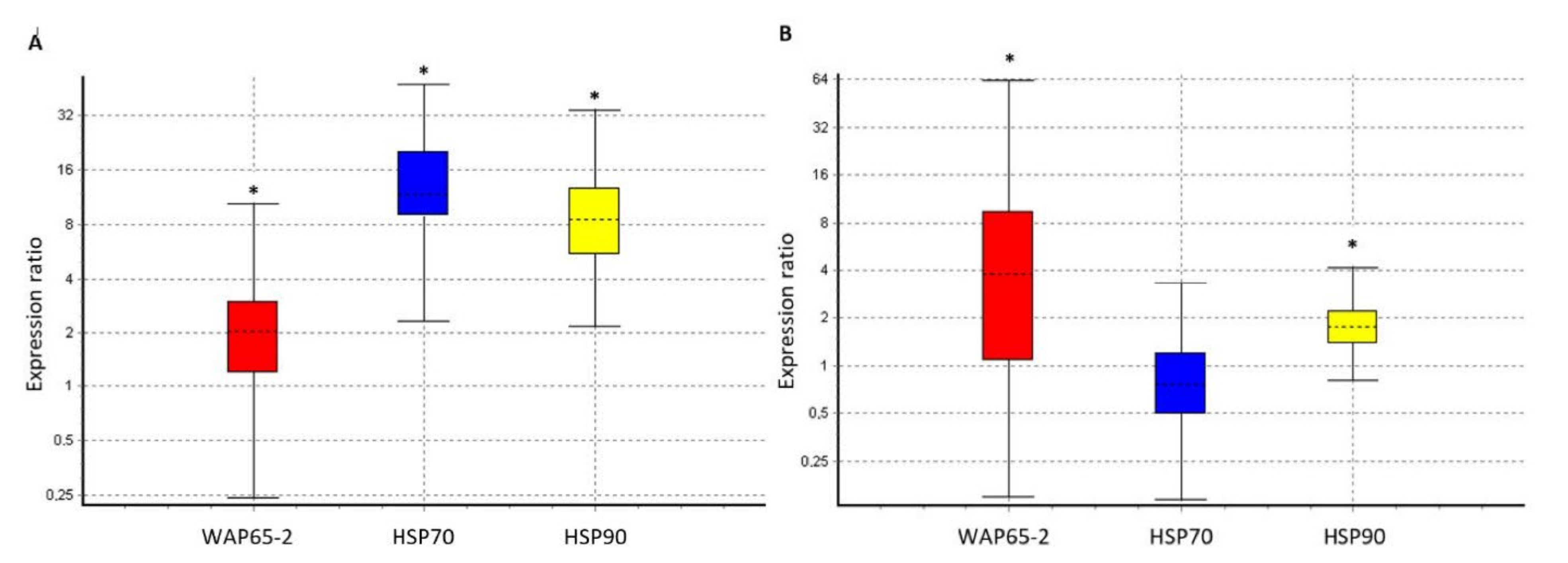
Stress-related genes expression in liver and kidney are presented in Figure 2. The expression of the WAP65-2 was 1.9-fold, HSP70 11.2-fold, and HSP90 was 8.2-fold higher in liver (Figure 2A), while WAP65-2 (3.9-fold) and HSP90 (1.7-fold) expression was higher in kidney (Figure 2B) of the wild-caught turbot compared to the farmed animals.

3.3. Histology Analyses

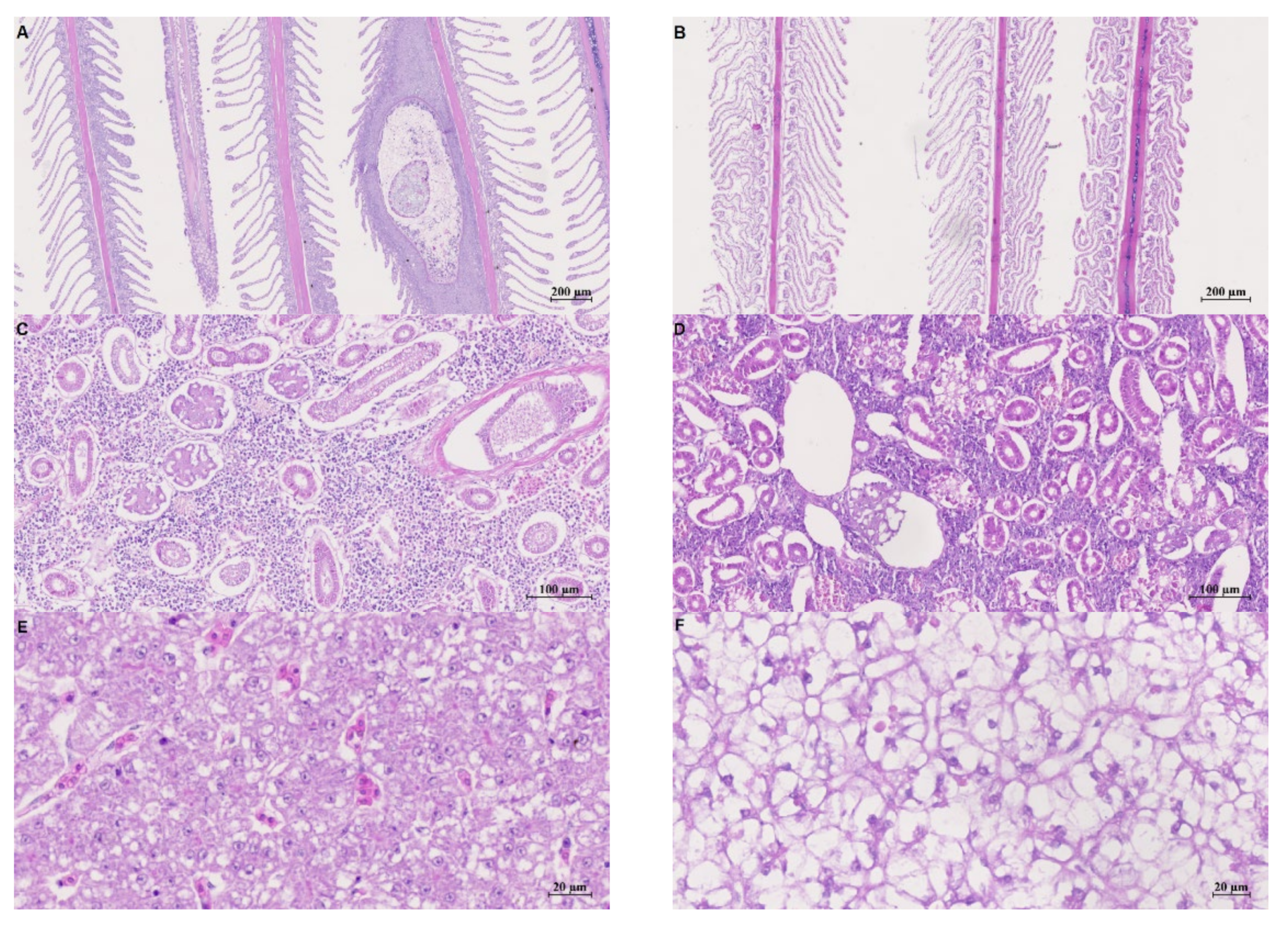
The histological examination of gill samples showed normal structure in the wild-caught turbot. Mucous cells were rich in mucus and randomly distributed from the interlamellar space to the tips of the lamellas. An infestation with digenean parasite Dactylogyrus sp. was noted. The parasite caused tissue reaction in terms of hyperplasia and hypertrophy of interlamellar cells (Figure 3A). In the farmed turbot (Figure 3B), gill tissue exhibited lamellar curling, presence of edema, and lifting of the epithelial layer. Increased number of mucous cells was noted in the interlamellar spaces. In the caudal kidney sections of the wild-caught turbot (Figure 3C), normal appearance of glomerulus was recorded with mild peritubular dilatation, but with extensive peritubular dilatation and tubular degeneration in farmed fish. Moreover, severe dilatation of Bowman capsule with different stages of atrophy and necrosis of glomerulus was noted, as well as hemorrhage in between tubules that suggests disruption of function (Figure 3D). Analysis of a wild-caught turbot liver (Figure 3E) exhibited scarce accumulation of glycogen in cells. Hepatocytes were densely packed, with presence of small lipid vacuoles indicating lower metabolic activity; moderate blood congestion was also observed. In the farmed turbot hepatocytes, increased accumulation of lipids and glycogen were recorded (Figure 3F). Analysis of wild-caught turbot intestines showed damaged and shortened intestinal villi caused by infestation of a tapeworm Bothriocephalus scorpii. Infestation of these parasites caused a progressive necrosis and atrophy response that extended throughout lamina epithelialis and lamina propria (Figure 3G). Intestine of farmed turbot exhibited normal length of intestinal villi and normal distribution and number of mucous cells. Ciliary layer of epithelial cells was well ordered (Figure 3H). Morphological structure of spleen tissue was normal with randomly distributed and well-organized melanomacrophage centers (MMC) in the wild-caught turbot (Figure 3I). In the farmed fish spleen tissue, the number of MMC was increased compared to the wild-caught population (Figure 3J).

4. Discussion

Fish respond to stress by activating the neuroendocrine system that leads to changes in plasma and tissue ion and metabolite levels, hematological profile, and HSPs [23]. Biochemical and hematological analyses thus provide important information about secondary response to stress in wild and captive conditions [6,24,25].
The GLU, TRIG, CHOL, and TP levels can reflect both the nutritional status and metabolic responses of fish to stress, since they represent the energy available during exposure to stressful conditions [23,26,27]. Our study has shown no significant difference in GLU level between examined groups, suggesting that GLU was not affected by farming condition and handling stress, which is consistent with results obtained by Liu et al. [17] and Waring et al. [28], but opposite to those of Čož-Rakovac et al. [5], who reported a significant elevation of GLU in farmed Dicentrarchus labrax compared to their wild counterparts. Continuous availability of high-energy feed (relatively high protein, lipids, and carbohydrates) and the reduction of activities in the culturing environment [29] resulted with significantly higher TRIG and TP concentrations, including ALB and GLOB fractions in farmed turbot. A wide range of GLU and TP observed in the wild-caught group (0.05–16.4 mmol/L and 10–46 g/L, respectively), combined with generally decreased other energy metabolites (TRIG), indicate possible starvation that is frequent in wild populations and could also be caused by Bothriocephalus scorpii detected in the wild-caught turbot intestine. B. scorpii, the most common parasite in turbot [30], causes a progressive intestine necrosis and atrophy, leading to malnutrition and lower resistance to stress [31] that could be the reason for significantly higher A/G ratio, which is often used as an indicator of stress response [27]. Reduction in plasma TRIG levels in the wild-caught fish could be attributed to lipolysis during starvation [32]. CHOL concentrations did not differ significantly between examined groups, which is in line with Aldrin et al. [33], who also did not record significant differences between farmed and wild turbot populations. Hill [34] reported that CHOL concentration increases as the fish size increases. However, we did not record such correlation (data not shown).
Urea and creatinine are end-products of protein metabolism, and changes in their concentrations could be caused by liver, kidney, or gill osmoregulatory disfunction [35,36,37]. We observed morphological changes in gill tissue of both groups (Figure 3A,B) but URE levels were not significantly different. However, manifold-higher CRE levels in the farmed group indicate disturbance of kidney filtration process, which is corroborated by detected pathohistological changes (Figure 3D). Ziskowski et al. [38] and Ajeniyi and Solomon [37] also stated that increased CRE concentrations suggest insufficiency of the kidney excretory functions. Significantly elevated TBIL levels in the farmed turbot in relation to its wild counterparts may indicate liver damage [39]. Histological analysis of the wild-caught turbot liver (Figure 3E) exhibited scarce accumulation of glycogen with densely packed hepatocytes, indicating lower metabolic activity, while the farmed turbot liver showed hepatocytes with increased accumulation of lipids and glycogen.
Plasma concentration of the electrolytes, such as Ca and P, have been used as indicators of secondary phase of stress response in fish, and their changes can be related to pathological disturbances [18,32]. Significantly decreased Ca and P levels in the farmed turbot, compared to the wild-caught, fish is a reflection of pathological changes detected in gills (that may affect the ionic permeability and cause decreased ionic levels in the blood) and kidney tissues. Renal dysfunction could also contribute to the hypocalcemia and hypophosphatemia [40]. The same changes induced multiple higher activity of intracellular enzyme CK that is released after cell damage and is therefore often used as an indicator of tissue damage [7,41]. Activity of ALP can be elevated following various stresses [42], and elevated activity of this enzyme in the wild-caught turbot is most likely caused by infestation with tapeworm B. scorpii [43].
Exposure of fish to unfavorable conditions induces oxidative stress and activation of enzymatic antioxidant defense mechanisms. Superoxide dismutase (SOD) is the first line of defense system and the most powerful antioxidant in the cell that catalyzes the decomposition reaction of the superoxide radical to the oxygen molecule and hydrogen peroxide. Hydrogen peroxide is further decomposed to water and oxygen by GPx. It plays an important role in preventing lipid peroxidation and membrane damage and also has microbicidal properties [44,45]. Antioxidant defenses in fish may be affected by many factors, such as the feeding behavior, nutritional factors, exposure to hypoxia, and parasitic infestations leading to increase in SOD and GPx activities [46]. Results of our study revealed significantly higher SOD and lower GPx activity in farmed turbot. The reason for higher SOD activity in farmed turbot could be feeding the fish diets with a high lipid level [47], while higher activity level of GPx in the wild-caught turbot could be due to parasite infestations, a higher level of membrane damage [46], and consequent malnutrition/starvation. This is consistent with the findings of Morales et al. [48], who showed significant increases of GPx level in sea bass deprived of food for 5 weeks.
Stress conditions can lead to change of leukocyte profile, causing lymphopenia accompanied or not by neutrophilia [49]. Therefore, the relative proportion of neutrophils to lymphocytes (N:L ratio) as an index of secondary stress response are increasingly used as a complementary method of physiological stress assessment [50]. Although we expected higher values in the farmed population, a significantly increased ratio was observed in the wild-caught turbot, which is most likely a consequence of infestation with B. scorpii or the stress hormones produced because of the infestation. There is evidence that this ratio could be influenced by diseases, including parasitic infestation [51].
Heat shock or stress proteins (HSPs) are another sensitive and reliable indicator of molecular response of fish to different stressors such as pathogens, inflammation, starvation, hypoxia, cold shock, environmental contaminants, and husbandry stressors [7,23]. Changes in gene expression of HSP-70, HSP-90, and WAP-65 associated with stress response to environmental (salinity, temperature, oxygen, hypoxia, and hyperoxia) and husbandry stressors (confinement, handling, overcrowding) have been detected in different fish species [52,53]. In turbot, many studies have focused on the temperature effects on HSP gene expression [54,55], whereas very little is known about how this species responds to different living regime within the optimal temperature range. In our study, we analyzed HSP70, HSP90, and WAP-65 in kidney and liver tissue and expected higher expression in the farmed population because of multiple farming stressors. However, HSP70, HSP90, and WAP-65 expression was several times higher in the wild-caught than in the farmed turbot. This is consistent with Liu et al. [16], who did not find influence of reared condition (increased stocking density) on HSP90 gene expression. However, bacterial infection [56,57] and starvation [58] can induce fish HSP gene expression, which can explain upregulation of HSP in the wild-caught turbot invaded by B. scorpii, which could affect nutrient resorption, leading to starvation and, consequently, upregulation of HSP.
Melanomacrophage centers (MMCs) are good biomarkers for stress assessment since the number and size of MMCs change in relation to several factors (stress, pollutants, diseases, starvation). It is thought that MMCs participate in both immune defense and nonimmunological, physiological processes [31,59]. Histopathological analyses of farmed turbot spleen tissue (Figure 3I) showed an increase in MMC density, which is in line with findings of Kurtović et al. [60] on sea bass, where cultured fish had increased MMC density compared to the wild population.
Tertiary stress response refers to aspects of whole-body performance. Condition factor is a suitable indicator for assessment of fish/population wellbeing [7,23]. Because of continuous, intensive feeding regime and limited space in captivity, farmed turbot were in a significantly better condition than the wild-caught group.

5. Conclusions

This is the first study comparing the stress response of farmed and wild-caught adult turbot on molecular, cellular, and whole-body levels. The results show that the morphological changes in gills, kidneys, liver, and spleen tissue observed in farmed turbot, also reflected in the measured biochemical parameters, are mainly influenced by aquaculture conditions, while wild populations are more susceptible to biotic stressors, such as parasite infestation, which was reflected in changes of gene expression of HSP. The overall results indicate that it is useful to use different parameters to assess the physiological responses and wellbeing of turbot because different types of stressors trigger different stress responses. In addition, the present study may be helpful to obtain general values for turbot plasma parameters and improve the knowledge of the biological responses of this species to different living conditions, which will lead to a better understanding of the biological and ecological characteristics of this species. Since the research was conducted during spring, future studies should focus on other seasons, especially in the warmer part of the year when the sea temperatures can reach values that are above optimal for this species.

Author Contributions

Conceptualization, I.S.-P., D.M., R.Č.-R. and N.T.P.; investigation, S.K., J.B., B.B.-L., M.B., S.B. and N.T.P.; methodology, J.B.; validation, B.B.-L., S.B., D.M., N.T.P. and K.B.; formal analysis, S.K., B.B.-L., K.B., I.S.-P., S.B. and M.B.; resources, R.Č.-R. and S.K.; writing—original draft preparation, S.K., J.B. and M.B.; writing—review and editing, I.S.-P., N.T.P., D.M., B.B.-L., S.B. and K.B.; visualization, J.B., M.B., K.B. and D.M.; supervision, I.S.-P. and R.Č.-R.; funding acquisition, R.Č.-R. All authors have read and agreed to the published version of the manuscript.

Funding

This research was supported by the Scientific Centre of Excellence for Marine Bioprospecting–BioProCro, a project co-financed by the Croatian Government and the European Union through the European Regional Development Fund: The Competitiveness and Cohesion Operational Programme (KK.01.1.1.01). The funders had no role in study design, data collection and analysis, decision to publish, or preparation of the manuscript.

Institutional Review Board Statement

Ethical review and approval were waived for this study due to procedures conducted according to European Directive 2010/63/EU (Article 1).

Informed Consent Statement

Not applicable.

Data Availability Statement

Not applicable.

Acknowledgments

The authors thank the employees of Cromaris d.o.o. (farm Budava) and M.Sc. Mario Lovrinov for technical assistance in turbot sampling.

Conflicts of Interest

The authors declare no conflict of interest.

References

  1. Petitjean, Q.; Jean, S.; Gandar, A.; Côte, J.; Laffaille, P.; Jacquin, L. Stress responses in fish: From molecular to evolutionary processes. Sci. Total Environ. 2019, 684, 371–380. [Google Scholar] [CrossRef] [PubMed]
  2. Montero, D.; Izquierdo, M.; Tort, L.; Robaina, L.; Vergara, J. High stocking density produces crowding stress altering some physiological and biochemical parameters in gilthead seabream, Sparus aurata, juveniles. Fish Physiol. Biochem. 1999, 20, 53–60. [Google Scholar] [CrossRef]
  3. Harper, C.; Wolf, J.C. Morphologic Effects of the Stress Response in Fish. ILAR J. 2009, 50, 387–396. [Google Scholar] [CrossRef] [PubMed]
  4. Sneddon, L.U.; Wolfenden, D.C.C.; Thomson, J.C. Stress management and welfare. In Biology of Stress in Fish—Fish Physiology 35; Schreck, C.B., Tort, L., Farrell, A.P., Brauner, C.J., Eds.; Academic Press: Cambridge, MA, USA, 2016; pp. 463–539. [Google Scholar]
  5. Čož-Rakovac, R.; Strunjak-Perović, I.; Topić Popović, N.; Hacmanjek, M.; Šmuc, T.; Jadan, M.; Lipej, Z.; Homen, Z. Cage culture effects on fish mullets (Mugilidae) liver histology and blood chemistry profile. J. Fish Biol. 2008, 72, 2557–2569. [Google Scholar] [CrossRef]
  6. Fazio, F.; Marafioti, S.; Arfuso, F.; Piccione, G.; Faggio, C. Comparative study of the biochemical and haematological parameters of four wild Tyrrhenian fish species. Veterinární Med. 2013, 58, 576–581. [Google Scholar] [CrossRef] [Green Version]
  7. Sopinka, N.M.; Donaldson, M.R.; O’Connor, C.M.; Suski, C.D.; Cooke, S.J. Stress Indicators in Fish. Fish Physiol. 2016, 35, 405–462. [Google Scholar] [CrossRef]
  8. Jardas, I. Jadranska Ihtiofauna; Školska Knjiga: Zagreb, Croatia, 1996; Volume 533. [Google Scholar]
  9. Person-Le Ruyet, J. Turbot (Scophthalmus maximus) Grow-out in Europe: Practices, Results, and Prospects. Turk. J. Fish. Aquat. Sci. 2002, 2, 29–39. [Google Scholar]
  10. Labatut, R.A.; Olivares, J.F. Culture of turbot (Scophthalmus maximus) juveniles using shallow raceways tanks and recirculation. Aquac. Eng. 2004, 32, 113–127. [Google Scholar] [CrossRef]
  11. Danancher, D.; Garcia-Vazquez, E. Turbot—Scophthalmus maximus. In Genetic Impact of Aquaculture Activities on Native Populations; Svåsand, T., Crosetti, D., García-Vázquez, E., Verspoor, E., Eds.; 2007; pp. 55–61. Available online: https://www.vliz.be/imisdocs/publications/318461.pdf (accessed on 23 November 2021).
  12. Bouza, C.; Vandamme, S.; Hermida, M.; Cabaleiro, S.; Volckaert, F.; Martinez, M. AquaTrace species Turbot (Scophthalmus maximus). Aquatrace.eu. World Wide Web Electronic Publication. 2014. Available online: https://fishreg.jrc.ec.europa.eu/web/aquatrace/leaflets/turbot (accessed on 23 November 2021).
  13. FAO. Scophthalmus maximus. Cultured Aquatic Species Information Programme. Text by Rodríguez Villanueva, J.L., Fernández Souto, B. Fisheries and Aquaculture Division [Online]. Rome. 2022. Updated 6 June 2005. Available online: https://www.fao.org/fishery/en/culturedspecies/psetta_maxima/ (accessed on 9 February 2022).
  14. Foss, A.; Imsland, A.K.D.; Van De Vis, H.; Abbink, W.; Lambooij, B.; Roth, B. Physiological response of temperature shocks in turbot and sole. J. Appl. Aquac. 2019, 31, 34–47. [Google Scholar] [CrossRef]
  15. Jia, Y.; Chen, X.; Wang, Z.; Meng, Z.; Huang, B.; Guan, C. Physiological response of juvenile turbot (Scophthalmus maximus. L.) during hyperthermal stress. Aquaculture 2020, 529, 735645. [Google Scholar] [CrossRef]
  16. Liu, B.; Jia, R.; Han, C.; Huang, B.; Lei, J.-L. Effects of stocking density on antioxidant status, metabolism and immune response in juvenile turbot (Scophthalmus maximus). Comp. Biochem. Physiol. Part C Toxicol. Pharmacol. 2016, 190, 1–8. [Google Scholar] [CrossRef] [PubMed]
  17. Liu, B.; Zhao, K.; Lei, J.; Huang, B.; Jia, R.; Wang, G. Stocking density effects on growth and stress response of juvenile turbot (Scophthalmus maximus) reared in land-based recirculating aquaculture system. Acta Oceanol. Sin. 2016, 36, 31–38. [Google Scholar] [CrossRef]
  18. Person-le Ruyet, J.; Lamers, A.; Le Roux, A.; Sévère, A.; Boeuf, G.; Mayer-Gostan, N. Long-term ammonia exposure of turbot: Effects on plasma parameters. J. Fish Biol. 2003, 62, 879–894. [Google Scholar] [CrossRef]
  19. Mota, V.C.; Hop, J.; Sampaio, L.A.; Heinsbroek, L.T.N.; Verdegem, M.C.J.; Eding, E.H.; Verreth, J.A.J. The effect of low pH on physiology, stress status and growth performance of turbot (Psetta maxima L.) cultured in recirculating aquaculture systems. Aquac. Res. 2018, 49, 3456–3467. [Google Scholar] [CrossRef] [Green Version]
  20. Jia, R.; Han, C.; Lei, J.-L.; Liu, B.-L.; Huang, B.; Huo, H.-H.; Yin, S.-T. Effects of nitrite exposure on haematological parameters, oxidative stress and apoptosis in juvenile turbot (Scophthalmus maximus). Aquat. Toxicol. 2015, 169, 1–9. [Google Scholar] [CrossRef]
  21. Mumford, S.; Heidel, J.; Smith, C.; Morrison, J.; MacConnell, B.; Blazer, V. Fish Histology and Histopathology, 4th ed.; US Fish & Wildlife Service: Elkins, WV, USA, 2007. [Google Scholar]
  22. Švob, M. Histološke i Histokemijske Metode; Svjetlost: Sarajevo, Bosnia and Herzegovina, 1974; pp. 142–205. [Google Scholar]
  23. Barton, B.A.; Morgan, J.D.; Vijayan, M.M. Physiological and condition-related indicators of environmental stress in fish. In Biological Indicators of Aquatic Ecosystem Stress; Adams, S.M., Ed.; American Fisheries Society: Bethesda, MD, USA, 2002; pp. 111–148. [Google Scholar]
  24. Wedemeyer, G.H.A.; Barton, B.A.; McLeay, D.J. Stress and acclimation. In Methods for Fish Biology; Schreck, C.B., Moyle, P.B., Eds.; American Fisheries Society: Bethesda, MD, USA, 1990; pp. 451–489. [Google Scholar]
  25. Zhou, X.; Li, M.; Abbas, K. Comparison of haemtology and serum biochemistry of cultured and wild Dojo loach Misgurnus anguillicaudatus. Fish Physiol. Biochem. 2009, 35, 435–441. [Google Scholar] [CrossRef]
  26. Congleton, J.L.; Wagner, T. Blood biochemistry indicators of nutritional status in juvenile salmonids. J. Fish Biol. 2006, 69, 473–490. [Google Scholar] [CrossRef]
  27. Jia, Y.; Gao, Y.; Chen, X.; Huang, B. Determination of optimal fasting time before blood sampling to get baseline data on serum biochemical characteristics in juvenile turbot (Scophthalmus maximus). Aquaculture 2018, 487, 83–88. [Google Scholar] [CrossRef]
  28. Waring, C.P.; Stagg, R.M.; Poxton, M.G. Physiological responses to handling in the turbot. J. Fish Biol. 1996, 48, 161–173. [Google Scholar] [CrossRef]
  29. Martínez, B.; Miranda, J.; Nebot, C.; Rodriguez, J.; Cepeda, A.; Franco, C. Differentiation of Farmed and Wild Turbot (Psetta maxima): Proximate Chemical Composition, Fatty Acid Profile, Trace Minerals and Antimicrobial Resistance of Contaminant Bacteria. Food Sci. Technol. Int. 2010, 16, 435–441. [Google Scholar] [CrossRef]
  30. Skrzypczak, M.; Rolbiecki, L. Helmintofauna of turbot Scophthalmus maximus (Linneaues, 1758) from the southern Baltic Sea including new data. Pol. J. Vet. Sci. 2015, 18, 599–605. [Google Scholar] [CrossRef] [Green Version]
  31. Sitjà-Bobadilla, A.; Estensoro, I.; Sánchez, J.P. Immunity to gastrointestinal microparasites of fish. Dev. Comp. Immunol. 2016, 64, 187–201. [Google Scholar] [CrossRef] [Green Version]
  32. Peres, H.; Santos, S.; Oliva-Teles, A. Blood chemistry profile as indicator of nutritional status in European seabass (Dicentrarchus labrax). Fish Physiol. Biochem. 2014, 40, 1339–1347. [Google Scholar] [CrossRef] [PubMed]
  33. Aldrin, J.; Messager, J.; Saleun, S. Analyses sanguines de turbots d’elevage immatures (Scophthalmus maximus L.). Aquaculture 1984, 40, 17–25. [Google Scholar] [CrossRef]
  34. Hille, S. A literature review of the blood chemistry of rainbow trout, Salmo gairdneri Rich. J. Fish Biol. 1982, 20, 535–569. [Google Scholar] [CrossRef]
  35. Wood, C.M.; McDonald, M.D.; Sundin, L.; Laurent, P.; Walsh, P.J. Pulsatile urea excretion in the gulf toadfish: Mechanisms and controls. Comp. Biochem. Physiol. Part B Biochem. Mol. Biol. 2003, 136, 667–684. [Google Scholar] [CrossRef]
  36. Kulkarni, R.S.; Barad, V.S. Effect of starvation on haematological and serum biochemical changes in the freshwater fish, Notopterus notopterus (Pallas). Int. J. Innov. Stud. Auat. Biol. Fish. 2015, 1, 24–29. [Google Scholar]
  37. Ajeniyi, S.A.; Solomon, R.J. Urea and creatinine of Clarias gariepinus in three different commercial ponds. Nat. Sci. 2014, 12, 124–138. [Google Scholar]
  38. Ziskowski, J.; Mercaldo-Allen, R.; Pereira, J.; Kuropat, C.; Goldberg, R. The effects of fin rot disease and sampling method on blood chemistry and hematocrit measurements of winter flounder, Pseudopleuronectes americanus from New Haven Harbor (1987–1990). Mar. Pollut. Bull. 2008, 56, 740–750. [Google Scholar] [CrossRef]
  39. Jee, J.-H.; Kim, S.-G.; Kang, J.-C. Effects of phenanthrene on growth and basic physiological functions of the olive flounder, Paralichthys olivaceus. J. Exp. Mar. Biol. Ecol. 2004, 304, 123–136. [Google Scholar] [CrossRef]
  40. Srivastav, A.; Rai, R.; Srivastav, S.K.; Suzuki, N.; Mishra, D. Effects of lead on the plasma electrolytes of a freshwater fish, Heteropneustes fossilis. Int. Aquat. Res. 2013, 5, 4. [Google Scholar] [CrossRef] [Green Version]
  41. Henry, J.B. Book Review: Clinical Diagnosis and Management by Laboratory Methods. Lab. Med. 2001, 32, 700. [Google Scholar] [CrossRef]
  42. Benjamin, M.M.; Mckelvie, D.H. Clinical Biochemistry. In Pathology of Laboratory Animal; Springer: New York, NY, USA, 1978; Volume II, pp. 1750–1815. [Google Scholar]
  43. Dick, T.A.; Chambers, C.; Isinguzo, I. Cestoidea (phylum Platyhelminthes). In Fish Diseases and Disorders; Woo, P.T.K., Ed.; CAB International: Wallingford, UK, 2006; pp. 391–416. [Google Scholar]
  44. Tocher, D.R.; Mourente, G.; Van der Eecken, A.; Evjemo, J.O.; Diaz, E.; Bell, J.G.; Geurden, I.; Lavens, P.; Olsen, Y. Effects of dietary vitamin E on antioxidant defense mechanisms of juvenile turbot (Schophthalmus maximus L.), halibut (Hippoglossus hippoglossus L.) and sea bream (Sparus aurata L.). Aquac. Nutr. 2002, 8, 195–207. [Google Scholar] [CrossRef]
  45. Ighodaro, O.M.; Akinloye, O.A. First line defense antioxidants-superoxide dismutase (SOD), catalase (CAT) and glutathione proxidase (GPX): Their fundamental role in the entire antioxidant defense grid. Alex. J. Med. 2018, 54, 287–293. [Google Scholar]
  46. Martínez-Álvarez, R.M.; Morales, A.E.; Sanz, A. Antioxidant Defenses in Fish: Biotic and Abiotic Factors. Rev. Fish Biol. Fish. 2005, 15, 75–88. [Google Scholar] [CrossRef]
  47. Rueda-Jasso, R.; Conceição, L.; Dias, J.; De Coen, W.; Gomes, E.; Rees, J.; Soares, F.; Dinis, M.; Sorgeloos, P. Effect of dietary non-protein energy levels on condition and oxidative status of Senegalese sole (Solea senegalensis) juveniles. Aquaculture 2004, 231, 417–433. [Google Scholar] [CrossRef]
  48. Morales, A.E.; Pérez-Jiménez, A.; Hidalgo, M.C.; Abellán, E.; Cardenete, G. Oxidative stress and antioxidant defenses after prolonged starvation in Dentex dentex liver. Comp. Biochem. Physiol. Part C Toxicol. Pharmacol. 2004, 139, 153–161. [Google Scholar] [CrossRef]
  49. Davis, A.K.; Maney, D.L.; Maerz, J.C. The use of leukocyte profiles to measure stress in vertebrates: A review for ecologists. Funct. Ecol. 2008, 22, 760–772. [Google Scholar] [CrossRef]
  50. Zebral, Y.D.; Zafalon-Silva, B.; Wiegand Mascarenhas, M.; Berteaux Robaldo, R. Leucocyte profile and growth rate as indicators of crowding stress in pejerey fingerlings (Odontesthes bonariensis). Aquac. Res. 2015, 46, 2270–2276. [Google Scholar] [CrossRef]
  51. Davis, A.K.; Maney, D.L. The use of glucocorticoid hormones or leukocyte profiles to measure stress in vertebrates: What’s the difference? Methods Ecol. Evol. 2017, 9, 1556–1568. [Google Scholar] [CrossRef]
  52. Eissa, N.; Wang, H.P. Transcriptional stress responses to environmental and husbandry stressors in aquaculture species. Rev. Aquac. 2016, 8, 61–88. [Google Scholar] [CrossRef]
  53. Wu, L.; Wang, Y.; Han, M.; Song, Z.; Song, C.; Xu, S.; Li, J.; Wang, Y.; Li, X.; Yue, X. Growth, stress and non-specific immune responses of turbot (Scophthalmus maximus) larvae exposed to different light spectra. Aquaculture 2020, 520, 734950. [Google Scholar] [CrossRef]
  54. Huang, J.-F.; Xu, Q.-Y.; Chang, Y.-M. Effects of temperature and dietary protein on gene expression ofHsp70andWap65and immunity of juvenile mirror carp (Cyprinus carpio). Aquac. Res. 2014, 46, 2776–2788. [Google Scholar] [CrossRef]
  55. Huang, Z.; Ma, A.; Wang, X.; Lei, J.; Li, W.; Wang, T.; Yang, Z.; Qu, J. Interaction of temperature and salinity on the expression of immunity factors in different tissues of juvenile turbot Scophthalmus maximus based on response surface methodology. Chin. J. Oceanol. Limnol. 2014, 33, 28–36. [Google Scholar] [CrossRef]
  56. Forsyth, R.B.; Candido, E.P.M.; Babich, S.L.; Iwama, G.K. Stress Protein Expression in Coho Salmon with Bacterial Kidney Disease. J. Aquat. Anim. Heal. 1997, 9, 18–25. [Google Scholar] [CrossRef]
  57. Diaz-Rosales, P.; Pereiro, P.; Figueras, A.; Novoa, B.; Dios, S. The warm temperature acclimation protein (Wap65) has an important role in the inflammatory response of turbot (Schophthalmus maximus). Fish Shellfish Immunol. 2014, 14, 80–92. [Google Scholar] [CrossRef] [Green Version]
  58. Yengkokpam, S.; Pal, A.; Sahu, N.; Jain, K.; Dalvi, R.; Misra, S.; Debnath, D. Metabolic modulation in Labeo rohita fingerlings during starvation: Hsp70 expression and oxygen consumption. Aquaculture 2008, 285, 234–237. [Google Scholar] [CrossRef]
  59. Steinel, N.; Bolnick, D.I. Melanomacrophage Centers As a Histological Indicator of Immune Function in Fish and Other Poikilotherms. Front. Immunol. 2017, 8, 827. [Google Scholar] [CrossRef] [Green Version]
  60. Kurtović, B.; Teskeredžić, E.; Teskeredžić, Z. Histological comparison of spleen and kidney tissue from farmed and wild European sea bass (Dicentrarchus labrax L.). Acta Adriat. 2008, 49, 147–154. [Google Scholar]
Figure 1. Sampling location in the Adriatic Sea, Croatia.
Applsci 12 04201 g001
Figure 2. Relative expression of WAP65-2, HSP70, and HSP90 genes in liver (A) and kidney (B) of the wild-caught, compared to the farmed, turbot Scophthalmus maximus. Ct values obtained by quantitative real time PCR were analyzed with REST 2008. Data are presented relative to farmed turbot (expression set as 1) as whisker–box plots in order to see expression median and distribution of permutated expression values with 95% confidence level. Therefore, the expression ratio values of >1 denote higher fold-change in wild-caught than in farmed turbot and values of <1 denote lower fold-change in wild-caught than in farmed turbot. Significant changes are marked as * p < 0.05.
Applsci 12 04201 g002
Figure 3. Histopathological changes in tissues of the wild-caught ((A)—gills; (C)—caudal kidney; (E)—liver; (G)—intestine; (I)—spleen) and the farmed ((B)—gills; (D)—caudal kidney; (F)—liver; (H)—intestine; (J)—spleen) turbot Scophthalmus maximus.
Applsci 12 04201 g003aApplsci 12 04201 g003b
Table 1. Primers and reaction conditions used in the experiments.
GenesPrimers Sequence (5′-3′)Size (bp)Reaction Condition
s18FW: ACTGAGGATGAGGTTGAGAG
RV: TCCAGACCATTAGCAAGGA
13359 °C
3.5 mM MgCl2
WAP65-2FW: GTTAGACGCCATCACCACTG
RV: CGCATGTAGACTGGACCTGA
8763 °C
3 mM MgCl2
HSP70FW: GTGTGCTCATTCAGGTCTATG
RV: ACCCTTGTCGTTTGTGATG
8159 °C
3 mM MgCl2
HSP90FW: GAAGTGTCTGGAGCTCTTTG
RV: ATGCGGGACAGGTATTCT
18260 °C
3.5 mM MgCl2
Table 2. Biochemistry and leukocyte profiles of the farmed and the wild-caught turbot Scophthalmus maximus from the northern Adriatic Sea.
Plasma ParameterFarmed TurbotWild-Caught Turbotp-Value
Median25th–75th PercentileMin–MaxMedian25th–75th PercentileMin–Max
Glucose (mmol/L)2.311.50–2.940.66–3.881.610.75–4.210.05–16.40.367
Triglycerides (mmol/L)7.545.69–9.731.26–14.461.490.95–2.060.37–5.17<0.0001
Cholesterol (mmol/L)3.853.37–4.772.64–5.444.213.42–5.990.87–12.320.1917
Total protein (g/L)40.537–4334–492622.25–33.7510–46<0.0001
Albumin (g/L)11.511–129–1476–103–14<0.0001
Globulin (g/L)28.528–3125–351916.25–23.507–32<0.0001
A/G ratio0.390.37–0.410.32–0.460.420.38–0.450.30–0.54<0.05
Bilirubin (µmol/L)1.030.79–1.580.19–4.470.0520.048–0.420.047–1.38<0.0001
Urea (mmol/L)2.261.85–2.621.13–3.562.421.76–3.470.64–5.180.295
Creatinine (µmol/L)10768–12212–17327.4023.75–32.37514.5–58<0.0001
Calcium (mmol/L)3.142.87–3.342.63–3.753.973.46–4.993.05–6.98<0.0001
Phosphorus (mmol/L)5.254.30–5.983.1–7.46.916.16–9.083.59–11.28<0.0001
Alkaline phosphatase (U/L)21.517–3312–399570.75–12739–402<0.0001
Creatine kinase (U/L)211.578–115818–4032164–54.51–795<0.0001
Superoxide dismutase (U/mL)0.8810.423–1.7920.179–3.5790.2320.003–0.3340–0.649<0.0001
Glutathione peroxidase (U/L)210185.5–240.5127–317582283–870108–2914<0.0001
Leucocyte profile
Lymphocytes (%)81.3067.40–84.2244.90–91.6767.2055.55–78.1333.33–89.160.053
Neutrophils (%)4.170.39–8.000–16.0017.0310.45–24.473.78–56.41<0.001
Monocytes (%)15.7510.87–28.318.33–39.7916.398.29–20.934.42–27.820.334
N:L ratio0.050.005–0.100–0.360.230.13–0.500.05–1.69<0.0001
Publisher’s Note: MDPI stays neutral with regard to jurisdictional claims in published maps and institutional affiliations.