Article

Design and Implementation of Anatomically Inspired Mesenteric and Intestinal Vascular Patterns for Personalized 3D Bioprinting

1
Department of Biomedical Engineering, Purdue School of Engineering and Technology, Indiana University–Purdue University Indianapolis, Indianapolis, IN 46202, USA
2
3D Tissue Bioprinting Core Laboratory, Indiana Institute for Medical Research, Richard L. Roudebush VA Medical Center, Indianapolis, IN 46202, USA
3
Department of Surgery, IU School of Medicine, Indianapolis, IN 46202, USA
*
Author to whom correspondence should be addressed.
Appl. Sci. 2022, 12(9), 4430; https://doi.org/10.3390/app12094430
Submission received: 21 March 2022 / Revised: 11 April 2022 / Accepted: 19 April 2022 / Published: 27 April 2022
(This article belongs to the Special Issue 3D Bioprinted Tissues for Personalized Medicine Approaches)

Abstract

:
Recent progress in bioprinting has made possible the creation of complex 3D intestinal constructs, including vascularized villi. However, for their integration into functional units useful for experimentation or implantation, the next challenge is to endow them with a larger-scale, anatomically realistic vasculature. In general, the perfusion of bioprinted constructs has remained difficult, and the current solution is to provide them with mostly linear and simply branched channels. To address this limitation, here we demonstrated an image analysis-based workflow leading through computer-assisted design from anatomic images of rodent mesentery and colon to the actual printing of such patterns with paste and hydrogel bioinks. Moreover, we reverse-engineered the 2D intestinal image-derived designs into cylindrical objects, and 3D-printed them in a support hydrogel. These results open the path towards generation of more realistically vascularized tissue constructs for a variety of personalized medicine applications.

1. Introduction

Intestinal diseases represent a significant segment of heath care, both in the US and worldwide [1]. An example is the necrotizing enterocolitis of newborns, where even a small graft applied before other solutions are found could make the difference between an infant’s life or death [2]. However, the field still lacks an adequate technology for the generation of models needed to tissue engineer intestinal constructs for in vivo implantation or for in vitro mechanistic studies and drug testing [3].
3D Bioprinting has emerged as one of the most promising approaches to fill this gap due to its precision, reproducibility, and high throughput [4]. In spite of conceptual and technical difficulties, the bioprinting of tissues and organs progresses slowly but surely. In particular, there are notable developments in bioprinting of intestinal tissue as simple epithelial-fibroblasts monolayers (useful as generic constructs for toxicological applications [5]). More recently, iPSC-derived gastrointestinal organoids self-assembled in cylindrical structures [6], or cylindrical ‘bio-patches’, were functionally integrated with resected rat intestines [7]. Thus, bioprinting of intestinal tissues is gaining increased attention for these and other reasons [8]. Of particular interest is the report of successful generation of intestinal mucosa with ‘vascularized’ finger-like villi [9,10]. However, these structures need the connection to a perfusion system, preferably following the cylindrical organization of this tube-like tissue, and further to the supporting mesentery, at the time when this will also be considered for in vivo implantation and/or attachment to the recipient abdominal cavity.
In order to stay alive, any cell in animal tissues is located no further than about 200 μm from a nearby capillary [11]. The microvascular networks are supplied by larger vessels distributed in space in a fractal manner [12], and composed of segments with increasingly complex cellular composition as the diameter of the vessels increases. Yet, the bioprinting of constructs with vascularization (and even more so with innervation) lags behind, although critical for their survival and function [13]. At least in part, this is due to the origination of bioprinting from the technology of additive manufacturing [14], which focuses on the objects’ external shape with less consideration for their inner structure, which, in general, is supposed to be filled with more or less homogenously distributed materials, or with cells expected to perform self-assembling [15].
So far, most of the ‘vascularization’ of bioprinted constructs was provided by linear channels, occasionally lined by an endothelial layer [13]. Of note, simple vascular channels have been incorporated in the bioprinted model of the intestinal finger-like villi, fabricated using a collagen bioink alone [9], or, in an improved version, collagen supplemented with extracellular matrix from the small intestinal submucosa [10]. The next step would be to connect these microvessels to blood supply networks with the structural properties of the intestinal vasculature and, desirably, also to mesenteric ones, preferably in a patient-personalized manner; however, to our knowledge such constructs have not been created yet.
To address this limitation, we recently suggested to directly import, in the bioprinted models, anatomically realistic tissue-specific structures [16] and vascular patterns [17] derived from microscopic or macroscopic images, respectively. One of the conspicuous features of vascularization is the fractal organization of its branched architecture, with dimensions and cellular composition dependent of the branching order, also known as ‘branch generation’ [18]. Thus we asked whether it is possible to capture and transfer this refined structural information in the printed vascular patterns using a dedicated image analysis program called VESGEN 2D [17], and we showed its utility in printing such patterns extracted from human eye fundus [19]. Here we applied this same workflow specifically to intestinal (both colonic and mesenteric) vasculature. The expectation is that after deploying a cell-containing bioink in linear patterns on a surface, the cells will self-assemble in vascular cords [20], and eventually will generate perfusable lumens [21].
Towards this goal, here we showed how to extract branch-specific vascular information from microscopic images, and direct-write print them for generating anatomically similar mesenteric and intestinal vascular patterns. To this end, we used the semi-automated image analysis platform VESGEN 2D [18], which has been previously applied to the analysis of intestinal [22], retinal [23], and other vascular fields [24]. Using this program, here we demonstrated a practical workflow of integrated image analysis with the preparation of intestinal vascular patterns for 3D (bio)printing. In the process, we also had to overcome several technical limitations of the current bioprinting technology, and thus we hope this work will inspire other users to seek and find similar or even more efficient solutions.
3D printed models derived from tissue-specific vascular structures also show promise for in vitro and in silico testing [25,26]. Thus, the anatomic vasculature-derived 3D printed constructs make gains not only toward the ultimate goal of tissue engineering to create whole synthetic tissues, but also for studying blood flow dynamics and the mechanisms of tissue regeneration (e.g., neovascularization) in culture, and for other applications.

2. Materials and Methods

2.1. Sources of Images

A rat mesenteric image was obtained from ref. [27], and one representing a montage of 148 images capturing 3.5 cm of a normal mouse colon for the assessment of the vascular network stained with Alexa 647-conjugated lectin from ref. [22].

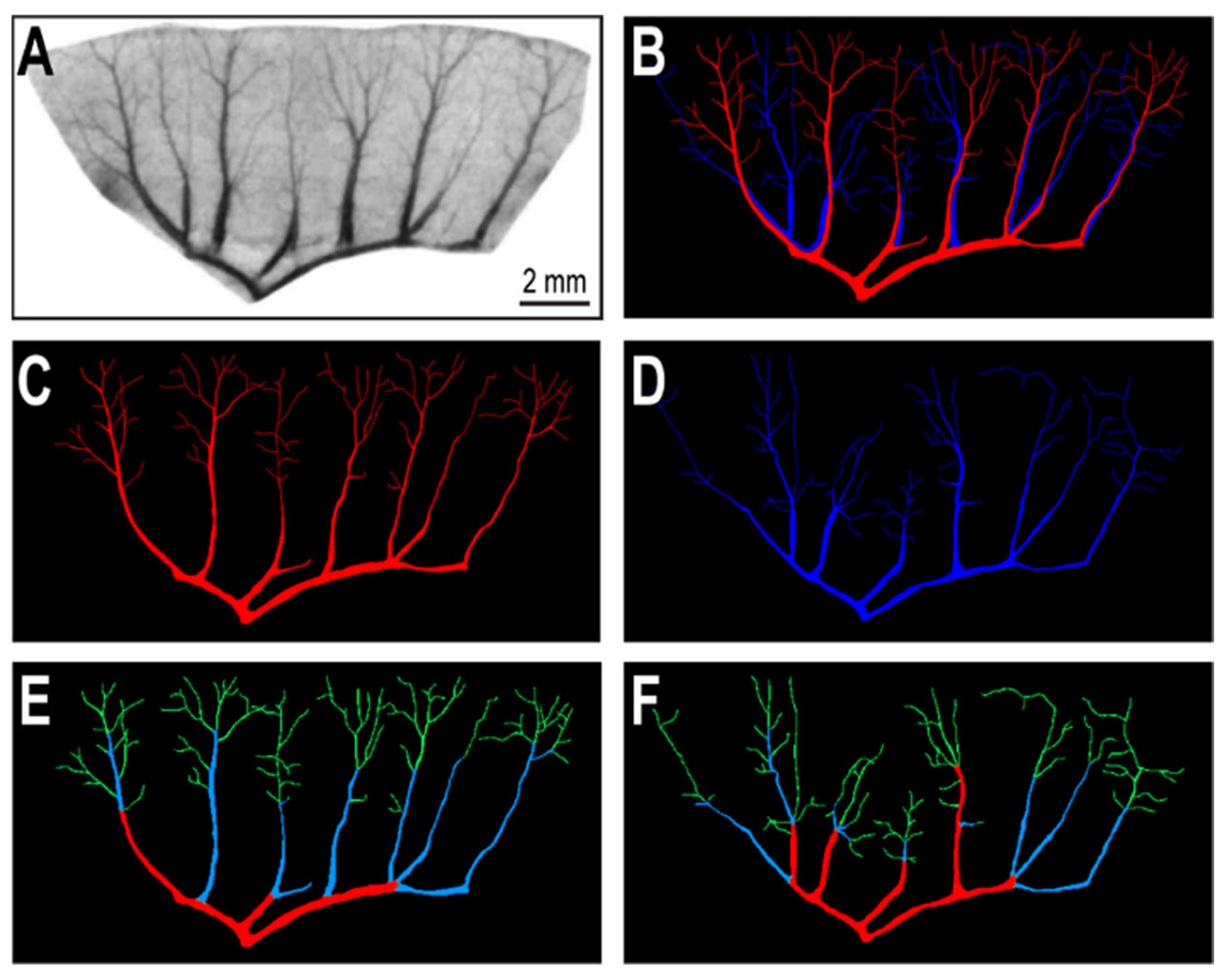
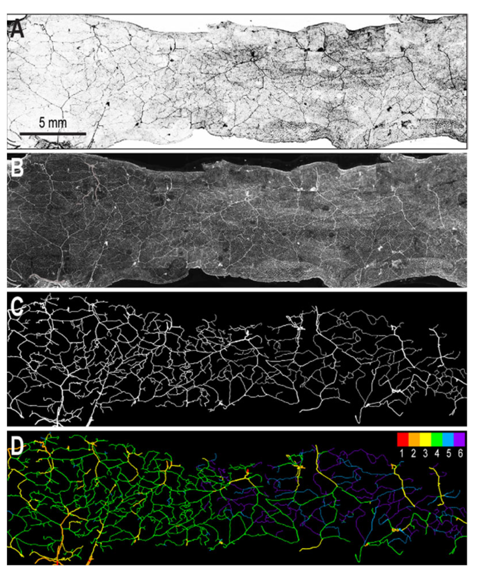
2.2. Segmentation of Vascular Patterns and VESGEN 2D Image Processing

The vasculature images were segmented in two steps: first manually (to identify the arterial and venous vessels and to isolate them into separate binary images) using Adobe Photoshop, followed by contrast adjustment, branch assignment, and artificial coloring, which were automatically performed by the VESGEN 2D program in several steps, as originally described in ref. [18]. The process to extract the vasculature from the colon image was the same as presented in detail for retina patterns, shown in Figure 3 in our publication [19]. However, due to the ‘network’ rather than ‘branching’ pattern of the colon vascularization, there is no method to differentiate between arterial and venous sides in this pattern, and therefore the colon vascular network was analyzed based on ‘generations’ only. The intestinal patterns were processed with VESGEN 2D using the ‘Vascular Tree’ morphology option, and the colon pattern was analyzed using the ‘Tree-Network Composite’ module, respectively. For reasons further explained below, the branch generations produced in the first analysis were then grouped into three generations using the ‘Group Generations’ function of the VESGEN 2D program to allow specific vascular segments to be printed with different biomaterials depending on desired composition, as addressed in the subsequent sections.

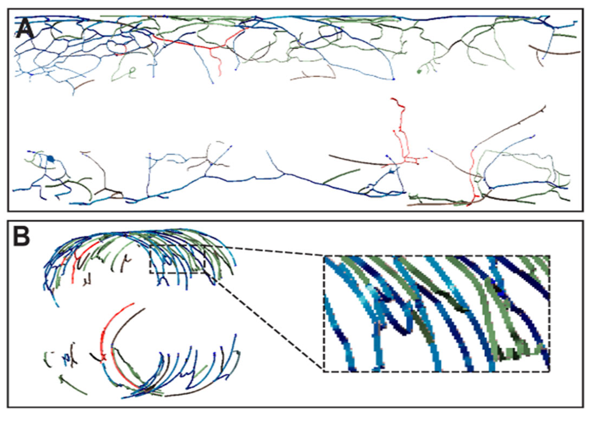
2.3. Preparation of Images for Printing

After reduction of generations to three groups, the colors were redefined in images using Adobe Photoshop as red (generation 1), blue (generation 2), and green (generation 3). Then, each generation was isolated into a separate image and converted to a binary image using Adobe Photoshop. The binary images were converted from .png format to .svg format via an online image converter (https://image.online-convert.com/convert-to-svg, accessed on 20 March 2022). The .svg formatted images were then imported into Autodesk Fusion 360 program as sketches. Selecting the surface of each .svg sketch, the surfaces were extruded by 1 mm. After exporting the resulting files in .stl format, the constructs were re-imported for printing into the BioCAM software of the 3DBiofactory bioprinter (from regenHU, Villaz-St-Pierre, Switzerland).
For 3D printing of the colon pattern, a cylinder with the same length of the colon image and circumference diameter of the image width was created in Fusion 360. Using the Sheet Metal Tool, the cylinder was unfolded to create a flat rectangle with the same dimensions of the colon image in mm. Then the surface of each imported .svg file, mentioned previously, was projected onto the surface of the rectangle using the Split Face function and subsequently extruded 1 mm from the surface. After repeating this process for all three generations, the rectangle was refolded into its original cylindrical configuration. Taking another cylinder with the same diameter of the original, the cylinder was removed, leaving the 3D vascular network. The three colon vascular patterns were exported as .stl files and re-imported into regenHU’s BioCAM program for printing.

2.4. Printing of Vascular Patterns

For proof-of-concept, the .stl files were printed on glass slides or on the bottom of tissue culture plate wells with a layer height of 1 mm (to prevent the printhead from scratching the printing surface). To this end, we used the syringe plunger printhead of the bioprinter with the commonly used glycerin-based Nivea paste (Nivea Crème, Hamburg, Germany) as a bioink surrogate [28], to which oil-based red, blue, and green food dyes were added as colorants. In selected experiments, the flat vascular patterns were also printed on the bottom of tissue culture plate wells in ‘direct-write’ mode [15], with acidic (LifeInk 240) or neutral (LifeInk 200) collagen bioinks (both from Advanced BioMatrix, San Diego, CA, USA), or with an alginate-nanocellulose bioink (from Cellink USA, Blacksburg, VA, USA). Alternatively, the cylindrical patterns were printed with Nivea paste within a polyacrylic hydrogel (Carbopol 980, from Lubrizol, Cleveland, OH, USA) [29], similarly to the ‘Free Reversible Embedding of Suspended Hydrogel’ (FRESH) method [30].
Human EC line EA.hy926 (ATTC Cat. #CRL-2922) was expanded according to the vendor’s instructions and labeled with Cell Tracker Green and Red (ThermoFisher Scientific/Molecular Probes, Cat. #C2925 and Cat. #C34565, respectively), and then suspended in LifeInk 240 at a concentration of 106 cells/mL. To this end, the medium- and small-sized branches of the patterns were consolidated in one generation and recolored in green, with the larger branches remaining to be labeled with red color. These larger branches were then printed with a feed rate of 2 mm/s and syringe extrusion velocity of 0.0075 mm/s, and the smaller ones with 1.75 mm/s feed rate at 0.0075 mm/s extrusion velocity. Images were acquired with an Olympus IX73 microscope with a 10x objective with an automatic scanning platform and a DP80 digital camera set at 4 × 4 binning. The images were subsequently processed in MetaMorph (v.7.7.0.0; Molecular Devices, San Jose, CA, USA), and displayed overlapping in phase contrast and on the green or red fluorescence channels.
Given that the CellTracker labels are also ‘vital stains’ (initially invisible but converted into a fluorescent form only inside of living cells), the post-printing survival of the cells was shown by the detectable fluorescence. However, we did not follow up the fate of the cells in long-term culture, since the inclusion of HUVEC in the printing process was only to see how their presence affects the printability of the collagenous bioink.

3. Results

In order to show how to transfer a vascular pattern from an image of a rat mesenteric vascular bed, obtained after the in vivo perfusion of an optical contrast enhancer to a printed construct (Figure 1A) [27], we used the procedure employed in our recent publication [19]. After background color reversal and segmentation, the vascular segments were identified (Figure 1B) and split in two separate arterial and venous groups (Figure 1C,D) respectively. To these vascular fields, we then applied the branch analysis provided by the VESGEN 2D software, which identified three branch generations (in colors in Figure 1E,F).
These files served for branch-specific .stl file generation (Figure 2A,B); then, we proceeded to direct-write printing with colored Nivea paste as a surrogate bioink (Figure 2C,D). We also printed these mesenteric vascular patterns with hydrogels (Figure S1), specifically with acidic (Figure S1A,B) and neutral (Figure S1C,D) collagen bioinks, with alginate-nanocellulose bioink (Figure S1E,F), and also with two-colors labeled EC in collagen bioink (1:1 with culture medium) (Figure S1G,H). Altogether, we found that acidic collagen best preserved the lines’ morphology and resolution, as in our previous study [19].
Similarly, to assess the usefulness of this workflow for generation of printable vascular patters with a different organization, we applied the same protocol to a segment of mouse colon. As shown in Figure 3, compared to that of the mesentery, the colon microvasculature has a crisscrossed and a more uniform network-like (rather than branched) organization. In this case, we could not separate the arterial from venous components, as there was no identifiable branch-originating (‘trunk’) vessel visible in these images, taken from the luminal side of the colon (Figure 3A). However, by applying the standard VESGEN 2D image processing protocol in the ‘network analysis’ mode (Figure 3B–D), and after generation reduction (Figure S2A), the program still identified a gradient of vessel dimensions and connections, suggesting a progressive branching going from left to right in Figure S2B–D.
For printing, we selected a central region of the image containing the overlap of these three ‘pseudo-generations’ (Figure 4A). The background was reverted again for clarity of the image (Figure 4B), and the three generations were separated and converted in individual stl files (Figure 4C). These files were then printed in contiguity with the correspondingly colored surrogate bioink (Figure 4D), as well as with two common bioinks, one based on collagen (Figure S3A) and one on nanocellulose-containing alginate (Figure S3B).
The colonic vascular network was also projected on a cylinder, as described in Materials and Methods. For this, the VESGEN-processed, branch-assigned intestinal network (Figure 5A) was rolled virtually onto a cylinder with the diameter required for providing the network’s continuity at the cylinder’s fusion line (Figure 5B). In preparation for actual 3D printing, the three generations were also separated and rolled independently (Figure 5C–E).
For proof of concept, we isolated the selected region of Figure 4 from the 3D model in Figure 5 and reconfigured it as in Figure 6. To avoid the spatial crossing of the printhead’s paths in between the upper and deeper levels of the model, the CAD was virtually sectioned by a horizontal plane in two semi-cylinders, retaining the generation allocations performed before (Figure 6A).
Of note, our and other bioprinters’ software decompose the continuous lines representing the vascular patterns in short linear segments, which after recombination generate lines that were thicker than the original design (Figure 6B), as discussed in more detail below. Then, these separate upper and lower 3D constructs were printed in contiguity in a supporting hydrogel by the FRESH method with the colored bioink surrogate, as shown from both the top (Figure S3C) and the bottom (Figure S3D) of the construct.

4. Discussion

To assign and transfer in printable constructs branch-specific information, here we recapitulated an image analysis-based workflow to process small portions of microscopic rodent mesenteric and intestinal tissues images, using the semi-automatic network analysis program VESGEN 2D [18], as we previously showed for human retinal vascular patterns [19]. To materialize this procedure, we printed the patterns in 2D and in the 3D mode with a colored bioink surrogate, with alginate-nanocellulose bioink, as well as with a cell-containing collagen bioink.
The logic behind choosing the printable materials to test was to move sequentially from a ‘practice level’ colored paste used for showing how the ‘branch generation’ information can be passed on from VESGEN 2D analysis to an actual print, to biological ones usable for bioprinting. For the latter, we started with a commercial collagenous bioink (“LifeInk”) as the most suitable support for microvascular cells, which is available in two formulations: (i) LifeInk 240, the acidic and thus more fluid form (suitable for hydrogel-embedded bioprinting where it undergoes jellification by taking the support bath’s pH); (ii) LifeInk 200, which has a neutral pH but is more viscous, yet appropriate for direct-write bioprinting of endothelial cells. In our hands, this second formulation led to a loss of printing resolution as compared with the acidic version, and for this reason we also tried another largely used alginate-nanocellulose bioink [31]. However, this did not provide a better printing resolution, with the additional inconvenience of being less transparent than collagenous bioinks, a feature needed for imaging the fluorescently labeled cells. Therefore, we returned to the most physiologic and convenient bioink (neutral collagen), to which we added EC for the final printing.
Admittedly, our work did not generate perfusable structures, which is the next phase of our research. However, a hydrogel line-confined self-assembling of microvascular structures from ether primary EC, or from the ‘stromal vascular fraction’ has been previously suggested [15]. Looking forward, it is worth considering the same approach as in the recently described ‘Bioprinting-Assisted Tissue Emergence’ (BATE) method, which uses very high densities of stem cells that spontaneously self-organize within hydrogels lines [6]. In these, the stem cells-derived tissue constructs are determined to organize according to the printing-imposed geometry, as anticipated elsewhere as well [16,17].
Another limitation of this study is that the printed images were only topological equivalents, and not exact representations of the original images. In part, this is due to the difficulty to detect and extract the finest microvessels from low-magnification microscopic images, as employed here. In addition, during VESGEN 2D processing one may intentionally reduce the number of vascular generations, as also illustrated here. This simplification is highly desirable in some circumstances, for example when splitting them down into too many groups is counterproductive, considering the additional lengthening of the procedure and of the printing time. These considerations are true for the vascular patterns of both intestine and mesentery (here treated distinctly, because of their different vascular organization and functions [32]).
A reason for the departure of the printed patterns from the initial images is also due to a technical limitation related to the current status of the printing technology. On some instruments (including ours) the straight or other geometric lines can be printed continuously with a resolution close to the diameter of the printheads (which in extrusion mode can be 0.25 mm or less). However, an arbitrarily shaped, tortuous line is treated by the path-generation software as a very elongated ‘object’, which is then further decomposed in shorter, parallel segments. This rendering generates thicker lines than the original ones, even when printed in a single layer. Added to this complication are the printability and hardening of the hydrogel (with or without a crosslinker), as well as its adhesivity to the substrate which, when too high, will lead to additional widening of the lines.
However, all these artifacts may have a lesser impact on the thickness of the actual cell cords generated within the printed hydrogel patterns. In fact, the ultimate arbiters of cells organization in cords are: (i) their phenotypes and composition (e.g., if adding or not supporting pericytes), (ii) the dynamics of self-assembling, and (iii) the in situ maturation process. Moreover, in vivo, these vasculogenic processes will be further sculptured by the contribution of inflammatory and/or repairing cells, mainly by the macrophages’ M1/M2 ratio, and possibly by spontaneously recruited progenitor cells [33], or by intentionally adding iPSC-derived vascular cells [34].
An alternative method to generate microvascular conduits in vitro relies on microfluidic devices containing microvascular channels, which demonstrate great benefits for in vitro experimentation for mechanistic studies and drug discovery [35]. Among their advantages are: design is straightforward to implement; conveniently grouped as either linear (or geometrically-branched) channels directly lined by endothelial cells, or hydrogel microdomains spontaneously colonized with microvascular cells (endothelial, smooth muscle cells and/or pericytes) by sprouting or self-assembly; controllable placement as related to their surroundings; predictable and stable conduit diameters; easy to connect to the outside fluid supply and to perfuse; ability to integrate with multiple parenchymal cell types, generating single or interconnected ‘organs on chip’ [36]; in-plane confinement for high quality microscopic imaging, etc.
These possibilities are derived from embedding the microfluidic channels in plastic material supports with pre-designed spatial features implemented by photolithography. However, from the standpoint of vascularization this technology has the following limitations: it may be constrained to the use of only pre-determined cell types capable to initially adhere to the walls of the microchannels; the presence of plastic materials could interfere with the inter-cellular interactions of the microvascular and parenchymal cells; due to the presence of plastics, it is not suitable for in vivo implantation; the active areas are limited to small (‘chip’) size ranges; further assembly is possible only by stacking and/or inter-device coupling, and this cannot generate natural 3D shapes; the devices have a predetermined, rigid spatial distribution, limiting the cells’ ability to self-organize only to the hydrogel microdomains and spontaneously adopt co-localization optimal for their function; the shapes of the microfluidic channels were in general linear or with simple geometric branches, constrained by the need of maintain a laminar flow inside of them, while in principle being possible to design more physiologic designs, etc. Some of these problems with microfluidic channels may not be encountered when using the bioprinted constructs envisioned here, when the method will mature enough to generate functional, perfusable (micro)vessels, and when the printing is performed on a transferable, biological ‘biopaper’ [37,38].
In conclusion, our study opens the way towards bioprinting of anatomically realistic mesenteric and intestinal patterns with flat (2D) or spatial (3D) distributions, as intended for a variety of personalized (i.e., authentic, patient-derived) in vitro and in vivo applications.

Supplementary Materials

The following supporting information can be downloaded at: https://www.mdpi.com/article/10.3390/app12094430/s1: Figure S1. Printing with bioinks of the mesenteric vascular patterns; Figure S2. Additional processing of the mouse colon vascular pattern for direct-write printing; Figure S3. 3D Printing of the colonic vascular patterns with hydrogels.

Author Contributions

Conceptualization: N.I.M.; investigation: R.C., D.R. and N.I.M.; methodology: R.C., D.R. and N.I.M.; supervision: N.I.M.; visualization: R.C. and L.M.; writing: N.I.M.; review and editing: R.C., L.M. and N.I.M. All authors have read and agreed to the published version of the manuscript.

Funding

This research received no external funding.

Institutional Review Board Statement

Not applicable.

Informed Consent Statement

Not applicable.

Data Availability Statement

Not applicable.

Acknowledgments

The authors are grateful to the Indiana Institute for Medical Research for supporting the 3D Tissue Bioprinting Core at Richard L. Roudebush VA Medical Center, to the Center for Research and Learning at IUPUI for the funding of a MURI project, to J. Dairaghi for ancillary help and to P. Parsons-Wingerter for useful discussions.

Conflicts of Interest

The authors declare no conflict of interest.

References

  1. Peery, A.F.; Crockett, S.D.; Murphy, C.C.; Lund, J.L.; Dellon, E.S.; Williams, J.L.; Jensen, E.T.; Shaheen, N.J.; Barritt, A.S.; Lieber, S.R.; et al. Burden and Cost of Gastrointestinal, Liver, and Pancreatic Diseases in the United States: Update 2018. Gastroenterology 2019, 156, 254–272.e11. [Google Scholar] [CrossRef] [PubMed] [Green Version]
  2. Shelby, R.D.; Raab, R.; Besner, G.E.; McElroy, S.J. Hope on the horizon: Promising novel therapies for necrotizing enterocolitis. Pediatr. Res. 2020, 88, 30–34. [Google Scholar] [CrossRef] [PubMed]
  3. Hewes, S.A.; Wilson, R.L.; Estes, M.K.; Shroyer, N.F.; Blutt, S.E.; Grande-Allen, K.J. In vitro models of the small intestine: Engineering challenges and engineering solutions. Tissue Eng. Part B Rev. 2020, 26, 313–326. [Google Scholar] [CrossRef] [PubMed]
  4. Qi, D.; Shi, W.; Black, A.R.; Kuss, M.A.; Pang, X.; He, Y.; Liu, B.; Duan, B. Repair and regeneration of small intestine: A review of current engineering approaches. Biomaterials 2020, 240, 119832. [Google Scholar] [CrossRef] [PubMed]
  5. Madden, L.R.; Nguyen, T.V.; Garcia-Mojica, S.; Shah, V.; Le, A.V.; Peier, A.; Visconti, R.; Parker, E.M.; Presnell, S.C.; Nguyen, D.G.; et al. Bioprinted 3D Primary Human Intestinal Tissues Model Aspects of Native Physiology and ADME/Tox Functions. iScience 2018, 2, 156–167. [Google Scholar] [CrossRef] [Green Version]
  6. Brassard, J.A.; Nikolaev, M.; Hübscher, T.; Hofer, M.; Lutolf, M.P. Recapitulating macro-scale tissue self-organization through organoid bioprinting. Nat. Mater. 2020, 20, 22–29. [Google Scholar] [CrossRef]
  7. Maina, R.M.; Barahona, M.J.; Geibel, P.; Lysyy, T.; Finotti, M.; Isaji, T.; Wengerter, B.; Mentone, S.; Dardik, A.; Geibel, J.P. Hydrogel-based 3D bioprints repair rat small intestine injuries and integrate into native intestinal tissue. J. Tissue Eng. Regen. Med. 2020, 15, 129–138. [Google Scholar] [CrossRef]
  8. Wengerter, B.C.; Emre, G.; Park, J.Y.; Geibel, J. Three-dimensional Printing in the Intestine. Clin. Gastroenterol. Hepatol. 2016, 14, 1081–1085. [Google Scholar] [CrossRef]
  9. Kim, W.; Kim, G. Intestinal Villi Model with Blood Capillaries Fabricated Using Collagen-Based Bioink and Dual-Cell-Printing Process. ACS Appl. Mater. Interfaces 2018, 10, 41185–41196. [Google Scholar] [CrossRef]
  10. Kim, W.; Kim, G.H. An intestinal model with a finger-like villus structure fabricated using a bioprinting process and collagen/SIS-based cell-laden bioink. Theranostics 2020, 10, 2495–2508. [Google Scholar] [CrossRef]
  11. Folkman, J. Tumor angiogenesis: Therapeutic implications. N. Engl. J. Med. 1971, 285, 1182–1186. [Google Scholar] [CrossRef] [PubMed]
  12. Popovic, N.; Vujosevic, S.; Popovic, T. Regional Patterns in Retinal Microvascular Network Geometry in Health and Disease. Sci. Rep. 2019, 9, 16340. [Google Scholar] [CrossRef] [PubMed]
  13. Miri, A.K.; Khalilpour, A.; Cecen, B.; Maharjan, S.; Shin, S.R.; Khademhosseini, A. Multiscale bioprinting of vascularized models. Biomaterials 2018, 198, 204–216. [Google Scholar] [CrossRef] [PubMed]
  14. Zadpoor, A.A.; Malda, J. Additive Manufacturing of Biomaterials, Tissues, and Organs. Ann. Biomed. Eng. 2017, 45, 1–11. [Google Scholar] [CrossRef] [PubMed]
  15. Chang, C.C.; Boland, E.D.; Williams, S.K.; Hoying, J.B. Direct-write bioprinting three-dimensional biohybrid systems for future regenerative therapies. J. Biomed. Mater. Res. Part B Appl. Biomater. 2011, 98, 160–170. [Google Scholar] [CrossRef] [Green Version]
  16. Moldovan, N.I. Three-Dimensional Bioprinting of Anatomically Realistic Tissue Constructs for Disease Modeling and Drug Testing. Tissue Eng. Part C Methods 2021, 27, 225–231. [Google Scholar] [CrossRef]
  17. Lagatuz, M.; Vyas, R.J.; Predovic, M.; Lim, S.; Jacobs, N.; Martinho, M.; Valizadegan, H.; Kao, D.; Oza, N.; Theriot, C.A.; et al. Vascular Patterning as Integrative Readout of Complex Molecular and Physiological Signaling by VESsel GENeration Analysis. J. Vasc. Res. 2021, 58, 207–230. [Google Scholar] [CrossRef]
  18. Vickerman, M.B.; Keith, P.A.; McKay, T.L.; Gedeon, D.J.; Watanabe, M.; Montano, M.; Karunamuni, G.; Kaiser, P.K.; Sears, J.E.; Ebrahem, Q.; et al. VESGEN 2D: Automated, User-Interactive Software for Quantification and Mapping of Angiogenic and Lymphangiogenic Trees and Networks. Anat. Rec. 2009, 292, 320–332. [Google Scholar] [CrossRef] [Green Version]
  19. Cadle, R.; Moldovan, L.; Parsons-Wingerter, P.; Moldovan, N.I. An Image Analysis-Based Workflow for 3D Bioprinting of Anatomically Realistic Retinal Vascular Patterns. Bioprinting 2021, 23, e00152. [Google Scholar] [CrossRef]
  20. Merfeld-Clauss, S.; Gollahalli, N.; March, K.L.; Traktuev, D.O. Adipose Tissue Progenitor Cells Directly Interact with Endothelial Cells to Induce Vascular Network Formation. Tissue Eng. Part A 2010, 16, 2953–2966. [Google Scholar] [CrossRef] [Green Version]
  21. Bogorad, M.I.; DeStefano, J.; Karlsson, J.; Wong, A.D.; Gerecht, S.; Searson, P.C. Review: In vitro microvessel models. Lab Chip 2015, 15, 4242–4255. [Google Scholar] [CrossRef] [PubMed] [Green Version]
  22. Parsons-Wingerter, P.; Reinecker, H.-C. For application to human spaceflight and ISS experiments: VESGEN mapping of microvascular network remodeling during intestinal inflammation. Gravit. Space Biol. Bull. 2012, 26, 2–12. [Google Scholar] [PubMed]
  23. Parsons-Wingerter, P.; Radhakrishnan, K.; Vickerman, M.B.; Kaiser, P.K. Oscillation of Angiogenesis with Vascular Dropout in Diabetic Retinopathy by VESsel GENeration Analysis (VESGEN). Investig. Opthalmol. Vis. Sci. 2010, 51, 498–507. [Google Scholar] [CrossRef] [PubMed]
  24. Parsons-Wingerter, P.; Mckay, T.L.; Leontiev, D.; Vickerman, M.B.; Condrich, T.K.; Dicorleto, P.E. Lymphangiogenesis by blind-ended vessel sprouting is concurrent with hemangiogenesis by vascular splitting. Anat. Rec. Part A Discov. Mol. Cell. Evol. Biol. 2006, 288, 233–247. [Google Scholar] [CrossRef]
  25. Scardulla, F.; Pasta, S.; D’Acquisto, L.; Sciacca, S.; Agnese, V.; Vergara, C.; Quarteroni, A.M.; Clemenza, F.; Bellavia, D.; Pilato, M. Shear stress alterations in the celiac trunk of patients with a continuous-flow left ventricular assist device as shown by in-silico and in-vitro flow analyses. J. Heart Lung Transplant. 2017, 36, 906–913. [Google Scholar] [CrossRef]
  26. Scardulla, F.; Bellavia, D.; D’Acquisto, L.; Raffa, G.M.; Pasta, S. Particle image velocimetry study of the celiac trunk hemodynamic induced by continuous-flow left ventricular assist device. Med. Eng. Phys. 2017, 47, 47–54. [Google Scholar] [CrossRef]
  27. Unthank, J.L.; Bohlen, H.G. Intestinal microvascular growth during maturation in diabetic juvenile rats. Circ. Res. 1988, 63, 429–436. [Google Scholar] [CrossRef] [Green Version]
  28. Paxton, N.; Smolan, W.; Böck, T.; Melchels, F.; Groll, J.; Jungst, T. Proposal to assess printability of bioinks for extrusion-based bioprinting and evaluation of rheological properties governing bioprintability. Biofabrication 2017, 9, 044107. [Google Scholar] [CrossRef]
  29. Bhattacharjee, T.; Zehnder, S.M.; Rowe, K.G.; Jain, S.; Nixon, R.M.; Sawyer, W.G.; Angelini, T.E. Writing in the granular gel medium. Sci. Adv. 2015, 1, e1500655. [Google Scholar] [CrossRef] [Green Version]
  30. Hinton, T.J.; Jallerat, Q.; Palchesko, R.N.; Park, J.H.; Grodzicki, M.S.; Shue, H.-J.; Ramadan, M.H.; Hudson, A.R.; Feinberg, A.W. Three-dimensional printing of complex biological structures by freeform reversible embedding of suspended hydrogels. Sci. Adv. 2015, 1, e1500758. [Google Scholar] [CrossRef] [Green Version]
  31. Markstedt, K.; Mantas, A.; Tournier, I.; Martínez Ávila, H.; Hägg, D.; Gatenholm, P. 3D Bioprinting Human Chondrocytes with Nanocellulose–Alginate Bioink for Cartilage Tissue Engineering Applications. Biomacromolecules 2015, 16, 1489–1496. [Google Scholar] [CrossRef] [PubMed]
  32. Coffey, J.C.; O’Leary, D.P. Defining the mesentery as an organ and what this means for understanding its roles in digestive disorders. Expert Rev. Gastroenterol. Hepatol. 2017, 11, 703–705. [Google Scholar] [CrossRef] [PubMed] [Green Version]
  33. Anghelina, M.; Krishnan, P.; Moldovan, L.; Moldovan, N.I. Monocytes/Macrophages Cooperate with Progenitor Cells during Neovascularization and Tissue Repair: Conversion of Cell Columns into Fibrovascular Bundles. Am. J. Pathol. 2006, 168, 529–541. [Google Scholar] [CrossRef] [PubMed] [Green Version]
  34. Cho, H.; Macklin, B.L.; Lin, Y.-Y.; Zhou, L.; Lai, M.J.; Lee, G.; Gerecht, S.; Duh, E.J. iPSC-derived endothelial cell response to hypoxia via SDF1a/CXCR4 axis facilitates incorporation to revascularize ischemic retina. JCI Insight 2020, 5, e131828. [Google Scholar] [CrossRef] [Green Version]
  35. Tronolone, J.J.; Jain, A. Engineering New Microvascular Networks On-Chip: Ingredients, Assembly, and Best Practices. Adv. Funct. Mater. 2021, 31, 2007199. [Google Scholar] [CrossRef]
  36. Maoz, B.M.; Herland, A.; Fitzgerald, E.A.; Grevesse, T.; Vidoudez, C.; Pacheco, A.; Sheehy, S.P.; Park, T.-E.; Dauth, S.; Mannix, R.; et al. A linked organ-on-chip model of the human neurovascular unit reveals the metabolic coupling of endothelial and neuronal cells. Nat. Biotechnol. 2018, 36, 865–874. [Google Scholar] [CrossRef]
  37. Hakam, M.S.; Imani, R.; Abolfathi, N.; Fakhrzadeh, H.; Sharifi, A.M. Evaluation of fibrin-gelatin hydrogel as biopaper for application in skin bioprinting: An in-vitro study. Bio-Med. Mater. Eng. 2016, 27, 669–682. [Google Scholar] [CrossRef]
  38. Lee, W.; Bae, C.Y.; Kwon, S.; Son, J.; Kim, J.; Jeong, Y.; Yoo, S.-S.; Park, J.-K. Cellular Hydrogel Biopaper for Patterned 3D Cell Culture and Modular Tissue Reconstruction. Adv. Health Mater. 2012, 1, 635–639. [Google Scholar] [CrossRef]
Figure 1. Preparation and analysis of rat vascular mesenteric images. (A) The background-inverted fluorescence image (from [27]). (B) This image was segmented and separated into the arterial (red) and venular (blue) components. (C) Separated arterial pattern. (D) Separated venous pattern. (E) VESGEN 2D assignment of branching in the arterial patterns to three generations. (F) Similar assignment of the venous pattern branches to three generations. Bar: 2 mm for all images.
Applsci 12 04430 g001
Figure 2. Conversion of mesenteric vascular branches into printable objects. The individual classes were separated and converted in printable arterial (A) and venous (B) .stl files, and direct-write printed with the colored bioink surrogate ((C) and (D), respectively). Bars: 5 mm.
Applsci 12 04430 g002
Figure 3. Image processing of a mouse colon vascular pattern. (A) Original light microscopy image. (B) Image’s background color inversion. (C) Image segmentation. (D) Identification with VESGEN 2D of six generations of vascular branches (color code in the insert). Bar in A: 5 mm for all images. The original length of the segment in this image is 3.5 cm (from [22]).
Applsci 12 04430 g003
Figure 4. 3D Printing of the colonic vascular pattern. (A) The central region of the image, containing the overlap of the three ‘pseudo-generations’, was selected for further processing. (B) Inversion of the background for clarity and aid in generating .stl files in (C). (C) Separation and conversion of these three generations in individual .stl files. (D) Printing of the files in contiguity (overlapped) with a correspondingly colored surrogate bioink. Bar: 5 mm. Magnification in (AC) is the same as in Figure 3.
Applsci 12 04430 g004
Figure 5. Cylindrical conversion of the colon image for printing. (A) Flat image from Figure 3 with the three separated branch generations in colors, displayed as prepared for rolling on a cylinder (inclined presentation to provide additional perspective). (B) Cylinder-rolled image of the vascular pattern. (CE) Rolled monocolor rendering of the .stl files of separated images of each vascular branch generation.
Applsci 12 04430 g005
Figure 6. Preparation of the central portion of cylindrical colon pattern for printing. (A) Virtual sectioning of the CAD model in two horizontal semi-cylinders. (B) Oblique view of the model, and selection of a portion for higher magnification to show the decomposition of the continuous lines in short segments (insert).
Applsci 12 04430 g006
Publisher’s Note: MDPI stays neutral with regard to jurisdictional claims in published maps and institutional affiliations.

