Article

Musculoskeletal and Sociodemographic Gender Differences between Vocational Ballet Students

by
Tobias Almasi
1,
Elisabeth Exner-Grave
2,
Daniela Ohlendorf
1 and
Eileen M. Wanke
1,*
1
Institute for Occupational, Social and Environmental Medicine, Johann Wolfgang Goethe University, 60590 Frankfurt am Main, Germany
2
Department of Health, Consumer Protection and Hazard Prevention, 59494 Soest, Germany
*
Author to whom correspondence should be addressed.
Appl. Sci. 2023, 13(1), 108; https://doi.org/10.3390/app13010108
Submission received: 25 October 2022 / Revised: 3 December 2022 / Accepted: 15 December 2022 / Published: 22 December 2022
(This article belongs to the Section Applied Biosciences and Bioengineering)

Abstract

:
Introduction: Studies of vocational ballet students are sparce. In particular, there is a lack of gender comparisons. The aim of the present study, therefore, was to give a musculoskeletal and sociodemographic description of the typical vocational ballet student in gender comparison. Methods: In this study, n = 414 female and n = 192 male students of the John Cranko School (JCS), aged between 5 and 22 years (Mean ± SD: 13.9 ± 3.5), were examined by an experienced orthopedist and dance physician. Results: Males started ballet (5.8/8.2 years, p < 0.001) and training at later age than females (13.5/14.6 years, p < 0.05). There was a high prevalence of low body weight among both sexes; however, particularly among female participants (58.4/16.2%, p < 0.001). Both sexes showed a large external rotation of the hip (f/m: 59/62°, p < 0.001), a large turnout (f/m: 82/86°, p < 0.01), high values for plantarflexion of the ankle joint (f/m: 72/68°, p < 0.001) and dorsiflexion of the metatarsophalangeal joint of the big toe (f/m: 90/87°, p < 0.001). Discussion: Differences in ballet-specific characteristics between genders (f/m) are converging and are smaller than described in the past. The particularly high prevalence of low body weight among students in the vocational training sector, particularly among females, highlights the need for deeper diagnostic investigation.

1. Introduction

While the pedagogy of classical ballet dates back to the 19th century, the developments, strict selection and systematization of long training beginning in childhood have led to enormous technical and stylistic development [1]. Although the technical fundamentals have remained unchanged, the demand for virtuosity, acrobatics, precision, speed and versatility, in terms of mastering different styles, has greatly increased in the 21st century [2,3,4]. Thus, in addition to the artistic-aesthetic requirements, above all, the psychological and physical stresses, which have an effect on the growing ballet student and are comparable to competitive sports, must be taken into account [5,6]. The stresses are also so high because, in classical ballet, there are hardly any aids that facilitate the work process and protect the dancers. The stresses act directly on the body, which makes the unrestricted and uncompromising suitability of the body for this profession all the more important [5,7,8]. Some claim that limited suitability favors acute injuries, incorrect loading and overuse damage [4,9,10]. The main sites of injury, incorrect loading and overload damage in ballet are the joint and ligament structures of the lower extremities. These are often tendinopathies, the degeneration of cartilage and bone in the hip, knee, and ankle joint, or stress fractures. Less common are injuries to the upper extremities [5,11,12,13,14]. The physical characteristics of professional ballet dancers are well described in the dance medicine literature [7,9,15]. For example, above-average flexibility is a characteristic of professional ballet dancers, as it is in acrobatic arts and gymnastics [7]. Furthermore, the external rotation of the hips plays a particularly important role in ballet, as all movements in classical ballet are based on outwardly rotated legs (turnout). Extensive studies of professional ballet dancers and vocational ballet students are still insufficient in the literature [9,16,17,18,19,20,21]. In particular, very little is known about the musculoskeletal characteristics of vocational male ballet students [19,22]. It has been suggested that male ballet dancers have lower physical suitability than females [9,23]. The aim of the present study was therefore to investigate gender differences between students at a vocational school for classical ballet and to describe their sociodemographic and musculoskeletal characteristics in order to provide a broad and up-to-date database that would allow comparisons to the normal population, previous and future studies of ballet students and adult professional ballet dancers. This article focuses on gender differences and shows an excerpt of a larger study containing more data and analysis on the subject. The other findings of the underlying study are not included. The complete study is the subject of a medical dissertation, which is expected to be defended by mid−2023.

2. Materials and Methods

Between 1998 and 2017, vocational ballet students of the John Cranko School (JCS), Germany, were examined by an orthopedist and dance physician. Affiliated with the Stuttgart Ballet of the Württemberg State Theater, this school trains vocational ballet students and is one of the most renowned ballet schools in the world [24]. The present study examined n = 606 students (414 female, 192 male), of an average age of 13.9 years (SD (Range): 3.5 (5–22 years)). High cross-cultural diversity was evident (49 countries, 5 continents). Those examined were in the ongoing admission process for ballet school (pre-school, grades 1–6), vocational school/state ballet academy, or had already been admitted. The subjects were examined for musculoskeletal and sociodemographic characteristics. Missing data in each characteristic category resulted in different numbers (n). In retrospect, it remains unclear why every student was not screened for every characteristic. For a good overview and transparency, the respective N are given in Table 1, and in the other tables after each category. Characteristic expressions were surveyed quantitatively or qualitatively. Data collection and documentation were carried out by history taking, inspection, and examination using a laptop, examination table, length measuring device, tape measure, body scale, and Jules Rippstein’s Plurimeter system: Pluri-Ligament for measuring mobility of the index metacarpophalangeal joint (iMCP), Puritor C for measuring external and internal rotation of the hip (ER/IR), Pluritor T for tibial torsion (TT) and Pluri-Ped for ankle joint ROM (Figure 1, Figure 2, Figure 3 and Figure 4) [7,15,25].
The sociodemographic characteristics were obtained by history taking. Each subject was asked if they were using a vision aid. “Age” was calculated using the birthdate. “Commencing with ballet” is the age at which the first ballet class was attended and “commencing with training” is the age when vocational training started. Body height and weight were taken and used to calculate the BMI (kg/m2) and BMI Percentile after Kromeyer-Hauschild [26] for those younger than 18 years old. A low body weight was defined by a BMI below 18.5 kg/m2 or below 10th BMI percentile. Severely low body weight was defined as a BMI below 17 kg/m2 or below 3rd BMI percentile [27,28]. The dorsiflexion of iMCP was measured with the non-dominant hand on the plate of the Pluri-Ligament (Figure 1). The shoulder girdle, waist triangle, spine, position of the pelvis, leg axis, intercondylar distance, rearfoot axis (RFA), instep, arches of the foot, foot shape and big toe axis were assessed by inspection (clinical judgement of the experienced physician) in a parallel stance, with feet hip width apart. The mobility of the tarsus was evaluated by observing the Windlass mechanism when performing a forefoot stance. The mobility of the spine was assessed by lateral flexion, forward flexion, backward flexion and a rotational movement. Scoliosis was assessed using the Adam’s forward bend test. If it was possible to actively or passively make scoliotic deformities disappear, it was considered a scoliotic malposition. Leg length, extension of the knee joint, range of motion (ROM) of the ankle joint (Figure 4) and metatarsophalangeal joint of the big toe (MTP) were measured in the supine position. The length of both legs was measured from the anterior superior iliac spine to the medial malleolus. Differences in these lengths were calculated. The ROM of MTP was measured using a goniometer. The ER and IR of the hip were measured in prone position with hips extended and knee flexed (Figure 2). The ROM (ER + IR) was calculated. TT was measured in a quadruped stance with feet extending over the edge of the examination table (Figure 3). Data preparation and analysis were performed by Microsoft Excel 30/15/2017 and IBM SPSS Statistics Version 26 for Mac. Significance was tested by two-sided T test, Mann-Whitney U test and the Chi2 test, with the significance level being set at 5.0%. Pearson’s correlation was used to evaluate the association between two variables.

3. Results

3.1. Sociodemographic Characteristics

The female examinees were younger than the males (13.4/15.1 years, U = 28097.50, Z = −5.625, p < 0.001) (Table 2). At the time of the study, 23.1% of the female and 20.6% of the male participants were dependent on a vision aid. This difference was not significant (χ2(1) = 0.44, p = 0.51). Figure 5 illustrates two frequency peaks for female and one for male students in the age groups examined.

3.2. Body Weight

The prevalence of low body weight was significantly higher among females (f/m: 58.4/16.2%, χ2(1) = 93.70, p < 0.001). Gender differences by age group were evident (Figure 6). Among the group with low body weight, female subjects accounted for 88.5%. The higher the severity of the low body weight, the higher the proportion of female students (< 10./ < 3./ < 1.Perc. according to Kromeyer-Hauschild [26]: 86.9/93.3/95.2%, BMI <18.5/ < 17.0 kg/m2: 72.2/100%). Severely low body weight (< 3. Perc./BMI < 17 kg/m2) was observed in 22.7% of female and 3.1% of male subjects (Figure 7).

3.3. Physical Examination

3.3.1. Dorsiflexion of the Index Finger

The female examinees showed significantly higher values for dorsiflexion than the males (92.0/81.4°, t(256) = 8.721, p < 0.001). Younger students were less likely to have iMCP joint mobility assessed (</≥14 years: f: 26.8/88.8%, m: 48.4/91.3%) and showed significantly higher mobility (</≥14 years: f: 95.0°/91.1°, t(241) = 2.565, p = 0.01, m: 86.0°/80.2°, t(145) = 2.295, p = 0.02).

3.3.2. Trunk

The trunk of the male examinees appeared significantly more symmetrical and erect (Table 3). The differences in spinal mobility were not significant (f/m: very good: 21.4/18.3%, physiological: 74.1/75.8%, limited: 4.5/5.9%, χ2(3) = 2.28, p = 0.52). Scoliosis was most frequently manifested thoracically in those examined. There was a difference in the second most frequent level of manifestation (f/m: thoracic: 44.1/41.7%, thoracolumbar: 39.6/25.0%, lumbar: 16.3/33.3%). Anterior pelvic tilt was observed significantly more often in younger students (</≥10years: f: 47.1/8.8%, χ2(1) = 67.844, p < 0.001, m: 40.0/4.9%, χ2(1) = 27.495, p < 0.001). The differences between genders in this age group were not significant (f/m: 47.1/40.0%, χ2(1) = 0.33, p = 0.57).

3.3.3. Legs and Hips

The differences in terms of relevant leg length difference (≥1.5 cm) (f/m: 4.7/2.1%, χ2(1) = 2.34, p = 0.13), straight leg axis (f/m: 93.3/89.2%, χ2(1) = 2.82, p = 0.09), genua vara (f/m: 3.7/7.0%, χ2(1) = 2.98, p = 0.09), valga (f/m: 3.0/3.8%, χ2(1) = 0.25, p = 0.62) and recurvata were not significant (f/m: 16.2/12.5%, χ2(1) = 1.31, p = 0.25). The male students showed significantly smaller IR of the hip (Table 4) and greater turnout (f/m: 82.3°/85.6°, t(390) = −3.10, p < 0.01). Younger students showed greater IR (<11/11–14/14–18/≥18: f: 53.6/47.6/41.6/41.3°, m: 50.3/43.7/32.5/32.1°) and smaller ER than older examinees (f: 56.3/58.5/61.2/58.8°, m: 57.1/62.0/63.1/61.9°). Figure 8 shows the differences between age groups in turnout, compared by sex.

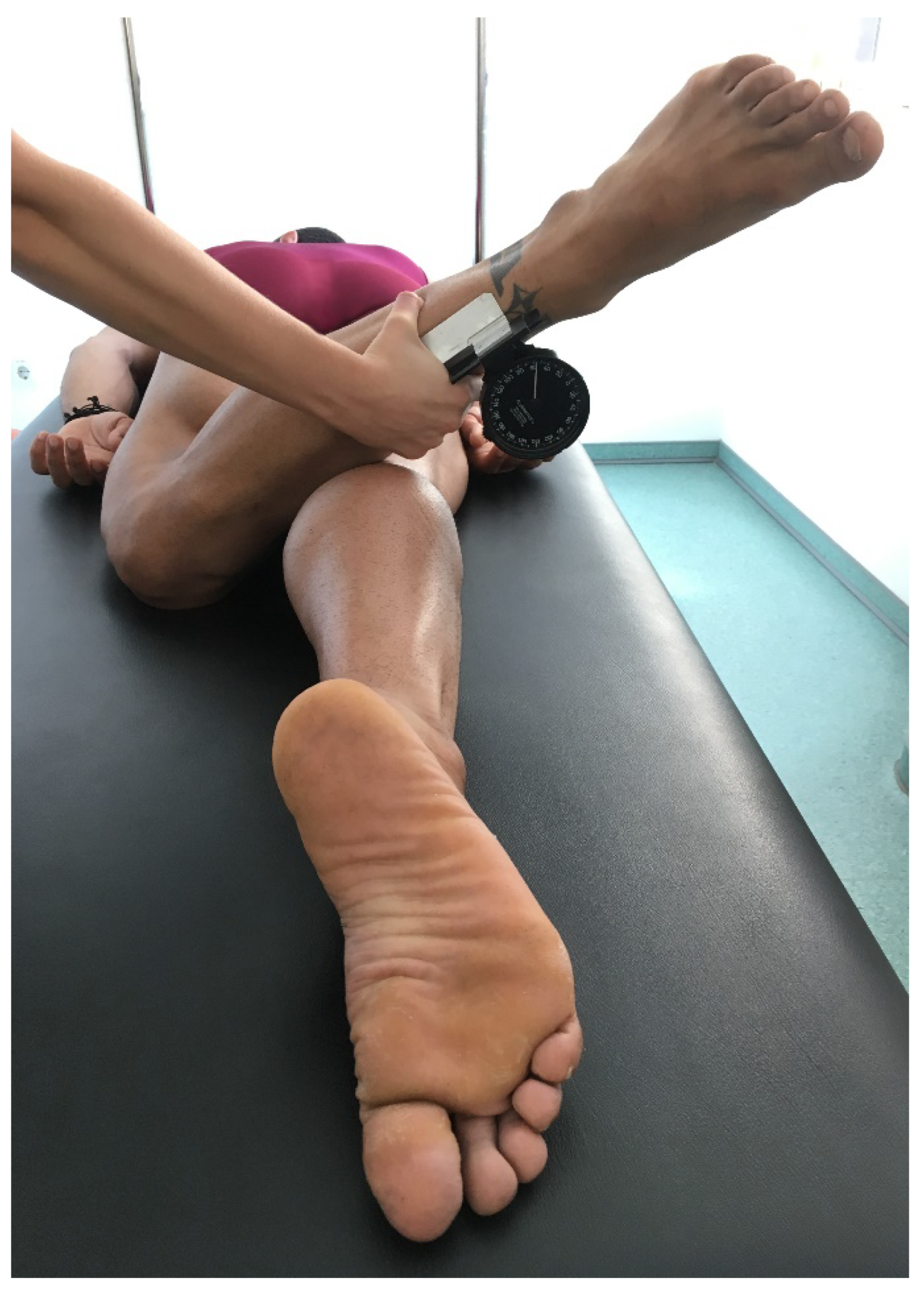
3.3.4. Foot-Ankle Complex

Differences in the foot inspection were not significant (Table 5). Splayed feet occurred with a higher prevalence among older students (</≥10 years: f: 36.3/59.1%, m: 20.0/53.3%). Here, there was no significant difference between genders in the group of older students (f/m: 59.1/53.3%, χ2(1) = 1.47, p = 0.23). Females showed a valgus big toe axis significantly more often (f/m: 20.8/7.9 %, χ2(1) = 15.30, p < 0.001). In single cases, severe forms were observed (juvenile hallux valgus (<10years): n = 3 (f), severe hallux valgus (>40°): n = 3 (m), hallux rigidus: n = 1 (m)).
Female students had significantly greater ROM of the ankle joint (Table 6). Among the females, older participants showed significantly greater values (</≥10years: Ex: 22.9/24.4°, t(356) = −2.52, p = 0.01, Flex: 67.7/72.2°, t(356) = −5.76, p < 0.001), while for the male examinees, this effect was not observed (</≥10years: Ex: 22.9/22.6°, t(173) = 0.23, p = 0.82, Flex: 67.4/67.9°, t(173) = 0.31, p = 0.76). For females, there was a correlation with age (right/left: Ex: f: r = 0.122/0.102, p = 0.020/.054, m: r = −0.039/−0.040, p = 0.611/0.600, Flex: f: r = 0.215/0.215, p < 0.001/0.001, m: r = 0.093/0.071, p = 0.218/0.351). Among females, passive dorsiflexion in the MTP joint was significantly smaller among older students (</≥10years: f: 91.4°/89.6, U = 11013.50, Z = −3.266, p = 0.001, m: 89.7°/86.3°, U = 1238.50, Z = −1.667, p = 0.1). This difference was even greater compared to the adult group and more evident among males (≥ 18years: f/m: 88.8/85.1°). There was a correlation with age (right/left: f: r = −0.245/−0.267, p = 0.001/0.001, m: r = −0.192/−0.184, p = 0.009/0.013).

4. Discussion

Studies describing the musculoskeletal profile of ballet and dance students are rare and often outdated, limited to the female gender, specific age groups, or certain characteristics [16,17]. Male ballet students are described even less frequently in the literature [19,22]. The present study collected the sociodemographic and physical characteristics of female and male vocational ballet students.

4.1. Age

The John Cranko School’s training program is divided by age, proficiency, and intensity levels (preschool, junior, intermediate, senior, professional level) [29]. In the present study, approximately half of the female examinees were of preschool and junior age, whereas the male examinees tended to be of intermediate age and older. The actual peak of male examinees did not occur until the senior and professional level. The generally high number of students in these age groups may be explained by the JCS’s good international reputation, which makes graduation from JCS particularly attractive if students have previously attended another school [30]. In addition, we suppose that attending senior level, with its intensified training, may increase the chances of entering the professional level of the school. The male subjects in the present study were, on average, slightly older than females. Any cross-age comparisons, therefore, should take into account an age-dependent developmental advantage of the males.

4.2. Body Weight

According to Tamaschke, a 10-year-old female vocational ballet student develops around the same percentile (height and weight) in the course of training [31]. Therefore, already at the aptitude test, a decision could be made about the usefulness of admission to training [31]. He describes her “ideal” body length (1.35–1.42 m) and weight (26.5–29.5 kg) and he claims a maximum of 50 kg to be the “ideal” weight of the fully-grown female professional ballet dancer [31]. These models reflect a leptosome-gracile (f) and athletic-gracile body type (m) described as favorable for classical dance [7]. Accordingly, the present study showed an extremely high and alarming prevalence of low body weight among students. Moreover, more than one-third were of severely low body weight. In the normal population, the prevalence of low body weight among 3–17 year old individuals was 7.6% [32]. There was no difference between genders [26]. In the present study, the prevalence for the same age group was approximately seven times higher (52.5%) and low body weight was significantly more common among females. This proportion increased with the increasing severity of low body weight. One possible cause is eating disorders or disordered eating behavior, the prevalence of which has been described as greatly increased in the professional educational sector [33,34]. Here, it is essential to ensure that the present body image was not brought about by dietary measures and corresponds to the constitutional type, as it is not generally a low BMI, as is frequently found in dance, but only a low BMI as a result of an eating disorder that is associated with an increased rate of injury and hormonal disturbances (sec. amenorrhea, delayed menarche, fatigue fractures, osteoporosis) [35,36,37,38,39,40,41,42]. The results of the present study underscore the need to determine BMI and percentile in vocational ballet students in the first instance so that low body weight does not go undetected. Furthermore, we see an importance in including screenings for eating disorders and appropriate nutrition uptake.

4.3. Beginning with Ballet and Training

On average, females in the present study began ballet earlier (5.8 vs. 8.2 years) and entered vocational training (13.5 vs. 14.6 years) before the male students. In a study by Hamilton et al., female dancers began ballet four years earlier than males, compared with only two years in the present study [9]. Compared with previous studies (1992 and 1974: ages 11 and 16, respectively), the males of the present study began their first ballet classes much earlier (age 8) [9,43]. Male students, in particular, commence with ballet earlier. Some claim that a late start with training might favor overload injuries [43,44]. As a cause, here, a lower adaptation to dance-specific loads in relation to the malleability of the bone and joint system, as well as a lower development of dance-specific coordination and basic motor skills with increasing age are discussed [7,18,44]. With a late start of training, the training time is also shortened, which further increases the load. The complete training period is eight to nine years and usually begins around the age of 10 [29,45]. The examinees in the present study started later, on average, but entered the JCS at least as early as 11.7 and 13.6 years (female and male subjects, respectively). Applicants who enter one of the advanced training classes must have previous dance-technical knowledge in order to qualify for the respective level [45]. Thus, it can be assumed that even older examinees who entered JCS or began training had sufficient previous dance training.

4.4. Physical Examination

4.4.1. Mobility of the Index Finger

Hypermobility has previously been shown to be a possible risk for acute injury and chronic overuse injuries, and ballet students are more likely to be hyperflexible than professional dancers [15,19,35,46,47]. However, opinions on the topic of hypermobility in dance remain divided [9,46,48,49,50,51]. This is due to the different methods used to assess mobility, as well as the interpretation of the results. For example, the Beighton score is controversial, because forward flexion can be improved through specific training. No correlation can be shown between the mobility of the base joint of the little finger and general mobility, and dancers are generally more flexible than the normal population [9,50,51,52]. Huwyler [15] declares that the iMCP joint of the non-dominant hand is the optimal location for measuring innate general mobility because it would not be particularly trained by any activity. He emphasizes that low values would not allow conclusions about the mobility of the joints important for dance and that values could only give an indication of the basic tendencies in joint mobility and should be understood as a warning in the case of particularly high values [15]. He mentions values above 105° to be possibly “critical” and values above 115° to be possibly “pathological”, as they might indicate a deficiency in the muscular stabilization of the joints [15,23]. However, studies to verify those statements are still lacking in the literature.
Hypermobile, isokinetic trunk strength tests can be used to test the extent to which the examined can ensure safe coordination of hypermobile joints through sufficient core stability [7]. Furthermore, knowledge and understanding of body types and the existence of hypermobility are important injury-preventing factors [19]. Thus, Benign Joint Hypermobility Syndrome would not be an obstacle to vocational training, as hypermobile dancers could develop strategies to meet the demands of their profession [19]. In the present study, none of the examinees showed values above 115°, and only for a few female examinees were very high values of dorsiflexion measured, among which one suffered from a recurrent dislocation of the patella. In general, female dancers are expected to be more flexible than males [23]. Accordingly, the female students in the present study also showed significantly higher values of dorsiflexion. The values only become meaningful around the age of 14 years, whereas higher values are generally expected in children [23]. In the present study, therefore, significantly fewer examinees in the younger group were examined by the Pluri-Ligament test, who, in turn, showed significantly higher values compared with the older group.

4.4.2. Trunk

A majority of the examinees in the present study showed a symmetrical, plumb and erect trunk. Asymmetries were not uncommon among both sexes, simultaneously. They may indicate scoliosis [7]. A lifetime prevalence of 5.2% (0–17-year-olds) has been described for scoliosis [53]. In comparison, the prevalence of scoliosis was found to be greatly increased in the present study. In addition, scoliotic malpositions were not infrequently observed. Among professional ballet dancers, an even higher prevalence of scoliosis has been described (f/m: 50/27%) [9]. In addition, scoliosis occurs more frequently in the pubertal growth phase (14–17-year-olds: 11.1%) and the difference between the sexes increases with age (f > m) [53,54]. This difference between age groups and sexes was also shown in the present study. At the same time, the present study observed a high prevalence of juvenile scoliosis. Scoliosis is generally not a criterion for exclusion from vocational classical ballet, although it is associated with an increased prevalence of spinal injury [35]. Some report that structural scoliosis with a Cobb angle greater than twenty degrees (lumbar or high thoracic) or scoliosis with an indication for surgical therapy would be incompatible with a professional ballet career [7,55]. However, studies to verify those statements are lacking in the literature. In the present study, as well as in Hamilton et al., scoliosis was detected and assessed by a clinical forward flexion test [9]. In addition, comparative data of standard values of the upper body posture for men and women could also be used here for comparison from an older age [56,57].
In the present study, anterior pelvic tilt was rarely seen with suspected bony fixation of an increased sacral base angle and was otherwise due to an inadequate ability to muscularly stabilize the pelvis. This is particularly supported by the significantly higher prevalence among younger students, who often still have insufficient core stability. The Palucca School in Dresden implemented X-ray diagnostics of the spine to exclude spondylosis for the entrance examination, from 1980 until the reunification of Germany [20]. Lohrer emphasizes that X-rays should be avoided before the age of 11 (absence of apophyseal bone nuclei) and describes a clinical test battery (suspension test, lumbosacral stabilization test) [58]. In addition, spondylosis and spondylolisthesis in competitive sports and dance are often asymptomatic [10,55].

4.4.3. Legs and Hips

A beautiful leg line in classical ballet is emphasized by the curve of a genu recurvatum [7]. Accordingly, in the present study, all of those examined showed at least full extension of the knee joints and almost all showed a straight leg axis. The prevalence of genu recurvata showed no significant gender difference. These results underline the typical characteristic of a beautiful leg line in ballet. Even more characteristic is the outward rotation of the legs from the hips (turnout) [7,9]. A reduced IR, in favor of an increased ER, is typical here and has been reported for professional ballet dancers [9]. The males of the present study showed a relevantly smaller IR and ROM in gender comparison. At the same time, the participants of the present study showed a larger IR and ROM than professional dancers [9]. The reason for this difference could be the significantly younger population of the present study. The development from childhood to adulthood leads to an increase in ER and a decrease in IR, via the physiological detorsion of the femur, and a reduction in the angle of antetorsion [7,15,18]. However, a strongly reduced IR can be pathological [59]. In the present study, a severely reduced IR, with a documented suspected diagnosis of hip dysplasia, epiphysiolysis capitis femoris, or femoroacetabular impingement, was documented for 2.2% of those examined. However, the mean IR (≥15 years) was within the normal range (30–40°) [60].
ER of the hip is a major contributor to turnout [9]. In the present study, this showed a statistically significant, but not clinically relevant, difference between the sexes (f/m: 59/62°). The values were much higher than in previous studies of female ballet students and professional dancers (49 and 52°) [9,18]. This may be due to the perfected ballet-specific selection, but also to the increasingly earlier onset of ballet and training, which allows for increased skeletal shaping. Together with a large ER, a relatively large TT allows for the turnout required for classical ballet (90° per leg) [7]. The mean TT of the present study (22°) showed no difference between the sexes and was above the average of the normal population (15–20°) [7]. None of the students had values (>40°) associated with an increased risk of damage to the knee joint (high shear forces) [7]. There was also a significant, but clinically irrelevant, difference in turnout between the sexes, with generally high values (f/m: 82/86°). If turnout is forced because its range is anatomically too small, an anterior pelvic tilt and, consequently, a provoked hyperlordosis of the lumbar spine, a forced rotation of the knee joints, a hyperpronation of the feet with valgus of the inner ankles and lateral foot edges that lose contact with the ground (rolling-in) are clinically evident [7,61]. Such mechanisms of compensation may achieve an additional 25° turnout, on average, but, at the same time, seem to increase the risk of acute injury, laxity of the joint-stabilizing ligamentous apparatus and chronic degenerative cartilage damage [10,43,61,62,63]. Huwyler emphasizes that for the calculation of the individual turnout, the anatomical ability of the knees for external rotation must therefore not be included [15].

4.4.4. Foot-Ankle Complex

A “beautiful foot” completes the desired leg line in classical ballet. A large plantar flexion of the ankle joint particularly contributes to this aesthetic effect. Compared to normal values (40–50°), the examinees of the present study showed significantly higher values [60]. Hamilton et al. describe the “beautiful foot” as an inert “must have” of the professional female ballet dancer, which would not be required of her male colleagues [9]. Although the females in the present study demonstrated significantly greater plantar flexion of the ankle joint, similarly high values were also measured for the males (71/68°). This result emphasizes the importance of large plantar flexion in aesthetic and functional terms for professional classical ballet. For the plantar flexion of the ankle joint, skeletal shaping and enlargement through training has been reported [9,15]. This could explain why the older female participants showed significantly greater plantar flexion than the younger students (weak positive correlation with age). In contrast, this effect was not seen for males. In addition to a large plantar flexion of the ankle joint, a high instep is associated with a “beautiful foot” [7]. In the present study, the female and male examinees showed a high instep with equal frequency. In contrast, the prevalence of a flexible tarsus was higher among females, and that of a low instep and a rigid tarsus was relevantly higher among males, which may indicate that the “beautiful foot” is still more frequently required by female ballet students than by males. A high instep should be distinguished from a hollow foot, which, according to Huwyler, always needs to be neurologically clarified and is not suitable for ballet [15]. It has been described that repetitive dorsiflexion of the ankle joint forced to the maximum can lead to symptoms of chronic anterior impingement in dancers and that the anatomy of a pes cavus, in particular, provokes an anterior impingement of the ankle joint [64,65,66]. Exner-Grave states that a hollow foot with unrestricted dorsiflexion of the ankle joint and a flexible tarsus is suitable for professional classical ballet [7]. According to this, a good joint guidance of the ankle joint with unrestricted joint play is essential for classical ballet in order to be able to sufficiently cushion the countless repetitive, aestheticized knee bends (i.e., pliés, fondus), small, medium, and large jumps. In the present study, the average passive dorsiflexion in both sexes (f/m: 24/23°) was within the normal range of the general population (20–30°) [60]. It has been observed that dancers in a muscularly warmed-up state can achieve significantly higher values [15]. The participants in the present study were not always muscularly warmed-up. Amongst the female students, there was also a weak positive correlation with age. With dorsiflexion in the ankle joint, force is optimally developed and transmitted via straight RFA. Valgus or varicose RFA, on the other hand, are associated with overuse injuries and injuries to the foot and ankle in dance, and also in the cranial chain at the lower leg, knee, hip, and back [59,62]. The majority of those examined in the present study demonstrated straight RFA. Valgus RFA was observed relevantly more frequently in female examinees (32/24%). Valgus RFA, in combination with a flat foot (pes planovalgus), is characteristic of childhood and, moreover, is a very common deformity in adults [7,59]. Both are also shown in the results of the present study.
The development of a splay foot in classical ballet is favored by the repetitive toe stance (relevé) and is not prevented by the material properties of the ballet shoes [4,7,10]. This may explain the high prevalence of splayfoot in the present study. A protective effect has been described for the square toe box of the pointe shoe in this regard [8]. In the present study, however, no significant difference in the prevalence of splayfoot between the sexes was found that would confirm a protective effect of the toe box, which is worn only by the female sex. Splayfoot, in turn, is associated with the development of HAV, which is considered to be the most common deformity of the lower extremity [59]. The prevalence of HAV among the 18-65- year old population is reported in the literature to be 23% and positively correlated with age [67]. In the considerably younger population of the present study, the prevalence of a valgic axis of the big toe was much lower. Furthermore, the literature describes a gender-typical difference (f > m), which the present study was also able to find [59]. A straight axis of the big toe and a large dorsiflexion in the MTP joint of the big toe guarantee the correct execution of the relevé and the elevation to the tip of the toe. In ballet, the MTP joint of the big toe is as important as the major joints of the body [15]. Those examined in the present study exhibited a desirably large dorsal extension for classical ballet (f/m: 90/87°). It turned out to be greater than normal values (70°) [68]. Huwyler emphasizes that it cannot be increased by training [44]. The present study could even show a weak negative correlation with age. The foot can be divided into four categories, according to its shape. Exner-grave [7] claims that a Roman or Egyptian shape, with short toes of equal length, provides an even load distribution on the toes’ tips in the female dancer’s pointe shoe. Huwyler [15] claims that its shape is important for female dancers (pointe work), but also of medical importance for the male dancer. The Greek shape, with an elongated second metatarsal bone, would lead to hammer toes, clavi and, due to punctual overloading, to a severely painful clinical picture of the metatarsus in both sexes [15]. However, in their study, Davidson et al. [69] did not show an increased risk for stress fracture due to a Greek shape of the foot. In the present study, a Greek shape with elongated os metatarsale II was observed in many of the students. In contrast, a Roman shape was rather rare among the examinees. There is still a lack of studies that investigate complaints of the foot based on foot shape in ballet.

5. Conclusions

The present study gives a broad description of the sociodemographic and musculoskeletal profile of female and male vocational ballet students. At present, male vocational ballet students begin dancing at a much younger age and eventually differ less than before from females regarding ballet-specific characteristics (anthropometry of legs, ER, turnout, ROM of ankle joint, dorsiflexion of MTP joint). The prevalence of low body weight, particularly among female vocational ballet students, is alarming and should prompt further diagnostic testing to rule out eating disorders and disordered eating behavior.

Author Contributions

Conceptualization, E.E.-G. and E.M.W.; methodology, E.E.-G. and E.M.W.; software, T.A. and E.M.W.; validation, T.A.; formal analysis, T.A.; investigation, E.E.-G.; resources, E.E.-G.; data curation, E.E.-G.; writing—original draft preparation, T.A. and E.M.W.; writing—review and editing, E.M.W. and D.O.; visualization, T.A.; supervision, E.M.W. All authors have read and agreed to the published version of the manuscript.

Funding

This research received no external funding.

Institutional Review Board Statement

Ethic Committee Name: Goethe University Frankfurt, Main, Germany; Approval Code: 2021-63; Approval Date: 16 February 2021.

Informed Consent Statement

Not applicable.

Data Availability Statement

The datasets used and/or analyzed during the current study are available from the corresponding author on reasonable request. The Figure 1, Figure 2 and Figure 3 were used in this article with the kind permission of Thieme group (Citation: Almasi et al., Muskuloskelettale Eignung für den professionellen klassischen Bühnentanz: Voraussetzungen–Besonderheiten–Untersuchungen Sportverletz Sportschaden 2019; 33(04): 192-202 DOI: 10.1055/a-0973-8753) © Thieme.

Acknowledgments

Sabrina Schlesinger: Statistic support; Anneli Chasemore: Native English language support.

Conflicts of Interest

The authors declare no conflict of interest.

References

  1. Liechtenhan, R. Vom Tanz zum Ballett: Geschichte und Grundbegriffe des Bühnentanzes, 2nd ed.; Belser: Stuttgart, Germany, 1993. [Google Scholar]
  2. Tarassow, N.I. Klassischer Tanz: Die Schule des Tänzers; Henschel: Berlin, Germany, 1974. [Google Scholar]
  3. Rossol, M.; Hinkamp, D. Hazards in the theater. Occup. Med. 2001, 16, 595–608. [Google Scholar] [PubMed]
  4. Arendt, Y.D.; Kerschbaumer, F. Verletzungen und Überlastungserscheinungen im professionellen Ballett. Z. Orthop. Ihre. Grenzgeb. 2003, 141, 349–356. [Google Scholar] [CrossRef] [PubMed]
  5. Wanke, E.M.; Mill, H.; Groneberg, D.A. Ballett als Leistungssport—Gesundheitliche Gefährdungen am Beispiel akuter Verletzungen bei Tanzschülern. Sport. Sportschaden 2012, 26, 164–170. [Google Scholar] [CrossRef] [PubMed]
  6. Wolf, A.S. Zur Problematik des Leistungssports bei Heranwachsenden. Arch. Gynecol. Obs. 1987, 242, 936–943. [Google Scholar] [CrossRef]
  7. Exner-Grave, E. TanzMedizin: Die Medizinische Versorgung Professioneller Tänzer; Schattauer: Stuttgart, Germany, 2008. [Google Scholar]
  8. Wanke, E.M.; Exner-Grave, E. Tanz mit Spitzenschuhen: Besonderheiten, körperliche Voraussetzungen und Tauglichkeitskriterien. Sport. Sportschaden 2017, 31, 213–221. [Google Scholar] [CrossRef]
  9. Hamilton, W.G.; Hamilton, L.H.; Marshall, P.; Molnar, M. A profile of the musculoskeletal characteristics of elite professional ballet dancers. Am. J. Sport. Med. 1992, 20, 267–273. [Google Scholar] [CrossRef] [PubMed]
  10. Fuchs, E.; Hess, H.; Kunz, M. Akute Verletzungen und chronische Überlastungen im klassischen Ballett. Sports Sportschaden 2003, 17, 123–131. [Google Scholar] [CrossRef]
  11. Smith, P.J.; Gerrie, B.J.; Varner, K.E.; McCulloch, P.C.; Lintner, D.M.; Harris, J.D. Incidence and Prevalence of Musculoskeletal Injury in Ballet: A Systematic Review. Orthop. J. Sport. Med. 2015, 3, 2325967115592621. [Google Scholar] [CrossRef] [Green Version]
  12. Yau, R.K.; Golightly, Y.M.; Richardson, D.B.; Runfola, C.D.; Waller, A.E.; Marshall, S.W. Potential Predictors of Injury Among Pre-Professional Ballet and Contemporary Dancers. J. Danc. Med. Sci. 2017, 21, 53–63. [Google Scholar] [CrossRef]
  13. Caine, D.; Goodwin, B.J.; Caine, C.G.; Bergeron, G. Epidemiological Review of Injury in Pre-Professional Ballet Dancers. J. Danc. Med. Sci. 2015, 19, 140–148. [Google Scholar] [CrossRef]
  14. Ekegren, C.L.; Quested, R.; Brodrick, A. Injuries in pre-professional ballet dancers: Incidence, characteristics and consequences. J. Sci. Med. Sports 2014, 17, 271–275. [Google Scholar] [CrossRef] [PubMed]
  15. Huwyler, J. Der Tänzer und Sein Körper: Aspekte des Tanzens aus Ärztlicher Sicht, 2nd ed.; PERIMED-Spitta: Balingen, Germany, 1995. [Google Scholar]
  16. Kabakci, A.G.; Yücel, A.H.; Ayvazoglu, S. Physical characteristics of students to receive ballet training. Cukurova Med. J. 2017, 41, 55–60. [Google Scholar] [CrossRef] [Green Version]
  17. Kadel, N.J.; Donaldson-Fletcher, E.A.; Gerberg, L.F.; Micheli, L.J. Anthropometric Measurements of Young Ballet Dancers. J. Danc. Med. Sci. 2005, 9, 84–90. [Google Scholar]
  18. Hamilton, D.; Aronsen, P.; Løken, J.H.; Berg, I.M.; Skotheim, R.; Hopper, D.; Clarke, A.; Briffa, N.K. Dance training intensity at 11–14 years is associated with femoral torsion in classical ballet dancers. Br. J. Sport. Med. 2006, 40, 299–303; discussion 303. [Google Scholar] [CrossRef]
  19. McCormack, M.; Briggs, J.; Hakim, A.; Grahame, R. Joint laxity and the benign joint hypermobility syndrome in student and professional ballet dancers. J. Rheumatol. 2004, 31, 173–178. [Google Scholar]
  20. Sonnenkalb, E. Einstellungsuntersuchungen für die Palucca-Schule Ergebnisse und Schlussfolgerungen. Arb. Inf. Für Theater Und Orch. Des Betr. Der Berl. Bühnen Und Der Arb. Berat. Der Theater Und Orch. Der DDR (BABB/AHB) 1984, 1, 24–28. [Google Scholar]
  21. Tamaschke, C. Anthropometrische Körperbaustudien an Schülerinnen der Staatlichen Ballettschule Berlin. Ärztliche Jugendkd. 1973, 69, 9–17. [Google Scholar]
  22. Nilsson, C.; Wykman, A.; Leanderson, J. Spinal sagittal mobility and joint laxity in young ballet dancers. A comparative study between first-year students at the Swedish Ballet School and a control group. Knee Surg Sports Traumatol. Arthrosc. 1993, 1, 206–208. [Google Scholar] [CrossRef]
  23. Huwyler, J. Ärztliche Eignungsprüfung zu einer tänzerischen Berufsausbildung. Ballettjournal 1989, 37, 56–61. [Google Scholar]
  24. Geschichte. Available online: http://www.john-cranko-schule.de/ueber-uns/geschichte/ (accessed on 14 December 2022).
  25. Rippstein, J. Le Plurimètre V64: Un nouvel instrument de mensuration. Ann. De Kinésithérapie 1983, 10, 37–45. [Google Scholar]
  26. Kromeyer-Hauschild, K.; Wabitsch, M.; Kunze, D.; Geller, D.; Geiss, H.; Hesse, V.; von Hippel, A.; Jaeger, U.; Johnsen, D.; Korte, W.; et al. Perzentile für den Body-mass-Index für das Kindes- und Jugendalter unter Heranziehung verschiedener deutscher Stichproben. Mon. Kinderheilkd. 2001, 149, 807–818. [Google Scholar] [CrossRef] [Green Version]
  27. BMI Rechner. Available online: https://adipositas-gesellschaft.de/bmi/ (accessed on 21 November 2021).
  28. Herpertz, S.; Hagenah, U.; Vocks, S.; von Wietersheim, J.; Cuntz, U.; Zeeck, A. S3-Leitlinie Diagnostik und Therapie der Essstörungen. 2018. Available online: https://osnadocs.ub.uni-osnabrueck.de/bitstream/urn:nbn:de:gbv:700-2016081114851/5/Deutsches_Aerzteblatt_2011_Herpetz_Zeitschriftenartikel.pdf (accessed on 14 December 2022).
  29. Ausbildung. Available online: https://www.john-cranko-schule.de/ausbildung/allgemeine-informationen/ (accessed on 20 August 2022).
  30. Liechtenhan, R. Ballettgeschichte im Überblick; Noetzel: Wilhelmshaven, Germany, 1990. [Google Scholar]
  31. Tamaschke, C. Untersuchungen zur Frage einer Prognosemöglichkeit der Körperlichen Entwicklung von Ballettschülerinnen; Humboldt-Universität: Berlin, Germany, 1982. [Google Scholar]
  32. Schienkiewitz, A.; Damerow, S.; Schaffrath Rosario, A.; Kurth, B.-M. Body-Mass-Index von Kindern und Jugendlichen: Prävalenzen und Verteilung unter Berücksichtigung von Untergewicht und extremer Adipositas. Bundesgesundheitsblatt Gesundh. Gesundh. 2019, 62, 1225–1234. [Google Scholar] [CrossRef] [PubMed]
  33. Abraham, S. Eating and weight controlling behaviours of young ballet dancers. Psychopathology 1996, 29, 218–222. [Google Scholar] [CrossRef]
  34. Ringham, R.; Klump, K.; Kaye, W.; Stone, D.; Libman, S.; Stowe, S.; Marcus, M. Eating disorder symptomatology among ballet dancers. Int. J. Eat Disord. 2006, 39, 503–508. [Google Scholar] [CrossRef] [PubMed]
  35. Steinberg, N.; Siev-Ner, I.; Peleg, S.; Dar, G.; Masharawi, Y.; Zeev, A.; Hershkovitz, I. Injuries in female dancers aged 8 to 16 years. J. Athl. Train. 2013, 48, 118–123. [Google Scholar] [CrossRef] [Green Version]
  36. Thomas, J.J.; Keel, P.K.; Heatherton, T.F. Disordered eating and injuries among adolescent ballet dancers. Eat Weight Disord 2011, 16, e216–e222. [Google Scholar] [CrossRef]
  37. Joy, E.; De Souza, M.J.; Nattiv, A.; Misra, M.; Williams, N.I.; Mallinson, R.J.; Gibbs, J.C.; Olmsted, M.; Goolsby, M.; Matheson, G.; et al. 2014 female athlete triad coalition consensus statement on treatment and return to play of the female athlete triad. Curr. Sports Med. Rep. 2014, 13, 219–232. [Google Scholar] [CrossRef]
  38. Torstveit, M.K.; Sundgot-Borgen, J. Low bone mineral density is two to three times more prevalent in non-athletic premenopausal women than in elite athletes: A comprehensive controlled study. Br. J. Sport. Med. 2005, 39, 282–287; discussion 282–287. [Google Scholar] [CrossRef] [Green Version]
  39. Allaway, H.C.; Southmayd, E.A.; De Souza, M.J. The physiology of functional hypothalamic amenorrhea associated with energy deficiency in exercising women and in women with anorexia nervosa. Horm. Mol. Biol. Clin. Investig. 2016, 25, 91–119. [Google Scholar] [CrossRef]
  40. Barrack, M.T.; Gibbs, J.C.; De Souza, M.J.; Williams, N.I.; Nichols, J.F.; Rauh, M.J.; Nattiv, A. Higher incidence of bone stress injuries with increasing female athlete triad-related risk factors: A prospective multisite study of exercising girls and women. Am. J. Sport. Med. 2014, 42, 949–958. [Google Scholar] [CrossRef]
  41. De Souza, M.J.; Williams, N.I. Beyond hypoestrogenism in amenorrheic athletes: Energy deficiency as a contributing factor for bone loss. Curr. Sports Med. Rep. 2005, 4, 38–44. [Google Scholar] [CrossRef] [PubMed]
  42. Warren, M.P.; Voussoughian, F.; Geer, E.B.; Hyle, E.P.; Adberg, C.L.; Ramos, R.H. Functional Hypothalamic Amenorrhea: Hypoleptinemia and Disordered Eating. J. Clin. Endocrinol. Metab. 1999, 84, 873–877. [Google Scholar] [CrossRef] [PubMed]
  43. Miller, E.H.; Schneider, H.J.; Bronson, J.L.; McLain, D. A new consideration in athletic injuries. The classical ballet dancer. Clin. Orthop. Relat. Res. 1975, 111, 181–191. [Google Scholar] [CrossRef] [PubMed]
  44. Huwyler, J. Veränderungen am Skelett des Mittelfußes durch professionelles klassisches Tanzen. Sport Sportschaden 1989, 3, 14–20. [Google Scholar] [CrossRef] [PubMed]
  45. Ausbildung. Available online: http://ballettschule-berlin.de/buehnentanz/ausbildung/ (accessed on 7 September 2022).
  46. Wanke, E. Prävention von Unfällen im Professionellen Bühnentanz (Rahmenempfehlungen). 2014. Available online: https://publikationen.dguv.de/widgets/pdf/download/article/2009. (accessed on 14 December 2022).
  47. Briggs, J.; McCormack, M.; Hakim, A.J.; Grahame, R. Injury and joint hypermobility syndrome in ballet dancers—A 5-year follow-up. Rheumatology 2009, 48, 1613–1614. [Google Scholar] [CrossRef] [Green Version]
  48. Steinberg, N.; Hershkovitz, I.; Zeev, A.; Rothschild, B.; Siev-Ner, I. Joint Hypermobility and Joint Range of Motion in Young Dancers. J. Clin. Rheumatol. 2016, 22, 171–178. [Google Scholar] [CrossRef]
  49. Klemp, P.; Stevens, J.E.; Isaacs, S. A hypermobility study in ballet dancers. J. Rheumatol. 1984, 11, 692–696. [Google Scholar]
  50. Klemp, P.; Learmonth, I.D. Hypermobility and injuries in a professional ballet company. Br. J. Sport. Med. 1984, 18, 143–148. [Google Scholar] [CrossRef] [Green Version]
  51. Klemp, P.; Chalton, D. Articular mobility in ballet dancers. A follow-up study after four years. Am. J. Sport. Med. 1989, 17, 72–75. [Google Scholar] [CrossRef]
  52. Chan, C.; Hopper, L.; Zhang, F.; Pacey, V.; Nicholson, L.L. The prevalence of generalized and syndromic hypermobility in elite Australian dancers. Phys. Ther. Sport. 2018, 32, 15–21. [Google Scholar] [CrossRef]
  53. Kamtsiuris, P.; Atzpodien, K.; Ellert, U.; Schlack, R.; Schlaud, M. Prävalenz von somatischen Erkrankungen bei Kindern und Jugendlichen in Deutschland: Ergebnisse des Kinder- und Jugendgesundheitssurveys (KiGGS). Bundesgesundheitsblatt-Gesundh. Gesundh. 2007, 50, 686–700. [Google Scholar] [CrossRef] [PubMed] [Green Version]
  54. Krause, L.; Kuntz, B.; Lampert, T.; Kamtsiuris, P.; Seeling, S.; Ziese, T. KiGGS Welle 1—Landesmodul Thüringen. Gesundheitsberichterstattung des Bundes. 2016, Volume 1. Available online: https://www.semanticscholar.org/paper/KiGGS-Studie-zur-Gesundheit-von-Kindern-und-in-zum-Krause-Kuntz/9bec15ea436d19079139cad72080c5f289ee1234. (accessed on 14 December 2022).
  55. Schönle, C. Prävention und Rehabilitation: Sportbedingte Schäden der Wirbelsäule und Empfehlungen zum Training. Sport-Orthopädie Sport-Traumatol. 2002, 18, 11–18. [Google Scholar] [CrossRef]
  56. Ohlendorf, D.; Fisch, V.; Doerry, C.; Schamberger, S.; Oremek, G.; Ackermann, H.; Schulze, J. Standard reference values of the upper body posture in healthy young female adults in Germany: An observational study. BMJ Open 2018, 8, e022236. [Google Scholar] [CrossRef] [PubMed] [Green Version]
  57. Ohlendorf, D.; Adjami, F.; Scharnweber, B.; Schulze, J.; Ackermann, H.; Oremek, G.M.; Kopp, S.; Groneberg, D.A. Standard values of the upper body posture in male adults. Adv. Clin. Exp. Med. 2018, 27, 1521–1528. [Google Scholar] [CrossRef] [Green Version]
  58. Lohrer, H.; Arentz, S. Grundlagen der orthopädischen Untersuchung im Kunstturnen. Sports Sportschaden 2001, 15, 55–58. [Google Scholar] [CrossRef]
  59. Niethard, F.U.; Biberthaler, P.; Pfeil, J. Orthopädie und Unfallchirurgie, 8th ed.; Thieme: Stuttgart, Germany, 2017. [Google Scholar]
  60. Füeßl, H.; Middeke, M.; Würtemberger, G. Anamnese und Klinische Untersuchung, 5th ed.; Thieme: Stuttgart, Germany, 2014. [Google Scholar]
  61. Goertzen, M.; Ringelband, R.; Schulitz, K.P. Verletzungen und Überlastungsschäden beim klassischen Ballett-Tanz. Z. Orthop. Ihre. Grenzgeb. 1989, 127, 98–107. [Google Scholar] [CrossRef]
  62. Nowacki, R.M.; Air, M.E.; Rietveld, A.B. Hyperpronation in Dancers Incidence and Relation to Calcaneal Angle. J. Danc. Med. Sci. 2012, 16, 126–132. [Google Scholar]
  63. Coplan, J.A. Ballet dancer’s turnout and its relationship to self-reported injury. J. Orthop. Sports Phys. 2002, 32, 579–584. [Google Scholar] [CrossRef]
  64. Russell, J.A.; Kruse, D.W.; Koutedakis, Y.; Wyon, M.A. Pathoanatomy of Anterior Ankle Impingement in Dancers. J. Danc. Med. Sci. 2012, 16, 101–108. [Google Scholar]
  65. Stoller, S.M.; Hekmat, F.; Kleiger, B. A Comparative Study of the Frequency of Anterior Impingement Exostoses of the Ankle in Dancers and Nondancers. Foot Ankle Int. 1984, 4, 201–203. [Google Scholar] [CrossRef]
  66. Kennedy, J.G.; Hodgkins, C.W.; Columbier, J.-A.; Hamilton, W.G. Chapter 21—Foot and Ankle Injuries in Dancers. In Baxter’s the Foot and Ankle in Sport, 2nd ed.; Mosby: Maryland Heights, MO, USA, 2008; pp. 469–483. [Google Scholar]
  67. Nix, S.; Smith, M.; Vicenzino, B. Prevalence of hallux valgus in the general population: A systematic review and meta-analysis. J. Foot Ankle Res. 2010, 3, 21. [Google Scholar] [CrossRef] [PubMed] [Green Version]
  68. Aumüller, G.; Aust, G.; Doll, A.; Engele, J.; Kirsch, J.; Mense, S.; Reißig, D.; Salvetter, J.; Schmidt, W.; Schmitz, F.; et al. Duale Reihe Anatomie, 1st ed.; Georg Thieme Verlag: Stuttgart, Germany, 2007. [Google Scholar]
  69. Davidson, G.; Pizzari, T.; Mayes, S. The influence of second toe and metatarsal length on stress fractures at the base of the second metatarsal in classical dancers. Foot Ankle Int. 2007, 28, 1082–1086. [Google Scholar] [CrossRef] [PubMed]
Figure 1. Non-dominant hand on plate of Pluri-Ligament with index finger pulled into dorsiflexion with 1 kg of pulling force (figure with the kind permission of Thieme group, © Thieme).
Applsci 13 00108 g001
Figure 2. Subject in prone and axis of Pluritor C aligned with margo anterior tibiae to measure rotation of the hip (figure with kind permission of Thieme group, ©Thieme).
Applsci 13 00108 g002
Figure 3. Subject in quadruped stance and tips of the fork of Pluritor T touching malleoli from dorsal to measure tibial torsion (figure with kind permission of Thieme group, ©Thieme).
Applsci 13 00108 g003
Figure 4. Subject in supine and plate of Pluri-Ped firmly pressed against sole of the foot to measure range of motion in ankle joint.
Applsci 13 00108 g004
Figure 5. Frequency of those examined per age group in percent.
Applsci 13 00108 g005
Figure 6. Low body weight by age group (ages < 10–17 years: <10th BMI percentile [26]; age ≥ 18 years: BMI <18.5 kg/m2 [28]).
Applsci 13 00108 g006
Figure 7. Severely low body weight by age group (ages <10–17 years: <3rd BMI percentile [26]; age ≥18 years: BMI <17 kg/m2 [28]).
Applsci 13 00108 g007
Figure 8. Average turnout by age group and gender.
Applsci 13 00108 g008
Table 1. Surveyed Characteristics with respective n.
Characteristic (Unit) n (f/m)
Age (a)412/191
Sociodemographic Vision aid (yes/no)403/189
Commenced withBallet (a)392/185
Training (a)124/69
AnthropometryBodyHeight (cm)412/191
Weight (kg)412/191
Shoulder girdleSymmetry *397/186
Waist triangleSymmetry *402/190
PelvisAnterior/Posterior tilt *382/184
Lateral tilt *400/187
SpineCurvature *403/189
LegAxis *402/186
Length (cm)396/188
Intercondylar distance (TF)380/172
Tibial torsion (°)267/125
FootRearfoot axis *402/189
Instep *59/27
Longitudinal arch *374/177
Transversal arch *392/185
Big toe axis *404/189
Shape *397/188
FunctionalityHandiMCP dorsiflexion (°)244/148
SpineMobility *402/186
HipER/IR (°)412/192
KneeExtension (°)390/184
Ankle jointExtension/Flexion (°)358/176
TarsusMobility *105/59
MTP joint Extension (°) 392/184
Flexion (°) 391/184
* Qualitative, TF = transverse finger, iMCP = index metacarpophalangeal joint, ER/IR = external/internal rotation, MTP = metatarsophalangeal. a = years.
Table 2. Sociodemographic characteristics, Mean ± SD (Range).
Characteristic (Unit)FemaleMalen (f/m)
Age (a) 13.4 ± 3.5 (5.8–20.4)15.1 *** ± 3.2 (7.2–22.6)412/191
Body height (cm) 152.4 ± 16.0 (113–179)166.2 *** ± 16.7 (121–197.5)412/191
Body weight (kg) 38.5 ± 11.4 (17–64.6)52.7 *** ± 14.6 (21–95)412/191
BMI (kg/m2) 16.1 ± 2.0 (11.3–22.2)18.5 *** ± 2.3 (13.3–24.4)411/192
BMI percentile ** a 11.5 ± 12.0 (< 1–73)31.3 *** ± 20.5 (< 1–86)385/155
Commenced with
Ballet (a) 5.8 ± 2.3 (2–14)8.2 *** ± 2.9 (2–18)392/185
Training (a) 13.5 ± 3.1 (6–18)14.6 * ± 3.1 (5–20)124/69
a = years, *** p < 0.001, ** according to Kromeyer-Hauschild [26], * p < 0.05.
Table 3. Differences in torso inspection in percent.
Characteristic FemaleMalen (f/m)
Shoulder girdlesymmetrical76.686.6 **397/186
asymmetrical23.413.4 **
Waist trianglesymmetrical71.183.2 *402/190
asymmetrical28.916.9 *
Anterior/Posteriorphysiological80.485.3382/184
pelvic tiltanterior tilt17.38.7 **
posterior tilt2.46
Lateral pelvic tiltstraight83.887.2400/187
tilted16.312.9
Spinevertical58.865.6403/189
Scoliosis27.520.1
scol. malposition10.47.4
flatback15.3 **
Kyph./Hyperlord.2.21.6
* p < 0.05, ** p < 0.01, scol. = scoliotic, Kyph./Hyperlord. = Kyphosis/Hyperlordosis.
Table 4. Legs and hips by gender, Mean ± SD (Range).
Characteristic (Unit)FemaleMalen (f/m)
Leg Length (cm)82.8 ± 9.7 (59.0–101.0)90.7 *** ± 10.1 (62.0–110.0)396/188
ICD (TF)0.7 ± 1.1 (0–5)1.1 *** ± 1.4 (0–7)380/172
TT (°)22.2 ± 6.4 (2–40)22.8 * ± 5.9 (5–38)267/125
ER (°)59.1 ± 7.7 (35–75)61.9 *** ± 6.8 (40–80)412/192
IR (°)46.4 ± 10.1 (18–72)37.1 *** ± 11.1 (10–70)412/192
ROM (ER + IR) (°)105.5 ± 9.6 (78–138.5)99.0 *** ± 10.6 (72.5–130.0)412/192
*** p < 0.001, ICD = Intercondylar distance, TF = Transverse finger, ER/IR = External/Internal rotation of the hip, TT = Tibial torsion, * not significant p < 0.05.
Table 5. Examinations of the foot by gender in percent.
Characteristic FemaleMalen (f/m)
RFAvalgic32.124.3402/189
straight5759.3
varic10.916.4
Instephigh3940.759/27
normal23.711.1
low37.348.1
Arch (l)lowered8487374/177
preserved14.410.7
sperelevated1.62.3
Arch (t)splayed53.849.7392/185
not splayed46.250.3
Shapeegyptian42.348.9397/188
greek37.837.8
roman11.18
interform8.85.3
Tarsusflexible75.261105/59
normal14.318.6
rigid10.520.3
RFA = Rear foot axis, l/t = longitudinal/transverse, p < 0.05 for all.
Table 6. Passive ROM of ankle and MTP joint in degrees, Mean ± SD (Range).
Characteristic FemaleMalen (f/m)
Ankle jointdorsiflexion
plantarflexion
24.1 ± 4.8 (5–40)22.7 *** ± 4.7 (10–45)358/176
71.2 ± 7.4 (40–90)67.9 *** ± 7.6 (50–90)358/176
MTP jointDorsiflexion
plantarflexion
90.0 ± 4.6 (70–110)86.7 *** ± 8.4 (40–100)392/184
62.4 ± 5.1 (50–90)61.9 ± 5.4 (50–80)391/184
ROM = Range of Motion, MTP = Metatarsophalangeal joint of the big toe, *** p < 0.001.
Disclaimer/Publisher’s Note: The statements, opinions and data contained in all publications are solely those of the individual author(s) and contributor(s) and not of MDPI and/or the editor(s). MDPI and/or the editor(s) disclaim responsibility for any injury to people or property resulting from any ideas, methods, instructions or products referred to in the content.

