Article

Effects of Bilberry Extract on Hepatic Cholesterol Metabolism in HepG2 Cells

1
Department of Food Science and Nutrition, Pusan National University, Busan 46241, Republic of Korea
2
Department of Food and Nutrition, Changwon National University, Changwon 51140, Republic of Korea
*
Authors to whom correspondence should be addressed.
Appl. Sci. 2023, 13(1), 516; https://doi.org/10.3390/app13010516
Submission received: 25 May 2022 / Revised: 26 July 2022 / Accepted: 27 July 2022 / Published: 30 December 2022
(This article belongs to the Special Issue Functional Foods in Disease Prevention and Health Promotion)

Abstract

:
Bilberry (Vaccinium myrtillus L.), rich in polyphenols, has been claimed to have lipid-lowering effects, but its underlying mechanisms remain unclear. The effects of bilberry extract (BE) with antioxidant properties on hepatic lipid metabolism were investigated by measuring the genes for cholesterol biosynthesis and flux in HepG2 cells. The mRNA and protein levels of genes involved in cholesterol biosynthesis such as sterol regulatory element-binding protein 2 and 3-hydroxy-3-methylglutaryl coenzyme A reductase were decreased in BE-treated cells. BE posttranscriptionally upregulated low-density lipoprotein receptor in HepG2 cells. There was a marked reduction in genes for very low-density lipoprotein assembly by BE treatment. Furthermore, the expression of canalicular transporter for cholesterol and bile acids, such as ABCG8 and ABCB11, was significantly elevated by BE treatment. Downregulation of lipogenic genes and upregulation of fatty acid oxidation-related genes were observed in BE-treated HepG2 cells. The expressions of sirtuins were altered by BE treatment. These results support that the effects of BE on hepatic cholesterol metabolism may be attributed to the regulation of genes for hepatic cholesterol biosynthesis, transport and efflux.

Graphical Abstract

1. Introduction

Cardiovascular disease (CVD) is the primary cause of mortality [1]. The Centers for Disease Control and Prevention (CDC) estimated that 34% of premature deaths (before age 80) from CVD could be prevented. Modifiable risk factors are primarily responsible for this premature mortality from CVD [2]. Hyperlipidemia, especially hypercholesterolemia, is one of the most critical and modifiable risk factors for CVD [3]. The plasma cholesterol concentration is precisely regulated by intracellular cholesterol metabolism [4]. Therefore, disrupted regulation of cholesterol homeostasis attributed to hypercholesterolemia can accelerate CVD development. Cholesterol homeostasis is tightly controlled by endogenous and exogenous pathways in the liver and intestine [5]. The intricate regulatory mechanisms of cholesterol include de novo biosynthesis, assembly and transport via lipoproteins, uptake by lipoprotein receptors, storage via esterification and conversion to bile acids for elimination [6]. Excess cholesterol excretion is critical in cholesterol homeostasis maintenance, as there are no enzymes for the degradation of cholesterol in humans [7]. High-density lipoprotein-facilitated reverse cholesterol transport (RCT) and recently reported transintestinal cholesterol excretion (TICE) are the two pathways to remove cholesterol as fecal-neutral sterols from the body [8]. Under normal conditions, hepatobiliary cholesterol excretion mediated by RCT accounts for more than 70% of neutral sterol excretion despite the contribution of nonbiliary cholesterol excretion mediated by TICE [9]. Therefore, targeting the regulation of hepatic cholesterol metabolism can be a promising strategy to lower hypercholesterolemia [10]. The types of commonly prescribed cardiovascular medications include statins, bile acid sequestrants, adenosine triphosphate-citrate lyase (ACL) inhibitors, PCSK9 inhibitors, cholesterol absorption inhibitors, fibrates and niacin. These effective cholesterol-lowering drugs available on the market have unwanted and possible side effects like any other drugs [11,12,13,14,15,16]. Therefore, natural products and their bioactive components exerting protective properties on hypercholesterolemia received significant attention.
Natural products rich in polyphenols and anthocyanins are claimed to have cardioprotective effects by exerting antioxidant, anti-inflammatory and hypolipidemic properties [17,18,19]. Bilberry (Vaccinimu myrtillus L.), one of the most abundant polyphenol sources, especially anthocyanins, has been administered as herbal medicine in European countries. Traditionally, it has been used to treat diarrhea, scurvy, dysentery and mouth and throat irritation [20]. It has been eaten as fresh fruit or used to make jams, juices and bases for liqueurs. Furthermore, it is widely used as an ingredient for supplementation and nutraceuticals to improve oxidative stress, inflammation and night vision [21,22]. Antioxidant, anti-inflammatory and hypolipidemic properties of BE related to the prevention of CVD are reported in vitro and in animals [23,24,25,26,27]. The hypocholesterolemic effects of bilberry are reported in the diet-induced mice model for nonalcoholic fatty liver disease, Zucker diabetic fatty rats and streptozocin- and alloxan-induced diabetes in rats [28,29,30,31]. In clinical trials, bilberry juice improved plasma inflammatory markers in participants with a higher risk of CVD, and the intake of anthocyanin purified from bilberry improved cholesterol profiles and efflux capacity in the subjects with hypercholesterolemia [32,33]. In addition, we reported the potential hypocholesterolemic effects of bilberry by modulating the genes related to the stimulation of TICE [34]. However, little is understood about the protective effects and mechanistic insight of BE on the regulation of hepatic cholesterol metabolism. In the present study, we investigated whether BE with antioxidant properties can alter the genes that regulate hepatic cholesterol synthesis, uptake and efflux in HepG2 cells.

2. Materials and Methods

2.1. Raw Material

Bilberry extract (BE), the spray-dried and standardized extract to 25% of anthocyanin contents, for the experiment was generously provided by Artemis International (Fort Wayne, IN, USA). The contents of anthocyanin, phenolic and flavonoids of BE were previously characterized [34].

2.2. Total Antioxidant Capacity

Total antioxidant capacity was measured using 2,2-diphenyl-1-picrylhydrazyl (DPPH), L-ascorbic acid, 2,2′-Azobis(2-methylpropionamidine) dihydrochloride, 2,4,6-Tris(2-pyridyl)-s-triazine, Iron(II) sulfate heptahydrate and Iron(III)chloride hexahydrate from Sigma Aldrich (St. Louis, MO, USA) and 2,2′-Azino-bis(3-ethylbenzothiazoline-6-sulfonic acid) diammonium salt (ABTS) from Roche (Mannheim, Germany)).
DPPH and ABTS radical scavenging activity was determined as previously described [35]. In brief, 2.95 mL of 100 μM DPPH solution was mixed with BE for 30 min. The absorbance was measured at 510 nm. The ABTS radical solution was generated by mixing 1.0 mM of AAPH and 2.5 mM ABTS in 100 mL of phosphate buffer solution and incubating for 30 min at 70 °C. Then, 245 μL of the prepared ABTS radical solution was mixed with 5 μL of BE and then incubated for 10 min at 37 °C. The absorbance was measured at 734 nm. The scavenging activity was expressed as mg Vitamin C equivalents/g dry weight. As previously described, the ferric reducing antioxidant power (FRAP) of BE was evaluated by reacting 250 μL of FRAP solution with 5 μL of diluted BE. The FRAP solution was a mixed solution with 10 volumes of 300 mM of acetate buffer (pH 3.6), 1 volume of 10 mM of 2,4,6-Tris(2-pyridyl)-s-triazine, 1 volume of 20 mM of FeCl3 and 1.2 volume of distilled water. The absorbance was measured at 593 nm. The reducing power was as mM FeSO4 equivalent/g dry weight [35].

2.3. HepG2 Cell Culture and Cytotoxicity

HepG2 cells (ATCC, Manassas, VA, USA), a human hepatocarcinoma cell line, were maintained in 100 U/mL of penicillin, 100 μg/mL of streptomycin, 1× vitamins and 10% fetal bovine serum supplemented Dulbecco’s Modified Eagle’s Medium. The cells were maintained at 37 °C in a humidified CO2 incubator. HepG2 cells were used within 10 passages in the present experiments. The concentrations of BE for the treatment were evaluated using Cell Count kit-8 (Dojindo Molecular Technologies, Rockville, MD, USA). The cells were treated with indicated concentrations of BE for 24 h, and cells without BE treatment served as a control for all analyses. Unless indicated, all reagents for the cell culture experiment were obtained from Hyclone (South Logan, UT, USA).

2.4. Quantitative Real-Time Polymerase Chain Reaction (qRT-PCR)

As previously described, the gene expression was measured by qRT-PCR analysis in HepG2 cells treated with BE [34,35]. Briefly, the concentrations of total RNA extracted by Trizol (Life Technologies, Carlsbad CA, USA) were determined using Cytation 1 (BioTek, Winooski, VT, USA). MMLV reverse transcriptase (Promega, Madison, WI, USA) was used to reverse-transcribe the cDNA from the RNA samples. The gene expression analysis was performed by SYBR Green-based qRT-PCR using a CFX96 real-time PCR detection system (Bio-Rad, Hercules, CA, USA). Glyceraldehyde-3-phosphate dehydrogenase (GAPDH), the endogenous control, was used for the normalization of the target gene expression. The Beacon Designer software (Premier 20 Biosoft, Palo Alto, CA, USA) was used to design the RNA primer sequences following the GeneBank database.

2.5. Western Blot Analysis

The changes in the target protein levels in BE-treated HepG2 cells were measured by conducting a Western blot analysis [34]. Briefly, 50 μg of cellular protein isolated using cell lysis buffer was separated in 4–16% SDS PAGE gel. The proteins were transferred to the PVDF membrane from the gel. The blots were incubated with primary antibodies overnight at 4 °C followed by the incubation of secondary antibodies at room temperature for 1 h. Chemiluminescent substrate (ThermoFisher Scientific, Waltham, MA, USA) was used to detect the target protein from the blots, and images were captured and analyzed using the Chemidoc XRS+ system and Image Lab software (Bio-Rad). The antibodies for ATP-binding cassette transporter A8 (ABCG8), ABCB11, 3-hydroxy-3-methylglutaryl coenzyme A reductase (HMGR), low-density lipoprotein receptor (LDLR), Niemann-Pick C1 Like 1 (NPC1L1), proprotein convertase subtilisin/kexin type 9 (PCSK9), scavenger receptor class B type 1 (SR-B1) and sterol regulatory element-binding protein 2 (SREBP2) were obtained from Abcam (Cambridge, MA, USA). β-actin (Sigma, St. Louis, MO, USA), one of the housekeeping proteins, served as a protein loading control.

2.6. Statistical Analysis

The significance was detected by one-way analysis of variance (ANOVA) and Newman–Keuls post hoc analysis using GraphPad Prism 7 (GraphPad Software, La Jolla, CA, USA). p values < 0.05 were considered statistically significant.

3. Results

3.1. Antioxidant Effects of BE

The antioxidant effects of BE were analyzed by evaluating radical scavenging activity and reducing power. BE showed 282.6 ± 13.5 mM VCE/g of DPPH and 560.8 ± 62.6 mM VCE/g of ABTS radical scavenging activity. The reducing power of BE was 4.73 ± 0.33 FeSO4/g of FRAP (Table 1).

3.2. Cytotoxicity of BE-Treated HepG2 Cells

The cytotoxicity of BE was investigated by measuring the cell viability of HepG2 cells with increasing concentrations from 0 to 200 μg/mL of BE treatment. Up to 100 μg/mL of BE treatment was not significantly different in HepG2 cells (Figure 1). Therefore, HepG2 cells were treated with either 50 or 100 μg/mL of BE in the following experiments.

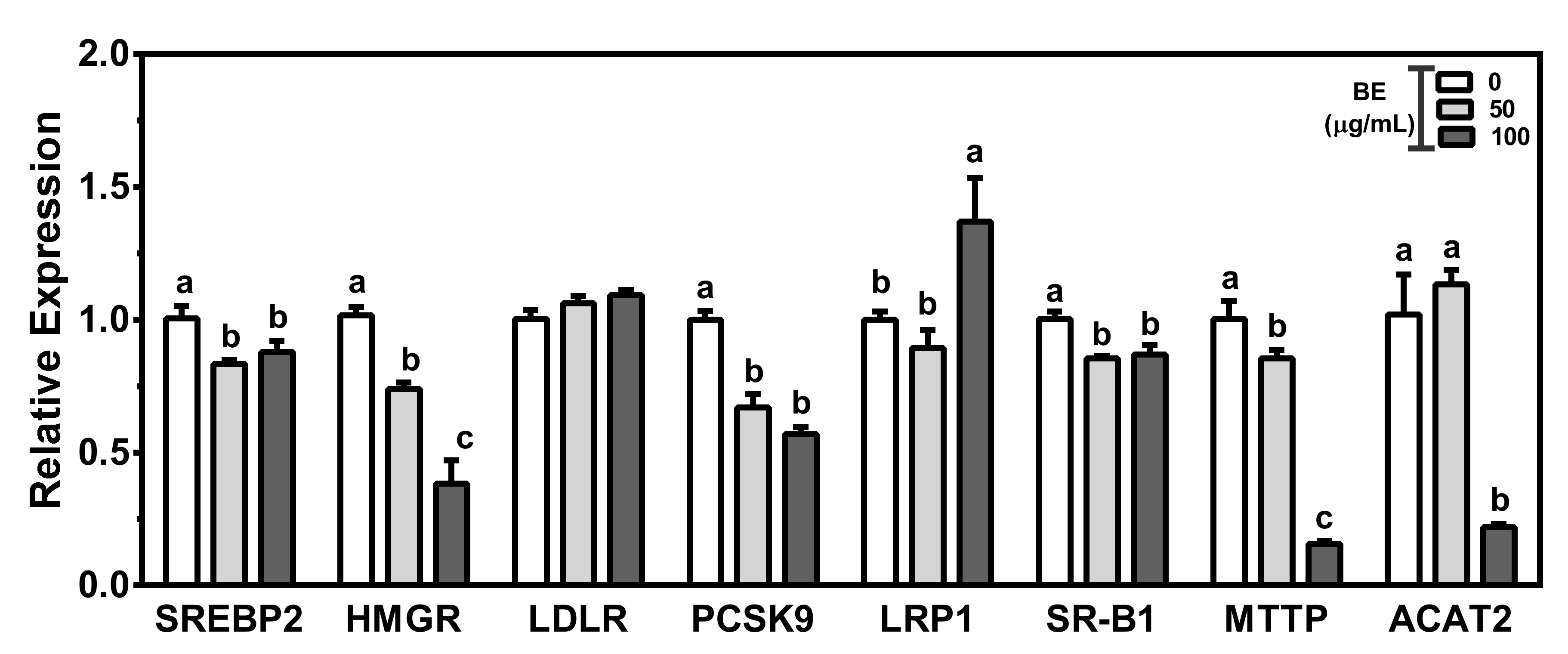
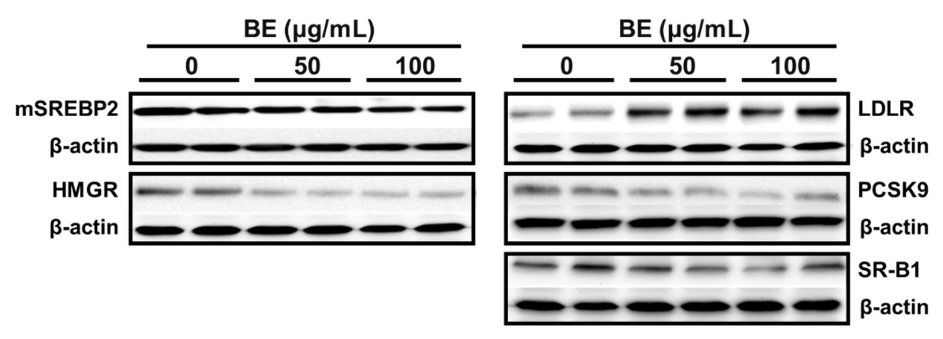
3.3. BE Altered the Expression of Genes for Biosynthesis and Flux of Cholesterol

To examine the effects of BE on hepatic cholesterol metabolism, we first assessed the mRNA abundance of genes for cholesterol biosynthesis and uptake in HepG2 cells treated with BE. In response to intracellular cholesterol deprivation, SREBP2 upregulates the transcription of HMGR and LDLR. Conversion of HMG-CoA to mevalonic acid by HMGR is the rate-limiting reaction in cholesterol de novo synthesis. The expression of SRBP2, the transcription factor for cholesterol metabolism, was significantly decreased in BE-treated HepG2 cells. There was a dose-dependent and significant reduction in HMGR expression by BE treatment. Hepatic lipoprotein receptors, i.e., LDLR, LDL-related protein 1 (LRP1) and SR-B1, play prominent roles in cellular cholesterol homeostasis by removing lipoprotein-derived cholesterol in the circulation. There were no changes in the mRNA abundance of LDLR, the receptor for LDL, in BE-treated cells. The mRNA levels of PCSK9, an enzyme responsible for LDLR degradation, were significantly decreased by BE treatment. The mRNA abundance of LRP1 was significantly upregulated, whereas the expression of SR-B1 was downregulated in 100 μg/mL of BE-treated cells. Hepatic very low-density lipoprotein (VLDL) assembly and secretion are essential for maintaining plasma cholesterol and TG levels. The expression of microsomal triglyceride transfer protein (MTTP), essential for the hepatic assembly of VLDL particles, was dose-dependently and significantly decreased by BE treatment. There was a significant reduction in ACAT2, a significant cholesterol esterification enzyme, by 100 μg/mL of BE treatment (Figure 2). The protein expressions of mature SREBP2 and HMGR were noticeably reduced in BE-treated HepG2 cells. BE markedly increased the protein levels of LDLR without alteration of mRNA levels. Moreover, PCSK9 protein levels were noticeably reduced by BE treatment. There was no induction of SR-B1 protein level in BE-treated HepG2 cells (Figure 3).

3.4. Regulation of Genes for Biliary Cholesterol Efflux by BE

The genes involved in the canalicular cholesterol efflux were measured in HepG2 cells to examine the effects of BE on biliary cholesterol flux. The transporters present in the canalicular membrane, i.e., NPC1L1 and ABCG5/G8, are involved in biliary cholesterol efflux in hepatocytes. NPC1L1 facilitates cholesterol absorption from bile, while the heterodimer ABCG5/G8 flux cholesterol mediates free cholesterol secretion into bile. There was a significant reduction in NPC1L1 expression in BE-treated HepG2 cells. A significant upregulation of ABCG8 was observed in treatment with 100 μg/mL of BE. Bile acid synthesis and secretion play essential roles in maintaining cholesterol homeostasis. There was a significant and dose-dependent increase in mRNA expression of CYP7A1, the rate-limiting enzyme for converting cholesterol to bile in BE-treated cells. Furthermore, 100 μg/mL of BE treatment significantly increased the expression of ABCB11, the canalicular transporter for bile acid secretion. A significant upregulation of ABCB4, a transporter for biliary phospholipid secretion, was observed in 100 μg/mL of BE-treated cells (Figure 4a). Consistent with mRNA data, the protein expressions of canalicular transporters for biliary cholesterol efflux were altered by BE treatment. There was a noticeable reduction in NPC1L1 protein abundance in BE-treated HepG2 cells. The protein levels of transporters involved in biliary cholesterol secretion, i.e., ABCG8 and ABCB11, were markedly induced by BE (Figure 4b).

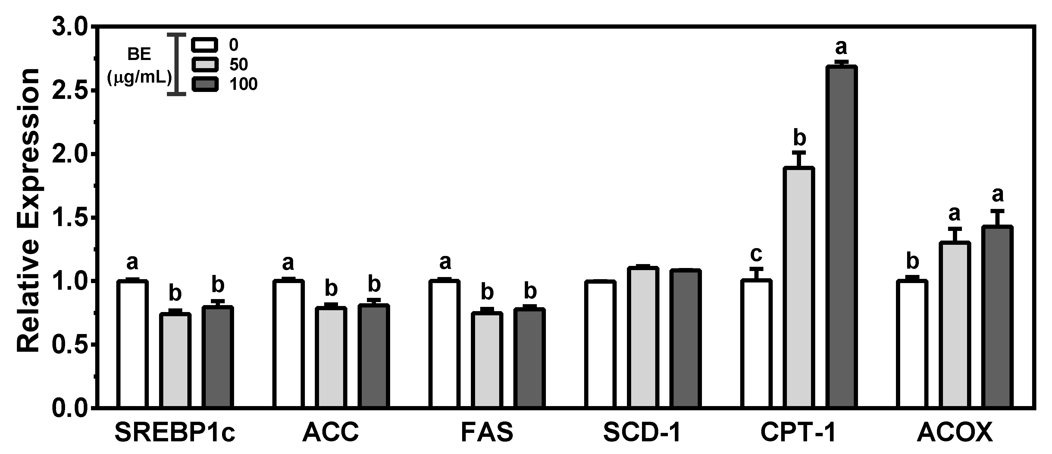
3.5. BE on Genes for Fatty Acid Metabolism

The mRNA levels of genes for hepatic lipogenesis and fatty acid oxidation were measured to examine whether BE can alter fatty acid metabolism in HepG2 cells. The lipogenic transcription SREBP1c can activate genes for de novo fatty acid biosynthesis such as acyl-CoA carboxylase (ACC), fatty acid synthase (FAS) and stearoyl-CoA desaturase-1 (SCD-1). In 100 μg/mL of BE-treated HepG2 cells, the expression of SREBP1c was significantly attenuated, with concomitant decreases in the mRNA level of ACC and FAS. There was no significant alteration of SCD-1 by BE treatment. The expression of CPT1, the key regulatory enzyme for mitochondrial fatty acid oxidation, was significantly and dose-dependently increased in BE-treated HepG2 cells. Furthermore, BE significantly increased the mRNA abundance of ACOX, the essential enzyme for peroxisomal fatty acid oxidation (Figure 5).

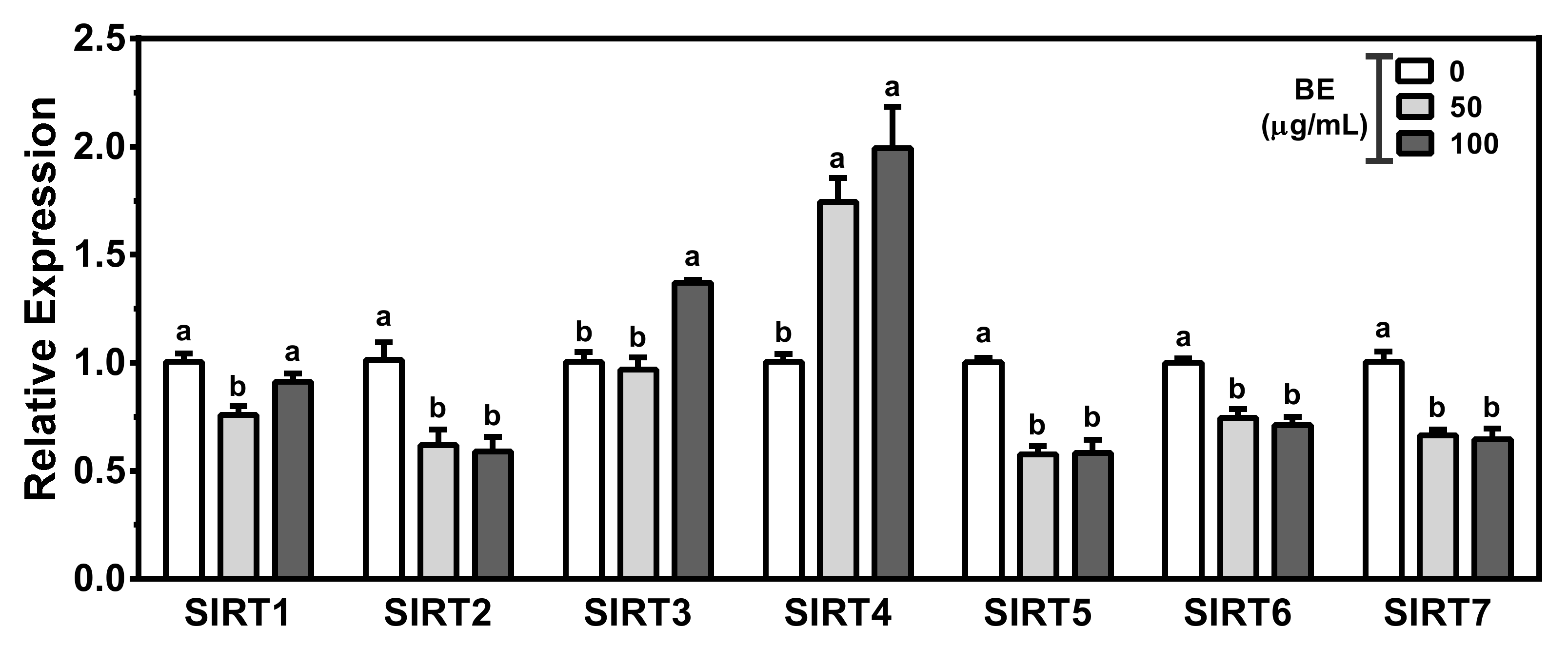
3.6. Alteration of Sirtuins by BE Treatment

The mRNA expression of seven isoforms of sirtuins (SIRTs) was evaluated to investigate the effects of BE on the regulation of SIRTs. The expression of SIRT1 was significantly decreased in 50 μg/mL of BE-treated HepG2 cells. There were significant reductions in the SIRT2, SIRT5, SIRT6 and SIRT7 by BE treatment. In contrast, there was significant induction of SIRT3 and SIRT4 expression in 100 μg/mL of BE-treated cells (Figure 6).

4. Discussion

Hypercholesterolemia is a major modifiable risk factor for the prevention of CVD [36]. Plasma cholesterol level is tightly associated with the regulation of cellular cholesterol metabolism [37]. Natural products and their bioactive components have preventative effects on CVD by exerting antioxidant, anti-inflammatory and hypocholesterolemic properties. Among natural products, polyphenol intake improved metabolic syndrome, a cluster of conditions associated with CVD risk factors, in animal and human clinical trials [38,39,40,41]. With high contents of polyphenols and anthocyanins, bilberry is known for its antioxidant and anti-inflammatory effects [42,43,44]. Previously, we reported the hypocholesterolemic effects of BE by altering the genes for the stimulation of TICE in Caco-2 cells [34]. The stimulation of TICE is an attractive strategy for the prevention of CVD [8]. However, the primary pathway for cholesterol removal from the body is mediated by hepatobiliary cholesterol driven by RCT [9,10]. Furthermore, the liver is the principal site of cholesterol metabolism, accounting for 70% of fecal sterol excretion under normal conditions [45]. Therefore, we investigated whether the hypocholesterolemic effects of BE are attributed to the regulation of hepatic genes involved in cholesterol biosynthesis, uptake and efflux in HepG2 cells.
The previous study confirmed that BE, the standardized bilberry extract to anthocyanin contents of 25%, contains 237.9 ± 17.1 mg cyanidin-3-glucoside equivalents/g of total anthocyanin [34]. Consistent with other studies, BE with high anthocyanin contents exerted antioxidant effects by scavenging DPPH and ABTS radicals and reducing power. The ethanolic extract of bilberry exhibited antioxidant effects by exerting significant DPPH and ABTS radical scavenging activity and suppressing nitric oxide in LPS-induced Raw 264.7 macrophages [46]. These data support that bilberry with high contents of anthocyanin can be used as a natural antioxidant.
Regulation of cholesterol biosynthesis is one of the regulatory mechanisms of maintaining cholesterol homeostasis. SREBPs are the transcription factor that regulates lipid metabolism [47]. SREBP2, the master regulator for cholesterol metabolism, is primarily responsible for the transcription of HMGR and LDLR [48]. HMGR, the key rate-limiting enzyme for de novo cholesterol synthesis, converts HMGCoA to mevalonate, the key intermediate in cholesterol biosynthesis [49]. The most commonly prescribed drug to lower cholesterol is statins, the HMGR inhibitors. Decreasing hepatic cholesterol biosynthesis by statins will lead to the upregulation of LDLR responsible for the clearance of LDL from plasma [11]. In the present study, the maturation of SREBP2 was significantly decreased with concomitant decreases in HMGR mRNA and protein levels by BE treatment. Consistent with these findings, natural products rich in polyphenols, including flavone and anthocyanin, modulated expressions of HMGR and SREBP2. Bergamot rich in flavones decreased HMGR levels and flavone luteolin decreased HMGR transcription by suppressing the nuclear translocation of SREBP2 in HepG2 cells [50,51]. Mulberry extract rich in anthocyanin ameliorated mRNA and protein abundance of HMGR in HepG2 cells [52]. Anthocyanin-rich red raspberry supplementation lowered HMGR expression, leading to decreased total cholesterol concentrations in high-fat-diet-induced hyperlipidemic mice [53]. Supplementation of Solanum nigrum polyphenols ameliorated the hepatic HMGR and SREBP2 protein levels in high-fat-diet-induced obese mice [54]. These results support that the reduction of HMGR and SREBP2 by BE may be attributed to its high polyphenol contents.
Hepatic LDLR regulates the uptake of apoB- and apoE-containing lipoproteins [55]. There was a marked induction of LDLR protein levels without changes in mRNA abundance in BE-treated HepG2 cells. Furthermore, LDLR induction by BE was not dependent on SREBP2 transcriptional regulation. These results indicate that BE increased LDLR by post-transcriptional regulation mechanisms. PCSK9, the negative post-transcriptional regulator of LDLR, binds and regulates the lysosomal degradation of LDLR [56,57]. Therefore, we examined the mRNA and protein expressions of PCSK9 to address underlying mechanisms of noticeable upregulation of LDLR protein by BE. Both PCSK9 mRNA and protein levels were significantly abolished in BE-treated HepG2 cells. A black raspberry extract containing flavonoids and phenolic acids exerted upregulation of LDLR and downregulation of PCSK9 in HepG2 cells [58]. LRP1 and SR-B1, the central regulators of plasma lipid concentration, play critical roles in the uptake of chylomicron remnant and HDL, respectively. BE treatment upregulated the expression of LRP1, whereas the expression of SR-B1 was downregulated. Polyphenol-rich compounds such as red wine concentrate, propolis and quercetin altered the hepatic SR-B1 expression in HepG2 cells and mice [59,60,61].
MTTP and ACAT2 play an essential role in the formation and secretion of the hepatic apoB-containing VLDL [62]. Hepatic MTTP, a lipid transfer protein, transfers TG and cholesterol esters for VLDL assembly and secretion [63]. ACAT2 catalyzes the esterification reaction that converts intracellular free cholesterol to cholesterol ester assembled into VLDL particles for secretion in the liver [64]. Dietary factors that suppress the secretion of VLDLs can be candidates for the prevention of dyslipidemia. The expression of both MTTP and ACAT2 was significantly decreased by BE. Consistent with our results, polyphenol-rich foods decreased apoB secretion attributed to the reduction in MTTP in HepG2 cells. Ellagic acid, a natural polyphenolic compound, suppressed apoB secretion in HepG2 cells [65]. White tea extract rich in EGCG downregulated apoB and MTTP expression, followed by a reduction in VLDL production [66].
Hepatic biliary cholesterol efflux is governed by canalicular cholesterol transporters, such as NPC1L1 and the heterodimer ABCG5/G8 [67]. Highly expressed in the intestine and liver, NPC1L1 functions as a cholesterol absorption transporter in humans [68]. One of the commonly prescribed medicine to lower plasma cholesterol is ezetimibe, the NPClL1 inhibitor that blocks intestinal cholesterol absorption [69,70]. Furthermore, NPC1L1 in hepatocyte counterbalances biliary cholesterol secretion mediated by ABCG5/G8 [71]. The hepatic ABCG5/G8 facilitates the direct export of free cholesterol from the liver to bile. The mRNA and protein levels of NPC1L1 were reduced, whereas ABCG8 was markedly elevated in BE-treated HepG2 cells. The NPC1L1 expression was directly and indirectly inhibited by luteolin and phenolic-compound-rich Opuntia ficus indica [72,73]. Similar to this study, persimmon tannin and coffee arabica pulp increased the expression of ABCG5/G8 in HepG2 cells [74,75]. Bile acid conversion from cholesterol is an essential pathway for cholesterol elimination. CYP7A1 and ABCB11 are required for biliary cholesterol excretion. In the liver, CYP7A1 initiates cholesterol degradation by converting cholesterol to bile acids [76]. The bile salt export pump (BSEP), ABCB11, facilitates canalicular bile acid secretion [77]. There was a marked upregulation of genes for rate-limiting control of bile acid biosynthesis and secretion in BE-treated HepG2 cells. Polyphenols, i.e., resveratrol and quercetins, induced CYP7A1 expression in HepG2 cells [78]. All of the genes for cholesterol transport were changed in the direction of canalicular biliary cholesterol secretion in BE-treated HepG2 cells. The results from the present study support that the protective effects of BE on hypercholesterolemia can be attributed to the stimulation of genes for biliary cholesterol excretion in the liver.
Increased hepatic de novo lipogenesis is known to contribute to hyperlipidemia. The transcription factor SREBP1c regulates ACC, FAS and SCD-1, the enzymes for fatty acid biosynthesis [47]. The expressions of hepatic SREBP1c and its downstream lipogenic genes ACC and FAS were significantly decreased by BE treatment. CPT-1 and ACOX are the key enzymes responsible for mitochondrial and peroxisomal fatty acid oxidation, respectively [79,80]. BE significantly upregulated both CPT-1 and ACOX expression in HepG2 cells. Consistent with these results, polyphenols increased CPT-1 in HepG2 cells.
Epigenetic modulation of cholesterol metabolism-related genes such as HMGR, LDLR and NPC1L1 are reported. BE significantly altered genes involved in cholesterol metabolism, i.e., biosynthesis, transport, bile acid conversion and secretion in the liver. Therefore, we investigated whether BE can alter sirtuin expression in HepG2 cells. The expression of SIRT1, SRIT2, SIRT5, SIRT6 and SIRT7 was downregulated, whereas SIRT3 and SIRT4 expressions were upregulated by BE treatment. Further study is needed to investigate the mechanistic insight of BE on the alteration of SIRTs.

5. Conclusions

In HepG2 cells, BE altered the expression of genes that regulate hepatic cholesterol synthesis, uptake and efflux of cholesterol and bile acids. All of the gene alterations induced by BE were in the direction of hepatobiliary cholesterol excretion. Further study is needed to evaluate whether polyphenols, especially anthocyanins in BE, are responsible for altering genes for hepatic cholesterol metabolism. These results indicate that the bilberry with antioxidant and hypolipidemic effects may be beneficial for the prevention of CVD.

Author Contributions

Conceptualization, M.B. and B.K.; methodology, J.H., M.B. and B.K.; validation, J.H. and B.K.; formal analysis, J.H., M.B. and B.K.; investigation, J.H., M.B. and B.K.; resources, B.K.; data curation, J.H., M.B. and B.K.; writing—original draft preparation, J.H. and B.K.; writing—review and editing, M.B. and B.K.; supervision, B.K.; funding acquisition, B.K. All authors have read and agreed to the published version of the manuscript.

Funding

This research received no external funding.

Institutional Review Board Statement

Not applicable.

Informed Consent Statement

Not applicable.

Data Availability Statement

The data presented in this study are available in the article.

Conflicts of Interest

The authors declare no conflict of interest.

References

  1. Wang, Y.; Ma, X.; Ye, J.; Zhang, S.; Chen, Z.; Jiang, S. Effects of Dietary Supplementation with Bilberry Extract on Growth Performance, Immune Function, Antioxidant Capacity, and Meat Quality of Yellow-Feathered Chickens. Animals 2021, 11, 1989. [Google Scholar] [CrossRef]
  2. Cannon, C.P. Cardiovascular disease and modifiable cardiometabolic risk factors. Clin. Cornerstone 2007, 8, 11–28. [Google Scholar] [CrossRef]
  3. Nelson, R.H. Hyperlipidemia as a Risk Factor for Cardiovascular Disease. Prim. Care 2013, 40, 195–211. [Google Scholar] [CrossRef] [Green Version]
  4. Lange, Y.; Steck, T.L. Cholesterol homeostasis and the escape tendency (activity) of plasma membrane cholesterol. Prog. Lipid. Res. 2008, 47, 319–332. [Google Scholar] [CrossRef] [Green Version]
  5. Groen, A.K.; Bloks, V.W.; Verkade, H.; Kuipers, F. Cross-talk between liver and intestine in control of cholesterol and energy homeostasis. Mol. Aspects Med. 2014, 37, 77–88. [Google Scholar] [CrossRef]
  6. Luo, J.; Yang, H.; Song, B.L. Mechanisms and regulation of cholesterol homeostasis. Nat. Rev. Mol. Cell Biol. 2020, 21, 225–245. [Google Scholar] [CrossRef]
  7. D’Arcy, B.M.; Swingle, M.R.; Schambeau, L.; Pannell, L.; Prakash, A.; Honkanen, R.E. Development of a Synthetic 3-ketosteroid Delta(1)-dehydrogenase for the Generation of a Novel Catabolic Pathway Enabling Cholesterol Degradation in Human Cells. Sci. Rep. 2019, 9, 5969. [Google Scholar] [CrossRef] [Green Version]
  8. Temel, R.E.; Brown, J.M. A new framework for reverse cholesterol transport: Non-biliary contributions to reverse cholesterol transport. World J. Gastroenterol. 2010, 16, 5946–5952. [Google Scholar] [CrossRef]
  9. Grefhorst, A.; Verkade, H.J.; Groen, A.K. The TICE Pathway: Mechanisms and Lipid-Lowering Therapies. Methodist Debakey Cardiovasc. J. 2019, 15, 70–76. [Google Scholar] [CrossRef]
  10. Temel, R.E.; Brown, J.M. A new model of reverse cholesterol transport: enTICEing strategies to stimulate intestinal cholesterol excretion. Trends Pharmacol. Sci. 2015, 36, 440–451. [Google Scholar] [CrossRef]
  11. Simonson, W. Update on statin drugs for lipid disorders. Geriatr. Nurs. 2018, 39, 350–351. [Google Scholar] [CrossRef]
  12. Kashani, A.; Sallam, T.; Bheemreddy, S.; Mann, D.L.; Wang, Y.; Foody, J.M. Review of side-effect profile of combination ezetimibe and statin therapy in randomized clinical trials. Am. J. Cardiol. 2008, 101, 1606–1613. [Google Scholar] [CrossRef]
  13. Viljoen, A.; Wierzbicki, A.S. Colesevelam: An improved bile acid sequestrant for treating hypercholesterolemia and improving diabetes. Expert Rev. Endocrinol. Metab. 2010, 5, 825–834. [Google Scholar] [CrossRef]
  14. Van Bruggen, F.H.; Nijhuis, G.B.J.; Zuidema, S.U.; Luijendijk, H. Serious adverse events and deaths in PCSK9 inhibitor trials reported on ClinicalTrials.gov: A systematic review. Expert Rev. Clin. Pharmacol. 2020, 13, 787–796. [Google Scholar] [CrossRef]
  15. Lemus, H.N.; Mendivil, C.O. Adenosine triphosphate citrate lyase: Emerging target in the treatment of dyslipidemia. J. Clin. Lipidol. 2015, 9, 384–389. [Google Scholar] [CrossRef]
  16. Sprecher, D.L. Raising high-density lipoprotein cholesterol with niacin and fibrates: A comparative review. Am. J. Cardiol. 2000, 86, 46L–50L. [Google Scholar] [CrossRef]
  17. Zafra-Stone, S.; Yasmin, T.; Bagchi, M.; Chatterjee, A.; Vinson, J.A.; Bagchi, D. Berry anthocyanins as novel antioxidants in human health and disease prevention. Mol. Nutr. Food Res. 2007, 51, 675–683. [Google Scholar] [CrossRef]
  18. Quinones, M.; Miguel, M.; Aleixandre, A. Beneficial effects of polyphenols on cardiovascular disease. Pharmacol. Res. 2013, 68, 125–131. [Google Scholar] [CrossRef]
  19. Tangney, C.C.; Rasmussen, H.E. Polyphenols, inflammation, and cardiovascular disease. Curr. Atheroscler. Rep. 2013, 15, 324. [Google Scholar] [CrossRef] [Green Version]
  20. Chu, W.; Cheung, S.C.M.; Lau, R.A.W.; Benzie, I.F.F. Bilberry (Vaccinium myrtillus L.). In Herbal Medicine: Biomolecular and Clinical Aspects; Benzie, I.F.F., Wachtel-Galor, S., Eds.; CRC Press/Taylor & Francis: Boca Raton, FL, USA, 2011. [Google Scholar]
  21. Ulbricht, C.; Basch, E.; Basch, S.; Bent, S.; Boon, H.; Burke, D.; Costa, D.; Falkson, C.; Giese, N.; Goble, M.; et al. An evidence-based systematic review of bilberry (Vaccinium myrtillus) by the Natural Standard Research Collaboration. J. Diet. Suppl. 2009, 6, 162–200. [Google Scholar] [CrossRef]
  22. Brasanac-Vukanovic, S.; Mutic, J.; Stankovic, D.M.; Arsic, I.; Blagojevic, N.; Vukasinovic-Pesic, V.; Tadic, V.M. Wild Bilberry (Vaccinium myrtillus L., Ericaceae) from Montenegro as a Source of Antioxidants for Use in the Production of Nutraceuticals. Molecules 2018, 23, 1864. [Google Scholar] [CrossRef] [Green Version]
  23. Grohmann, T.; Litts, C.; Horgan, G.; Zhang, X.; Hoggard, N.; Russell, W.; de Roos, B. Efficacy of Bilberry and Grape Seed Extract Supplement Interventions to Improve Glucose and Cholesterol Metabolism and Blood Pressure in Different Populations—A Systematic Review of the Literature. Nutrients 2021, 13, 1692. [Google Scholar] [CrossRef] [PubMed]
  24. Kowalska, K.; Olejnik, A.; Szwajgier, D.; Olkowicz, M. Inhibitory activity of chokeberry, bilberry, raspberry and cranberry polyphenol-rich extract towards adipogenesis and oxidative stress in differentiated 3T3-L1 adipose cells. PLoS ONE 2017, 12, e0188583. [Google Scholar] [CrossRef] [Green Version]
  25. Roth, S.; Spalinger, M.R.; Muller, I.; Lang, S.; Rogler, G.; Scharl, M. Bilberry-derived anthocyanins prevent IFN-gamma-induced pro-inflammatory signalling and cytokine secretion in human THP-1 monocytic cells. Digestion 2014, 90, 179–189. [Google Scholar] [CrossRef] [PubMed] [Green Version]
  26. Luo, H.; Lv, X.D.; Wang, G.E.; Li, Y.F.; Kurihara, H.; He, R.R. Anti-inflammatory effects of anthocyanins-rich extract from bilberry (Vaccinium myrtillus L.) on croton oil-induced ear edema and Propionibacterium acnes plus LPS-induced liver damage in mice. Int. J. Food Sci. Nutr. 2014, 65, 594–601. [Google Scholar] [CrossRef]
  27. Kropat, C.; Mueller, D.; Boettler, U.; Zimmermann, K.; Heiss, E.H.; Dirsch, V.M.; Rogoll, D.; Melcher, R.; Richling, E.; Marko, D. Modulation of Nrf2-dependent gene transcription by bilberry anthocyanins in vivo. Mol. Nutr. Food Res. 2013, 57, 545–550. [Google Scholar] [CrossRef]
  28. Nakano, H.; Wu, S.; Sakao, K.; Hara, T.; He, J.; Garcia, S.; Shetty, K.; Hou, D.X. Bilberry Anthocyanins Ameliorate NAFLD by Improving Dyslipidemia and Gut Microbiome Dysbiosis. Nutrients 2020, 12, 3252. [Google Scholar] [CrossRef]
  29. Brader, L.; Overgaard, A.; Christensen, L.P.; Jeppesen, P.B.; Hermansen, K. Polyphenol-rich bilberry ameliorates total cholesterol and LDL-cholesterol when implemented in the diet of Zucker diabetic fatty rats. Rev. Diabet. Stud. 2013, 10, 270–282. [Google Scholar] [CrossRef] [Green Version]
  30. Stefanut, M.N.; Cata, A.; Pop, R.; Tanasie, C.; Boc, D.; Ienascu, I.; Ordodi, V. Anti-hyperglycemic effect of bilberry, blackberry and mulberry ultrasonic extracts on diabetic rats. Plant Foods Hum. Nutr. 2013, 68, 378–384. [Google Scholar] [CrossRef]
  31. Asgary, S.; RafieianKopaei, M.; Sahebkar, A.; Shamsi, F.; Goli-malekabadi, N. Anti-hyperglycemic and anti-hyperlipidemic effects of Vaccinium myrtillus fruit in experimentally induced diabetes (antidiabetic effect of Vaccinium myrtillus fruit). J. Sci. Food Agric. 2016, 96, 764–768. [Google Scholar] [CrossRef]
  32. Karlsen, A.; Paur, I.; Bohn, S.K.; Sakhi, A.K.; Borge, G.I.; Serafini, M.; Erlund, I.; Laake, P.; Tonstad, S.; Blomhoff, R. Bilberry juice modulates plasma concentration of NF-kappaB related inflammatory markers in subjects at increased risk of CVD. Eur. J. Nutr. 2010, 49, 345–355. [Google Scholar] [CrossRef] [PubMed]
  33. Zhu, Y.; Huang, X.; Zhang, Y.; Wang, Y.; Liu, Y.; Sun, R.; Xia, M. Anthocyanin supplementation improves HDL-associated paraoxonase 1 activity and enhances cholesterol efflux capacity in subjects with hypercholesterolemia. J. Clin. Endocrinol. Metab. 2014, 99, 561–569. [Google Scholar] [CrossRef] [PubMed] [Green Version]
  34. Hong, J.; Kim, M.; Kim, B. The Effects of Anthocyanin-Rich Bilberry Extract on Transintestinal Cholesterol Excretion. Foods 2021, 10, 2852. [Google Scholar] [CrossRef] [PubMed]
  35. Jeon, S.; Lee, S.; Choi, Y.; Kim, B. The Effects of Polyphenol-Rich Black Elderberry on Oxidative Stress and Hepatic Cholesterol Metabolism. Appl. Sci. 2021, 11, 10018. [Google Scholar] [CrossRef]
  36. Mommersteeg, P.M.; Denollet, J.; Spertus, J.A.; Pedersen, S.S. Health status as a risk factor in cardiovascular disease: A systematic review of current evidence. Am. Heart J. 2009, 157, 208–218. [Google Scholar] [CrossRef]
  37. Kapourchali, F.R.; Surendiran, G.; Goulet, A.; Moghadasian, M.H. The Role of Dietary Cholesterol in Lipoprotein Metabolism and Related Metabolic Abnormalities: A Mini-review. Crit. Rev. Food Sci. Nutr. 2016, 56, 2408–2415. [Google Scholar] [CrossRef]
  38. Chiva-Blanch, G.; Badimon, L. Effects of Polyphenol Intake on Metabolic Syndrome: Current Evidences from Human Trials. Oxid. Med. Cell Longev. 2017, 2017, 5812401. [Google Scholar] [CrossRef] [Green Version]
  39. Rienks, J.; Barbaresko, J.; Nothlings, U. Review Association of Polyphenol Biomarkers with Cardiovascular Disease and Mortality Risk: A Systematic Review and Meta-Analysis of Observational Studies. Nutrients 2017, 9, 415. [Google Scholar] [CrossRef] [Green Version]
  40. Mendonca, R.D.; Carvalho, N.C.; Martin-Moreno, J.M.; Pimenta, A.M.; Lopes, A.C.S.; Gea, A.; Martinez-Gonzalez, M.A.; Bes-Rastrollo, M. Total polyphenol intake, polyphenol subtypes and incidence of cardiovascular disease: The SUN cohort study. Nutr. Metab. Cardiovas. 2019, 29, 69–78. [Google Scholar] [CrossRef]
  41. Quinones, M.; Miguel, M.; Aleixandre, A. The polyphenols, naturally occurring compounds with beneficial effects on cardiovascular disease. Nutr. Hosp. 2012, 27, 76–89. [Google Scholar] [CrossRef]
  42. Bornsek, S.M.; Ziberna, L.; Polak, T.; Vanzo, A.; Ulrih, N.P.; Abram, V.; Tramer, F.; Passamonti, S. Bilberry and blueberry anthocyanins act as powerful intracellular antioxidants in mammalian cells. Food Chem. 2012, 134, 1878–1884. [Google Scholar] [CrossRef] [PubMed]
  43. Yao, N.; Lan, F.; He, R.R.; Kurihara, H. Protective effects of bilberry (Vaccinium myrtillus L.) extract against endotoxin-induced uveitis in mice. J. Agric. Food Chem. 2010, 58, 4731–4736. [Google Scholar] [CrossRef] [PubMed]
  44. Bao, L.; Yao, X.S.; Tsi, D.; Yau, C.C.; Chia, C.S.; Nagai, H.; Kurihara, H. Protective effects of bilberry (Vaccinium myrtillus L.) extract on KBrO3-induced kidney damage in mice. J. Agric. Food Chem. 2008, 56, 420–425. [Google Scholar] [CrossRef] [PubMed]
  45. Wang, D.Q.; Portincasa, P.; Tso, P. Transintestinal cholesterol excretion: A secondary, nonbiliary pathway contributing to reverse cholesterol transport. Hepatology 2017, 66, 1337–1340. [Google Scholar] [CrossRef] [Green Version]
  46. Bayazid, A.B.; Chun, E.M.; Al Mijan, M.; Park, S.H.; Moon, S.-K.; Lim, B.O. Anthocyanins profiling of bilberry (Vaccinium myrtillus L.) extract that elucidates antioxidant and anti-inflammatory effects. Food Agric. Immunol. 2021, 32, 713–726. [Google Scholar] [CrossRef]
  47. Brown, M.S.; Goldstein, J.L. Sterol regulatory element binding proteins (SREBPs): Controllers of lipid synthesis and cellular uptake. Nutr. Rev. 1998, 56, S1–S3. [Google Scholar] [CrossRef]
  48. Madison, B.B. Srebp2: A master regulator of sterol and fatty acid synthesis. J. Lipid. Res. 2016, 57, 333–335. [Google Scholar] [CrossRef] [Green Version]
  49. Goldstein, J.L.; Brown, M.S. Regulation of the mevalonate pathway. Nature 1990, 343, 425–430. [Google Scholar] [CrossRef]
  50. Huang, Y.; Tocmo, R.; Nauman, M.C.; Haughan, M.A.; Johnson, J.J. Defining the Cholesterol Lowering Mechanism of Bergamot (Citrus bergamia) Extract in HepG2 and Caco-2 Cells. Nutrients 2021, 13, 3156. [Google Scholar] [CrossRef]
  51. Wong, T.Y.; Lin, S.M.; Leung, L.K. The Flavone Luteolin Suppresses SREBP-2 Expression and Post-Translational Activation in Hepatic Cells. PLoS ONE 2015, 10, e0135637. [Google Scholar] [CrossRef]
  52. Liu, L.K.; Chou, F.P.; Chen, Y.C.; Chyau, C.C.; Ho, H.H.; Wang, C.J. Effects of mulberry (Morus alba L.) extracts on lipid homeostasis in vitro and in vivo. J. Agric. Food Chem. 2009, 57, 7605–7611. [Google Scholar] [CrossRef] [PubMed]
  53. Tu, L.; Sun, H.; Tang, M.; Zhao, J.; Zhang, Z.; Sun, X.; He, S. Red raspberry extract (Rubus idaeus L. shrub) intake ameliorates hyperlipidemia in HFD-induced mice through PPAR signaling pathway. Food Chem. Toxicol. 2019, 133, 110796. [Google Scholar] [CrossRef] [PubMed]
  54. Peng, C.H.; Cheng, J.J.; Yu, M.H.; Chung, D.J.; Huang, C.N.; Wang, C.J. Solanum nigrum polyphenols reduce body weight and body fat by affecting adipocyte and lipid metabolism. Food Funct. 2020, 11, 483–492. [Google Scholar] [CrossRef] [PubMed]
  55. Van de Sluis, B.; Wijers, M.; Herz, J. News on the molecular regulation and function of hepatic low-density lipoprotein receptor and LDLR-related protein 1. Curr. Opin. Lipidol. 2017, 28, 241–247. [Google Scholar] [CrossRef] [Green Version]
  56. Park, S.W.; Moon, Y.A.; Horton, J.D. Post-transcriptional regulation of low density lipoprotein receptor protein by proprotein convertase subtilisin/kexin type 9a in mouse liver. J. Biol. Chem. 2004, 279, 50630–50638. [Google Scholar] [CrossRef] [Green Version]
  57. Lagace, T.A. PCSK9 and LDLR degradation: Regulatory mechanisms in circulation and in cells. Curr. Opin. Lipidol. 2014, 25, 387–393. [Google Scholar] [CrossRef] [Green Version]
  58. Song, K.H.; Kim, Y.H.; Im, A.R.; Kim, Y.H. Black Raspberry Extract Enhances LDL Uptake in HepG2 Cells by Suppressing PCSK9 Expression to Upregulate LDLR Expression. J. Med. Food 2018, 21, 560–567. [Google Scholar] [CrossRef]
  59. Lee, D.-H.; Choi, S.-S.; Kim, B.-B.; Kim, S.-Y.; Kang, B.-S.; Lee, S.-J.; Park, H.-J. Effect of alcohol-free red wine concentrates on cholesterol homeostasis: An in vitro and in vivo study. Process. Biochem. 2013, 48, 1964–1971. [Google Scholar] [CrossRef]
  60. Yu, Y.; Si, Y.; Song, G.; Luo, T.; Wang, J.; Qin, S. Ethanolic extract of propolis promotes reverse cholesterol transport and the expression of ATP-binding cassette transporter A1 and G1 in mice. Lipids 2011, 46, 805–811. [Google Scholar] [CrossRef]
  61. Ren, K.; Jiang, T.; Zhao, G.J. Quercetin induces the selective uptake of HDL-cholesterol via promoting SR-BI expression and the activation of the PPARgamma/LXRalpha pathway. Food Funct. 2018, 9, 624–635. [Google Scholar] [CrossRef]
  62. Shelness, G.S.; Sellers, J.A. Very-low-density lipoprotein assembly and secretion. Curr. Opin. Lipidol. 2001, 12, 151–157. [Google Scholar] [CrossRef] [PubMed]
  63. Tiwari, S.; Siddiqi, S.A. Intracellular trafficking and secretion of VLDL. Arterioscler. Thromb. Vasc. Biol. 2012, 32, 1079–1086. [Google Scholar] [CrossRef] [PubMed] [Green Version]
  64. Parini, P.; Davis, M.; Lada, A.T.; Erickson, S.K.; Wright, T.L.; Gustafsson, U.; Sahlin, S.; Einarsson, C.; Eriksson, M.; Angelin, B.; et al. ACAT2 is localized to Hepatocytes and is the major cholesterol-esterifying enzyme in human liver. Circulation 2004, 110, 2017–2023. [Google Scholar] [CrossRef] [PubMed] [Green Version]
  65. Ieda, A.; Wada, M.; Moriyasu, Y.; Okuno, Y.; Zaima, N.; Moriyama, T. Ellagic Acid Suppresses ApoB Secretion and Enhances ApoA-1 Secretion from Human Hepatoma Cells, HepG2. Molecules 2021, 26, 3885. [Google Scholar] [CrossRef] [PubMed]
  66. Luo, K.; Ma, C.; Xing, S.; An, Y.; Feng, J.; Dang, H.; Huang, W.; Qiao, L.; Cheng, J.; Xie, L. White tea and its active polyphenols lower cholesterol through reduction of very-low-density lipoprotein production and induction of LDLR expression. Biomed. Pharmacother. 2020, 127, 110146. [Google Scholar] [CrossRef]
  67. Kidambi, S.; Patel, S.B. Cholesterol and non-cholesterol sterol transporters: ABCG5, ABCG8 and NPC1L1: A review. Xenobiotica 2008, 38, 1119–1139. [Google Scholar] [CrossRef]
  68. Jia, L.; Betters, J.L.; Yu, L.Q. Niemann-Pick C1-Like 1 (NPC1L1) Protein in Intestinal and Hepatic Cholesterol Transport. Annu. Rev. Physiol. 2011, 73, 239–259. [Google Scholar] [CrossRef] [Green Version]
  69. Chang, T.Y.; Chang, C. Ezetimibe blocks internalization of the NPC1L1/cholesterol complex. Cell Metab. 2008, 7, 469–471. [Google Scholar] [CrossRef] [Green Version]
  70. Garcia-Calvo, M.; Lisnock, J.M.; Bull, H.G.; Hawes, B.E.; Burnett, D.A.; Braun, M.P.; Crona, J.H.; Davis, H.R.; Dean, D.C.; Detmers, P.A.; et al. The target of ezetimibe is Niemann-Pick Cl-Like 1 (NPC1L1). Proc. Natl. Acad. Sci. USA 2005, 102, 8132–8137. [Google Scholar] [CrossRef] [Green Version]
  71. Temel, R.E.; Tang, W.; Ma, Y.; Rudel, L.L.; Willingham, M.C.; Ioannou, Y.A.; Davies, J.P.; Nilsson, L.M.; Yu, L. Hepatic Niemann-Pick C1-like 1 regulates biliary cholesterol concentration and is a target of ezetimibe. J. Clin. Investig. 2007, 117, 1968–1978. [Google Scholar] [CrossRef]
  72. Kobayashi, S. The Effect of Polyphenols on Hypercholesterolemia through Inhibiting the Transport and Expression of Niemann-Pick C1-Like 1. Int. J. Mol. Sci. 2019, 20, 4939. [Google Scholar] [CrossRef] [PubMed] [Green Version]
  73. Ressaissi, A.; Attia, N.; Pacheco, R.; Falé, P.L.; Serralheiro, M.L.M. Cholesterol transporter proteins in HepG2 cells can be modulated by phenolic compounds present in Opuntia ficus-indica aqueous solutions. J. Funct. Foods 2020, 64, 103674. [Google Scholar] [CrossRef]
  74. Ge, Z.Z.; Zhu, W.; Peng, J.M.; Deng, X.Y.; Li, C.M. Persimmon tannin regulates the expression of genes critical for cholesterol absorption and cholesterol efflux by LXR alpha independent pathway. J. Funct. Foods 2016, 23, 283–293. [Google Scholar] [CrossRef]
  75. Ontawong, A.; Pasachan, T.; Trisuwan, K.; Soodvilai, S.; Duangjai, A.; Pongchaidecha, A.; Amornlerdpison, D.; Srimaroeng, C. Coffea arabica pulp aqueous extract attenuates oxidative stress and hepatic lipid accumulation in HepG2 cells. J. Herb. Med. 2021, 29, 100465. [Google Scholar] [CrossRef]
  76. Chiang, J.Y. Regulation of bile acid synthesis: Pathways, nuclear receptors, and mechanisms. J. Hepatol. 2004, 40, 539–551. [Google Scholar] [CrossRef] [PubMed]
  77. Hayashi, H.; Sugiyama, Y. Bile salt export pump (BSEP/ABCB11): Trafficking and sorting disturbances. Curr. Mol. Pharmacol. 2013, 6, 95–103. [Google Scholar] [CrossRef] [PubMed]
  78. Chambers, K.F.; Day, P.E.; Aboufarrag, H.T.; Kroon, P.A. Polyphenol Effects on Cholesterol Metabolism via Bile Acid Biosynthesis, CYP7A1: A Review. Nutrients 2019, 11, 2588. [Google Scholar] [CrossRef] [Green Version]
  79. Hoppel, C. The role of carnitine in normal and altered fatty acid metabolism. Am. J. Kidney Dis. 2003, 41, S4–S12. [Google Scholar] [CrossRef]
  80. Christensen, E.; Woldseth, B.; Hagve, T.A.; Poll-The, B.T.; Wanders, R.J.; Sprecher, H.; Stokke, O.; Christophersen, B.O. Peroxisomal beta-oxidation of polyunsaturated long chain fatty acids in human fibroblasts. The polyunsaturated and the saturated long chain fatty acids are retroconverted by the same acyl-CoA oxidase. Scand. J. Clin. Lab. Investig. Suppl. 1993, 215, 61–74. [Google Scholar] [CrossRef]
Figure 1. Cell viability of BE-treated HepG2 cells. Cells were treated with increasing concentrations from 0 to 200 μg/mL of BE for 24 h. Different letters on the data points represent significant differences (p < 0.05). Values are means ± S.E.M. n = 6.
Applsci 13 00516 g001
Figure 2. The effects of BE on the mRNA expression of genes involved in cholesterol de novo synthesis and uptake in HepG2 cells. Cells were treated with either 50 or 100 μg/mL of BE for 24 h. The mRNA abundance was determined by qRT-PCR analysis. Bars with different letters are significantly different (p < 0.05). Values are means ± S.E.M. n = 6.
Applsci 13 00516 g002
Figure 3. The effects of BE on the protein levels for cholesterol de novo synthesis and uptake in HepG2 cells. Cells were treated with either 50 or 100 μg/mL of BE for 24 h. The representative images of Western blot are shown. β-actin served as a loading control.
Applsci 13 00516 g003
Figure 4. The effect of BE on the genes regulating biliary cholesterol efflux in HepG2 cells. The cells were treated with either 50 or 100 μg/mL of BE for 24 h. (a) The mRNA abundance was determined by qRT-PCR analysis. Bars with different letters are significantly different (p < 0.05). Values are means ± S.E.M. n = 6. (b) The representative image of the Western blot is shown. β-actin served as a loading control.
Applsci 13 00516 g004
Figure 5. The expression of genes involved in fatty acid synthesis and oxidation in BE-treated HepG2 cells. Cells were treated with either 50 or 100 μg/mL of BE for 24 h. The mRNA abundance was determined by qRT-PCR analysis. Bars with different letters are significantly different (p < 0.05). Values are means ± S.E.M. n = 6.
Applsci 13 00516 g005
Figure 6. Modulation of SIRTs in BE-treated HepG2 cells. The cells were treated with either 50 or 100 μg/mL of BE for 24 h. The mRNA expression of SIRT isoforms was measured by qRT-PCR. Bars with different letters are significantly different (p < 0.05). Values are means ± S.E.M. n = 6.
Applsci 13 00516 g006
Table 1. Total antioxidant capacity of BE.
Total Antioxidant Capacity
DPPH (mM VCE/g)ABTS (mM VCE/g)FRAP (mM FeSO4/g)
282.6 ± 13.5560.8 ± 62.64.73 ± 0.33
Data represent mean ± SEM. VCE, vitamin C equivalents.
Disclaimer/Publisher’s Note: The statements, opinions and data contained in all publications are solely those of the individual author(s) and contributor(s) and not of MDPI and/or the editor(s). MDPI and/or the editor(s) disclaim responsibility for any injury to people or property resulting from any ideas, methods, instructions or products referred to in the content.

