Article

Body Composition in Karate: A Dual-Energy X-ray Absorptiometry Study

Laboratory of Anthropometry and Body Composition, Department of Neurosciences, Biomedicine, and Movement Sciences, University of Verona, Via Casorati 43, I-37134 Verona, Italy
*
Author to whom correspondence should be addressed.
Appl. Sci. 2023, 13(1), 559; https://doi.org/10.3390/app13010559
Submission received: 1 December 2022 / Revised: 24 December 2022 / Accepted: 29 December 2022 / Published: 31 December 2022

Abstract

:
Karate is a widely practiced combat sport. Karatekas’ body composition has typically been obtained in small groups using skinfolds or bioelectric impedance. In this work, we assessed three-compartment body composition using the accurate dual-energy X-ray absorptiometry technique (QDR Horizon, Hologic) in a large sample (n = 58; 74% males) of black belt karatekas. Stature-adjusted body composition indices (fat mass index; fat-free mass index; bone mineral apparent density) were calculated. The Student’s t-test was used for group–group analysis. Correlation was assessed using the Pearson’s r. The ability of fat-free soft tissue mass to predict bone mineral content and areal bone mineral density was assessed with linear regression. Reference mean and quartile values for whole-body and regional body composition were obtained for the male athletes. The body composition indices were generally more favorable in the male than female karatekas. The bone mineral apparent density was similar in the males and females at all sites except the right leg. The fat-free soft tissue mass predicted the bone mineral content and areal bone mineral density with good accuracy (R2 = 0.542–0.827; p < 0.001 for all models). The data presented in this paper are expected to be of use for karate coaches, physical trainers, and participants interested in assessing and monitoring athletes’ body composition.

1. Introduction

Karate is a martial art born in Japan, which is nowadays practiced all over the world. Recent data [1] show that the number of karate practitioners around the world is >100 million, belonging to about 200 national federations. Moreover, the program of the Tokyo 2020 Olympic Games included karate as an additional sport. Karate is an intermittent combat sport, which is physically demanding, dynamic, and high intensity, involving repeated bouts of strikes and defenses, performed as explosive and technically demanding sequences [2]. Accordingly, karate requires both aerobic [3] and anaerobic [4] capabilities. Speed, agility, coordination and balance, and a high level of other motor and functional abilities are required to successfully perform in karate [5]. It has been shown that continuative karate training improves physical qualities including flexibility, strength, balance, and cardiorespiratory fitness [6].
It is well known that athletic success is influenced by diverse factors, from morphological and body composition characteristics to sport-specific skills [7,8]. In particular, it has been shown that the practiced discipline affects the athletes’ physical characteristics. While these physical traits (e.g., stature and skeletal muscle cell number) are largely determined by genetic factors, they are, to some extent, dependent on environmental factors, e.g., systematic conditioning and dieting, the most observable effects affecting body composition with an increase of lean body mass (and muscular strength), and a concomitant reduction in fat mass [9]. Karate has weight competition categories ranging from <60 kg to >84 kg for male and from <50 kg to >68 kg for female athletes.
Ten years ago, a review [10] on the anthropometric, physical, and physiological profile of elite karate athletes showed that information on the body composition of karatekas came essentially from anthropometry and prediction equations. Percent body fat was the most investigated variable, ranging from 7.4 to 18.6% (males and females). Only one paper assessed three-compartment body composition using dual-energy X-ray absorptiometry (DXA) in a small (n = 14) sample of male athletes [11]. In the following years, a limited number of papers dealt with the body composition of adult (>18 y) karatekas, typically using bioelectric impedance analysis (BIA) [12,13,14] or skinfolds [15,16,17]. Currently, the gold standard for the assessment of body composition is the four-compartment method (4C); however, DXA outputs are accurate and precise enough to reliably differentiate lean and fat tissues in addition to mineral [18,19]. Instead, two review papers recently showed that the predictive equations developed using BIA lead to large variability in the estimated value of fat-free mass vs. DXA being population- and device-specific [20,21]. It has also been shown that BIA may overestimate fat-free mass and underestimate FM vs. DXA in athletes [22,23,24,25]. Based on the above, the present work aimed at defining the body composition of karatekas in a large sample of male and female athletes using the laboratory reference method DXA. Given the possible relevance of asymmetries in the body composition of martial sports athletes [26,27], we also conducted a preliminary study comparing right and left upper and lower limb compartments.

2. Materials and Methods

2.1. Participants and Study Design

For this cross-sectional study, a convenience sample of Caucasian karate (kata and kumite) athletes was recruited nationwide. The study was approved by the local ethics committee. Written, informed consent was obtained from all athletes. The inclusion criteria were: aged between 18 and 35 y; karate participation for at least 1 y; and competing at the national or international level. The exclusion criteria were the presence of: ongoing pathology of the locomotor apparatus; significant injury in the last six months; being on pharmacological treatment for any reason; and, for females, amenorrhea (i.e., the absence of menses for a period longer than three months). Information about sport experience (belt, years of practice, training hours) was obtained by interview. A total of 60 karatekas were recruited. Two athletes were excluded due incomplete data. Accordingly, 58 karatekas (43 males, mean age 22.3 ± 4.12 years; 15 females, mean age 20.0 ± 1.81 years) were included in the analysis. The frequencies for weight category (kg) were as follows. Males: −60, n = 3; −67, n = 12; −75, n = 9; −84, n = 16; 84+, n = 3. Females: −50, n = 3; −55, n = 2; −61, n = 7; −68, n = 3; 68+, n = 0.

2.2. Anthropometry and Body Composition Analysis

An electronic scale (Tanita electronic scale BWB-800 MA) was used to measure body mass to the nearest 0.1 kg; a Harpenden stadiometer (Holtain Ltd., Crymych, UK) was used to measure stature to the nearest mm according to standard procedures [28] (intra-rater error ≤0.15% for body mass and ≤0.10% for stature). Body mass index (BMI) was calculated as weight (kg)/height (m2). Body composition was evaluated in terms of bone mineral content (BMC) (and bone areal density (aBMD)), fat-free soft tissue mass (FFSTM), and fat mass (FM) by means of a total body DXA scanner (QDR Horizon, Hologic, Marlborough, MA, USA; fan-beam technology, software version 13.6.05). For the quality control checking of baseline drift, the reference phantom obtained from the manufacturer was used daily. For the sake of consistency, the same researcher carried out all examinations. The participants were scanned in the late morning after voiding when fasted for at least 4 h. The participants were required not to exercise vigorously in the 24 h before examination. DXA scans were performed at least 10 days before or after competition, thus representing close to “competition body composition”, yet before acute weight loss/gain had begun. The participants were in light clothing and removed any metal or reflective material. All measurements were performed according to Nana et al. [29]. Scans were taken of the whole body (WB). The in vivo measurement short-term precision was calculated according to standard criteria [30]; in particular, repeated (n = 2) scanning of 30 subjects with repositioning was carried out. The precision (percent coefficient of variation) was 2.12%, 0.59%, 0.75%, 2.19%, and 0.68% for FM, FFSTM, BMC, %FM, and aBMD, respectively. The following regions were identified in the WB scans with the aid of the Hologic software and eventual correction by one researcher: head, trunk, upper limb (left and right), and lower limb (left and right). The BMC and aBMD values in the total body less head (TBLH) region were used for analysis as recommended in [31], also considering that a large proportion of total body mineral is contained in the skull, which is not easily amenable to changes induced by physical activity [32]. However, WB values for BMC and aBMD were also reported in the results for the sake of comparison with the previous literature. For the purpose of this work, FM, FFSTM, BMC, and aBMD were also calculated for the lower limbs together (Legs) and the four limbs together (Appendicular). In our sample of karate participants, stature and body mass were confounding variables, especially for bone measurements, because DXA is a projectional technique and DXA-derived measurements do not adequately correct for body and/or bone size [33]. Accordingly, the soft tissue body composition variables were also expressed either as a proportion to total body mass or normalized by square stature (fat mass index, FMI; fat-free mass index, FFMI) [34]. The bone mineral was expressed as bone mineral apparent density (BMAD, g/cm3) according to the formula proposed by Katzman et al. [35]: BMAD = BMC/(bone area2/body stature).

2.3. Statistical Analysis

The normality of the data was assessed using the Shapiro–Wilk test. The data are presented as mean ± SD. Sample size calculation (G*Power 3.1) for regression analysis using f2 = 0.15, alpha = 0.05, power = 0.80 as the input parameters yielded n = 55. Correlation analysis was carried out calculating the Pearson’s r. The criteria indicated by Hopkins [36] were used to assess the strength of correlation, i.e., almost perfect (0.90–1), very large (0.70–0.89), large (0.50–0.69), moderate (0.31–0.49), and small (0–0.30). A comparison of body composition in the male and female karatekas and in the right and left limb were carried out with the unpaired- and paired-sample, two-tailed t-test, respectively. The effect size (Cohen’s d) was calculated and rated according to Cohen [37] as small (0.2), medium (0.5), and large (0.8). The ability of FFSTM to predict bone variables was assessed with linear regression analysis. The IBM-SSPS statistical package (v. 26) was used for all analysis. Statistical significance was set at p ≤ 0.05.

3. Results

Valid, complete results were obtained for 58 participants (74% males). All of them were black belt. The data were collapsed across weight divisions and analyzed within sex or side of the body (right, left) as needed. In the whole sample of athletes, the mean age was 21.7 ± 3.78 years, mean body mass was 68.1 ± 10.70 kg, mean stature was 171.3 ± 8.41 cm, and mean BMI was 23.1 ± 2.46 kg/m2; mean karate participation was 14.5 ± 4.10 years (range: 5–28 years). The participants trained 8.0 ± 3.42 h/w (range: 2–20 h) in 4.2 ± 1.97 sessions/w (range: 2–10). The main characteristics of the participant karatekas (males and females) are shown in Table 1. The male and female karatekas were not statistically significantly different in terms of age, BMI, years of karate participation, weekly volume of training, and number of training sessions per week. The males were statistically significantly older (+2.3 years), heavier (+16.6 kg), and taller (+12.9 cm) than the females.
The body composition karatekas (males and females) are shown in Table 2. Males showed statistically significantly higher values (effect size: large) for all bone mineral variables except for thoracic and lumbar spine aBMD. Females showed statistically significantly higher %FM at all regional sites and at the TBLH level with a large effect size. FFSTM was statistically significantly higher in males at all regional sites and at the TBLH level with a large effect size.
The stature-adjusted indices of body composition for males and females are reported in Table 3. All body composition indices were statistically significantly different in males and females except for trunk FMI. FMI was higher in the females and FFMI in males with a large effect size.
BMAD (Table 4) was not statistically significantly different in the males and females, except for right leg BMAD, which was higher in the males (p = 0.024, effect size: medium). The average lumbar spine BMAD was higher in the females, but the difference was at the limit of statistical significance (p = 0.082, effect size: small).
The quartile (25°, 50°, 75°) values of the body composition variables for the males are presented in Table 5.
The data on body composition symmetry in the male and female karatekas are reported in Table 6. While a number of statistically significant differences were present, they showed a trivial (<0.2) or small to moderate effect size.
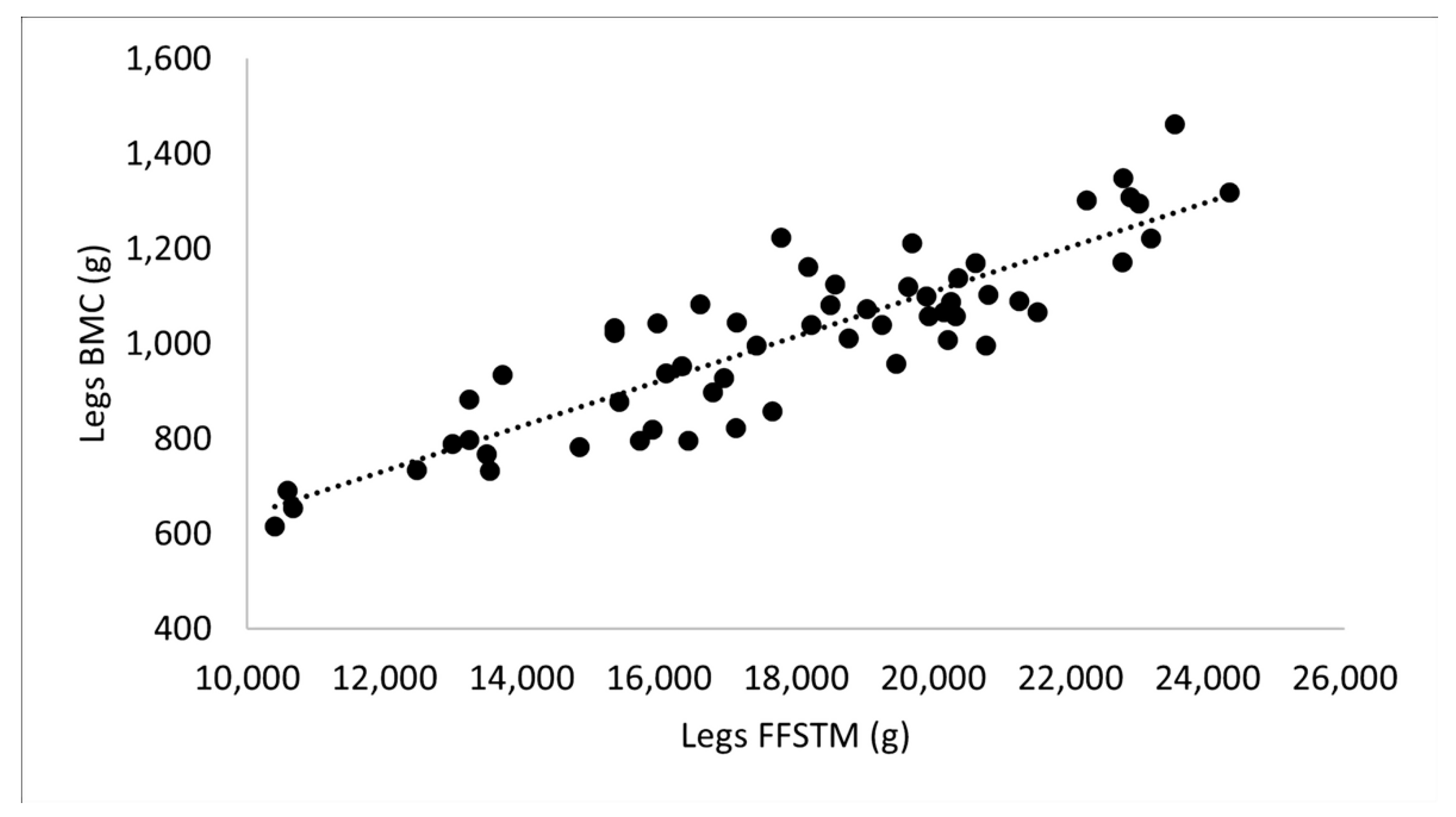
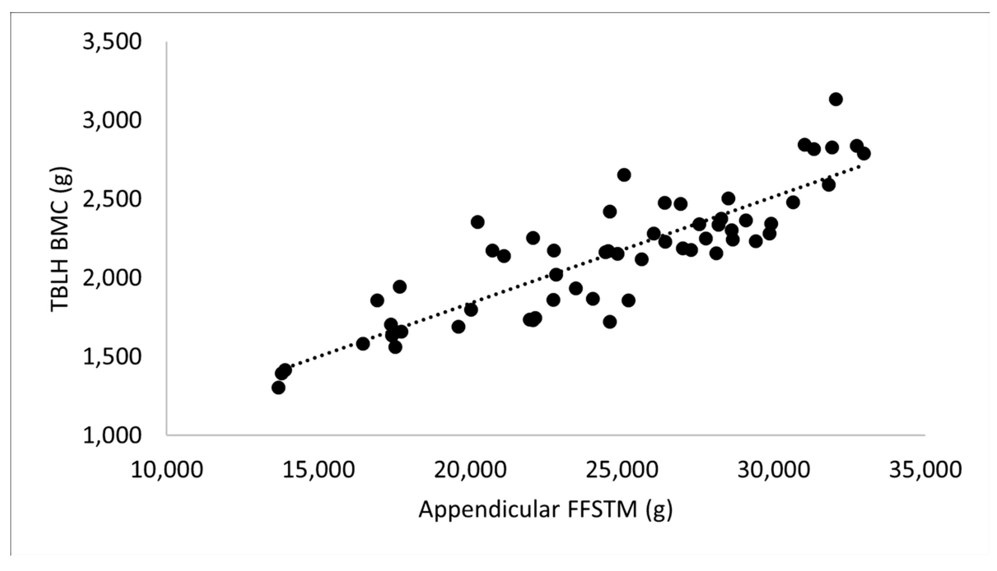
When the relationship between the bone mineral variables and FFSTM was investigated (Figure 1, Figure 2 and Figure 3), a very large correlation was found between TBLH BMC and TBLH FFSTM (r = 0.866) as well as Legs BMC and Legs FFSTM (r = 0.879), and almost perfect for Arms BMC and Arms FFSTM (r = 0.911).
The correlation between FFSTM and BMD was very high for all skeletal sites (r, 0.742–0.844).
The results of the regression analysis using FFSTM as the predictor variable of BMC and aBMD are reported in Table 7. All models were statistically significant (p < 0.001 for all). The R2 was higher for the BMC (range: 0.745–0.827) than the aBMD (range: 0.542–0.707) prediction equations. SEE was 5.28–9.45% of the mean value of the predicted variable. Forcing sex into the equations as a predictor variable did not change the F value to any statistically significant extent.

4. Discussion

This work investigated the body composition of a large sample (n = 58) of karate participants using the accurate DXA technology. To the best of our knowledge, the sample size in this work is unparalleled in previous literature investigating body composition in adult karatekas. All participants were black belt, currently competing at the national and international level, and DXA scans were taken at least 10 days before or after competition, i.e., presumably before/after acute weight loss/recovery (a practice frequently adopted by athletes in weight category sports) [38] had taken place. Therefore, we are confident that the reported data reflect the current body composition of karatekas. The reference values for WB and regional body composition were produced for males across weight divisions. In the whole sample of athletes (males and females), the findings showed possible right–left asymmetry in the limb body composition and the ability of appendicular FFSTM (which fairly approximate the total body amount of skeletal muscle [39]) to reliably predict the content and areal density of bone mineral, especially in the limbs.
Previous work using DXA [11] in a small number (n = 14) of male karatekas showed lower WB FM and higher WB FFSTM in comparison with the male karatekas (n = 43) participating in this study (8.3% vs. 15.1% and 61.2 kg vs. 57.4 kg, respectively). Higher average values were also found for WB BMC (3.7 kg vs. 2.8 kg) and aBMD (1.36 g/cm2 vs. 1.25 g/cm2). Age and BMI were similar in the two groups. An explanation for such a discrepancy can be the different volume of training in the two groups (at least 18 h/w vs. 8 h/w on the average; participants in the Andreoli et al. [11] study were admittedly highly trained athletes), which is expected to have an important effect on soft tissue and bone mineral, associated with higher energy expenditure, muscle mass adaptation to high training, and higher overall impact load on bone. As reported by Chaabène et al. [10], the WB %FM of elite male karatekas (estimated by skinfolds) ranged from 7.4 to 16.8%, showing high variability among countries and suggesting that higher %FM “does not prevent from high-level performance” in karate. In females, WB %FM (average value, 18.6%) was only reported for seven Botswana national-level athletes. Subsequent studies employing BIA [12,13,14] or skinfolds [15,16,17] for body composition assessment in adult (>18 years) karatekas showed WB %FM ranging from about 10% to 16% in males and from 23% to 24% in females. Our data confirm and expand on these finding by using a more accurate method such as DXA to show that karate participants competing at the national and international level are well fit (especially males), but they do not typically reach the low (<10%) WB %FM found in other sports (e.g., cycling). Our findings are supported by DXA data collected in other combat sports showing WB %FM averaging 14% and 25% (for males and females, respectively) in judo, and 13% and 22% in wrestling participants [40]. Similarly, Stantos and colleagues [41] showed that the median (50th) percentile for WB %FM was about 12% (males) and 23% (females) in a sample of judo and wrestling athletes.
The stature-adjusted body composition indices (FMI, FFMI) were statistically significantly lower (FMI) and higher (FFMI) in the male than female karatekas, with the exception of trunk FMI, which was similar by sex (males, 1.56 kg/m2; females, 1.82 kg/m2). This indicates that sex-related differences in adipose tissue distribution in karatekas are essentially located in the limbs, independently of stature. The WB FMI and FFMI were 3.6 kg/m2 and 4.9 kg/m2, and 17.7 kg/m2 and 16.2 kg/m2 in the males and females, respectively. These figures are comparable to those reported by Reale and colleagues [40] in judo and wrestling participants (FMIjudo: males, 3.9 kg/m2; females 6.3 kg/m2; FMIwrestling: males, 3.3 kg/m2; females, 5.6 kg/m2. FFMIjudo: males, 20.8 kg/m2; females, 18.1 kg/m2; FFMIwrestling: males, 19.8 kg/m2; females 19.0 kg/m2), but worse that those reported for boxing and taekwondo. Using BIA, Sterkowicz-Przbycien et al. [15] found an FFMI of 21.6 kg/m2 in polish national karate team athletes, a figure comparable with that presented herein using DXA. According to Santos et al. [41], the median (50th) percentile for WB FFMI (DXA) for judo and wrestling participants was 20.6 kg/m2 for males and 16.3 kg/m2 for females. Overall, it can be concluded that karate has a limited impact on the stature-independent accrual of fat and lean mass in comparison with other combat sports, e.g., boxing and taekwondo.
Stature is one of the most important aspects affecting aBMD because it “inherently overestimates the BMD of tall people and underestimates the mineral density of short people” [35]. Since the karatekas in our sample were distributed over a wide range of statures (151.5–192.9 cm), with females being statistically significantly shorter than males (Table 1), stature-adjusted BMAD was calculated to obtain insight into possible bone size-independent differences in the mineral density of bone between the sexes. The results (Table 4) showed that BMAD is almost identical in males and females at the TBLH and most regional levels, showing that bone size difference is critically involved in the statistically significant differences in aBMD found in the male and female karatekas (Table 2). Interestingly, the lower limb BMAD was similar between the sexes in the left leg (males, 0.599 ± 0.036 g/cm3; females, 0.589 ± 0.043 g/cm3; p = 0.353) and higher in males in the right leg (males, 0.603 ± 0.037 g/cm3; females, 0.577 ± 0.047 g/cm3; p = 0.024). This suggests that karate participation is able to induce bone size-independent improvement of the mineral density of bone in one of two legs. Further research is needed to clarify the reason(s) for that. Notably, BMAD showed a tendency to be higher in the female than the male karatekas at the lumbar spine (p = 0.082; ES = 0.478), thereby implying a possible positive, bone size-independent effect of female sex in regional bone mineralization in karatekas. However, higher BMAD was recently found at the lumbar spine in female vs. male flat jockers [42], showing that such sex effects might be present across sports with different physical demands.
Table 5 presents the quartile values for the body composition variables of male karatekas, expanding on data presented by Santos et al. [41] in a sample of wrestling and judo athletes. These data can be useful to coaches, physical trainers, and athletes to assess individual body composition.
Asymmetry is a debated issue in sports science [43]. While sporting-associated asymmetries are not expected for a non-laterally dominant sport such as karate, previous findings revealed some functional asymmetries in the limbs of combat sports [44,45,46] as well as running, which were able to affect performance. The data in Table 6 showed the presence of several statistically significant asymmetries in the limb body composition of the male and female karatekas, which were generally in favor of the right limb. This is in accordance with the overwhelming proportion of participating karatekas (95%) declaring the right lower limb as the dominant one. However, relative differences between the limb body composition were quite limited (Table 6) and the effect size was trivial or moderate. It should be taken into account that both kata and kumite karatekas were included in the study sample. While kata training involves relatively balanced fighting stances on both the left and right sides, kumite train laterally much more unilaterally (i.e., in a left or right stance). Accordingly, further investigation in separate groups of kata and kumite karatekas is warranted to better clarify the asymmetry issue in karate. While some degree of asymmetry can be acceptable in athletes, it has been suggested that a predisposition to injury incidence exists for functional asymmetry values greater than 15% between the limbs [43,44]. We did not investigate function in this study; work is in progress to assess the effect of limb body composition asymmetry on some key physiological variables (e.g., balance) in karate.
In the last three decades, the concept of the bone–muscle functional unit has emerged more and more frequently [47,48,49]. This concept hypothesizes that bone and muscle are two interconnected tissues mutually affecting each other through a complex crosstalk [50]. It has been shown that lean mass is positively associated with BMC and BMD [51], especially in youth [52]. A recent systematic review and meta-analysis [53] highlighted that muscular strength, which is especially related [54] to appendicular lean mass, is a marker of skeletal health during development and early adulthood. In our sample of karatekas, a close relationship was present between BMC and aBMD, and FFSTM (Figure 1, Figure 2 and Figure 3). In order to assess the structural bone–muscle unit in karate athletes, linear regression analysis was carried out of the whole sample of karatekas participating in this study using BMC or aBMD as the dependent variable and FFSTM as the predictor variable. The results (Table 7) showed that FFSTM effectively predicts BMC and BMD (R2 ranging from 0.542 to 0.827) at the TBLH as well as upper and lower limb level, with sex not affecting the FFSTM predictive ability. The equations presented in Table 7 are useful for inferring the bone changes associated with diet- and/or training-associated changes in skeletal muscle mass.
The main limitations and strengths of this work are itemized in the following. The limitations are: 1. The number of female karatekas in the sample was small, thereby preventing full analysis of the female karateka; 2. The body composition data were not split across weight categories; 3. We did not measure bone mineral at specific skeletal sites (e.g., forearm, hip), thereby preventing a more precise assessment of the effect of karate participation on the skeleton; 4. The dietary regimen of the participants was not recorded, making it impossible to relate caloric and nutrient intake with body composition; 5. No attempt was made to relate body composition and sport-specific skills. The strengths are: 1. The large number of black belt participants; 2. The use of DXA to accurately assess body composition with novel insights into the bone mineral status of the karatekas; 3. The production of reference body composition values for male karatekas across weight categories.

5. Conclusions

In conclusion, this work offered reliable data on the three-compartment body composition of elite karatekas, inclusive of reference body composition values for males. Further, we made predictive equations available to estimate bone variables as a function of the body’s lean mass and gave a preliminary account of asymmetries in the limb body composition. Overall, information presented herein is expected to be of use for karate coaches, physical trainers, and participants interested in assessing and monitoring athletes’ body composition.

Author Contributions

Conceptualization, C.M. and V.C.; methodology, V.C., C.M. and F.S.; software, C.Z.; validation, V.C. and C.M.; formal analysis, C.Z.; investigation, V.C., C.M. and F.S.; resources, C.M. and C.Z.; data curation, V.C., C.M. and F.S.; writing—original draft preparation, C.Z.; writing—review and editing, V.C., C.M. and F.S.; visualization, V.C.; supervision, C.Z.; project administration, C.M.; funding acquisition, C.Z. and C.M. All authors have read and agreed to the published version of the manuscript.

Funding

This research received no external funding.

Institutional Review Board Statement

The study was conducted in accordance with the Declaration of Helsinki, and approved by the ethics committee at the Department of Neurosciences Biomedicine and Movement Sciences (Prot. 26/19 of 19 November 2019).

Informed Consent Statement

Informed consent was obtained from all subjects involved in the study.

Data Availability Statement

Data supporting the results can be obtained from the authors upon reasonable request.

Acknowledgments

The authors would like to thank all of the participant karatekas for their kind cooperation.

Conflicts of Interest

The authors declare no conflict of interest.

References

  1. World Karate Federation|WKF. 12 September 2021. Available online: https://www.wkf.net/ (accessed on 17 January 2022).
  2. Sforza, C.; Turci, M.; Grassi, G.P.; Shirai, Y.F.; Pizzini, G.; Ferrario, V.F. Repeatability of mae-geri-keage in traditional karate: A three-dimensional analysis with black-belt karateka. Percept. Mot. Ski. 2002, 95, 433–444. [Google Scholar] [CrossRef]
  3. Imamura, H.; Yoshimura, Y.; Uchida, K.; Nishimura, S.; Nakazawa, A.T. Maximal oxygen uptake, body composition and strength of highly competitive and novice karate practitioners. Appl. Human Sci. 1998, 17, 215–218. [Google Scholar] [CrossRef] [PubMed] [Green Version]
  4. Bussweiler, J.; Hartmann, U. Energetics of basic karate kata. Eur. J. Appl. Physiol. 2012, 112, 3991–3996. [Google Scholar] [CrossRef] [PubMed]
  5. Filingeri, D.; Bianco, A.; Bde, D.; Daniele, Z.; Bd, A.; Paoli, A.; Palma, A. Is karate effective in improving postural control? Arch. Budo 2012, 8, 149–152. [Google Scholar] [CrossRef]
  6. Origua Rios, S.; Marks, J.; Estevan, I.; Barnett, L.M. Health benefits of hard martial arts in adults: A systematic review. J. Sport. Sci. 2018, 36, 1614–1622. [Google Scholar] [CrossRef]
  7. Kerr, D.A.; Stewart, A.D. Body Composition in Sport; Human Kinetics: Champaign, IL, USA, 2009; pp. 67–86. [Google Scholar]
  8. Carter, J.E.L.; Ackland, T. Somatotype in sport. In Applied Anatomy and Biomechanics in Sport; Ackland, T.R., Elliott, B.C., Bloomfield, J., Eds.; Human Kinetics Publishers: Champain, IL, USA, 2009; pp. 47–66. [Google Scholar]
  9. Burdukiewicz, A.; Pietraszewska, J.; Andrzejewska, J.; Stachoń, A. Morphological optimization of female combat sports athletes as seen by the anthropologists. Anthropol. Rev. 2016, 79, 201–210. [Google Scholar] [CrossRef] [Green Version]
  10. Chaabène, H.; Hachana, Y.; Franchini, E.; Mkaouer, B.; Chamari, K. Physical and physiological profile of elite karate athletes. Sport. Med. 2012, 42, 829–843. [Google Scholar] [CrossRef]
  11. Andreoli, A.; Monteleone, M.; Van Loan, M.; Promenzio, L.; Tarantino, U.; De Lorenzo, A. Effects of different sports on bone density and muscle mass in highly trained athletes. Med. Sci. Sport. Exerc. 2001, 33, 507–511. [Google Scholar] [CrossRef]
  12. Gligoroska, J.P.; Manchevska, S.; Sivevska, E.; Matveeva, N.; Kostovski, Z. Bioelectrical Impedance Analysis of Body Composition in Karate Athletes Regarding the Preparatory Period//Analiza Telesnog Sastava Karatista Bioelektričnom Impedansom Pre I Posle Pripremnog Perioda. Sport. Sci. Health 2016, 12, 81–86. [Google Scholar] [CrossRef] [Green Version]
  13. Gligoroska, J.P.; Todorovska, L.; Mancevska, S.; Karagjozova, I.; Petrovska, S. Bioelectrical Impedance Analysis in Karate Athletes: Bia Parameters Obtained with Inbody720 Regarding the Age. Res. Phys. Educ. Sport Health 2016, 5, 117–121. [Google Scholar]
  14. Gloc, D.; Plewa, M.; Nowak, Z. The effects of kyokushin karate training on the anthropometry and body composition of advanced female and male practitioners. J. Combat. Sport. Martial Arts 2012, 3, 63–71. [Google Scholar] [CrossRef]
  15. Sterkowicz-Przybycień, K. Body composition and somatotype of the top of polish male karate contestants. Biol. Sport 2010, 27, 195–201. [Google Scholar] [CrossRef] [Green Version]
  16. Shariat, A.; Shaw, B.; Kargarfard, M.; Shaw, I.; Lam, E. Kinanthropometric attributes of elite male Judo, Karate and Taekwondo athletes. Rev. Bras. Med. Esporte 2017, 23, 260–263. [Google Scholar] [CrossRef] [Green Version]
  17. Blerim, S.; Zarko, K.; Visar, G.; Agron, A.; Egzon, S. Differences in Anthropometrics Characteristics, Somatotype and Motor Skill in Karate and Non-Athletes//Razlike u antropometrijskim karakteristikama, somatotipu i motoričkim sposobnostima karatista i nesportista. Sport. Sci. Health 2017, 14, 62–65. [Google Scholar] [CrossRef] [Green Version]
  18. Albanese, C.V.; Diessel, E.; Genant, H.K. Clinical applications of body composition measurements using DXA. J. Clin. Densitom. 2003, 6, 75–85. [Google Scholar] [CrossRef]
  19. Day, K.; Kwok, A.; Evans, A.; Mata, F.; Verdejo-Garcia, A.; Hart, K.; Ward, L.C.; Truby, H. Comparison of a Bioelectrical Impedance Device against the Reference Method Dual Energy X-Ray Absorptiometry and Anthropometry for the Evaluation of Body Composition in Adults. Nutrients 2018, 10, 1469. [Google Scholar] [CrossRef] [Green Version]
  20. Marra, M.; Sammarco, R.; De Lorenzo, A.; Iellamo, F.; Siervo, M.; Pietrobelli, A.; Donini, L.M.; Santarpia, L.; Cataldi, M.; Pasanisi, F.; et al. Assessment of Body Composition in Health and Disease Using Bioelectrical Impedance Analysis (BIA) and Dual Energy X-Ray Absorptiometry (DXA): A Critical Overview. Contrast Media Mol. Imaging 2019, 2019, 3548284. [Google Scholar] [CrossRef]
  21. Beaudart, C.; Bruyère, O.; Geerinck, A.; Hajaoui, M.; Scafoglieri, A.; Perkisas, S.; Bautmans, I.; Gielen, E.; Reginster, J.Y.; Buckinx, F. Equation models developed with bioelectric impedance analysis tools to assess muscle mass: A systematic review. Clin. Nutr. ESPEN 2020, 35, 47–62. [Google Scholar] [CrossRef] [Green Version]
  22. De Lorenzo, A.; Bertini, I.; Iacopino, L.; Pagliato, E.; Testolin, C.; Testolin, G. Body composition measurement in highly trained male athletes: A comparison of three methods. J. Sport. Med. Phys. Fit. 2000, 40, 178–183. [Google Scholar]
  23. Nickerson, B.; Snarr, R.; Russel, A.; Bishop, P.; Esco, M. Comparison of Bia and DXA for estimating body composition in collegiate female athletes. J. Sport Hum. Perform. 2014, 2, 29–39. [Google Scholar] [CrossRef]
  24. Ploudre, A.; Arabas, J.L.; Jorn, L.; Mayhew, J.L. Comparison of Techniques for Tracking Body Composition Changes across a Season in College Women Basketball Players. Int. J. Exerc. Sci. 2018, 11, 425–438. [Google Scholar] [PubMed]
  25. Arias Téllez, M.J.; Carrasco, F.; España Romero, V.; Inostroza, J.; Bustamante, A.; Solar Altamirano, I. A comparison of body composition assessment methods in climbers: Which is better? PLoS ONE 2019, 14, e0224291. [Google Scholar] [CrossRef] [PubMed]
  26. Mala, L.; Maly, T.; Cabell, L.; Cech, P.; Hank, M.; Coufalova, K.; Zahalka, F. Body Composition and Morphological Limbs Asymmetry in Competitors in Six Martial Arts. Int. J. Morphol. 2019, 37, 568–575. [Google Scholar] [CrossRef] [Green Version]
  27. Šimenko, J.; Ipavec, M.; Vodicar, J.; Rauter, S. Body symmetry/asymmetry in youth judokas in the under 73 kg category. Ido Mov. Cult. 2017, 17, 51–55. [Google Scholar] [CrossRef]
  28. Lohman, T.G.; Roche, A.F.; Martorell, R. Anthropometric Standardization Reference Manual; Human Kinetics Books: Champaign, IL, USA, 1988. [Google Scholar]
  29. Nana, A.; Slater, G.J.; Stewart, A.D.; Burke, L.M. Methodology review: Using dual-energy X-ray absorptiometry (DXA) for the assessment of body composition in athletes and active people. Int. J. Sport Nutr. Exerc. Metab. 2015, 25, 198–215. [Google Scholar] [CrossRef] [Green Version]
  30. Hangartner, T.N.; Warner, S.; Braillon, P.; Jankowski, L.; Shepherd, J. The Official Positions of the International Society for Clinical Densitometry: Acquisition of dual-energy X-ray absorptiometry body composition and considerations regarding analysis and repeatability of measures. J. Clin. Densitom. 2013, 16, 520–536. [Google Scholar] [CrossRef]
  31. Guss, C.E.; McAllister, A.; Gordon, C.M. DXA in Children and Adolescents. J. Clin. Densitom. 2021, 24, 28–35. [Google Scholar] [CrossRef]
  32. Taylor, A.; Konrad, P.T.; Norman, M.E.; Harcke, H.T. Total body bone mineral density in young children: Influence of head bone mineral density. J. Bone Miner. Res. 1997, 12, 652–655. [Google Scholar] [CrossRef]
  33. Prentice, A.; Parsons, T.J.; Cole, T.J. Uncritical use of bone mineral density in absorptiometry may lead to size-related artifacts in the identification of bone mineral determinants. Am. J. Clin. Nutr. 1994, 60, 837–842. [Google Scholar] [CrossRef]
  34. Wells, J.C.K.; Cole, T.J.; ALSPAC study steam. Adjustment of fat-free mass and fat mass for height in children aged 8 y. Int. J. Obes. Relat. Metab. Disord. 2002, 26, 947–952. [Google Scholar] [CrossRef] [Green Version]
  35. Katzman, D.K.; Bachrach, L.K.; Carter, D.R.; Marcus, R. Clinical and anthropometric correlates of bone mineral acquisition in healthy adolescent girls. J. Clin. Endocrinol. Metab. 1991, 73, 1332–1339. [Google Scholar] [CrossRef] [PubMed]
  36. Hopkins, W.G.; Marshall, S.W.; Batterham, A.M.; Hanin, J. Progressive statistics for studies in sports medicine and exercise science. Med. Sci. Sport. Exerc. 2009, 41, 3–13. [Google Scholar] [CrossRef] [PubMed]
  37. Cohen, J. Statistical Power Analysis for the Behavioral Sciences, 2nd ed.; L. Erlbaum Associates: Hillsdale, NJ, USA, 1988. [Google Scholar]
  38. Reale, R.; Slater, G.; Burke, L.M. Acute-Weight-Loss Strategies for Combat Sports and Applications to Olympic Success. Int. J. Sport. Physiol. Perform. 2017, 12, 142–151. [Google Scholar] [CrossRef] [PubMed]
  39. Kim, J.; Wang, Z.; Heymsfield, S.B.; Baumgartner, R.N.; Gallagher, D. Total-body skeletal muscle mass: Estimation by a new dual-energy X-ray absorptiometry method. Am. J. Clin. Nutr. 2002, 76, 378–383. [Google Scholar] [CrossRef] [PubMed] [Green Version]
  40. Reale, R.; Burke, L.M.; Cox, G.R.; Slater, G. Body composition of elite Olympic combat sport athletes. Eur. J. Sport Sci. 2020, 20, 147–156. [Google Scholar] [CrossRef]
  41. Santos, D.A.; Dawson, J.A.; Matias, C.N.; Rocha, P.M.; Minderico, C.S.; Allison, D.B.; Sardinha, L.B.; Silva, A.M. Reference values for body composition and anthropometric measurements in athletes. PLoS ONE 2014, 9, e97846. [Google Scholar] [CrossRef] [Green Version]
  42. Jackson, K.A.; Sanchez-Santos, M.T.; MacKinnon, A.L.; Turner, A.; Kuznik, K.; Ellis, S.; Box, C.; Hill, J.; Javaid, M.K.; Cooper, C.; et al. Bone density and body composition in newly licenced professional jockeys. Osteoporos. Int. 2017, 28, 2675–2682. [Google Scholar] [CrossRef] [Green Version]
  43. Maloney, S.J. The Relationship Between Asymmetry and Athletic Performance: A Critical Review. J. Strength Cond. Res. 2019, 33, 2579–2593. [Google Scholar] [CrossRef]
  44. Bishop, C.; Turner, A.; Read, P. Effects of inter-limb asymmetries on physical and sports performance: A systematic review. J. Sport. Sci. 2018, 36, 1135–1144. [Google Scholar] [CrossRef] [Green Version]
  45. Bishop, C.; Read, P.; Stern, D.; Turner, A. Effects of Soccer Match-Play on Unilateral Jumping and Interlimb Asymmetry: A Repeated-Measures Design. J. Strength Cond. Res. 2022, 36, 193–200. [Google Scholar] [CrossRef]
  46. Kons, R.L.; Diefenthaeler, F.; Orssatto, L.B.R.; Sakugawa, R.L.; da Silva Junior, J.N.; Detanico, D. Relationship between lower limb asymmetry and judo-specific test performance. Sport Sci. Health 2020, 16, 305–312. [Google Scholar] [CrossRef]
  47. Frost, H.M.; Schönau, E. The “muscle-bone unit” in children and adolescents: A 2000 overview. J. Pediatr. Endocrinol. Metab. 2000, 13, 571–590. [Google Scholar] [CrossRef] [PubMed]
  48. Battafarano, G.; Rossi, M.; Marampon, F.; Minisola, S.; Del Fattore, A. Bone Control of Muscle Function. Int. J. Mol. Sci. 2020, 21, 1178. [Google Scholar] [CrossRef] [PubMed] [Green Version]
  49. Tagliaferri, C.; Wittrant, Y.; Davicco, M.-J.; Walrand, S.; Coxam, V. Muscle and bone, two interconnected tissues. Ageing Res. Rev. 2015, 21, 55–70. [Google Scholar] [CrossRef]
  50. Brotto, M.; Bonewald, L. Bone and muscle: Interactions beyond mechanical. Bone 2015, 80, 109–114. [Google Scholar] [CrossRef] [PubMed] [Green Version]
  51. Reid, I.R. Relationships among body mass, its components, and bone. Bone 2002, 31, 547–555. [Google Scholar] [CrossRef] [PubMed]
  52. Crabtree, N.J.; Kibirige, M.S.; Fordham, J.N.; Banks, L.M.; Muntoni, F.; Chinn, D.; Boivin, C.M.; Shaw, N.J. The relationship between lean body mass and bone mineral content in paediatric health and disease. Bone 2004, 35, 965–972. [Google Scholar] [CrossRef]
  53. Torres-Costoso, A.; López-Muñoz, P.; Martínez-Vizcaíno, V.; Álvarez-Bueno, C.; Cavero-Redondo, I. Association Between Muscular Strength and Bone Health from Children to Young Adults: A Systematic Review and Meta-analysis. Sport. Med. 2020, 50, 1163–1190. [Google Scholar] [CrossRef]
  54. Barbat-Artigas, S.; Plouffe, S.; Pion, C.H.; Aubertin-Leheudre, M. Toward a sex-specific relationship between muscle strength and appendicular lean body mass index? J. Cachexia Sarcopenia Muscle 2013, 4, 137–144. [Google Scholar] [CrossRef]
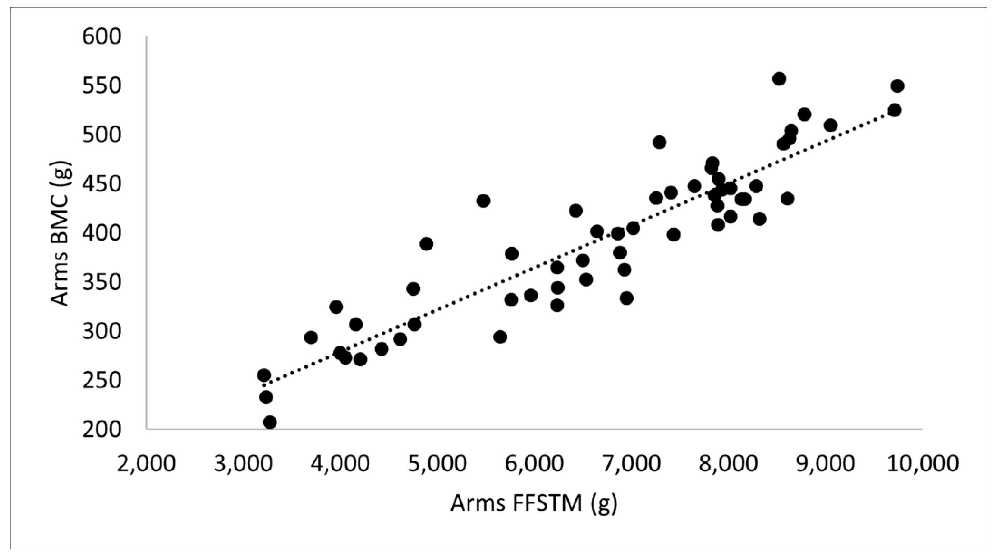
Figure 1. Relationship between fat-free soft tissue mass and bone mineral content in the arms assessed in 58 black belt karatekas competing at the national and international level. FFSTM, fat-free soft tissue mass; BMC, bone mineral content.
Applsci 13 00559 g001
Figure 2. Relationship between fat-free soft tissue mass and bone mineral content in the legs assessed in 58 black belt karatekas competing at the national and international level. FFSTM, fat-free soft tissue mass; BMC, bone mineral content.
Applsci 13 00559 g002
Figure 3. Relationship between appendicular fat-free soft tissue mass and bone mineral content at the total body less head region assessed in 58 black belt karatekas competing at the national and international level. FFSTM, fat-free soft tissue mass; TBLH, total body less head; BMC, bone mineral content.
Applsci 13 00559 g003
Table 1. Characteristics of male and female karatekas. Mean ± standard deviation (SD). Student’s t-test.
VariableMales
(n = 43)
Females
(n = 15)
t-Valuep-ValueES
MeanSDMeanSD
Age (years)22.34.120.01.82.0650.0440.722
Body mass (kg)72.48.055.87.47.021<0.0012.152
Stature (cm)174.66.6161.75.16.895<0.0012.188
BMI (kg/m2)23.72.021.32.83.5310.0010.986
Karate participation (y)14.94.413.12.71.4780.1450.491
Training (h/w)8.44.06.73.71.4660.1480.453
Sessions (n/w)4.42.13.61.61.4350.1570.438
ES, effect size; BMI, body mass index.
Table 2. Body composition (DXA) in male and female karatekas. Mean ± standard deviation (SD). Student’s t-test.
VariableMales
(n = 43)
Females
(n = 15)
t-Valuep-ValueES
MeanSDMeanSD
WB BMC (g)2843.0316.12265.0332.15.444<0.0011.783
WB BMD (g/cm2)1.2530.0841.1740.0913.060<0.0010.899
TBLH BMC (g)2298.8323.31720.1281.96.156<0.0011.878
TBLH aBMD (g/cm2)1.1370.0791.0140.0885.057<0.0011.471
Left arm BMC (g)207.530.7140.621.87.767<0.0012.498
Left arm aBMD (g/cm2)0.8790.0650.7510.0596.705<0.0012.064
Right arm BMC (g)220.432.0152.725.67.445<0.0012.336
Right arm aBMD (g/cm2)0.9040.0680.7810.0715.959<0.0011.767
Thoracic spine BMC (g)127.722.6103.817.13.725<0.0011.194
Thoracic spine aBMD (g/cm2)0.9990.1170.9350.1041.8770.0660.577
Lumbar spine BMC (g)68.713.159.312.22.4420.0180.741
Lumbar spine aBMD (g/cm2)1.1980.1441.1920.1440.1380.8910.041
Pelvis BMC (g)370.972.9276.768.94.322<0.0011.328
Pelvic aBMD (g/cm2)1.3460.1431.2120.1553.0580.0030.898
Left leg BMC (g)539.271.6400.561.66.676<0.0012.076
Left leg aBMD (g/cm2)1.3560.0841.2030.0905.969<0.0011.751
Right leg BMC (g)547.772.6408.164.55.378<0.0012.033
Right leg aBMD (g/cm2)1.3670.0901.2020.0926.068<0.0011.812
Trunk BMC (g)787.9128.5618.1122.24.459<0.0011.354
Trunk aBMD (g/cm2)1.0560.1050.9650.1112.8590.0060.842
Left arm FM (g)609.3221.3696.3250.01.2690.2100.368
Left arm FFSTM (g)3650.7542.62052.3379.210.520<0.0013.415
Left arm %FM13.74.823.97.06.306<0.0011.609
Right arm FM (g)547.3221.0643.2218.91.4500.1530.436
Right arm FFSTM (g)3865.0543.82179.1407.810.956<0.0013.508
Right arm %FM11.84.521.56.06.590<0.0011.820
Trunk FM (g)4779.71662.64769.61310.20.0210.9830.007
Trunk FFSTM (g)27,172.02917.419,495.52148.99.312<0.0012.996
Trunk %FM14.54.219.03.93.6440.0010.863
Left leg FM (g)2123.2799.32905.4982.73.0730.0030.873
Left leg FFSTM (g)9643.51147.76639.5907.49.169<0.0012.904
Left leg %FM17.05.128.66.77.066<0.0011.961
Right leg FM (g)2141.4843.32967.51003.93.1080.0030.891
Right leg FFSTM (g)9837.91225.16805.7945.08.707<0.0012.729
Right leg %FM16.85.428.66.46.940<0.0011.995
TBLH FM (g)10,201.03644.811,982.13574.21.6370.1070.493
TBLH FFSTM (g)54,159.26065.137,172.24658.59.859<0.0013.141
TBLH %FM15.14.523.25.05.859<0.0011.704
WB FM11,099.83674.312,766.63601.21.5200.1370.006
WB FFSTM57,427.86183.340,007.24739.39.921<0.0013.162
WB %FM15.44.222.94.65.827<0.0011.700
WB, whole body; ES, effect size; BMC, bone mineral content; aBMD, areal bone mineral density; FM, fat mass; FFSTM, fat-free soft tissue mass; TBLH, total body less head.
Table 3. Stature-adjusted indices of whole body (WB) and regional fat and fat-free mass in male and female karatekas. Mean ± standard deviation (SD). Student’s t-test.
Variable Males
(n = 43)
Females
(n = 15)
t-Valuep-ValueES
MeanSDMeanSD
WB FMI (kg/m2)3.61.24.91.43.3240.0021.004
WB FFMI (kg/m2)19.71.616.21.87.362<0.0012.078
TBLH FMI (kg/m2)3.31.24.61.43.3360.0021.012
TBLH FFMI (kg/m2)18.51.514.91.77.669<0.0012.210
Left arm FMI (kg/m2)0.200.0710.260.092.7680.0080.701
Left arm FFMI (kg/m2)1.260.160.830.159.068<0.0012.774
Right arm FMI (kg/m2)0.180.070.240.092.8750.0060.747
Right arm FFMI (kg/m2)1.340.150.890.169.632<0.0012.874
Trunk FMI (kg/m2)1.560.531.820.501.6270.1090.504
Trunk FFMI (kg/m2)9.150.737.690.806.541<0.0012.039
Left leg FMI (kg/m2)0.700.261.110.383.8890.0011.260
Left leg FFMI (kg/m2)3.330.302.690.336.954<0.0012.027
Right leg FMI (kg/m2)0.700.271.130.384.691<0.0011.288
Right leg FFMI (kg/m2)3.400.312.750.346.651<0.0011.974
ES, effect size; FMI, fat mass index; FFMI, fat-free mass index; TBLH, total body less head.
Table 4. Bone mineral apparent density (BMAD; g/cm3) in male and female karatekas. Mean ± standard deviation (SD). Student’s t-test.
Variable Males
(n = 43)
Females
(n = 15)
t-Valuep-ValueES
MeanSDMeanSD
TBLH BMAD (g/cm3)0.09880.00490.09740.00580.9300.3560.261
Left arm BMAD (g/cm3)0.6550.0420.6540.0500.0590.9530.022
Right arm BMAD (g/cm3)0.6520.0380.6510.0530.0440.9650.022
Thoracic spine BMAD (g/cm3)1.3800.2011.3690.1520.2010.8420.062
Lumbar spine BMAD (g/cm3)3.6930.5393.9090.3451.4500.0820.478
Pelvis BMAD (g/cm3)0.8660.0970.8770.1220.3540.7250.010
Left leg BMAD (g/cm3)0.5990.0360.5890.0440.9360.3530.247
Right leg BMAD (g/cm3)0.6050.0360.5770.0472.3180.0240.667
ES, effect size; TBLH, total body less head.
Table 5. Quartile (25th, 50th, 75th) values for the body composition of male karatekas.
Variable Quartile
255075
Body mass (kg)65.573.279.0
Stature (cm)169.4175.5179.9
BMI (kg/m2)22.2823.5625.23
WB bone area (cm2)2155.72259.22386.0
WB BMC (g)2646.62793.33010.2
WB aBMD (g/cm2)1.2001.2551.3024
TBLH BMC (g)2152.12252.02474.5
TBLH aBMD (g/cm2)1.0851.1301.177
Left arm BMC (g)186.9206.2223.7
Left arm aBMD (g/cm2)0.8360.8850.918
Right arm BMC (g)200.4221.5237.0
Right arm aBMD (g/cm2)0.8720.9120.932
Thoracic spine BMC (g)108.5127.0140.2
Thoracic spine aBMD (g/cm2)0.9150.9701.103
Lumbar spine BMC (g)59.565.478.6
Lumbar spine aBMD (g/cm2)1.0921.1961.276
Pelvis BMC (g)327.3371.7407.4
Pelvis aBMD (g/cm2)1.2671.3371.433
Left leg BMC (g)491.5530.6585.2
Left leg aBMD (g/cm2)1.2841.3341.414
Right leg BMC (g)501.4540.9586.0
Right leg aBMD (g/cm2)1.3081.3631.424
Trunk BMC (g)695.7758.3873.5
Trunk aBMD (g/cm2)0.9991.0471.118
Left arm FM (g)452.0564.3715.3
Left arm FFSTM (g)3209.63782.94019.3
Left arm %FM9.712.815.2
Right arm FM (g)382.3467.6626.1
Right arm FFSTM (g)3453.03933.44286.6
Right arm %FM8.310.713.9
Trunk FM (g)3352.74411.55585.5
Trunk FFSTM (g)24,709.327,755.529,957.5
Trunk %FM10.414.016.8
Left leg FM (g)1450.41957.92627.5
Left leg FFSTM (g)8687.79670.410,368.6
Left leg %FM12.716.420.6
Right leg FM (g)1392.91998.02657.9
Right leg FFSTM (g)8711.39879.610,625.4
Right leg %FM12.216.519.8
TBLH FM (g)7015.69508.212,947.3
TBLH FFSTM (g)49,302.254,771.158,453.1
TBLH %FM11.214.917.0
BMI, body mass index; WB, whole body; BMC, bone mineral content; aBMD, areal bone mineral density; FM, fat mass; FFSTM, fat-free soft tissue mass.
Table 6. Asymmetry in limb body composition for the whole sample of karatekas (n = 58). Student’s t-test.
VariableMales
(n = 43)
Females (n = 15)
LeftRightt-Valuep-ValueESLeft Right t-Valuep-ValueES
meanSDmeanSD
Arm BMC (g)207.5 30.7220.432.0−8.166<0.0010.41140.6 21.8152.6 24.6−5.703<0.0010.51
Arm aBMD (g/cm2)0.8790.0650.9040.068−6.114<0.0010.100.751 0.0590.781 0.071−3.6590.0030.46
Leg BMC (g)539.271.6543.777.6−1.3420.1870.06400.5 61.6408.1 64.5−2.5720.0220.12
Leg aBMD (g/cm2)1.3560.0841.3670.090−1.7770.0830.121.203 0.0901.201 0.0920.1290.9000.02
Arm FM (g)609.3221.3547.3221.06.988<0.0010.28696.3 250.0643.2 218.93.5090.0030.22
Arm FFSTM (g)3650.7542.63865.0543.8−8.376<0.0010.392052.3 379.22179.1 407.8−5.776<0.0010.32
Leg FM (g) 2123.2843.32141.41147.7−0.9190.3630.0182905.4 982.72967.5 1003.9−1.5550.101
Leg FFSTM (g)9643.51147.79837.91225.1−3.6540.0010.166639.5 907.46805.7 945.0−3.4250.0040.28
ES, effect size; BMC, bone mineral content; aBMD, areal bone mineral density; FM, fat mass; FFSTM, lean soft tissue mass.
Table 7. Results of linear regression analysis in the whole group of karatekas (n = 58).
Dependent VariablePredictor VariableAdjusted R2SEEConstantBeta Coefficient (95% CI)Beta Coefficientp-Value
Arms BMC (g)Arms FFSTM (g)0.82734.4107.30.043 (0.038–0.048)0.911<0.001
Arms aBMD (g/cm2)Arms FFSTM (g)0.7070.04590.5894.06 × 10−5 (3.4 × 10⁻⁵–4.8 × 10⁻⁵)0.844<0.001
Legs BMC (g)Legs FFSTM (g)0.76889.7166.340.047 (0.040–0.054)0.879<0.001
Legs aBMD (g/cm2)Legs FFSTM (g)0.6160.06970.8682.523 × 10−5 (2.0 × 10⁻⁵–3.0 × 10⁻⁵)0.789<0.001
TBLH BMC (g)Appendicular FFSTM (g)0.745203.2483.40.068 (0.057–0.078)0.866<0.001
TBLH aBMD (g/cm2)Appendicular FFSTM (g)0.5420.06560.7611.400 × 10−5 (1.1 × 10⁻⁵–1.7 × 10−50.742<0.001
SEE, standard error of estimate; CI, confidence interval; BMC, bone mineral content; FFSTM, fat-free soft tissue mass; aBMD, areal bone mineral density; TBLH, total body less head.
Disclaimer/Publisher’s Note: The statements, opinions and data contained in all publications are solely those of the individual author(s) and contributor(s) and not of MDPI and/or the editor(s). MDPI and/or the editor(s) disclaim responsibility for any injury to people or property resulting from any ideas, methods, instructions or products referred to in the content.