Article

Radiological Assessment of Extracranial Vertebral Artery Variations: A Computed Tomography Angiography Study

by
Faiza Al Hajri
1,
Bayan Al Yahya’ey
2,
Srinivasa Rao Sirasanagandla
3,
Sreenivasulu Reddy Mogali
4 and
Eiman Al-Ajmi
5,*
1
Radiology Residency Program, Oman Medical Specialty Board, Athaiba North, Muscat 132, Oman
2
College of Medicine and Health Sciences, Sultan Qaboos University, Al-Khoudh, Muscat 123, Oman
3
Department of Human and Clinical Anatomy, College of Medicine and Health Sciences, Sultan Qaboos University, Al-Khoudh, Muscat 123, Oman
4
Lee Kong Chian School of Medicine, Nanyang Technological University Singapore, Singapore 308232, Singapore
5
Department of Radiology and Molecular Imaging, College of Medicine and Health Sciences, Sultan Qaboos University, Al-Khoudh, Muscat 123, Oman
*
Author to whom correspondence should be addressed.
Appl. Sci. 2023, 13(10), 5822; https://doi.org/10.3390/app13105822
Submission received: 10 April 2023 / Revised: 2 May 2023 / Accepted: 4 May 2023 / Published: 9 May 2023
(This article belongs to the Special Issue Anatomical Techniques and Clinical Studies in the Human)

Abstract

:
We evaluated the incidence of intraforaminal and extraforaminal variations of the vertebral artery (VA) in the Omani population using computed tomography angiography (CTA). CTA results of 579 consecutive Omani patients (1158 VAs) were reviewed retrospectively for the analysis of unusual entrance transverse foramen (UE-V2), midline migration (MM), persistent first intersegmental artery (PFIA), and paracondylar process (PP) variations of VA. The anomalous origin and VA dominance were also determined. The sex and side differences in the incidence of these variations were analyzed using the Chi-square test. The incidence of UE-V2 was observed in 10.44% (121 out of 1158) of cases. The incidence of UE-V2 at different vertebral levels was observed in the sequence of C5 (71%) > C4 (20.7%) > C7 (7.43%) > C3 (0.8%). The incidence of MM variation was 1.29%. PFIA and PP variations were found in 0.17% and 0.60% of cases, respectively. Left-dominant VA was identified in 44.7% (259 out of 579) of subjects. The incidence of VA variations was not significantly associated with either sex or side. Anomalous VA origin from arch aorta (3.5%) and right common carotid artery (CCA) (0.08%) was identified. The incidence of UE-V2 in Omani subjects is comparatively higher than that reported in other Asian populations. A rare case of VA originating from the right CCA was also identified.

1. Introduction

The vertebral arteries (VAs), critical blood vessels of posterior brain circulation, can exhibit a variety of anatomical variations which have an important implication in medical procedures, specifically in the context of spine surgeries [1,2]. During cervical spine surgeries and endovascular interventions, the VA is at risk of injury due to its close relationship to the vertebrae and the presence of anomalies, such as variant origin [3,4,5], unusual entrance [6,7,8,9,10], and course in the cervical spine [11,12], hypoplasia [13], and tortuosity [14]. These anomalies make it more difficult to identify and access the VAs during surgeries and may lead to inadvertent injuries, which can have detrimental consequences for patients [2]. Therefore, these injuries need to be prevented. Research into a comprehensive understanding of the different types of VA anomalies and their clinical impact could help clinicians find patients who are at risk for VA injury during surgery, choose the best surgical approach, and improve patient clinical outcomes.
VA is known to present variations in its origin [3,4,5] and course [10,11]. Typically, each VA arises from the first part of each subclavian artery and then ascends to reach the level of the transverse foramen of the C6 vertebra. Then, it ascends through the transverse foramina of C6 vertebra to the C1 vertebra, curves medially behind the lateral mass of the atlas, and enters the cranial cavity through the foramen magnum. At the pontomedullary junction, the right and left VAs converge into the basilar artery [15]. The intracranial segment of VA gives various branches, including the anterior spinal artery, medial and lateral medullary branches, ramus choroideus ventriculi quarti, and posterior inferior cerebellar artery [16].
Classically, the VA course is divided into four segments: V0 (its origin), V1 is a short pre-foraminal segment, V2 (intraforaminal segment) extends between the transverse foramina of the C6 and C2 vertebrae, V3 runs between the C2 foramen and foramen magnum (extraforaminal segment), and V4 refers to the intracranial portion of the artery [11,17]. Both cadaveric [18] and radiographic studies (CT and MRI) [6,8,9,19,20] have been widely investigated for VA variations. In recent years, computed tomography angiography (CTA) has become a popular choice in research as it has the advantage of displaying all the characteristics of blood vessels and precise measurements of variations. Rarely, VA may arise from the aortic arch and right common carotid artery (CCA) [3]. The unusual entrance transverse foramen (UE-V2) and midline migration (MM) are the two important variations of the V2 segment [11,12]. On the other hand, persistent first intersegmental artery (PFIA) and paracondylar processes (PP) are frequently reported variations of the V3 segment [11,15,21,22]. Classically, VA dominance is defined as an unequal diameter of VA with a significant difference between both sides. It is also considered a congenital variation of VA [2].
The reported incidences of V2 and V3 segment variations varied among different studies worldwide [6,8,9,10,11,23,24]. The influence of regional, ethnic, and environmental factors on VA variability has been implicated [25]. Adequate radiological data on anatomical VA variations are important parts of the pre-surgical assessment of VA. Particularly, V3 and V2 segment variations are utmost important to avoid iatrogenic injuries during cervical spine surgical procedures, including corpectomy, laminectomy, and while implanting plates, screws, and artificial discs [11,26,27]. Knowledge about these variations is also clinically important, as their presence increases the risk of vertebra-basilar circulatory disorders [28,29]. Despite the potential problems posed by VAs variants, there are still knowledge gaps that must be addressed to enhance the understanding of VA anomalies and their associated clinical impact. These include small sample sizes, limited use of advanced imaging modalities, and lack of focus on a specific population. In the context of the Middle Eastern population, very few studies have reported V2 and V3 segment variations [23,24]. Furthermore, there is a lack of baseline data in the Omani population. Therefore, the present retrospective study investigated the incidence of V2 and V3 segment variations among Omani subjects (1158 VAs in 579 patients) referred to Sultan Qaboos University Hospital (SQUH) using the CTA technique. Additionally, we also investigated the unusual origin of VA and the dominance of VA based on a method described by Jeng et al. (2004) [30].

2. Materials and Methods

2.1. Study Population

A retrospective cross-sectional study was conducted on Omani subjects of either sex, aged ≥18 years, who were referred for CTA of the neck to the Department of Radiology and Molecular Imaging at SQUH, a tertiary health care hospital based in the Sultanate of Oman. The study included consecutive patients who visited the department during the period from January 2017 and December 2021. The average age of the patients was 57 ± 16.81 (18–98) years. The main clinical indications of the CTA examinations included stroke, transient ischemic attack, and trauma. Patients with suboptimal enhancement of the arteries, arterial luminal occlusion, history of neck surgery, tumors, or severe deformities of the cervical spine and non-Omani patients were excluded from the study. A total of 153 patients’ CTA scans were excluded from the study. Ethical approval was obtained prior to the study from the Medical and Research Ethics Committee at the College of Medicine and Health Sciences (Ref. No. SQU-EC/183/2022).

2.2. CTA Acquisition Protocol

The CTA scans of the neck for all patients were performed using a 64-slice multidetector CT scanner (Siemens Sensation 64) with a 100 kV peak voltage, pitch of 0.7 and detector configuration of 64 × 0.6 mm. The scan range was from the carina to the vertex. Picture Archiving and Communication System (PACS) (Synapse PACS, FUJIFILM Worldwide, version 5.7.102) was used for reviewing the scans. A slice thickness of 0.75 mm was used to assess the images in the axial plane with coronal and sagittal reformats.

2.3. Data Acquisition

The data were collected from the electronic medical records in the “TrackCare” Health Information System of SQUH. The data pertaining to the V2 and V3 segment variations were analyzed and recorded manually for each patient as described previously (Wang et al., 2021) [11]. The tools in the PACS were used while measuring the dimensions. Each CTA was analyzed to identify the UE-V2 and MM variations from the V2 segment and the PFIA and PP variations from the V3 segment. UE-V2 variation was recorded when VA entered the transverse foramen other than the C6 vertebra (Figure 1).
MM variation was recorded when the distance between the medial wall of the VA and the midline of the related cervical vertebra was less than 10 mm (Figure 2).
On the other hand, a PFIA variation was recorded when the artery showed an unusual course under the posterior arch of the atlas (Figure 3).
A PP variation was identified when there was a bony bridge between the upper articular process and posterior arch of the atlas (Figure 4).
A VA was considered dominant when a side-to-side diameter difference of 1.2 mm or more was present. If the difference is less than 1.2 mm, then it is considered co-dominant. The vertebral dominance (VD) was defined following a standard method (Jeng et al., 2004) [30]. The origin of VA in each case was noted and the frequency of unusual origin were documented. Only one observer, a senior radiology resident, was involved in the screening and data collection from each CTA. Data that were recoded included the total number of each possible variant, as well as the side of variation and sex. Patient’s data were first inputted into a Microsoft Excel spreadsheet, and then transferred to SPSS software.

2.4. Statistical Analysis

The data analysis was performed using Statistical Package for the Social Sciences (SPSS, version 26.0, IBM Corporation, Armonk, NY, USA). All the variables related the study were categorical. Hence, descriptive statistics (e.g., frequency and percentage) were employed to present the data. Chi-square test was used to determine the associations between the sex or side on frequency of V2 and V3 segments variations, and the VD. A p-value of <0.05 was considered statistically significant.

3. Results

After consideration of inclusion and exclusion criteria, a total of 579 patients were included in the current research. Among these patients, 338 were male (58.38%) and 241 were female (41.62%). The incidence of all variations was reported for 1158 courses (579 patients) of VA screened in the present study.

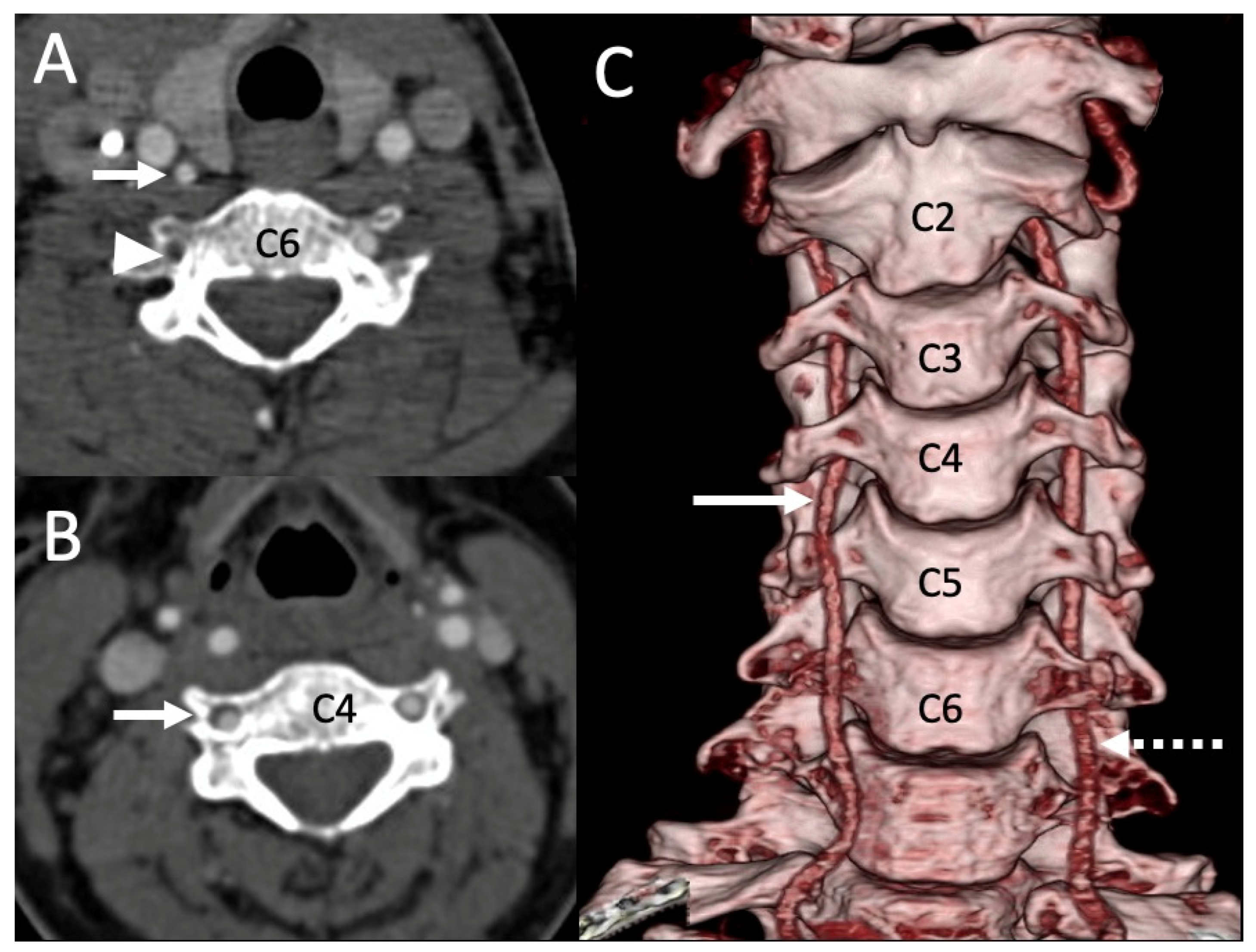
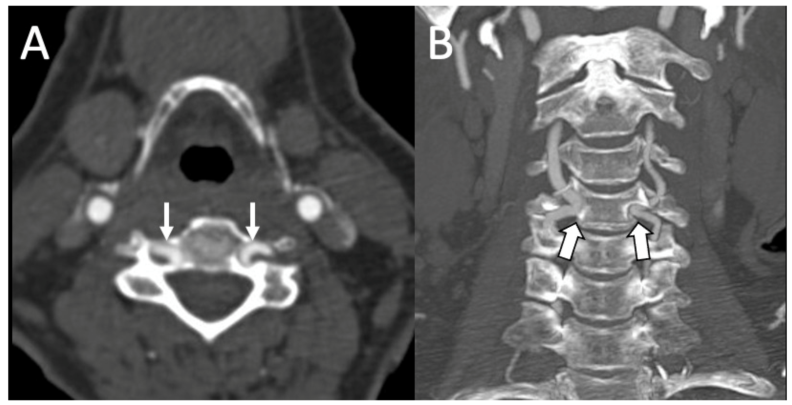
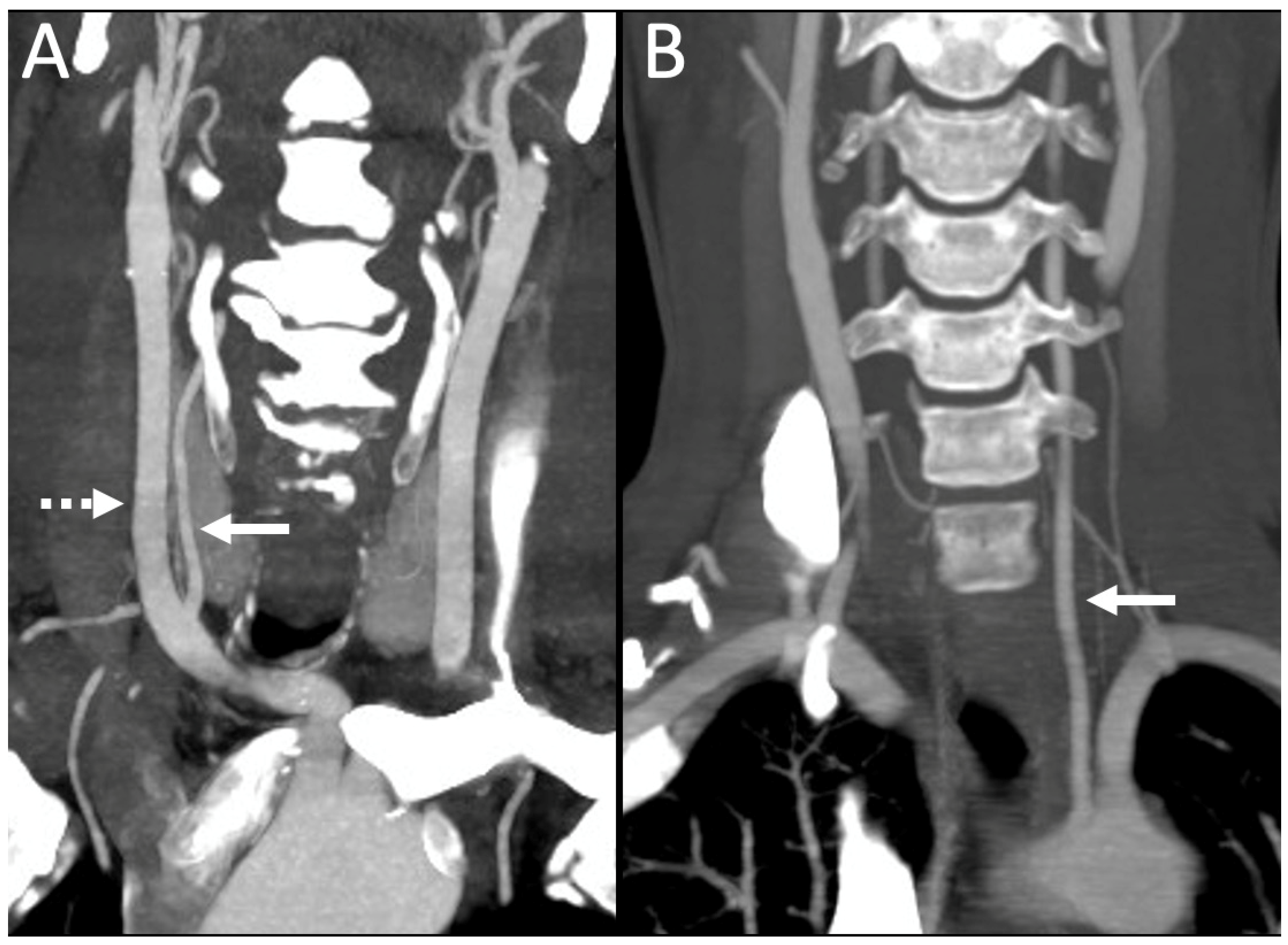
3.1. Variations in the V2 Segment

In the lower cervical spine region, UE-V2 and MM variations were observed. Among all variations, the incidence of UE-V2 was relatively high, with a frequency of 10.44% (121/1158). The incidence of UE-V2 in males was 10.35%, while in females it was 10.58%. The incidence of UE-V2 variation on the right and left sides was 5.44% and 5.01%, respectively. The incidence of UE-V2 at different vertebral levels was observed in the sequence of C5 > C4 > C7 > C3 with a frequency of 71% (86 out of 121), 20.7% (25 out of 121), 7.43% (nine out of 121), and 0.8% (one out of 121), respectively. Figure 5 shows UE-V2 at the third cervical vertebra, which is a rarely reported anomaly.
There were no statistical differences between the occurrence of UE-V2 incidence and sex (p = 0.979) or side (p = 0.701) (Table 1). The other variation of the V2 segment was MM, which refers to a VA closer to the midline of the cervical spine. The overall incidence of MM variation was 1.29% (15 out of 1158) of courses. It was observed in 1.18% and 1.45% of courses in males and females, respectively (Table 1). This variant was observed predominantly in older subjects. Except for one patient who was twenty-two years old, the average age of the patients with MM was 63.3 years with an age range of 46–87 years. The incidence of MM variation was highest on the left side with 0.95% (Table 1). There were no significant differences in MM incidence between sex (Table 1, p = 0.892) or side (Table 1, p = 0.119). The average distance of MM variation on the right side was 6.25 mm (range: 3.3–8.7) and 7.8 mm (range: 6.4–9.6) on the left side.

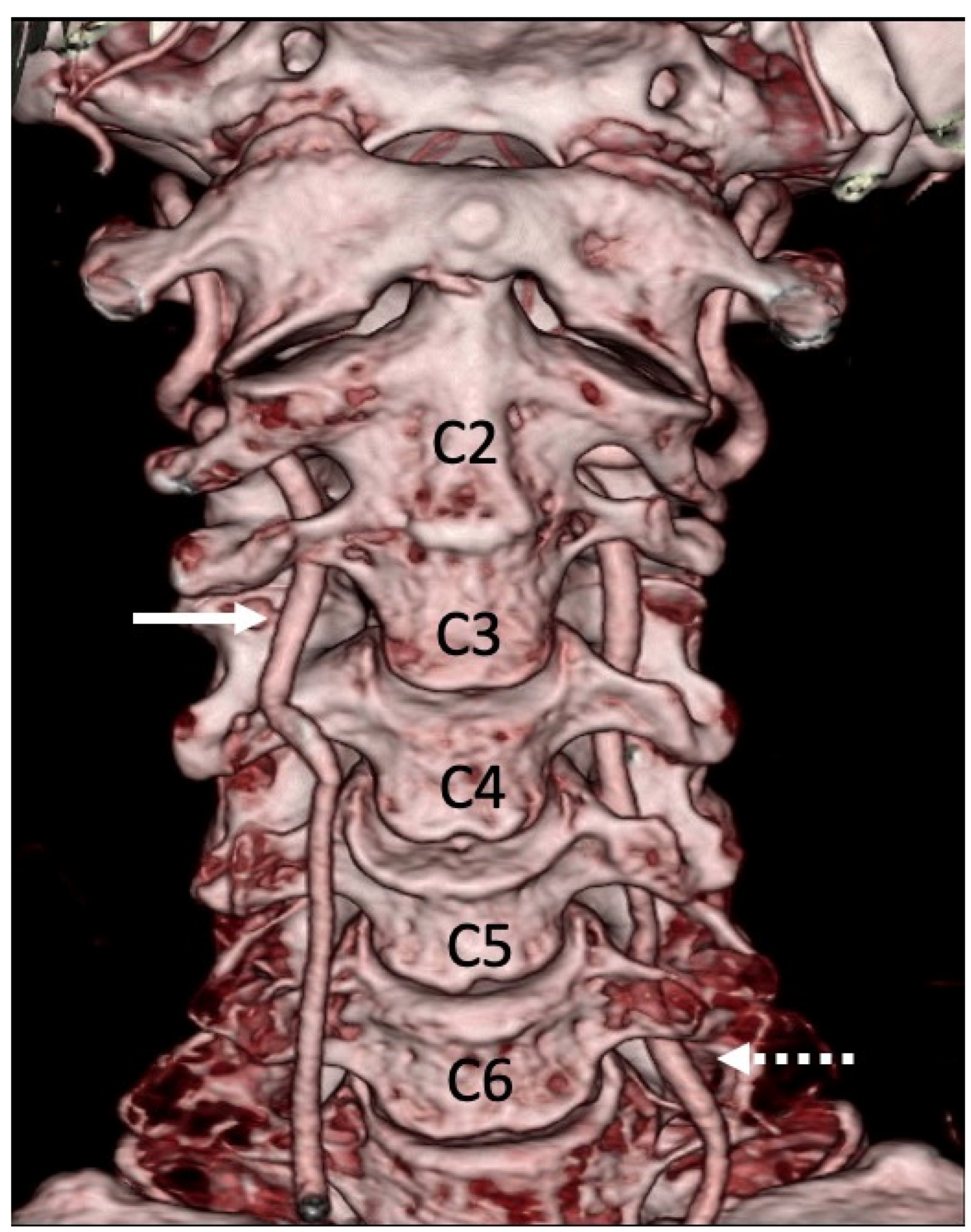
3.2. Variations of the V3 Segment

In the upper cervical spine region, the PFIA and PP were two kinds of variations identified. PFIA variation was observed in two cases (0.17%), and both cases were male and observed on the right side (Table 1). The PP variation of the V3 segment certainly refers to the anomalous course of the VA in relation to the posterior arch of the atlas. The PP variation was observed in 0.60% of cases. In the PP variation, additional bony growth of the occipital bone appeared as a bony bridge between the occipital condyle and posterior arch of the atlas, and the VA was often surrounded by this bridge. The incidence of this variation was high in females (1.04%; p = 0.135) and found on the left side (0.43%; p = 0.452) (Table 1).

3.3. Vertebral Artery Dominance (VAD)

In the present study, the differences in the diameter of VAs were observed. Among study subjects, the right dominant VA was found in 28.5% (165 out of 579) and left dominant VA was in 44.7% (259 out of 579) of subjects. The codominant VA was found in 26.8% (155 out of 579) of cases. The right dominant VA in males and females was 30.1% and 26.1% while the left dominant VA was 42% and 48.5% in males and females, respectively. On the other hand, the codominant VA was observed with a frequency of 27.8% and 25.3%, in males and females. There were no significant differences in VA diameter and sex (p > 0.05). VA arose from arch of aorta in 3.5% of courses (41 out of 1158), and all these were left-sided. In one case (0.08%), the right VA arose from the right common carotid artery (Figure 6).

4. Discussion

Based on the CTA studies, this study reported the various types of variations in vertebral anatomy and analyzed the incident rates based on sex and laterality. While racial differences in VA variations have been reported in the literature [6,8,9,10,11,20,23,24], to the best of our knowledge, very few studies from the Middle Eastern region [23,24] and no study from Oman have reported incidences of different kinds of VA variations. We report for the first time the incidence of VA variations in Omani subjects, particularly in the V2 and V3 segments, and discuss their clinical importance.
In previous CTA studies, the incidence of UE-V2 varied from 4.4% to 12.5% (Table 2).
Studies from Korea [9], Japan [8], and France [6] reported an incidence of 6.5%, 4.4%, and 7%, respectively. Two recent studies from China reported an incidence of 9.16% and 8.3% [10,11]. Other imaging studies reported an incidence of 7.8% in Germany [19], 12.5% in Republika Srpska [31], 7.5% in Turkey [32], and 5.3% of left VA and 0.8% of right VA in Thailand [20]. A study reported an incidence of 7% among 50 Saudi patients [23]. The reported incidence of UE-V2 in Omani subjects is slightly higher than that reported in most of the studies [6,8,9,19,20,23,32]. The characteristics of the UE-V2 observed in the present study were compared with those of previous studies and presented in Table 2. Consistent with previous studies, in the present study, the transverse process of C5 is the common entrance point. In studies by Kim et al. (2016) [9] and Wako et al. (2014) [8], the sequence of UE-V2 levels incidence was found to be C5 > C4 > C7 > C3 and C5 > C7 > C4 > C3, respectively. In a recent study, in Chinese subjects, the sequence was C5 > C4 > C7 > C3 [10]. We observed a similar incidence of sequence for UE-V2 levels observed in Chinese subjects [10]. The UE-V2 variation is also often referred to as an extra-foraminal VA anomaly. When such variation is present, the subaxial cervical spine procedures need to be performed with more caution as below the entrance, the artery is unprotected by the bone. The UE-V2 segment is at high risk of injury during surgeries in the cervical region, such as tumor resection and stellate ganglion block. Prior knowledge of baseline data and the characteristics of this variation is essential for successful surgeries and the prevention of life-threatening events [33].
The variability in UE-V2 incidences among different populations could be attributed to the racial influences on the embryonic development of VA. The development of VAs begins at the distal ends of the seventh intersegmental dorsal arteries, which usually give rise to subclavian arteries [3]. The anastomoses between the 1st and 6th dorsal intersegmental arteries will form the rest of the VAs [34]. Additionally, VA can anastomose with either the ascending or deep carotid arteries. Unusual entrance depends on the dominance of ascending or deep carotid artery anastomoses. If the longitudinal intersegmental anastomosis is completed by the ascending cervical artery, the developing VA will follow the ascending cervical artery and enter the transverse foramen at a higher level than the usual C6 level. On the other hand, if the longitudinal intersegmental anastomosis is completed by the deep cervical artery, then the VA will follow the deep cervical artery and enter into the transverse foramen at the usual C6 vertebral level [10,35].
The course of VA close to the midline in the V2 segment is referred to as MM. The incidence of MM observed in the present study is 1.29%. In a recent study, MM was observed in 2.63% of cases [11]. In another study, it was observed in 3.8% of cases (Eskander et al., 2009). Similar to the results observed by Eskander et al. (2009) [12], MM in the present study was seen predominantly in the older age group. Long-term degenerative and post traumatic changes in the cervical spine are possible logical mechanisms that explain the MM variation of VA [12,36]. The MM variation is known to be associated with sub-axial cervical spine surgeries. In this intra-foraminal variation, VA is at high risk of injury, particularly in routine corpectomy surgery [12]. Hence, preoperative identification of MM is crucial to avoid unexpected complications during surgeries and for successful outcomes.
Relatively high racial differences were observed in V3 segment variations when compared to V2 segment variations [7,37,38]. The occurrence of PFIA is due to the developmental failure of its obliteration [37]. Failed obliteration could be associated with genetic, hemodynamic, and environmental factors [37]. Two different studies reported a high incidence of 5% to 10% PFIA in Asian populations [7,38]. Another recent study reported 4.4% PFIA incidence in the Chinese population [11]. In contrast, we observed this only in 0.17% of cases. Similar to the present study, a low incidence of 0.01% was observed in the American population [37]. The differences in incidences of VA anomalies could be attributed to genetic variations between the populations.
The paracondylar process is the other variation of the V3 segment. This developmental anomaly is usually formed due to partial proatlas somite assimilation into the occipital bone [39]. In anthropological studies, PP incidence was reported in Maltese skulls (2.02%), South African skulls (0.9%), South Indian skulls (2.1%), and North Indian skulls (10.5%) [40,41,42]. In a recent study, PP incidence was reported in 5.9% of cases in Chinese subjects. Comparatively, a low incidence of PP (0.6%) was observed in the present study. Generally, it is an incidental finding [43], asymptomatic, and can be detected unilaterally or bilaterally [44]. In a few instances, PP may compress the C1 spinal nerve, causing occipito-cervical pain [45]. More often, PP mimics a calcified stylohyoid ligament. However, this ligament can be easily differentiated in coronal-reformatted CT [44].
In the present study, there were no sex or side differences in all the types of VA variations occurrence. Similar results were reported in previous research [11,46] except MM variation which was significantly higher in female [11]. This indicates that variations in the V2 and V3 segments of the VA are not substantially impacted by the individual’s sex or the investigated side of the body. Hence, more investigations with a larger sample may require a conclusive result in this regard.
Generally, the VA is left-dominant, and its diameter is typically larger than that of the right VA. In general, left VAD is found in 50% of people. In angiographic or post-mortem studies, equal-sized bilateral VAs were found in 6–26% of patients [30]. Consistent with previous studies, in our study, the incidence of left VAD was higher than the right side [30,47,48,49]. Although VAD was considered clinically irrelevant in the past, recent studies have demonstrated the association between the VAD and posterior circulation ischemic stroke [28,29]. VAD causes basilar artery bending by promoting an asymmetric flow pattern at the vertebrobasilar junction [50], which subsequently increases the risk of developing posterior circulation ischemic stroke [48,51]. The anomalous origin of VA is more common on the left side than the right side [3]. Usually, such anomalous origin is from the arch of aorta with an incidence rate of 2.4–5.8% [52,53]. VA originating from the right common carotid artery is rare and was reported only in a few cases with and without the persistence of brachycephalic trunk [3,4,5]. In the present study, we identified right VA arising from the right CCA. During embryonic development, the persistence of the 3rd–6th intersegmental artery would result in the origination of VA from the CCA [5].
The strengths of the present study were the larger sample size, the sample being representative of patients from different parts of Oman, the CTA technique, and the focus on the Omani population. Knowledge of baseline data of VA variations reported in the present study is of tremendous importance for the preoperative planning of endovascular interventions and to avoid inadvertent arterial injuries in neck surgeries. Furthermore, it may be advised that patients undergo a radiologic evaluation prior to any invasive procedure or neck surgeries to prevent accidental injuries of the VA.

Limitations

This is a retrospective study that was conducted in the Omani adult population based on CTA images. The findings of this study apply to the origin, V2 and V3 segments of the VA. Only one investigator identified all the variations of the VA. To ensure accuracy and quality control, standard measurement protocols were applied, and the investigator was blinded to the scans. Further, the investigator was trained on the techniques related to the measurements taken in this study. Future studies with a large sample need not only report the prevalence of VAs in the Middle Eastern population, but also their clinical implications and outcomes.

5. Conclusions

The incidence of UE-V2 variation in Omani subjects is comparatively higher than that reported in other Asian populations. An anomalous origin from the right common carotid artery and unusual entry at the third cervical vertebra were observed. Additionally, the incidence of these variations occurs without any sex or side differences. The baseline data of VA variations reported may be clinically important for the pre-surgical planning of cervical surgeries and successful outcomes.

Author Contributions

Conceptualization, E.A.-A. and S.R.S.; methodology, E.A.-A., F.A.H., and B.A.Y.; validation, E.A.-A. and F.A.H.; formal analysis, S.R.S. and B.A.Y.; investigation, F.A.H. and E.A.-A.; resources, E.A.-A. and F.A.H.; data curation, E.A.-A., F.A.H., B.A.Y. and S.R.S.; writing—original draft preparation, S.R.S., B.A.Y., and S.R.M.; writing—review and editing, E.A.-A. and S.R.M. All authors have read and agreed to the published version of the manuscript.

Funding

This research received no external funding.

Institutional Review Board Statement

Ethical approval was obtained prior to the study from the Medical and Research Ethics Committee at the College of Medicine and Health Sciences (Ref. No. SQU-EC/183/2022).

Informed Consent Statement

Patient consent was waived due to the reason that the study was a retrospective analysis of anatomical variations of vertebral artery in a computed tomography angiography and reported the anonymous data without revealing any identification details of patients. This statement was also mentioned in the institutional ethics committee application.

Data Availability Statement

Since the present study involves the patients’ data, its public availability was restricted. The anonymous raw data of the present study will be shared with readers and reviewers upon request to the corresponding author.

Conflicts of Interest

The authors declare no conflict of interest.

References

  1. Hsu, W.K.; Kannan, A.; Mai, H.T.; Fehlings, M.G.; Smith, Z.A.; Traynelis, V.C.; Gokaslan, Z.L.; Hilibrand, A.S.; Nassr, A.; Arnold, P.M.; et al. Epidemiology and Outcomes of Vertebral Artery Injury in 16 582 Cervical Spine Surgery Patients: An AOSpine North America Multicenter Study. Glob. Spine J. 2017, 7, 21S–27S. [Google Scholar] [CrossRef] [PubMed]
  2. Zhu, W.; Wang, Y.-F.; Dong, X.-F.; Feng, H.-X.; Zhao, H.-Q.; Liu, C.-F. Study on the correlation of vertebral artery dominance, basilar artery curvature and posterior circulation infarction. Acta Neurol. Belg. 2016, 116, 287–293. [Google Scholar] [CrossRef] [PubMed]
  3. Lazaridis, N.; Piagkou, M.; Loukas, M.; Piperaki, E.-T.; Totlis, T.; Noussios, G.; Natsis, K. A systematic classification of the vertebral artery variable origin: Clinical and surgical implications. Surg. Radiol. Anat. 2018, 40, 779–797. [Google Scholar] [CrossRef] [PubMed]
  4. Lemke, A.J.; Benndorf, G.; Liebig, T.; Felix, R. Anomalous origin of the right vertebral artery: Review of the literature and case report of right vertebral artery origin distal to the left subclavian artery. AJNR Am. J. Neuroradiol. 1999, 20, 1318–1321. [Google Scholar]
  5. Elnaggar, M.E.; Abduljawad, H.; Assiri, A.; Ebrahim, W.H. Anomalous origin of right vertebral artery from right common carotid artery. Radiol. Case Rep. 2021, 16, 1574–1579. [Google Scholar] [CrossRef]
  6. Bruneau, M.; Cornelius, J.F.; Marneffe, V.; Triffaux, M.; George, B. Anatomical variations of the V2 segment of the vertebral artery. Neurosurgery 2006, 59, ONS20–ONS24. [Google Scholar] [CrossRef]
  7. Hong, J.T.; Lee, S.W.; Son, B.C.; Sung, J.H.; Yang, S.H.; Kim, I.S.; Park, C.K. Analysis of anatomical variations of bone and vascular structures around the posterior atlantal arch using three-dimensional computed tomography angiography. J. Neurosurg. Spine 2008, 8, 230–236. [Google Scholar] [CrossRef]
  8. Wakao, N.; Takeuchi, M.; Nishimura, M.; Riew, K.D.; Kamiya, M.; Hirasawa, A.; Kawanami, K.; Imagama, S.; Sato, K.; Takayasu, M. Vertebral artery variations and osseous anomaly at the C1-2 level diagnosed by 3D CT angiography in normal subjects. Neuroradiology 2014, 56, 843–849. [Google Scholar] [CrossRef]
  9. Kim, J.T.; Lee, H.J.; Kim, J.H.; Hong, J.T. Quantitative analysis of unusual entrance of the vertebral artery into the cervical foramen (V2 segment) and its clinical implications. Eur. Spine J. 2016, 25, 4188–4194. [Google Scholar] [CrossRef]
  10. Yi, X.; Xie, P.; Zhang, L.; Lu, F.; Chen, H.; Liu, K. Entrance and origin of the extracranial vertebral artery found on computed tomography angiography. Sci. Rep. 2022, 12, 15274. [Google Scholar] [CrossRef]
  11. Wang, S.; Ren, W.-J.; Zheng, L.; Sun, S.-T.; Zhang, B.-H.; Chen, Y.; Xiang, L.-B. Anatomical Variations of the Vertebral Artery: Analysis by Three-Dimensional Computed Tomography Angiography in Chinese Population. Orthop. Surg. 2021, 13, 1556–1562. [Google Scholar] [CrossRef] [PubMed]
  12. Eskander, M.S.; Connolly, P.J.; Eskander, J.P.; Brooks, D.D. Injury of an aberrant vertebral artery during a routine corpectomy: A case report and literature review. Spinal Cord 2009, 47, 773–775. [Google Scholar] [CrossRef]
  13. Dinç, Y.; Özpar, R.; Emir, B.; Hakyemez, B.; Bakar, M. Vertebral artery hypoplasia as an independent risk factor of posterior circulation atherosclerosis and ischemic stroke. Medicine 2021, 100, e27280. [Google Scholar] [CrossRef]
  14. Naldemir, I.F.; Unlu, E.N.; Onbas, O. Tortuous vertebral artery triggering vertebral foramen expansion and radiculopathy in a 19-year-old patient: A case report. J. Med. Case Rep. 2020, 14, 170. [Google Scholar] [CrossRef] [PubMed]
  15. Omotoso, B.R.; Harrichandparsad, R.; Satyapal, K.S.; Moodley, I.G.; Lazarus, L. Radiological anatomy of the intracranial vertebral artery in a select South African cohort of patients. Sci. Rep. 2021, 11, 12138. [Google Scholar] [CrossRef] [PubMed]
  16. Zhu, S.-W.; Yang, Y.; Liu, Y.-G.; Cao, J.-W.; Li, F. Anatomical features and clinical significance of radiculomuscular artery variants involving the suboccipital segment of vertebral artery: Angiographic and cadaver studies. Clin. Neuroradiol. 2018, 28, 75–80. [Google Scholar] [CrossRef]
  17. Arslan, D.; Ozer, M.A.; Govsa, F.; Kitis, O. Surgicoanatomical aspect in vascular variations of the V3 segment of vertebral artery as a risk factor for C1 instrumentation. J. Clin. Neurosci. 2019, 68, 243–249. [Google Scholar] [CrossRef]
  18. Magklara, E.-P.; Pantelia, E.-T.; Solia, E.; Panagouli, E.; Piagkou, M.; Mazarakis, A.; Skandalakis, P.; Troupis, T.; Filippou, D. Vertebral artery variations revised: Origin, course, branches and embryonic development. Folia Morphol. 2021, 80, 1–12. [Google Scholar] [CrossRef]
  19. Meila, D.; Tysiac, M.; Petersen, M.; Theisen, O.; Wetter, A.; Mangold, A.; Schlunz-Hendann, M.; Papke, K.; Brassel, F.; Berenstein, A. Origin and course of the extracranial vertebral artery: CTA findings and embryologic considerations. Clin. Neuroradiol. 2012, 22, 327–333. [Google Scholar] [CrossRef]
  20. Woraputtaporn, W.; Ananteerakul, T.; Iamsaard, S.; Namking, M. Incidence of vertebral artery of aortic arch origin, its level of entry into transverse foramen, length, diameter and clinical significance. Anat. Sci. Int. 2019, 94, 275–279. [Google Scholar] [CrossRef]
  21. Zhang, H.; Chai, W.; Wang, S.; Wang, Y.; Li, H. Persistent first intersegmental artery (PFIA) visualized by three-dimensional computed tomography angiography in Chinese population. Int. J. Surg. 2018, 52, 233–236. [Google Scholar] [CrossRef] [PubMed]
  22. Yang, H.; Bai, X.; Huan, X.; Wang, T. Paracondylar process combined with persistent first intersegmental vertebral artery: An anatomic case report and literature review. Br. J. Neurosurg. 2020, 1–5. [Google Scholar] [CrossRef] [PubMed]
  23. Al-Habib, A.; Albadr, F.; Ahmed, J.; Aleissa, A.; Al Towim, A. Quantitative assessment of vertebral artery anatomy in relation to cervical pedicles: Surgical considerations based on regional differences. Neurosciences 2018, 23, 104–110. [Google Scholar] [CrossRef] [PubMed]
  24. Vaiman, M.; Beckerman, I. Importance of preoperative detection of vertebral artery anomalies in neck surgery. ANZ J. Surg. 2011, 81, 164–167. [Google Scholar] [CrossRef]
  25. Bhatia, K.; Ghabriel, M.N.; Henneberg, M. Anatomical variations in the branches of the human aortic arch: A recent study of a South Australian population. Folia Morphol. 2005, 64, 217–223. [Google Scholar]
  26. Yaprak, F.; Ozer, M.A.; Govsa, F.; Eraslan, C. Variations of the extracranial segment of vertebral artery as a bleeding risk factor. Surg. Radiol. Anat. 2021, 43, 1735–1743. [Google Scholar] [CrossRef]
  27. Elgueta, M.F.; Ortiz Jimenez, J.; Wang, N.N.; Pérez Lara, A.; Chankowsky, J.; Charghi, R.; Tran, D.Q.; Finlayson, R.J. Anatomical Variations of the Vertebral Artery in the Upper Cervical Spine: Clinical Relevance for Procedures Targeting the C1/C2 and C2/C3 Joints. Reg. Anesth. Pain Med. 2018, 43, 367–371. [Google Scholar] [CrossRef]
  28. Park, J.-H.; Kim, J.-M.; Roh, J.-K. Hypoplastic vertebral artery: Frequency and associations with ischaemic stroke territory. J. Neurolo. Neurosurg. Psychiatry 2007, 78, 954–958. [Google Scholar] [CrossRef]
  29. Chuang, Y.-M.; Huang, Y.-C.; Hu, H.-H.; Yang, C.-Y. Toward a further elucidation: Role of vertebral artery hypoplasia in acute ischemic stroke. Eur. Neurol. 2006, 55, 193–197. [Google Scholar] [CrossRef]
  30. Jeng, J.-S.; Yip, P.-K. Evaluation of vertebral artery hypoplasia and asymmetry by color-coded duplex ultrasonography. Ultrasound Med. Biol. 2004, 30, 605–609. [Google Scholar] [CrossRef]
  31. Vujmilović, S.; Spasojević, G.; Vujnović, S.; Malobabić, S.; Vujković, Z. Variability of the vertebral artery origin and transverse foramen entrance level-CT angiographic study. Folia Morphol. 2018, 77, 687–692. [Google Scholar] [CrossRef] [PubMed]
  32. Tasdemir, R.; Cihan, O.F. Multidetector computed tomography evaluation of origin, V2 segment variations and morphology of vertebral artery. Folia Morphol. 2022. ahead of print. [Google Scholar] [CrossRef] [PubMed]
  33. Narouze, S. Ultrasound-guided stellate ganglion block: Safety and efficacy. Curr. Pain Headache Rep. 2014, 18, 424. [Google Scholar] [CrossRef] [PubMed]
  34. Motomura, M.; Watanabe, K.; Tabira, Y.; Iwanaga, J.; Matsuuchi, W.; Yoshida, D.; Saga, T.; Yamaki, K.-I. A Case of Duplicated Right Vertebral Artery. Kurume Med. J. 2018, 64, 69–73. [Google Scholar] [CrossRef]
  35. Lin, C.-Y.; Liu, Y.-S.; Chen, Y.-C.; Shih, Y.-H.; Chang, C.-C.; Chuang, M.-T. Variations in the origin and course of the extracranial vertebral artery on multidetector computed tomography angiography. Iran. J. Radiol. 2018, 15, e61623. [Google Scholar] [CrossRef]
  36. Choi, J.-W.; Lee, J.-K.; Moon, K.-S.; Kim, Y.-S.; Kwak, H.-J.; Joo, S.-P.; Kim, J.-H.; Kim, S.-H. Endovascular embolization of iatrogenic vertebral artery injury during anterior cervical spine surgery: Report of two cases and review of the literature. Spine 2006, 31, E891–E894. [Google Scholar] [CrossRef]
  37. O’Donnell, C.M.; Child, Z.A.; Nguyen, Q.; Anderson, P.A.; Lee, M.J. Vertebral artery anomalies at the craniovertebral junction in the US population. Spine 2014, 39, E1053–E1057. [Google Scholar] [CrossRef]
  38. Yamazaki, M.; Koda, M.; Aramomi, M.A.; Hashimoto, M.; Masaki, Y.; Okawa, A. Anomalous vertebral artery at the extraosseous and intraosseous regions of the craniovertebral junction: Analysis by three-dimensional computed tomography angiography. Spine 2005, 30, 2452–2457. [Google Scholar] [CrossRef]
  39. Kaushal, P. Epitransverse process: A rare outgrowth from atlas vertebra. Int. J. Anat. Var. 2010, 3, 108–109. [Google Scholar]
  40. Taitz, C. Bony observations of some morphological variations and anomalies of the craniovertebral region. Clin. Anat. 2000, 13, 354–360. [Google Scholar] [CrossRef]
  41. Kaur, J.; Srivastava, D.; Singh, D.; Raheja, S. The study of hyperostosic variants: Significance of hyperostotic variants of human skulls in anthropology. Anat. Cell Biol. 2012, 45, 268–273. [Google Scholar] [CrossRef] [PubMed]
  42. Murlimanju, B.V.; Chettiar, G.K.; Krishnamurthy, A.; Pai, M.M.; Saralaya, V.V.; Prabhu, L.V.; Vadgaonkar, R. The Paracondylar Skull Base: Anatomical Variants and Their Clinical Implications. Turk. Neurosurg. 2015, 25, 844–849. [Google Scholar] [CrossRef] [PubMed]
  43. Narayanan, R.; Shankar, B.; Paruthikunnan, S.M.; Kulkarni, C.D. Paracondylar process of the occipital bone of the skull: A rare congenital anatomical variant. BMJ Case Rep. 2014, 2014, bcr2014205315. [Google Scholar] [CrossRef]
  44. Guebert, G.M.; Rowe, L.J.; Yochum, T.R. Congenital anomalies and normal skeletal variants. In Essentials of Skeletal Radiology, 3rd ed.; Yochum, T.R., Rowe, L.J., Eds.; Lippincott Williams & Wilkins: Philadelphia, PA, USA, 2005; pp. 262–264. [Google Scholar]
  45. McCall, T.; Coppens, J.; Couldwell, W.; Dailey, A. Symptomatic occipitocervical paracondylar process. J. Neurosurg. Spine 2010, 12, 9–12. [Google Scholar] [CrossRef] [PubMed]
  46. Zhang, M.; Dayani, F.; Purger, D.A.; Cage, T.; Lee, M.; Patel, M.; Singh, H. Extraforaminal Vertebral Artery Anomalies and Their Associated Surgical Implications: An Epidemiologic and Anatomic Report on 1000 Patients. World Neurosurg. 2020, 141, e971–e975. [Google Scholar] [CrossRef] [PubMed]
  47. Perren, F.; Poglia, D.; Landis, T.; Sztajzel, R. Vertebral artery hypoplasia: A predisposing factor for posterior circulation stroke? Neurology 2007, 68, 65–67. [Google Scholar] [CrossRef]
  48. Zhang, D.-P.; Zhang, S.-L.; Zhang, J.-W.; Zhang, H.-T.; Fu, S.-Q.; Yu, M.; Ren, Y.-F.; Ji, P. Basilar artery bending length, vascular risk factors, and pontine infarction. J. Neurol. Sci. 2014, 338, 142–147. [Google Scholar] [CrossRef]
  49. Sun, Y.; Shi, Y.-M.; Xu, P. The Clinical Research Progress of Vertebral Artery Dominance and Posterior Circulation Ischemic Stroke. Cerebrovasc. Dis. 2022, 51, 553–556. [Google Scholar] [CrossRef]
  50. Ngo, M.T.; Kwak, H.S.; Chung, G.H. Change in basilar artery length and bending according to aging and vertebral artery dominance: A longitudinal study. Sci. Rep. 2020, 10, 8904. [Google Scholar] [CrossRef]
  51. Hong, J.M.; Chung, C.S.; Bang, O.Y.; Yong, S.W.; Joo, I.S.; Huh, K. Vertebral artery dominance con-tributes to basilar artery curvature and peri-vertebrobasilar junctional infarcts. J. Neurol. Neurosurg. Psychiatry 2009, 80, 1087–1092. [Google Scholar] [CrossRef]
  52. Liu, Y.-D.; Li, Z.-Q.; Fu, J.-J.; E, Y.-J. A rare anomalous origin of right vertebral artery with double branch: First case report. Interv. Neuroradiol. 2018, 24, 225–228. [Google Scholar] [CrossRef] [PubMed]
  53. Yuan, S.-M. Aberrant Origin of Vertebral Artery and its Clinical Implications. Braz. J. Cardiovasc. Surg. 2016, 31, 52–59. [Google Scholar] [CrossRef] [PubMed]
Figure 1. (A,B) Axial CT angiography images show UE-V2 variant on the right side. The right vertebral artery is entering the transverse foramen at C4 (arrows). The right transvers foramen at C6 is small (arrowhead). (C) 3D reconstruction demonstrates UE-V2 on the right side at C4 (solid arrow), while the left vertebral artery is entering normally the transverse foramen at C6 (dashed arrow).
Applsci 13 05822 g001
Figure 2. (A) An axial CT angiography image shows medial migration of the vertebral arteries bilaterally at C4 (arrows). The distance between the medial wall of the vertebral artery on either side and the midline of the cervical vertebra is 7 mm. (B) Coronal maximum intensity projection image shows the medialization of the vertebral arteries at this level compared to other levels (arrows).
Applsci 13 05822 g002
Figure 3. (A,B) PFIA variant on the right side, as the right vertebral artery is coursing below the posterior arch of C1 (arrow) without passing through the right C1 transverse foramen, which is smaller compared to the left side (arrowhead). (C) Posterior view of a 3D reconstructed image in another patient shows the course of the right vertebral artery below the posterior arch of the C1 (solid arrows) while the dominant left vertebral artery courses normally above the arch (dashed arrows).
Applsci 13 05822 g003
Figure 4. (A) A coronal CT angiography image using the bone window shows left paracondylar process with the left vertebral artery (arrow) within a canal formed by an enlarged lateral mass of C1 (dashed arrow) merging with the occipital condyle. (B) A paracondylar process is seen in another patient in the axial plane with an enlarged left lateral mass of C1 (dashed arrow) articulating with the left occipital condyle (arrowhead). (C) 3D reconstructed image shows the paracondylar process on the left side (arrow).
Applsci 13 05822 g004
Figure 5. 3D reconstruction demonstrates UE-V2 on the right side at C3 (solid arrow), while the left vertebral artery is entering into the transverse foramen at C6 (dashed arrow).
Applsci 13 05822 g005
Figure 6. (A) A coronal CTA maximum intensity projection image shows the right vertebral artery (solid arrow) originating from the right common carotid artery (dashed arrow). Right-sided UE-V2 variant is also present in this patient. (B) The left vertebral artery is seen arising from the aortic arch in another patient (arrow).
Applsci 13 05822 g006
Table 1. Association between V2 and V3 segments variations with sex or side.
GenderMale (676)Female (482)Chi-Square Test
V2UE-V2 (%)10.35% (70/676)10.58% (51/482)X2 = 0.001
p = 0.979
MM (%)1.18% (8/676)1.45% (7/482)X2 = 0.018
p = 0.892
V3PFIA (%)0.30% (2/676)0% (0/482)X2 = 0.228
P = 0.541
PP (%)0.30% (2/676)1.04% (5/482)X2 = 1.489
p = 0.135
SideRight sideLeft side
V2UE-V2 (%)5.44% (63/1158)5.01% (58/1158)X2 = 0.148
p = 0.701
MM (%)0.35% (4/1158)0.95% (11/1158)X2 = 2.431
p = 0.119
V3PFIA (%)0.17% (2/1158)0X2 = 0.501
p = 0.500
PP (%)0.17% (2/1158)0.43% (5/1158)X2 = 0.575
p = 0.452
UE-V2: unusual entrance of VA; MM: midline migration; PFIA: persistent first intersegmental artery; PP: paracondylar processes; V2: intraformainal segment of vertebral artery; V3: extracranial segment of vertebral artery.
Table 2. The unusual entrance of vertebral artery variation incidences in different studies.
Author & YearBruneau et al., 2006 [6]Hong et al., 2008 [7]Wakao et al., 2014 [8]Kim et al., 2016 [9]Vujmilovic et al., 2018 [31]Yi et al., 2022 [10]Present study
CountryFranceKoreaJapanKoreaRepublika SrpskaChinaOman
Total Patients (n), Modality250 (200 MRI, 50 CT)350 (CTA)919 (CTA)2207 (CTA)112 (CTA)223 (CTA)579 (CTA)
Overall Unusual Entrance (%)7% (35/500)5.1% (36/700)4.4% (81/1838)6.5% (286/4414)12.5% (28/224)8.2% (37/446)10.4% (121/1158)
C3%2.9%--0.3%-2.7%0.8%
C4%14.3%30.6%12.3%18.2%3.1%29.7%20.7%
C5%71.4%63.9%70.4%74.8%8.9%56.8%71%
C7%11.4%5.5%17.3%6.7%0.4%10.8%7.4%
Right (n)/Left (n)17/1816/1640/41150/13516/1222/1572/49
Unilateral (%) vs. Bilateral (%)94 vs. 694 vs. 691 vs. 986.5 vs. 13.5NANA85 vs. 15
Disclaimer/Publisher’s Note: The statements, opinions and data contained in all publications are solely those of the individual author(s) and contributor(s) and not of MDPI and/or the editor(s). MDPI and/or the editor(s) disclaim responsibility for any injury to people or property resulting from any ideas, methods, instructions or products referred to in the content.

