Abstract
The Campylobacter species is considered as an emerging zoonotic threat to public health. C. jejuni and C. coli are the most studied species, yet a variety of other species of the same genus were found to be the causative agents of other diseases. Chicken meat has been described as an excellent vehicle for the transmission of some Campylobacter species but most of the relevant research has been conducted in urban populations and concerned meat of industrial-grade birds. To investigate the abundance and prevalence of the Campylobacter genus in rural free-grazing chicken, quantitative and qualitative methods at 37 and at 42 °C were employed. The possible correlation of the prevalence with certain epidemiological factors (size of the flock, presence of other poultry species, presence of small ruminants, feeding concentrates, or leftovers) has been also investigated. In total, 242–249 strains (depending on the method) belonging to the following 18 different Campylobacter species have been isolated: C. coli, C. rectus, C. hominis, C. helveticus, C. upsaliensis, C. jejuni, C. avium, C. fetus, C. hepaticus., C. lari, C. sputorum, C. mucosalis, C. gracilis, C. showae, C. hyointestinalis, C. concisus, C. cuniculorum, and C. ureolyticus. The size of the flock and the presence of small ruminants in the same household were the most influential factors affecting the prevalence of most species. Campylobacter species biodiversity can be attributed to environmental, zoonotic, or anthropogenic contamination. Rural populations should be educated about the importance of self-protection measures during their contact with their poultry and the necessity to cook sufficiently the meat.
1. Introduction
Backyard poultry breeding is very popular among the rural population in Greece as it is internationally. It is the easiest and cheapest form of animal breeding, it is accepted by all religions andsupplies the household with proteins of high nutritional value [1] and therefore it is not a surprise that chicken meat is combined into many traditional culinary dishes around the world. On the other hand, chicken and chicken meat can be easily contaminated from the stage of production and all along the logistic chain [2,3] let alone in the rural environment where the hygienic conditions are seriously compromised. The visceral organs of the birds, their skin, and their feathers, all harbor bacteria which can contaminate the meat during plucking and mainly during evisceration due to the rupture of the intestine [4,5,6,7,8].
Large populations of Campylobacter jejuni and Campylobacter coli live in the intestines of various animals such as sheep, cattle, and poultry, and can contaminate food and water resulting in human disease [9,10,11]. These species are considered as a leading cause of infection of the digestive tract ranging from self-limiting gastroenteritis to life-threatening bacteremia and enterocolitis [10,12]. Other clinical entities associated with the Campylobacter species such as Guillain–Barre syndrome (one-third of the cases are due to C. jejuni) and Miller–Fisher syndrome are of autoimmune origin [9,10]. C. jejuni is the prime concern for public health, being responsible for 90% of the campylobacteriosis cases encountered internationally [11,12]. Chicken meat consumption when raw or undercooked is the most frequent source of foodborne disease in humans [13,14]. From 2010 to 2017, there have been 236 foodborne Campylobacter outbreaks reported in the USA [15]. Campylobacteriosis is the most accounted gastrointestinal illness in the EU/EEA. In 2017, 250,161 cases of campylobacteriosis were checked in the 29 EU/EEA countries, while the overall EU/EEA report rate was 44.4 cases per 100.000 population [16].
C. jejuni and C. coli are perhaps the most investigated Campylobacter species due to their impact on public health, but other species of this genus are also detrimental to human health causing serious and—occasionally—life-threatening disease. To mention a few, C. concisus has been linked to cases of enterocolitis, periodontal disease, and inflammatory bowel disease (IBD) [17], C. fetus has been described as the causative agent of cervical osteomyelitis [18], and bacteremia [19], while C. upsaliensis and C. ureolyticus have been isolated from patients undergoing periodontitis, IBD, and gastroenteritis [20]. The list is much longer of course and still growing, raising alarm with respect to the genus Campylobacter.
Most of the research concerning Campylobacter in poultry in Greece and internationally is limited to industrial broilers and poultry slaughterhouses and retail establishments. EU legislation (Greek law complies) by Regulation 1495/2017 states that sampling once per week is obligatory in slaughterhouses and other establishments and that samples should be analyzed according to EN ISO 10272-2. This kind of priority is quite understandable since industrial poultry meat feeds most of the urban population worldwide and to a degree, it is expected that from this research the backyard household chicken has been excluded. Yet this subject is equally important from the perspective of public health, since practically all rural populations breed and consume backyard chicken meat.
The purpose of this study is to investigate the prevalence of the species of the Campylobacter genus in rural households’ chicken and its possible correlation to various epidemiological factors.
2. Materials and Methods
2.1. Samples Collection
Lists of registered rural households were provided by the municipal agencies of Arta, Epirus, Greece. From these lists and with the aid of random numbers a series of households were located as sampling points. In every household the aim of the study was explained to the owner through a telephone conversation the previous day and on the day of the visit he was asked to sacrifice between one and three birds depending on the size of the flock (3–15 chickens, 15–40 chickens, and more than 40). In case of absence or denial, the team contacted and visited the next listed household. The days chosen for visits were always Thursdays and Fridays because according to the local culinary tradition of the area, these are the days for slaughtering in order for the bird’s meat to season until Sunday, the day of its consumption in the family table. We decided that a maximum of 20 households for every group of combined criteria (see below) would be sufficient. All owners sacrificed the birds by cervical dislocation and immediately afterward our team took the necessary samples aseptically using sterile forceps and scissors as follows: (a) approximately 30 g of chicken skin, (b) 100 g of the pectoralis muscle, and (c) one swab of the visceral cavity and the matching liver. The samples were placed in a sterile polyethylene bag or small bottles containing a small quantity of 0.1% peptone water (Oxoid, Basingstoke, England) just to avoid dryness, and the samples were transported to the laboratory in insulated boxes with ice packs and processed within 1 h. A consent document was obtained from the head of each household for participation in this study.
Classification
The samples were classified with respect to the following four criteria (see Discussion):
- (A)
- The size of the flock: up to 15 birds (Gallus domesticus), 15–40 birds, and more than 40 birds to 60;
- (B)
- The presence or not in the same household of other poultry species like turkeys, ducks, etc;
- (C)
- The presence or not in the same household of small ruminants (sheep and goats) and pigs;
- (D)
- The administration of households’ leftovers of plant origin (potatoes, tomatoes etc.) or the administration of industrial-grade concentrated feeds (corn, barley, etc.).
From these 4 criteria, 15 study groups emerged (Table 1).

Table 1.
Sampling size and groups formed by the four criteria of the study.
2.2. Recovery of Presumptive Campylobacter spp. Isolates
All samples were analyzed in parallel by two assays, a quantitative (enumeration of total Campylobacter colony counts) and qualitative (presence/absence) analysis of the samples.
2.2.1. Quantitative Analysis
To investigate and quantify the presence of Campylobacter isolates in the stored samples, each sample was weighed, collected in an aseptic way by using sterile forceps and scissors and put in a sterile Stomacher blender bag under completely sterile conditions, and homogenized slowly for 2 min using a Stomacher 400 Circulator. Two (2) g samples (material: muscle, skin, or content from visceral cavity/liver) were placed into 18 mL diluting liquid in a sterile plastic bag and were again homogenized for 1 min using a stomacher. In the present study, the following materials were used as diluting agents: (a) 0.1% buffered peptone water-BPW (Oxoid, Basingstoke, England; (b) maximum recovery diluent (Oxoid Deutschland GmbH, Wesel, Germany); (c) Bolton Broth base (Oxoid, Basingstoke, England) containing Campylobacter selective supplement, with 5% lysed horse blood and (d) Bolton broth base, with 5% lysed horse blood (Oxoid) and rifampin (Sigma-Aldrich) concentrations 12.5 mg/L (R-BB). Before being added to the Bolton broth, a stock solution of the antibiotic Rifampin was prepared by dissolving it in dimethyl sulfoxide (Sigma-Aldrich) and was filtered through a 0.2-lm syringe filter (MACHEREY-NAGEL, Duren, Germany).
A volume of 1 mL from the sample (10−1), in triplicate, was spread-plated (0.3, 0.3, 0.3, and 0.1 mL) over four [21]: (a) modified charcoal cefoperazone deoxycholate agar plates (mCCDA) (Oxoid, Basingstoke, England); (b) CampyFood agar plates (CFA; Biomerieux, l’Etoile, France); and (c) Karmali Agar plates (Oxoid) with Campylobacter Selective Supplement (Karmali) SR0167. The first dilution (10−1) was followed by 10-fold dilutions in 0.1% buffered peptone water -BPW, until reaching a dilution of 10−5. Aliquots (0.1 mL) of these dilutions were spread using the direct plating method in triplicate on the surface of mCCDA, CampyFood agar, and Karmali agar (with supplement) plates. The first series of Petri dishes were incubated at 37 °C for 48 h under micro-aerobic conditions and the second series was incubated at 42 °C for 48 h under micro-aerobic conditions. Microaerophilic conditions (5% O2, 10% CO2, and 85% N2) were achieved by introducing sachets of CampyGen (Oxoid) in a rectangular jar (2.5 L capacity). Excess moisture during microaerobic incubation can lead to undesirable confluent or swarming growth of Campylobacter owing to its high motility. Accordingly, excess moisture was avoided by the addition of 4–5 drops of glycerol onto a piece of filter paper in an uncovered petri dish along with the plates in the chamber.
For each positive plate, and if necessary, up to five typical Campylobacter colonies were then subcultured onto plates of Columbia Blood Agar (Oxoid) for further characterization, in accordance with the standard procedure of the International Organization for Standardization (ISO) 10272 [22,23,24].
2.2.2. Qualitative Analysis
For the enrichment culture method, 10 mL from the initial homogenate sample were put into 90 mL: (a) Bolton Broth Base, with 5% lysed horse blood, (b) Bolton Broth Base, with 5% lysed horse blood (Oxoid) and rifampin concentrations 12.5 mg/L (R-BB), (c) Bolton broth (Oxoid, Basingstoke, Hampshire, UK), containing Campylobacter selective supplement, supplemented with 5% lysed horse blood, and (d) Preston broth (Nutrient broth No.2 CM0067 plus modified Preston Campylobacter selective supplement SR0204E, Oxoid, Basingstoke, England) with 5% sterile defibrinated sheep blood. The falcon tubes were placed in respective jars with CampyGen (Oxoid, Basingstoke, England) sachets to create a microaerobic atmosphere and were sealed. The falcon tubes were incubated at 37 °C for 4 to 6 h and then at 42 °C for 48 h. Enriched samples were plated onto (a) modified charcoal–cefoperazone–deoxycholate mCCD) agar (Oxoid) supplemented with CCDA Selective Supplement; (b) Preston agar (Oxoid) supplemented Modified Preston Campylobacter Selective Supplement (Oxoid™), and (c) CampyFood agar. Plates were incubated at 37 and 42 °C for 44 h. After microscopy, at least five typical colony, were taken from each selective agar and subcultured onto Columbia Blood Agar (Oxoid) to further analyze for oxidase, Hippurate hydrolysis test, and indoxyl acetate [22].
2.2.3. Species Identification
Isolates which presumptively were characterized as Campylobacter spp. were cultured on sheep Columbia Blood Agar, incubated in microaerobic conditions as described above at 42 °C for 24 h and subsequently through the methodology of Matrix-Assisted Laser Desorption Ionization Time of Flight Mass Spectrometry (MALDI-TOF MS; Bruker Biotyper Microflex; Bremen Germany), were identified at the species level. Analysis of the obtained spectra was performed by the MALDI Biotyper software package (MBT Compass version 4.1) and the reference database version (BDAL Rev. No 11, 10,833 database entries) (Bruker Daltonik GmbH, Bremen, Germany). The entire process, including MALDI-TOF mass spectrometry measurement to strain identification, was performed automatically without interference from any researcher. Although about 100 peaks were generated by the software, the threshold for peak acceptance was at a signal-to-noise ratio of 3. However and after alignment of the peaks, a mass-to-charge ratio difference of 2.00, was considered correct for the identification at the species level while a ratio between 1.7 and 1.999, was considered correct for the identification at the genus level and these differences were applied in the current study [25,26].
2.3. Statistical Analysis
Multilinear regression models were used to infer the impact of the four criteria (epidemiological factors) on the biodiversity of the Campylobacter species with a significance level of p < 0.05. The Chi-square test of independence was applied wherever necessary with a significance level p < 0.05.
3. Results
In general, and for both quantitative methods (incubation conditions: 42 and 37 °C) the counts ranged from 1.20 log CFU to 3.50 log CFU. In most cases where a species was isolated from more than one tissue, the counts in skin samples were slightly higher (but not significantly) than the counts from pectoral muscle samples which were also slightly higher than the ones in the liver and the visceral cavity.
In total 18 Campylobacter species were isolated by the four different analytical methods, 16 being common for all methods, while C. rectus was isolated at 42 °C by both isolation methods (quantitative and qualitative characterization) and C. hominis was isolated only by the qualitative method at 37 °C. By far, C. jejuni was the most abundant species, followed by C. coli. These two species accounted for more than 61% of the total isolated Campylobacter strains (Table 2 and Table 3).
Table 2.
Number of strains for every Campylobacter species isolated by the four different methods.
Table 3.
Isolations of Campylobacter strains from the skin, the pectoral muscle and the visceral cavity and liver of the birds (qualitative method at 42 °C).
Table 4 presents the birds (Gallus domesticus) found positive for any Campylobacter strain in any of the examined tissues. From a total of 600 birds, the positivity score varied between 40.33% and 41.50% depending on the method, with the qualitative method at 42 °C detecting the highest prevalence rate (41.50%). Interestingly, only 6 birds (1%) showed mixed infection by two Campylobacter species. Multilinear regression revealed that for all methods the size of the flock and the presence of small ruminants were decisive factors very strongly correlated to the prevalence of Campylobacter spp. (R > 0.943, p < 0.001). Particularly, the small size of the flock (3–15 birds) acted as a protective factor, while the larger size (more than 40 birds) was correlated with increased prevalence.
Table 4.
Number of positive birds per experimental group for every different method.
Table 5 shows the number of Campylobacter strains isolated with respect to the different epidemiological groups and the different methods applied. With the aid of the qualitative method at 42 °C more strains (n = 264) were isolated in comparison to the strains isolated by the other methods. For all methods multilinear regression points at the size of the flock as the sole factor affecting the overall abundance of the genus Campylobacter. The small size (3–15 birds) being reductive and the large size of the flock (more than 40 birds) increasing the number of isolations, regardless of the other factors.
Table 5.
Number of Campylobacter spp. strains isolated from every group by different methods.
Table 6 and Figure 1, Figure 2, Figure 3 and Figure 4 present the frequencies of isolations of Campylobacter spp. from the tissues of the birds with respect to the different epidemiological groups and the different methods. The number of isolations is different from the number of strains (Table 4) because a strain could be present at the same time in the skin and/or in the pectoral muscle and/or in the visceral cavity and the liver. Both qualitative methods (at 42 °C and at 37 °C) managed to produce more isolations than the quantitative ones. Yet for all methods, the skin was the tissue with most isolations and then followed the pectoral muscle and finally the visceral cavity and the liver. Figure 5, Figure 6 and Figure 7 show in a comparative manner the isolations per tissue for all the epidemiological groups. These tables and figures demonstrate that the discrepancies between the different analytical methods are more intense in groups where the Campylobacter strains are more abundant.
Table 6.
Isolations of Campylobacter spp. strains from the different tissues with respect to the group and to the method.
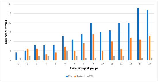
Figure 1.
Campylobacter spp. strains isolated by the qualitative method at 37 °C from the skin, the pectoral muscle and the visceral cavity, and the liver and their distribution in the epidemiological groups. (Enumeration of the epidemiological groups as in Table 3, Table 4, Table 5 and Table 6); V/L: visceral cavity/liver.
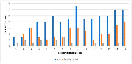
Figure 2.
Campylobacter spp. strains isolated by the quantitative method at 37 °C from the skin, the pectoral muscle and the visceral cavity, and the liver and their distribution in the epidemiological groups. (Enumeration of the epidemiological groups as in Table 3, Table 4, Table 5 and Table 6); V/L: visceral cavity/liver.
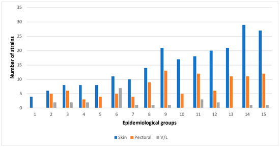
Figure 3.
Campylobacter spp. strains isolated by the qualitative method at 42 °C from the skin, the pectoral muscle and the visceral cavity, and the liver and their distribution in the epidemiological groups. (Enumeration of the epidemiological groups as in Table 3, Table 4, Table 5 and Table 6); V/L: visceral cavity/liver.
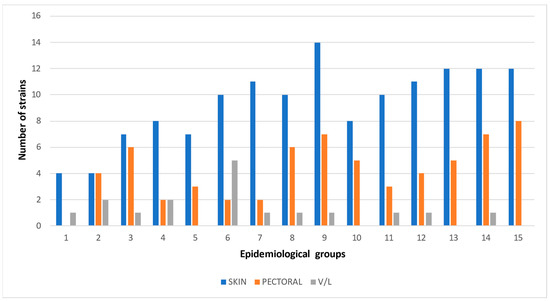
Figure 4.
Campylobacter spp. strains isolated by the quantitative method at 42 °C from the skin, the pectoral muscle and the visceral cavity, and the liver and their distribution in the epidemiological groups. (Enumeration of the epidemiological groups as in Table 3, Table 4, Table 5 and Table 6); V/L: visceral cavity/liver.
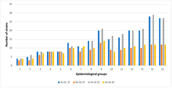
Figure 5.
Campylobacter spp. strains isolated from the skin and their distribution in the epidemiological groups. (Enumeration of the epidemiological groups as in Table 3, Table 4, Table 5 and Table 6). Sk Ql 37 °C: Skin qualitative method at 37 °C, Sk Qt 37 °C: Skin quantitative method at 37 °C, Sk Ql 42 °C: Skin qualitative method at 42 °C, Sk Qt 42 °C: Skin quantitative method at 42 °C.
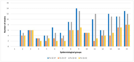
Figure 6.
Campylobacter spp. strains isolated from the pectoral muscle and their distribution in the epidemiological groups. (Enumeration of the epidemiological groups as in Table 3, Table 4, Table 5 and Table 6). Pe Ql 37 °C: Pectoral muscle qualitative method at 37 °C, Pe Qt 37 °C: Pectoral muscle quantitative method at 37 °C, Pe Ql 42 °C: Pectoral muscle qualitative method at 42 °C, Pe Qt 42 °C: Pectoral muscle quantitative method at 42 °C.
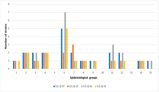
Figure 7.
Campylobacter spp. strains isolated from the visceral cavity and the liver and their distribution in the epidemiological groups. (Enumeration of the epidemiological groups as in Table 3, Table 4, Table 5 and Table 6). V/L Ql 37 °C: Visceral cavity/liver qualitative method at 37 °C, V/L Qt 37 °C: Visceral cavity/liver quantitative method at 37 °C, V/L Ql 42 °C: Visceral cavity/liver Qualitative method at 42 °C, V/L Qt 42 °C: Visceral cavity/liver Quantitative method at 42 °C.
Regression analysis showed that where different analytical methods produced different results, the statistical inference of the impact of the four criteria on the number of isolations could also be different. In the skin, the number of isolated Campylobacter strains was negatively affected by the small size of the flock (3–15 birds) for all methods. According to the qualitative method at 37 °C, the quantitative method at 37 °C, and the qualitative at 42 °C (but not the quantitative at 42 °C), the presence of small ruminants was very strongly positively correlated to the number of isolated Campylobacter (R > 0.883, p < 0.001). Likewise, for all methods except for the quantitative at 37 °C, the number of isolations from the pectoral muscle was strongly negatively correlated to the small size of the flock (3–15 birds) while for both quantitative methods, the presence of small ruminants was also strongly positively correlated (R > 0.604, p < 0.05). Regression analysis showed no significant correlations for the isolations by both qualitative and quantitative methods at 42 °C from the visceral cavity and the liver but moderate negative (R = 0.588, p = 0.02176) and strong negative (R = 0.608, p = 0.01606) correlations with the presence of small ruminants for the qualitative method at 37 °C and for the quantitative method at 37 °C, respectively.
Since the qualitative method at 42 °C was the most prolific and revealed the wider range of Campylobacter biodiversity, a further analysis was performed in the most abundant species as they are shown in Table 7. C. jejuni isolated from skin was very strongly positively correlated to the small size of the flock (3–15 birds) and to the presence of small ruminants (R = 0.811, p = 0.001586) while for C. jejuni isolated from the pectoral muscle, a strong positive correlation to the large size of the flock (more than 40 birds) and to the presence of small ruminants was observed (R = 0.739, p = 0.008737). C. coli isolated from skin showed a very strong negative correlation to the small size of the flock (3–15 birds) and a very strong positive correlation to the large size of the flock (more than 40 birds) (R = 0.846, p = 0.000535), while C. coli isolated from the pectoral muscle did not show any significant correlation to any of the four criteria. C. avium isolated from the skin showed a strong negative correlation to the presence of small ruminants (R = 0.654, p = 0.008164), C. fetus showed a strong positive correlation to the presence of small ruminants, and C. lari showed a moderate positive correlation to the large size of the flock (R = 0.533, p = 0.04077).
Table 7.
Distribution of the most abundant Campylobacter species with respect to the various groups and tissues.
The distribution of the isolated strains by the qualitative method at 42 °C among the three tissues was significantly different only for C. hepaticus (x2 = 7.616, p = 0.02219), C. coli (x2 = 5.384, p = 0.02032) and C. jejuni (x2 = 10.616, p = 0.00495) (Table 6).
4. Discussion
In this study, four criteria were used to assess the prevalence of Campylobacter species. Given that the research on this topic focuses almost entirely on industrial broilers and not on rural backyard chickens, the design of this study was similar to a navigation in uncharted waters. Rural husbandry and slaughter practices may vary significantly among different households. It is practically impossible to classify in detail all the different factors involved, so we formulated these four criteria with deductive logic. In Criterion A, the number of chickens in the household referred to the density of the birds and intraspecies transmission. Yet, one household may rear few birds but in close confinement while another household may rear more birds but in a larger space. In Criteria B and C, the presence in the same household of other species of poultry and of small ruminants respectively, were used to investigate possible interspecies transmission of Campylobacter strains. Of course, here too other factors are involved such as the size of the flock of the birds or of the small ruminants, the husbandry practices, or the degree of the contact. Finally, in Criterion D, the feeding practice (leftovers or concentrates) is important not only for the possibility of Campylobacter transmission through feeds, but also by affecting the general condition of the chicken. A further analysis of this criterion could involve factors such as the amount and the composition of concentrates fed. It is needless to say that further research is required and that the present research has also the character of a pilot study.
It seems that humans and chickens, despite their immense biological differences, share many Campylobacter species. The latter have specific growth requirements, regarding the temperature and this fact can be a limiting factor for their survival outside warm-blooded animals, but not an eliminating one. Their optimal growth is at either 42 °C (body temperature of chicken) or at 37 °C (human body temperature). A known epidemiological problem is the simultaneous presence of various Campylobacter species or types in human infections. What happens in such cases is that the diagnostic laboratories usually isolate and identify a single colony, missing thus the whole etiological picture of an outbreak. Likewise, when isolating Campylobacter from foods, enrichment should be performed at both temperatures, otherwise some strains present will not be identified.
Isolating Campylobacter spp. from samples such as raw food and environmental samples, which contain a plethora of species can be compromised by the antagonistic and inhibitory presence of more dominant species. Although the Campylobacter species have specific requirements for growth (such as temperature, pH, and microaerophile conditions) which make their survival not an easy task, yet they survive in environments where they are not expected, such as in refrigerated food. The Campylobacter species have been characterized as thermotolerant [27], or even thermophile due to its optimum growth at 41.5 °C. They cannot grow at temperatures lower than 30° [28]. Actually, the term “the Campylobacter conundrum” refers to this paradox. The very basic purpose of this study was to investigate the Campylobacter populations in free-grazing chicken in the rural environment. The skin of birds living outdoors is not the ideal niche for the Campylobacter species and this is the main reason that all accepted standard methods [24], both qualitative and quantitative were employed. The possibility of isolation of rare species in low counts or the existence of Campylobacter traumatized bacterial cells, underlined the necessity of an enrichment step before plating on solid agar [29,30]. A single ideal approach for the isolation of all Campylobacter species does not exist since some less-common species are inhibited by the selective media or at the incubation at 42 °C [7,31,32]. Therefore, where the occurrence of such organisms is expected or investigated, suitable cultivation conditions are required, such as membrane filtration, special atmospheric and temperature conditions, prolonged incubation, and subsequent plating on non-selective media [33,34].
Any references to skin or to meat contamination by Campylobacter spp. found in literature, refer to skin samples or meat samples originating from slaughter facilities or retail establishments but none from samples of rural origin. The reasons are understandable since (a) Campylobacter infections are among the principal causes of foodborne illnesses in urban areas where people consume industrial broilers and (b) industrial poultry meat targets the global food market with an annual turnover of billions of US$, while the rural poultry is consumed by local populations and visitors affecting much less both public health and economy. This may well be true, but the backyard chicken is remaining a global culinary practice, and the rural populations are still in danger of Campylobacter infections not only through ingestion of contaminated meat but also through direct contact with the birds or from the environment [35].
The occurrence rates of Campylobacter spp. in the birds of the present study varied depending on the analytical method and were calculated 40.33% for the quantitative method at 42 °C, 41.50% for the qualitative method at 42%, 41.33% for the quantitative method at 37 °C, and 41.00% for the qualitative method at 37 °C. These rates are far below the nearly 100% prevalence rate reported in the European industrial broiler farms [36,37] and USA broiler farms [38,39,40], or even from 60.30% in Sub-Saharan Africa [41], but higher than 0.6–13.1% reported in Northern European countries [42]. However, all these studies refer to samples from caeca or to fecal samples where the conditions are ideal for the main thermophilic Campylobacter species, and not to skin or to meat samples taken in the field as in our study [43,44].
A most obvious observation concerning our study is that three criteria out of the four, i.e., the presence of other species of poultry (criterion B), the presence of small ruminants (criterion C), and the feeding practice (criterion D) had no effect on the prevalence and on the distribution of the overall Campylobacter genus among the different epidemiological groups (Table 3). On a species level, however, some species were significantly affected by the presence of small ruminants. Criterion D (feeding practice), besides the possibility of transmission of Campylobacter from the environment through animal feed to poultry farms [13] was theoretically expected to affect indirectly the general condition of the birds since the concentrates provide more nutrients than the household’s leftovers of plant origin and consequently the immune system would function more efficiently keeping at bay at least some Campylobacter species. However, in rural areas, the birds graze freely and acquire whatever they need from the natural environment of the estate they live, and this appears to equalize the differences in the feeding practices. As for the presence of other species of poultry, it seems that birds that share the same habitat regardless of their species, carry similar microbiomes and so the same microorganisms circulate among them. Ethology factors perhaps could limit the close contact of birds of different species creating thus a sort of intraspecies barrier.
Small ruminants are a source of various Campylobacter species, shedding strains in their environment [45,46]. Possible routes of transmission are considered the pests (rodents mainly) and the personnel. Other authors however, like Ridley et al. (2011) propose a two-way direction of the Campylobacter contamination and they report an interaction between broilers and ruminants [47].
C. fetus colonizes the intestines of cattle and sheep which act as the reservoir of the microorganism [48,49]. It is a well-known pathogen in these species causing abortion and infertility [50,51]. Regression analysis confirmed the very strong correlation of C. fetus to the presence of small ruminants among the epidemiological groups (R = 0.814, p = 0.0059), a rather expected finding. In fact, C. fetus strains were only isolated from epidemiological groups where small ruminants were present in the same household as chicken. Yet in the same regression model, the large and the small number of birds were strongly negatively correlated to the number of strains of C. fetus, a finding implying that the size of the flock creates an ecosystem where the dynamics of antagonistic species regulate the prevalence rates of C. fetus. According to all four methods, 19 strains were isolated in total. Most of the isolations were from the skin and none from the visceral cavity and the liver. It is possible that this increased skin infection is due to environmental contact with small ruminants’ feces. By the qualitative method at 42 °C distribution (Table 6), the correlation to the presence of the small ruminants was strong (R = 0.629, p = 0.012).
The overall C. jejuni prevalence among the different groups was very strongly positively correlated to the large size of the flock (more than 30 birds) and to the presence of small ruminants (R = 0.832, p < 0.001). C. jejuni is known to colonize the digestive tract of warm-blooded hosts [48] infecting thus the poultry. Depending on the method, 96 to 98 strains were isolated making C. jejuni the most prevalent of all Campylobacter species (Table 2), a finding agreeing with similar findings of other authors who reported that this species is the most dominant and poultry is considered as its primary reservoir [49,50]. The distribution of C. jejuni strains was found significantly increased in skin as compared to the one in the pectoral muscle by the qualitative method at 42 °C (x2 = 10.6162, p = 0.004851) implying environmental contamination.
C. coli was the second dominant species in this study (Table 2) with 64 strains (both methods at 42 °C) or 63 strains (both methods at 37 °C). None of these strains was isolated from the visceral cavity and the liver. Most of the isolations were from the skin (x2 = 5.3841, p = 0.0203) as in the case of all other Campylobacter species. Interestingly, the quantitative methods at both temperatures (42 °C and 37 °C) revealed a strong positive correlation (R = 0.789, p = 0.003) to the the large size of the flock (more than 30 birds) while the qualitative methods (42 and 37 °C) showed a strong negative correlation (R = 0.725, p = 0.002) to the small size of the flock (3–10 birds). These differences can be attributed to the different distribution of strains according to the different analytical methods, but what these findings have in common is that the multitude of chickens in the flock plays an important role.
Although C. coli and C. jejuni are reported to survive in the guts of various mammals as well as in other poultry species caeca, our results do not show any increased prevalence in the epidemiological groups where small ruminants and/or other poultry species were present, implying that other environmental sources of contamination like rodents or the water.
C. avium has been isolated from chicken and turkeys [43,51,52,53,54] yet our results do not show any increased prevalence in the groups where other poultry species shared the same environment. All four analytical methods we used, concur that the presence of small ruminants was strongly negatively correlated with its prevalence rates (R > 0.721, p < 0.002), suggesting intense antagonistic dynamics from other Campylobacter strains shed by these animals.
C. lari was third after C. jejuni and C. coli prevalent species with twenty-four (24) isolated strains by three of the four methods (Table 2). This species is usually isolated from various sea birds like seagulls, from marine mammals and has been also isolated from domestic animals [55]. In our study, its isolations have been positively strongly correlated (R = 0.702, p = 0.00284) to the large size of the flock (more than 30 birds). As previously discussed for other species, the large size of the flock creates perhaps more opportunities for certain species to find niches.
C. hepaticus is a causative agent of Spotty Liver Syndrome (SLD), a disease of free-range laying hens [56,57]. All birds in our study were clinically healthy and obviously, the ones from which C. hepaticus was isolated were asymptomatic carriers. Most isolations occurred from liver samples. C. hepaticus shows lower growth rates in iron-depleted environments and this explains its tissue tropism [58]. Regression analysis showed a strong positive correlation to the large size of the flock (more than 30 birds) and a strong negative correlation to the presence of small ruminants (R = 0.742, p = 0.00831).
Six species were singletons since only one strain of them was isolated (Table 2). From these C. cuniculorum has been isolated from rabbits [59] and probably its source in our study was rabbits kept in the same household. Since C. rectus, C. helveticus, C. mucosalis, and C. showae originate from humans and other animals such as dogs, cats, and pigs, while C. hominis originates from humans [51], it is quite possible that the contamination of the birds in our study was either anthropogenic or coming from the household’s dog or from the omnipresent cats. Anthropogenic transmission seems likely since C. rectus and C. showae are involved in the pathogenesis of gingivitis and periodontitis [60,61]. It is interesting that in C. rectus, C. hominis and C. helveticus, isolated exclusively from the epidemiological group 1, where the flock was small (3–15 birds), there were neither other poultry species nor small ruminants present, and the birds were fed with leftovers. Possibly these households are small estates, and the birds live in close contact with humans and pets.
Two of the species from which two strains were isolated (Table 3), C. gracilis and C. concisus, are found in the oral cavity of humans in health and disease, e.g., periodontitis [62,63,64], so an anthropogenic contamination to the birds is possible, while C. upsaliensis has been isolated from dogs’ and cats’ healthy and diarrheic feces [65,66].
C. sputorum has been isolated from cattle, sheep, humans, and dogs [67], providing plenty of opportunities for transmission to birds, particularly in a rural environment. In our study, all four strains (Table 3) have been isolated from groups 3, 8, and 13, in which small ruminants were present. Similarly, C. hyointestinalis has been isolated from cattle, sheep, and deer [68,69] and in the present study, all four strains (Table 3) were isolated from groups 8, 13, and 14, where small ruminants are present. On the contrary, C. ureolyticus was isolated from groups 6 and 11 where other poultry species and ruminants were not present. This species has been isolated from cattle and horses [54] as well as from humans suffering from periodontitis and other diseases [70].
5. Conclusions
Chicken-rearing practices in rural households are very different and adapt to different environments in comparison to the more steadfast industrial ones. Hence, the research is more complicated, since there are many factors involved. From the present study, the following results were obtained:
- Eighteen species of the Campylobacter genus have been isolated from the free grazing chicken in the rural environment, an impressive abundance that suggests that this genus can survive in more environmental niches than has been thought.
- The isolation of Campylobacter strains raises public health issues as well as animal health issues, concerning rural nonindustrial environments.
- The multitude of birds in the flock of chickens was the most decisive factor which affected the prevalence of most Campylobacter species.
- The presence of small ruminants in the same household significantly affected the prevalence of certain species such as C. fetus.
- The presence of other species of poultry and the feeding practice (leftovers or concentrates) did not affect the prevalence of Campylobacter strains.
- Although the danger to human health is possible, the anthropogenic contamination of the birds cannot be excluded, particularly for some Campylobacter species involved in some human oral cavity ailments such as periodontitis.
- The qualitative methods were more proliferative in isolating the Campylobacter species, especially at 42 °C.
- Rural populations must be educated on the necessity to cook chicken meat well enough and maintain good personal hygiene practices.
Author Contributions
Conceptualization, C.V. and P.D.; methodology, A.D., G.R., C.V. and P.D.; software, A.D. and G.R.; validation, C.V. and P.D.; formal analysis, A.D., G.R. and C.V.; investigation, A.D., G.R. and C.V.; resources, A.D. and G.R.; data curation, A.D. and G.R.; writing—original draft preparation, A.D., G.R. and C.V.; writing—review and editing, A.D., K.A.-D., C.V. and P.D.; visualization, A.D. and C.V.; supervision, C.V., K.A.-D. and P.D.; project administration, C.V. and P.D.; funding acquisition, P.D. All authors have read and agreed to the published version of the manuscript.
Funding
This research received no external funding.
Institutional Review Board Statement
Not applicable.
Informed Consent Statement
Not applicable.
Data Availability Statement
Not applicable.
Conflicts of Interest
The authors declare no conflict of interest.
References
- Yulistiani, R.; Praseptiangga, D.; Supyani; Sudibya. Occurrences of Salmonella spp. and Escherichia coli in chicken meat, intestinal contents and rinse water at slaughtering place from traditional market in Surabaya, Indonesia. IOP Conf. Ser. Mater. Sci. Eng. 2019, 633, 012007. [Google Scholar] [CrossRef]
- Hessel, C.T.; de Oliveira Elias, S.; Pessoa, J.P.; Zanin, L.M.; Stedefeldt, E.; Tondo, E.C. Food safety behavior and handling practices during purchase, preparation, storage and consumption of chicken meat and eggs. Food Res. Int. 2019, 125, 108631. [Google Scholar] [CrossRef]
- Sequeira, M.; Signorini, L.; Frizzo, L.S. Occurrence of Thermotolerant Campylobacter spp. at Different Stages of the Poultry Meat Supply Chain in Argentina. N. Z. Vet. J. 2013, 61, 337–343. [Google Scholar]
- Goddard, A.D.; Arnold, M.E.; Allen, V.M.; Snary, E.L. Estimating the time at which commercial broiler flocks in Great Britain become infected with Campylobacter: A Bayesian approach. Epidemiol. Infect. 2014, 142, 1884–1892. [Google Scholar] [CrossRef]
- Mughini-Gras, L.; Penny, C.; Ragimbeau, C.; Schets, F.M.; Blaak, H.; Duim, B.; Wagenaar, J.A.; de Boer, A.; Cauchie, H.M.; Mossong, J.; et al. Quantifying potential sources of surface water contamination with Campylobacter jejuni and Campylobacter coli. Water Res. 2016, 101, 36–45. [Google Scholar] [CrossRef]
- Kaakoush, N.O.; Castaño-Rodríguez, N.; Mitchell, H.M.; Man, S.M. Global Epidemiology of Campylobacter Infection. Clin. Microbiol. Rev. 2015, 28, 687–720. [Google Scholar] [CrossRef]
- Igwaran, A.; Okoh, A.I. Human campylobacteriosis: A public health concern of global importance. Heliyon 2019, 5, e02814. [Google Scholar] [CrossRef]
- Facciolà, A.; Riso, R.; Avventuroso, E.; Visalli, G.; Delia, S.A.; Laganà, P. Campylobacter: From microbiology to prevention. J. Prev. Med. Hyg. 2017, 58, E79–E92. [Google Scholar]
- Finsterer, J. Triggers of Guillain–Barré Syndrome: Campylobacter jejuni Predominates. Int. J. Mol. Sci. 2022, 23, 14222. [Google Scholar] [CrossRef]
- Ang, C.W.; De Klerk, M.A.; Endtz, H.P.; Jacobs, B.C.; Laman, J.D.; Van Der Meché, F.G.A.; Van Doorn, P.A. Guillain-Barré Syndrome- and Miller Fisher Syndrome-Associated Campylobacter jejuni Lipopolysaccharides Induce Anti-GM 1 and Anti-GQ 1b Antibodies in Rab. Infect. Immun. 2001, 69, 2462–2469. [Google Scholar] [CrossRef]
- Campylobacter. Available online: https://www.efsa.europa.eu/en/topics/topic/campylobacter (accessed on 30 March 2023).
- Hadiyan, M.; Momtaz, H.; Shakerian, A. Prevalence, antimicrobial resistance, virulence gene profile and molecular typing of Campylobacter species isolated from poultry meat samples. Vet. Med. Sci. 2022, 8, 2482–2493. [Google Scholar] [CrossRef] [PubMed]
- Hakeem, M.J.; Lu, X. Survival and Control of Campylobacter in Poultry Production Environment. Front. Cell. Infect. Microbiol. 2021, 10, 615049. [Google Scholar] [CrossRef]
- Chicken and Food Poisoning. Available online: https://www.cdc.gov/foodsafety/chicken.html (accessed on 30 March 2023).
- Outbreaks Involving Campylobacter. Available online: https://www.cdc.gov/campylobacter/outbreaks/outbreaks.html (accessed on 25 March 2023).
- Campylobacteriosis Annual Epidemiological Report for 2021. Available online: https://www.ecdc.europa.eu/sites/default/files/documents/campylobacteriosis-annual-epidemiological-report-2021.pdf (accessed on 25 March 2023).
- Miljković-Selimović, B.; Babić, T.; Kocić, B.; Aleksić, E.; Malešević, A.; Tambur, Z. Campylobacter concisus. J. Infect. Dev. Ctries. 2021, 15, 1216–1221. [Google Scholar] [CrossRef]
- Awada, B.; Hindy, J.R.; Chalfoun, M.; Kanj, S.S. Cervical osteomyelitis potentially caused by Campylobacter fetus. J. Infect. Public Health 2021, 14, 1233–1236. [Google Scholar] [CrossRef]
- Dobrović, K.; Fila, B.; Janeš, A.; Civljak, R. Campylobacter fetus Bacteremia Related to Vascular Prosthesis and Pseudoaneurysm Infection: A Case Report and Review. Pathogens 2022, 11, 1536. [Google Scholar] [CrossRef]
- Bullman, S.; Corcoran, D.; O’Leary, J.; Lucey, B.; Byrne, D.; Sleator, R.D. Campylobacter ureolyticus: An emerging gastrointestinal pathogen? FEMS Immunol. Med. Microbiol. 2011, 61, 228–230. [Google Scholar] [CrossRef]
- Habib, I.; Sampers, I.; Uyttendaele, M.; Berkvens, D.; De Zutter, L. Baseline Data from a Belgium-Wide Survey of Campylobacter Species Contamination in Chicken Meat Preparations and Considerations for a Reliable Monitoring Program. Appl. Environ. Microbiol. 2008, 74, 5483–5489. [Google Scholar] [CrossRef]
- ISO 10272-1; Microbiology of Food and Animal Feeding Stuffs—Horizontal Method for Detection and Enumeration of Campylobacter spp.—Part 1: Detection Method. International Organization for Standardization: Geneva, Switzerland, 2006.
- EN ISO 10272–2:2006; Microbiology of Food and Animal Feeding Stuffs—Horizontal Method for Detection and Enumeration of Campylobacter spp.—Part 2: Colony-Count Technique. International Organization for Standardization: Geneva, Switzerland, 2007.
- EN ISO 10272-2:2017; Microbiology of the Food Chain—Horizontal Method for Detection and Enumeration of Campylobacter spp.—Part 2: Colony-Count Technique. International Organization for Standardization: Geneva, Switzerland, 2007.
- Tzora, A.; Nelli, A.; Voidarou, C.; Fthenakis, G.; Rozos, G.; Theodorides, G.; Bonos, E.; Skoufos, I. Microbiota “Fingerprint” of Greek Feta Cheese through Ripening. Appl. Sci. 2021, 11, 5631. [Google Scholar] [CrossRef]
- Tzora, A.; Nelli, A.; Voidarou, C.; Fotou, K.; Bonos, E.; Rozos, G.; Grigoriadou, K.; Papadopoulos, P.; Basdagianni, Z.; Giannenas, I.; et al. Impact of an Omega-3-Enriched Sheep Diet on the Microbiota and Chemical Composition of Kefalograviera Cheese. Foods 2022, 11, 843. [Google Scholar] [CrossRef]
- Levin, R.E. Campylobacter jejuni: A Review of its Characteristics, Pathogenicity, Ecology, Distribution, Subspecies Characterization and Molecular Methods of Detection. Food Biotechnol. 2007, 21, 271–347. [Google Scholar] [CrossRef]
- Silva, J.; Leite, D.; Fernandes, M.; Mena, C.; Gibbs, P.A.; Teixeira, P. Campylobacter spp. as a Foodborne Pathogen: A Review. Front. Microbiol. 2011, 2, 200. [Google Scholar] [CrossRef]
- Lazou, T.P.; Gelasakis, A.I.; Chaintoutis, S.C.; Iossifidou, E.G.; Dovas, C.I. Method-Dependent Implications in Foodborne Pathogen Quantification: The Case of Campylobacter coli Survival on Meat as Comparatively Assessed by Colony Count and Viability PCR. Front. Microbiol. 2021, 12, 604933. [Google Scholar] [CrossRef] [PubMed]
- Jacobs-Reitsma, W.; Lyhs, U.; Wagenaar, J. Campylobacter in the food supply. In Campylobacter; Nachamkin, I., Szymanski, C., Blaser, J., Eds.; ASM Press: Washington, DC, USA, 2008; pp. 627–644. [Google Scholar]
- Fitzgerald, C.; Whichard, J.; Fields, P.I. The Genus Campylobacter. In Practical Handbook of Microbiology, 2nd ed.; CRC Press: Boca Raton, FL, USA, 2008; pp. 563–574. [Google Scholar]
- Fitzgerald, C.; Whichard, J.; Nachamkin, I. Diagnosis and antimicrobial susceptibility of Campylobacter species. In Campylobacter, 3rd ed.; ASM Press: Washington, DC, USA, 2008; pp. 227–243. [Google Scholar]
- Debruyne, L.; Broman, T.; Bergström, S.; Olsen, B.; On, S.L.W.; Vandamme, P. Campylobacter volucris sp. nov., isolated from black-headed gulls (Larus ridibundus). Int. J. Syst. Evol. Microbiol. 2010, 60 Pt 8, 1870–1875. [Google Scholar] [CrossRef]
- Lastovica, A.J.; On, S.L.W.; Zhang, L. The Family Campylobacteraceae. In The Prokaryotes; Springer: Heidelberg/Berlin, Germany, 2014; pp. 307–335. [Google Scholar] [CrossRef]
- Epps, S.; Harvey, R.; Hume, M.; Phillips, T.; Anderson, R.; Nisbet, D. Foodborne Campylobacter: Infections, Metabolism, Pathogenesis and Reservoirs. Int. J. Environ. Res. Public Health 2013, 10, 6292–6304. [Google Scholar] [CrossRef]
- Schets, F.M.; Jacobs-Reitsma, W.F.; Van Der Plaats, R.Q.J.; Heer, L.K.-D.; Van Hoek, A.H.A.M.; Hamidjaja, R.A.; Husman, A.M.D.R.; Blaak, H. Prevalence and types of Campylobacter on poultry farms and in their direct environment. J. Water Health 2017, 15, 849–862. [Google Scholar] [CrossRef] [PubMed]
- Sibanda, N.; McKenna, A.; Richmond, A.; Ricke, S.C.; Callaway, T.; Stratakos, A.C.; Gundogdu, O.; Corcionivoschi, N. A Review of the Effect of Management Practices on Campylobacter Prevalence in Poultry Farms. Front. Microbiol. 2018, 9, 2002. [Google Scholar] [CrossRef]
- Thames, H.T.; Fancher, C.A.; Colvin, M.G.; McAnally, M.; Tucker, E.; Zhang, L.; Kiess, A.S.; Dinh, T.T.N.; Sukumaran, A.T. The Prevalence of Salmonella and Campylobacter on Broiler Meat at Different Stages of Commercial Poultry Processing. Animals 2022, 12, 2460. [Google Scholar] [CrossRef] [PubMed]
- Thames, H.T.; Sukumaran, A.T. A Review of Salmonella and Campylobacter in Broiler Meat: Emerging Challenges and Food Safety Measures. Foods 2020, 9, 776. [Google Scholar] [CrossRef]
- Tang, Y.; Sahin, O.; Pavlovic, N.; LeJeune, J.; Carlson, J.; Wu, Z.; Dai, L.; Zhang, Q. Rising fluoroquinolone resistance in Campylobacter isolated from feedlot cattle in the United States. Sci. Rep. 2017, 7, 494. [Google Scholar] [CrossRef]
- Gahamanyi, N.; Mboera, L.E.G.; Matee, M.I.; Mutangana, D.; Komba, E.V.G. Prevalence, Risk Factors, and Antimicrobial Resistance Profiles of Thermophilic Campylobacter Species in Humans and Animals in Sub-Saharan Africa: A Systematic Review. Int. J. Microbiol. 2020, 2020, 2092478. [Google Scholar] [CrossRef]
- Skarp, C.P.A.; Hänninen, M.-L.; Rautelin, H.I.K. Campylobacteriosis: The role of poultry meat. Clin. Microbiol. Infect. 2016, 22, 103–109. [Google Scholar] [CrossRef] [PubMed]
- Al Hakeem, W.G.; Fathima, S.; Shanmugasundaram, R.; Selvaraj, R.K. Campylobacter jejuni in Poultry: Pathogenesis and Control Strategies. Microorganisms 2022, 10, 2134. [Google Scholar] [CrossRef] [PubMed]
- Corry, J.E.; Atabay, H.I. Poultry as a source of Campylobacter and related organisms. Symp. Ser. Soc. Appl. Microbiol. 2001, 90, 96S–114S. [Google Scholar] [CrossRef]
- Shange, N.; Gouws, P.; Hoffman, L.C. Campylobacter and Arcobacter species in food-producing animals: Prevalence at primary production and during slaughter. World J. Microbiol. Biotechnol. 2019, 35, 146. [Google Scholar] [CrossRef] [PubMed]
- Thépault, A.; Rose, V.; Quesne, S.; Poezevara, T.; Béven, V.; Hirchaud, E.; Touzain, F.; Lucas, P.; Méric, G.; Mageiros, L.; et al. Ruminant and chicken: Important sources of campylobacteriosis in France despite a variation of source attribution in 2009 and 2015. Sci. Rep. 2018, 8, 9305. [Google Scholar] [CrossRef]
- Ridley, A.M.; Morris, V.K.; Cawthraw, S.A.; Ellis-Iversen, J.; Harris, J.A.; Kennedy, E.M.; Newell, D.G.; Allen, V.M. Longitudinal Molecular Epidemiological Study of Thermophilic Campylobacters on One Conventional Broiler Chicken Farm. Appl. Environ. Microbiol. 2011, 77, 98–107. [Google Scholar] [CrossRef]
- Awada, R.; Ghssein, G.; El Roz, A.; Farhat, M.; Nehme, N.; Hassan, H.F. Prevalence of Campylobacter spp. in broilers in North Lebanon. Vet. World 2023, 16, 322–328. [Google Scholar] [CrossRef]
- Silva, M.F.; Kienesberger, S.; Pereira, G.; Mateus, L.; Lopes-da-Costa, L.; Silva, E. Molecular diagnosis of bovine genital campylobacteriosis using high-resolution melting analysis. Front. Microbiol. 2022, 13, 969825. [Google Scholar] [CrossRef]
- Yde Aagaard, M.E.; Frahm Kirk, K.; Linde Nielsen, H.; Steffensen, R.; Nielsen, H. Campylobacter concisus from chronic inflammatory bowel diseases stimulates IL-8 production in HT-29 cells. Gut Pathog. 2023, 15, 5. [Google Scholar] [CrossRef]
- Costa, D.; Iraola, G. Pathogenomics of Emerging Campylobacter Species. Clin. Microbiol. Rev. 2019, 32, e00072-18. [Google Scholar] [CrossRef]
- Horrocks, S.M.; Anderson, R.C.; Nisbet, D.J.; Ricke, S.C. Incidence and ecology of Campylobacter jejuni and coli in animals. Anaerobe 2009, 15, 18–25. [Google Scholar] [CrossRef]
- Suzuki, H.; Yamamoto, S. Campylobacter Contamination in Retail Poultry Meats and By-Products in the World: A Literature Survey. J. Vet. Med. Sci. 2009, 71, 255–261. [Google Scholar] [CrossRef]
- Wayou, B.A.; Kassa, G.M.; Sori, T.; Mondin, A.; Tucciarone, C.M.; Cecchinato, M.; Pasotto, D. Molecular Survey and Identification of Campylobacter spp. in Layer Farms in Central Ethiopia. Trop. Med. Infect. Dis. 2022, 7, 31. [Google Scholar] [CrossRef]
- Miller, S.; Amadi, V.; Bekele, A.Z.; Zieger, U.; Hariharan, H.; Stone, D. Identification of Human and Poultry Campylobacter Sequence Types in Small Indian Mongooses (Herpestesauropunctatus) in Grenada, West Indies. Int. J. Vet. Med. Res. Rep. 2014, 2014, 676408. [Google Scholar] [CrossRef]
- Phung, C.; Vezina, B.; Anwar, A.; Wilson, T.; Scott, P.C.; Moore, R.J.; Van, T.T.H. Campylobacter hepaticus, the Cause of Spotty Liver Disease in Chickens: Transmission and Routes of Infection. Front. Vet. Sci. 2020, 6, 505. [Google Scholar] [CrossRef]
- Crawshaw, T.R.; Chanter, J.I.; Young, S.C.; Cawthraw, S.; Whatmore, A.M.; Koylass, M.S.; Vidal, A.B.; Salguero, F.J.; Irvine, R.M. Isolation of a novel thermophilic Campylobacter from cases of spotty liver disease in laying hens and experimental reproduction of infection and microscopic pathology. Vet. Microbiol. 2015, 179, 315–321. [Google Scholar] [CrossRef]
- Petrovska, L.; Tang, Y.; Jansen van Rensburg, M.J.; Cawthraw, S.; Nunez, J.; Sheppard, S.K.; Ellis, R.J.; Whatmore, A.M.; Crawshaw, T.R.; Irvine, R.M. Genome Reduction for Niche Association in Campylobacter Hepaticus, A Cause of Spotty Liver Disease in Poultry. Front. Cell. Infect. Microbiol. 2017, 7, 354. [Google Scholar] [CrossRef] [PubMed]
- Zanoni, R.G.; Debruyne, L.; Rossi, M.; Revez, J.; Vandamme, P. Campylobacter cuniculorum sp. nov., from rabbits. Int. J. Syst. Evol. Microbiol. 2009, 59, 1666–1671. [Google Scholar] [CrossRef] [PubMed]
- Lawson, A.J.; On, S.L.; Logan, J.M.; Stanley, J. Campylobacter hominis sp. nov., from the human gastrointestinal tract. Int. J. Syst. Evol. Microbiol. 2001, 51, 651–660. [Google Scholar] [CrossRef] [PubMed]
- Macuch, P.J.; Tanner, A.C. Campylobacter species in health, gingivitis, and periodontitis. J. Dent. Res. 2000, 79, 785–792. [Google Scholar] [CrossRef]
- Liu, M.M.; Boinett, C.J.; Chan, A.C.K.; Parkhill, J.; Murphy, M.E.P.; Gaynor, E.C. Investigating the Campylobacter jejuni Transcriptional Response to Host Intestinal Extracts Reveals the Involvement of a Widely Conserved Iron Uptake System. mBio 2018, 9, e01347-18. [Google Scholar] [CrossRef] [PubMed]
- Miller, W.G.; Yee, E. Complete Genome Sequence of Campylobacter gracilis ATCC 33236T. Genome Announc. 2015, 3, e01087-15. [Google Scholar] [CrossRef] [PubMed]
- Omara, S.T.; El Fadaly, H.A.; Barakat, A.M.A. Public Health Hazard of Zoonotic Campylobacter jejuni Reference to Egyptian Regional and Seasonal Variations. Res. J. Microbiol. 2015, 10, 343–354. [Google Scholar] [CrossRef]
- Couturier, B.A.; Hale, D.C.; Couturier, M.R. Association of Campylobacter upsaliensis with Persistent Bloody Diarrhea. J. Clin. Microbiol. 2012, 50, 3792–3794. [Google Scholar] [CrossRef][Green Version]
- Chaban, B.; Ngeleka, M.; Hill, J.E. Detection and quantification of 14 Campylobacter species in pet dogs reveals an increase in species richness in feces of diarrheic animals. BMC Microbiol. 2010, 10, 73. [Google Scholar] [CrossRef]
- Miller, W.G.; Yee, E.; Chapman, M.H.; Bono, J.L. Comparative Genomics of All Three Campylobacter sputorum Biovars and a Novel Cattle-Associated C. sputorum Clade. Genome Biol. Evol. 2017, 9, 1513–1518. [Google Scholar] [CrossRef]
- Wilkinson, D.A.; O’donnell, A.J.; Akhter, R.N.; Fayaz, A.; Mack, H.J.; Rogers, L.E.; Biggs, P.J.; French, N.P.; Midwinter, A.C. Updating the genomic taxonomy and epidemiology of Campylobacter hyointestinalis. Sci. Rep. 2018, 8, 2393. [Google Scholar] [CrossRef] [PubMed]
- Costa, D.; Lévesque, S.; Kumar, N.; Fresia, P.; Ferrés, I.; Lawley, T.D.; Iraola, G. Pangenome analysis reveals genetic isolation inCampylobacter hyointestinalissubspecies adapted to different mammalian hosts. Sci. Rep. 2021, 3431. [Google Scholar] [CrossRef]
- Vandamme, P.; Debruyne, L.; De Brandt, E.; Falsen, E. Reclassification of Bacteroides ureolyticus as Campylobacter ureolyticus comb. nov., and emended description of the genus Campylobacter. Int. J. Syst. Evol. Microbiol. 2010, 60 Pt 9, 2016–2022. [Google Scholar] [CrossRef]


Disclaimer/Publisher’s Note: The statements, opinions and data contained in all publications are solely those of the individual author(s) and contributor(s) and not of MDPI and/or the editor(s). MDPI and/or the editor(s) disclaim responsibility for any injury to people or property resulting from any ideas, methods, instructions or products referred to in the content. |
© 2023 by the authors. Licensee MDPI, Basel, Switzerland. This article is an open access article distributed under the terms and conditions of the Creative Commons Attribution (CC BY) license (https://creativecommons.org/licenses/by/4.0/).