Article

Volatile Organic Compounds Determination from Intestinal Polyps and in Exhaled Breath by Gas Chromatography–Mass Spectrometry

by
Antonella Maria Aresta
1,
Nicoletta De Vietro
1,*,
Arcangelo Picciariello
2,
Maria Teresa Rotelli
2,
Donato Francesco Altomare
2,
Agnese Dezi
2,
Gennaro Martines
2,
Alessia Di Gilio
1,
Jolanda Palmisani
1,
Gianluigi De Gennaro
1 and
Carlo Zambonin
1
1
Department of Biosciences, Biotechnologies and Environment, University “Aldo Moro”, Via Orabona 4, 70126 Bari, Italy
2
Department of Precision and Regenerative Medicine and Ionian Area, University “Aldo Moro”, Piazza G. Cesare 11–Polyclinic, 70124 Bari, Italy
*
Author to whom correspondence should be addressed.
Appl. Sci. 2023, 13(10), 6083; https://doi.org/10.3390/app13106083
Submission received: 20 April 2023 / Revised: 10 May 2023 / Accepted: 14 May 2023 / Published: 15 May 2023
(This article belongs to the Special Issue New Trends in Biosciences III)

Abstract

:
In this paper, a new protocol is described, based on solid phase microextraction (SPME) coupled with gas chromatography–mass spectrometry (GC-MS), to monitor ex vivo changes in endogenous volatile organic compounds (VOCs) released by surgically resected colonic tissues (normal colonic mucosa and adenomatous polyps) from seven patients undergoing operative colonoscopy to identify their molecular pattern. The exhalated volatile organic molecules from these patients were sampled by the ReCIVA® breath sampler, shortly before surgery, and analyzed by GC-MS. Comparing VOC patterns identified in the tissues and in the breath of the same patients, a possible correlation can be found between the levels of methylbenzene and benzaldehyde exhaled and the presence of colonic adenomatous polypoid lesions.

1. Introduction

SPME is an efficient, sensitive, rapid, and solvent-free sample preparation technique that merges, in one step, sampling, isolation, and enrichment of analytes. It is commonly joined with GC or liquid chromatography (LC) to separate and detect the extracted analytes [1,2,3,4,5]. The simplicity of sampling and the design flexibility of the probe make SPME a useful extraction technique for analyzing biological samples of different natures and sizes. SPME is a useful rapid diagnostic tool for in vitro/ex vivo and in vivo studies [2,3,4,5,6]. The development of minimally invasive SPME microfibers has made it possible to monitor target analytes in vivo or changes in the metabolome/lipidome of a cell or living organ [7,8].
Colorectal cancer (CRC) is one of the most common tumors, both in Europe and USA, and is associated with high mortality [9,10,11]. Following a multistep process involving genetic, histological, and morphological changes, CRC generally originates from adenomatous polyps. Early detection and endoscopic treatment of polypoid lesions are the primary CRC prevention and can significantly reduce CRC incidence and mortality [12,13].
Recently, GC-MS analysis of exhaled VOCs has been demonstrated to be able to differentiate between CRC patients and healthy people (negative colonoscopy) [14]. A correspondence between VOCs exhaled and released by surgically resected cancer tissue of the same CRC patient was found using the SPME-GC-MS technique [15].
In this study, we applied the same headspace (HS)- and direct immersion (DI)-SPME-GC-MS protocols, optimized in our previous work [15], to determine and monitor ex vivo, for one week, the endogenous VOCs produced from tissues (healthy mucosa and colorectal adenomatous polyps) obtained from patients undergoing operative colonoscopy and compared them with their exhaled VOCs, sample by ReCIVA® device and investigated by GC-MS, to search any possible correlation.

2. Materials and Methods

2.1. Patients and Polyp Characteristics

After obtaining written informed consent, the exhaled breath and fresh specimens of healthy colonic mucosa and resected adenomatous polyps of seven patients, enlisted to undergo endoscopic treatment, were analyzed. Table 1 resumes the demographic and co-morbidities of patients and polypoid lesions characteristics.

2.2. Tissues (Colonic Adenomatous Polypoid Lesion and Normal Mucosa) Analysis

2.2.1. Chemicals and SPME Sampling Device

Reference standards of benzaldehyde, ethylbenzene, indole, methylbenzene, phenol, and octanal (analytical grade; purity > 98%) were purchased from Sigma-Aldrich (Milano, Italy) to confirm the chemical structure of the molecules recognized by the National Institute of Standards and Technology (NIST) library. Stock solutions (10 mg/mL) of each compound were prepared in methanol (Sigma-Aldrich), stored at 8 °C, and daily diluted in fresh culture medium (Dulbecco’s Modified Eagle Medium, Euroclone, Milano, Italy) to prepare working standard mixtures. The SPME (Supelco, Milano, Italy) mounting kit included a manual holder and a 50/30 mm CAR/DVB/PDMS (carboxen, divinylbenzene, polydimethylsiloxane) fiber of 1 cm in length. Before use, the fiber was conditioned in the GC injector according to the supplier’s instructions.

2.2.2. SPME VOCs Extraction

Excised tissue was cut in biopsies weighing 0.020 ± 0.005 g and placed in a 1.7 mL screw top amber glass vial, hermetically sealed with a PTFE (polytetrafluoroethylene)/silicone septum (Sigma-Aldrich, St. Louis, MO, USA), containing 0.2 mL of sterile culture medium. Within 30 min from resection, the vials were incubated at 37 °C and analyzed immediately (time zero) and after 1, 4, and 7 days by HS- and DI-SPME. A “blank” vial containing 0.2 mL of culture medium was simultaneously subjected to the same protocol. A volume of 0.01 mL of standard working mixtures at appropriate concentrations was then added to a 1.7 mL amber glass vial equipped with PTFE /silicone septum and screw cap (Sigma-Aldrich, St. Louis, MO, USA), containing 0.2 mL of sterile culture medium. These samples were subjected to the same analytical procedures.
The extraction of the VOCs by HS-SPME was carried out by placing the fiber at 37 °C for 30 min.
DI-SPME was carried out by drawing a volume of 15 µL from HS vials with a microliter syringe from Hamilton (Sigma-Aldrich, St. Louis, MO, USA) and placed into a 1.7 mL vial containing 1.5 mL of 15% NaCl sterile solution and a magnetic stirrer bar (Sigma-Aldrich, St. Louis, MO, USA). After sealing with a cap equipped with PTFE/silicone septa, the fiber was immersed in the solution under agitation (700 rpm) for 60 min at room temperature. The volume subtracted from each HS vial was restored with sterile fresh culture medium before storing the vial in the incubator at 37 °C.

2.2.3. GC-MS Apparatus and Analysis Experimental Conditions

For this study, a TRACE GC Ultra (Thermo Scientific, Waltham, MA, USA) with an ion-trap mass spectrometer (Polaris Q, Thermo Scientific, Waltham, MA, USA) was used. A TRACE TR-5 MS-fused silica capillary column (30 m × 0.25 mm i.d., 0.25 mm film thickness; Thermo Scientific) was used for chromatographic separation, and helium (Rivoira, Torino, Italy) with a constant flow rate of 1.0 mL/min was used as carrier gas.
The chromatographic conditions were those optimized in our previous work [15]. The mass spectrometer was operated in the electron impact (EI+) mode with a source temperature of 200 °C, ionizing voltage of 70 eV, and transfer line temperature of 240 °C. Full scan mode (SCAN; 40–250 m/z with a total scan time of 0.34 s) was used for the mass analysis. Total ion count (TIC) chromatograms were acquired and extracted ion chromatograms (XIC) were obtained for individual analytes by selecting their characteristic m/z values. VOC identification was carried out using the MS database of the NIST.
After extraction, fibers were transferred into the injection port of the GC for 2 min in splitless mode. In ex vivo experiments, fibers were exposed for an additional 5 min in the GC injector port at 250 °C before a new sampling to avoid memory effects.

2.2.4. Linear Regression Analysis, Limits of Detection (LOD), and Quantification (LOQ)

The HS- and DI-SPME conditions employed were evaluated with linear regression analysis of peak area versus analyte concentration in culture medium, utilizing standard solutions of suitable concentration. For the selected compounds (benzaldehyde, ethylbenzene, indole, methylbenzene, octanal, and phenol), the linear ranges, calibration curve equations, as well as LOD and LOQ were determined with LOD ≅ (3·sda)/b and LOQ ≅ (10·sda)/b, where sda is the standard deviation of the y-intercept and b is the slope of the regression line.

2.3. Analysis of the Exhaled Breath VOCs

2.3.1. Breath Sampling and Characterization

The breath of seven patients with adenomatous colonic polyps was sampled with a ReCIVA® breath sampler (Owlstone Medical, Cambridge, UK). The breath-sampling kit consists of a mask and thermal desorption (TD) tubes ensuring reproducible collection of VOCs during real-time monitoring of the patient’s breathing. A mask made in medical-grade silicone, including a low-resistance bacterial filter, was fixed to the device before each sampling. A medical air canister was then connected via a plastic pressure reducer, set to 15 L·min−1. The exhaled breath was captured in four TD tubes (Markes International, Llantrisant, UK; biomonitoring sorbent tubes) capable of retaining carbon compounds from C4 to C30. The alveolar fraction of the exhaled breath was selected through infrared carbon dioxide detection. The ReCIVA® breath sampler was connected to a laptop equipped with breath-sampling software (Owlstone Medical, Cambridge, UK), designed to ensure the accurate monitoring of breathing air pressure (partial pressure of carbon dioxide). All subjects fasted for at least 4 h before breath sampling.
Sampling was always performed in the same room, aerated for 30 min before each procedure.
The mask was securely adhered to the face and patients were instructed to breathe the air released by the medical air canister. After a 60-s ReCIVA® device washout with pure air (purity 99.99%; SOL Group, Monza, Italy), patient breath was collected for 10 min under PC-dedicated program control.
Straight after sampling, the TD tubes were removed, covered with a plastic cap, and delivered to the chemistry department within 24 h for GC-MS analysis.
Three TD tubes containing room air were sample-tested before commencement of the breath sampling on each sampling day to exclude environmental contaminations.
VOCs collected in stainless steel TD tubes were desorbed with a thermal desorber (Unity-xr, Markes International), directly connected to the gas chromatograph with a heated transfer line. After heating each tube for 10 min at 220 °C, the desorbed VOCs were directly transferred into the gas chromatograph injector at 200 °C, operating in split mode (50% in and 50% out), utilizing helium as carrier gas, at a linear velocity of 0.5 cm·s−1. A gas chromatograph (Clarus 680, PerkinElmer, Waltham, MA, USA) coupled with a quadrupole mass spectrometer (Clarus SQ 8T, PerkinElmer) was used for separation and quantification of desorbed VOCs. A 60 m × 0.25 mm i.d., 1.4 µm film thickness, capillary column Rtx®-VMS (Restek, Bellefonte, PA, USA) was utilized at 50 °C for 5 min, then ramped 10 °C·min−1 to 160 °C, 5 min at 160 °C, ramped 10 °C·min−1 to 220 °C, and 5 min at 220 °C. The temperature of the transfer line was 280 °C, whereas the ion source of quadrupole was 220 °C. The MS was performed at 70 eV electron impact ionization energy, in full-scan mode (SCAN) with scan range of 40–250 amu. For compound identification and quantification SCAN monitoring mode was used. The Clarus SQ8 GC-MS software (PerkinElmer) was used for acquisition and elaboration of the data.
To prevent memory effects, after each analysis, two empty TD tubes (without adsorbent phase) were analyzed to remove eventual residues of the previous sample from the thermal desorber and analysis apparatus.
After each analysis, TD tubes were conditioned at 340 °C for 3 h, as recommended by the producer, capped, sealed with parafilm, and stored at 8 °C.

2.3.2. Linear Regression Analysis, LOD and LOQ

VOCs produced by normal colonic mucosa and adenomatous polyps, recognized also in the breath of the patients by MS NIST database, were tested by linear regression analysis. Stock solutions (1 mg·mL−1) of each chosen volatile molecule (purity ≥ 97%; Sigma-Aldrich) were prepared in methanol (purity ≥ 98%; Sigma-Aldrich), stored at 8 °C, and diluted to prepare working solutions. Working solution (1 µL) containing authentic standards (5, 10, 15, 25, 50, and 100 ng·mL−1) was added into a TD tube which was then analyzed following the procedure described above. The identification of VOCs was performed with the MS database of the NIST.
Linear regression analysis was performed plotting the peak area against the amount (ng) of each analyte in TD tube.
As previously described (Section 2.2.4), LOD and LOQ values were determined by LOD ≅ (3·sda)/b and LOQ ≅ (10·sda)/b, where, also in this case, sda is the standard deviation of the y-intercept and b is the slope of the regression line.

3. Experimental Results

VOCs produced by normal colonic mucosa and adenomatous polyp specimens were analyzed and monitored by HS- and DI-SPME-GC-MS procedures for seven days under ex vivo conditions. The analyses showed that both tissues of the same patient produced a similar VOC pattern but with different fingerprints. Figure 1 shows two typical HS-SPME-GC-MS chromatograms obtained analyzing the tissues of the same patient at time zero.
Thirty-two different VOCs were identified by inspecting the acquired chromatograms with those reported in the library of the NIST. The list of the compounds identified with their retention times (RT), the characteristic ions (m/z) used to recall the XIC, the numbers for each of them in the hit list (i.e., match factor and probability) created by NIST library for the identification of their chemical structures, the confirmation of identity by standards, and the frequency of appearance in the acquired HS- and DI-SPME-GC/MS chromatograms are reported in Table 2, which clearly shows that benzaldehyde, ethylbenzene, indole, methylbenzene, phenol, octanal, and oxime methoxy-phenyl are the most frequently detected compounds in chromatograms (Frequency > 50%). Benzaldehyde and oxime methoxy-phenyl were also found in chromatograms obtained from the culture medium (control) and their basal values were subtracted from those of tissues, indicating that all tissues exhale and/or metabolize these compounds.
For benzaldehyde, ethylbenzene, indole, and methylbenzene, clear differences were found between healthy and adenomatous tissue of the same patient when evolution over time was followed, ex vivo, by SPME-GC-MS. Figure 2 shows the typical time-dependent monitoring (0, 1, 4, and 7 days) of the seven selected compounds by DI-SPME-GC-MS in tissue samples from one of the patients (n° 2).
Comparative analysis of the kinetic profiles obtained showed that phenol and octanal were always present at time zero in both healthy tissues and polypoid lesions. In addition, for these two VOCs, a gradual reduction over time was constantly observed until their disappearance in all samples was analyzed. Oxime methoxy-phenyl also appeared in control samples, and its levels increased over time in all the cases. Benzaldehyde, ethylbenzene, indole, and methylbenzene were observed, for all samples, only after 24 h of incubation. Their levels, in general, increased progressively over the seven days but were significantly higher in adenomatous tissue than in healthy mucosa, whereas the opposite happens for ethylbenzene.
Table 3 shows the linear regression analysis results and the concentration ranges, estimated in this study, for standardly identified VOCs (i.e., benzaldehyde, ethylbenzene, indole, methylbenzene, phenol, and octanal) in culture media containing tissues (normal colonic mucosa and adenomatous polyps). No standard was available for oxime methoxy-phenyl. The F-test results showed that only for the concentration levels of benzaldehyde, phenol, and octanal there was no statistically significant difference (p < 0.05) between healthy mucosal and adenomatous polypoid lesion tissues.
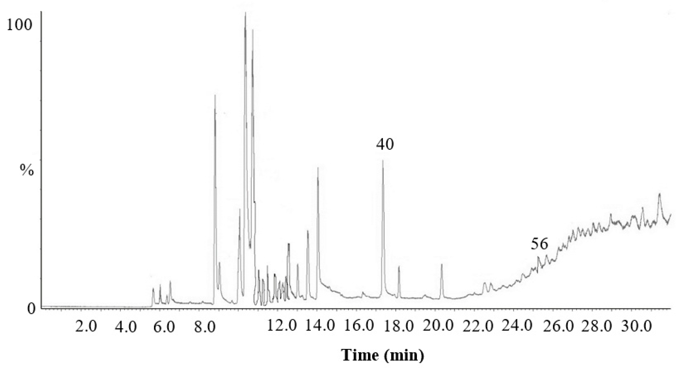
With reference to the exhaled breath, Figure 3 shows an example of the chromatogram of the same patient (n° 2; acquired in SCAN mode), before endoscopic treatment.
Sixty-six VOCs were detected (S/N ≥ 3) overall and sixty-one were identified and reported in Table 4. Benzaldehyde and methylbenzene, already evidenced in the colonic polyps and normal mucosa tissues, were also detected in the breaths of the same patients and, therefore, a possible correlation between their quantity in the breath and that released from tissues was hypothesized.
Table 5 shows the concentration ranges, estimated in this study, for these two standardly identified VOCs.

4. Discussion

In this study, seven volatile compounds, namely benzaldehyde, ethylbenzene, indole, methylbenzene, phenol, octanal, and oxime methoxy-phenyl were the most significant VOCs detected by SPME and CG-MS, for the different levels and kinetic profiles, in normal colonic mucosa and adenomatous polypoid lesion tissue of the same patient in ex vivo condition. Furthermore, benzaldehyde and methylbenzene, which are produced by both tissues, can also be detected in the expired breath of the same patients. High levels of methylbenzene (concentration range: >50 pg*mL−1) and absence/traces of benzaldehyde (concentration range: n.d.-LOD) were found in the breath of patients affected by adenomatous polypoid lesions (see Table 4). On the other hand, ethylbenzene, indole, phenol, octanal, and oxime methoxy-phenyl were not found in the corresponding expired breath samples, suggesting that they do not cross the capillary–alveolar barrier to be exhaled or that different metabolic pathways involving the intestinal microbiota could contribute to their production.
Comparing these data with those reported in another paper by our group, and performed with the same methodology, on CRC specimens, some of the VOCs highlighted in this study, such as benzaldehyde, ethylbenzene, methylbenzene, and octanal were identified in ex vivo CRC tissue samples [15]. When the kinetic profiles of these compounds are compared with those of CRC patients [15], it is evident that benzaldehyde, ethylbenzene, and methylbenzene levels are different, whereas the octanal profile looks the same. In addition, differences were also found between the concentration ranges of these compounds in polyps compared with colonic adenocarcinoma. Specifically, ethylbenzene and octanal concentrations were lower in adenomatous polypoid lesions compared to CRC, whereas methylbenzene values were comparable.
Moreover, the kinetic analysis by DI-SPME has highlighted several differences between the VOCs produced by colonic polyps and CRC cancer tissues, probably correlated to differences in the metabolic pathways involved in tumor progression. A decrease in ethylbenzene production may be related to increased consumption or binding of this compound with protein or lipid [16]. In fact, as in the literature reported, ethylbenzene undergoes α- and ω-oxidation by microorganisms, whereas the octanal metabolism could be linked to oxidative stress [17]. Klemenz et al. [16] showed that aldehydes, under different conditions, were consumed and/or bound by cultured human adipose tissue cells. In the case of benzaldehyde, Zimmermann et al. [18] described different metabolic pathways, such as tryptophan metabolism, in which this compound could be involved. Thanks to intestinal microbiota, tryptophan produces indole and a lot of its metabolites [19] precursors or signal molecules for many biologically active substances that play an important role in the “gut-brain axis” [20].
Phenol was considered for the first time in this study and showed comparable levels between normal mucosa and adenomatous polypoid lesion tissues. Phenol and its derivate, in general, because of their peroxidative capacity, could result in hematotoxic and hepatotoxic effects, facilitating mutagenesis and carcinogenesis in humans and other living organisms. Phenol is also produced during the natural decomposition of organic materials [21].
Furthermore, Oxime methoxy-phenyl is a typical chemical compound in body odor [22].
Finally, a recent paper analyzing the VOC pattern exhaled by CRC patients suggests that low levels of ethylbenzene and tetradecane, coupled with high levels of methylbenzene, could be predictive of a CRC [15]. A similar VOC pattern was also found in patients affected by colonic polyps; therefore, it could be speculated that this breath analysis might play a role as a mass screening tool since high levels of methylbenzene with no or very low traces of benzaldehyde and tetradecane seem to suggest the presence of colon neoplasia (polyp or cancer).

5. Conclusions

In this paper, an SPME-GC-MS-optimized protocol was advantageously employed to monitor ex vivo changes in endogenous VOCs produced by surgically resected colonic tissues (normal colonic mucosa and adenomatous polyp) from seven patients undergoing operative colonoscopy, to identify their secreted molecules.
To find a possible correlation between the pattern of VOCs exhaled by a patient affected by a polypoid lesion and the substances released by its colonic tissues, the breath of each patient was sampled by the ReCIVA® breath sampler, shortly before surgery, and analyzed by GC-MS.
Methylbenzene and benzaldehyde levels in the breath seemed to be connected to the presence of adenomatous colonic polypoid lesions.

Author Contributions

Conceptualization, A.M.A., N.D.V., A.P., D.F.A. and C.Z.; data curation, A.M.A. and N.D.V.; formal analysis, A.M.A., N.D.V., M.T.R., G.M., A.D.G. and J.P.; investigation, A.M.A. and N.D.V.; methodology, A.M.A., N.D.V., A.P., M.T.R., G.M., A.D.G., J.P. and C.Z.; resources, D.F.A., G.D.G. and C.Z.; supervision, D.F.A. and C.Z.; validation, A.M.A., N.D.V. and A.P.; writing—original draft, A.M.A., N.D.V., A.P. and D.F.A.; writing—review and editing, A.M.A., N.D.V., A.P., M.T.R., D.F.A., A.D., A.D.G., J.P., G.D.G. and C.Z. All authors have read and agreed to the published version of the manuscript.

Funding

This research received no external funding.

Institutional Review Board Statement

All methods were carried out in accordance with relevant guidelines and regulations. The experimental protocol was performed in compliance with the Declaration of Helsinki and approved by the ethics committee of the Azienda Ospedaliero-Universitaria Policlinico, Bari, Italy, and (cod. 0081215-29-10-2020).

Informed Consent Statement

Informed consent was obtained from all subjects involved in the study.

Data Availability Statement

The datasets used and/or analyzed during the current study are available from the corresponding author upon reasonable request.

Conflicts of Interest

The authors declare no conflict of interest.

References

  1. Zambonin, C.; Aresta, A. Review: Recent applications of solid phase microextraction coupled to liquid chromatography. Separations 2021, 8, 34. [Google Scholar] [CrossRef]
  2. Boiko, B. Solid-phase microextraction: A fit-for-purpose technique in biomedical analysis. Anal. Bioanal. Chem. 2022, 414, 7005–7013. [Google Scholar]
  3. Aresta, A.; Cotugno, P.; De Vietro, N.; Massari, F.; Zambonin, C. Determination of polyphenols and vitamins in wine-making by-products by supercritical fluid extraction (SFE). Anal. Lett. 2020, 53, 2585–2595. [Google Scholar] [CrossRef]
  4. Aresta, A.; Di Grumo, F.; Zambonin, C. Determination of major isoflavones in soy drinks by solid-phase micro extraction coupled to liquid chromatography. Food Anal. Methods 2016, 9, 925–933. [Google Scholar] [CrossRef]
  5. De Vietro, N.; Aresta, A.; Rotelli, M.T.; Zambonin, C.; Lippolis, C.; Picciariello, A.; Altomare, D.F. Relationship between cancer tissue derived and exhaled volatile organic compound from colorectal cancer patients. Preliminary results. J. Pharm. Biomed. Anal. 2020, 180, 113055–113065. [Google Scholar] [CrossRef] [PubMed]
  6. Musteata, F.; Musteata, M.; Pawliszyn, J. Fast in vivo microextraction: A new tool for clinical analysis. Clin. Chem. 2006, 52, 708–715. [Google Scholar] [CrossRef] [PubMed]
  7. Piri-Moghadam, H.; Ahmadi, F.; Gómez-Ríos, G.A.; Boyacı, E.; Reyes-Garcés, N.; Aghakhani, A.; Bojko, B.; Pawliszyn, J. Fast quantitation of target analytes in small volumes of complex samples by matrix-compatible solid-phase microextraction devices. Angew. Chem. Int. Ed. Engl. 2016, 55, 7510–7514. [Google Scholar] [CrossRef]
  8. Jaroch, K.; Taczyńska, P.; Czechowska, M.; Bogusiewicz, J.; Łuczykowski, K.; Burlikowska, K.; Bojko, B. One extraction tool for in vitro-in vivo extrapolation? SPME-based metabolomics of in vitro 2D, 3D, and in vivo mouse melanoma models. J. Pharm. Anal. 2021, 11, 667–674. [Google Scholar] [CrossRef]
  9. Arnold, M.; Sierra, M.S.; Laversanne, M.; Soerjomataram, I.; Jemal, A.; Bray, F. Global patterns and trends in colorectal cancer incidence and mortality. Gut 2017, 66, 683–691. [Google Scholar] [CrossRef]
  10. Siegel, R.L.; Miller, K.D.; Goding Sauer, A.; Fedewa, S.A.; Butterly, L.F.; Anderson, J.C.; Cercek, A.; Smith, R.A.; Jemal, A. Colorectal cancer statistics. Cancer J. Clin. 2020, 70, 145–164. [Google Scholar] [CrossRef]
  11. Armstrong, H.; Bording-Jorgensen, M.; Wine, E. Review: The multifaceted roles of diet, microbes, and metabolites in cancer. Cancers 2021, 13, 767. [Google Scholar] [CrossRef] [PubMed]
  12. Aarons, C.B.; Shanmugan, S.; Bleier, J.I.S. Management of malignant colon polyps: Current status and controversies. World J. Gastroenterol. 2014, 20, 16178–16183. [Google Scholar] [CrossRef] [PubMed]
  13. Altomare, D.F.; Porcelli, F.; Picciariello, A.; Pinto, M.; Di Lena, M.; Caputi Iambrenghi, O.; Ugenti, I.; Guglielmi, A.; Vincenti, L.; De Gennaro, G. The use of the PEN3 e-nose in the screening of colorectal cancer and polyps. Tech. Coloproctol. 2016, 20, 405–409. [Google Scholar] [CrossRef] [PubMed]
  14. Altomare, D.F.; Picciariello, A.; Rotelli, M.T.; De Fazio, M.; Aresta, A.M.; Zambonin, C.G.; Vincenti, L.; Trerotoli, P.; De Vietro, N. Chemical signature of colorectal cancer: Case–control study for profiling the breath print. BJS Open 2020, 4, 1189–1199. [Google Scholar] [CrossRef] [PubMed]
  15. De Vietro, N.; Aresta, A.M.; Picciariello, A.; Rotelli, M.T.; Zambonin, C. Determination of vocs in surgical resected tissues from colorectal cancer patients by solid phase microextraction coupled to gas chromatography–mass spectrometry. Appl. Sci. 2021, 11, 6910. [Google Scholar] [CrossRef]
  16. Klemenz, A.C.; Meyer, J.; Ekat, K.; Bartels, J.; Traxler, S.; Schubert, J.K.; Kamp, G.; Miekisch, W.; Peters, K. Differences in the emission of volatile organic compounds (vocs) between non-differentiating and adipogenically differentiating mesenchymal stromal/stem cells from human adipose tissue. Cells 2019, 8, 697. [Google Scholar] [CrossRef]
  17. Berthe-Corti, L.; Fetzner, S. Bacterial metabolism of n-alkanes and ammonia under oxic, suboxic and anoxic conditions. Acta Biotechnol. 2002, 22, 299–336. [Google Scholar] [CrossRef]
  18. Zimmermann, D.; Hartmann, M.; Moyer, M.P.; Nolte, J.; Baumbach, J.I. Determination of volatile products of human colon cell line metabolism by GC/MS analysis. Metabolomics 2007, 3, 13–17. [Google Scholar] [CrossRef]
  19. Zhang, Q.F.; Xiao, H.M.; Zhan, J.T.; Yuan, B.F.; Feng, Y.Q. Simultaneous determination of indole metabolites of tryptophan in rat feces by chemical labeling assisted liquid chromatography-tandem mass spectrometry. Chin. Chem. Lett. 2022, 33, 4746–4749. [Google Scholar] [CrossRef]
  20. Morais, L.H.; Schreiber, H.L.T.; Mazmanian, S.K. The gut microbiota–brain axis in behaviour and brain disorders. Nat. Rev. Microbiol. 2021, 19, 241–255. [Google Scholar] [CrossRef]
  21. Michałowicz, J.; Duda, W. Phenols—Sources and Toxicity. Pol. J. Environ. Stud. 2007, 16, 347–362. [Google Scholar]
  22. Jh, S.K.; Hayashi, K. Molecular structural discrimination of chemical compounds in body odor using their GC–MS chromatogram and clustering methods. Int. J. Mass Spectrom. 2017, 423, 1–14. [Google Scholar] [CrossRef]
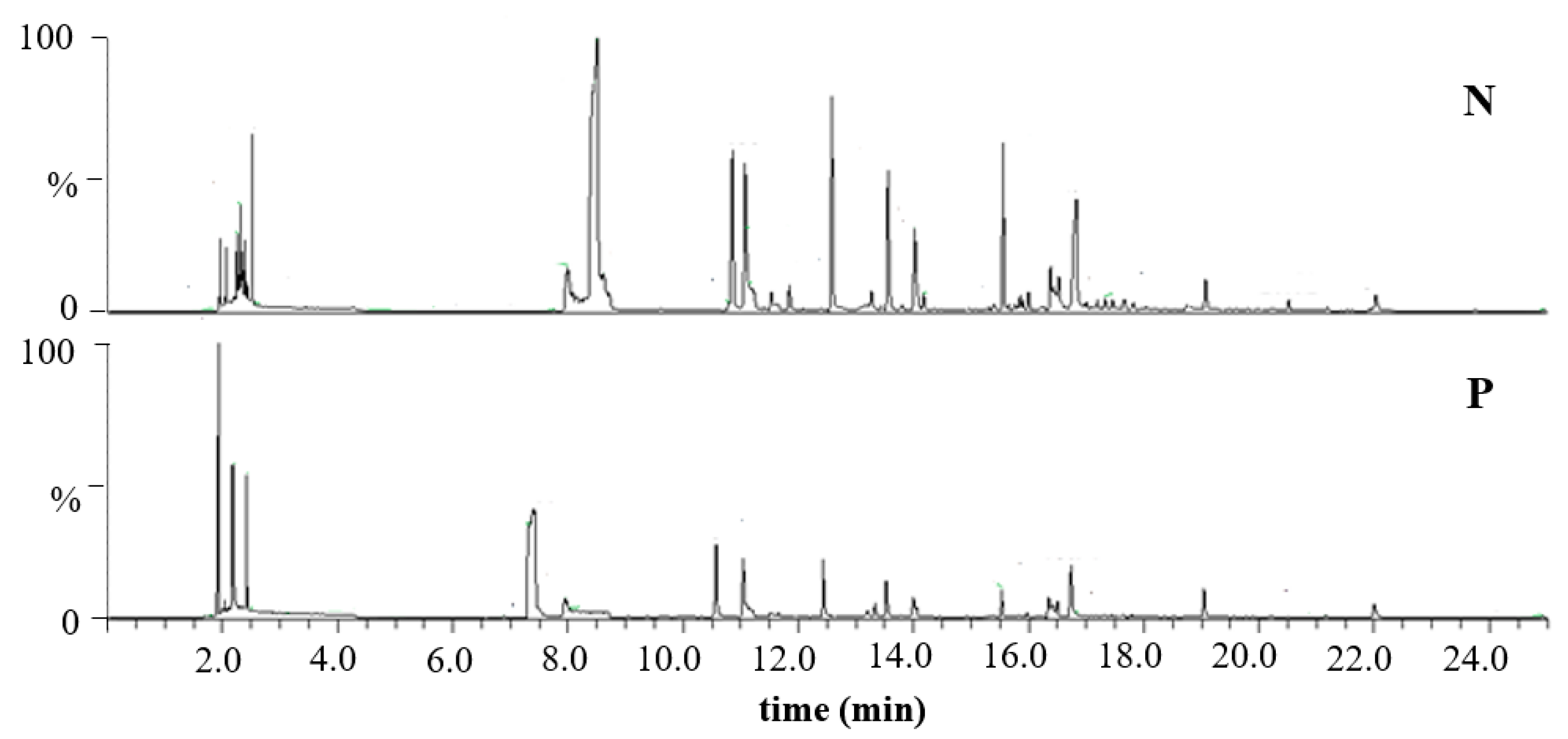
Figure 1. HS-SPME-GC-MS chromatograms of VOCs released by colon normal mucosa (N) and adenomatous polyp (P) tissues from the same patient (n° 2) at time zero.
Applsci 13 06083 g001
Figure 2. Time-dependent monitoring (0, 1, 4, and 7 days) peak area of benzaldehyde, ethylbenzene, indole, methylbenzene, phenol, octanal, and oxime methoxy-phenyl by DI-SPME-GC-MS in tissue samples (normal colonic mucosa and adenomatous polyp) from the same patient (n° 2), before undergoing curative surgery.
Applsci 13 06083 g002
Figure 3. Chromatographic profile (SCAN mode) of the exhaled breath of patient (n° 2) affected by an adenomatous colonic polypoid lesion before endoscopic treatment.
Applsci 13 06083 g003
Table 1. Demographics and co-morbidities of adenomatous colonic polypoid lesion-affected patients (n = 7) and polyp characteristics.
Demographics and Co-Morbidities of Adenomatous Colonic Polypoid Lesion-Affected Patients (n = 7)
Mean Age (years)63
Sex ratio (M:F)5:2
Hypertension2
Diabetes0
Hypothyroidism0
Smoker1
Polyp characteristics
Polyp size2.0 ± 0.5 cm
Hystology7 adenomatous polyps
Grading2 moderate and 7 severe dysplasia
Table 2. Lists of compounds, RT, characteristic ions (m/z), match factor, probability, standard identity confirmation, and frequency of appearance in the acquired HS- and DI-SPME-GC-MS chromatograms related to secreted compounds by normal colonic mucosa and adenomatous polypoid lesion.
#RT (min)CompoundCharacteristic Ions (m/z)MatchProb.
(%)
Standard Identity ConfirmationFrequency (%)
HSDI
12.02 ± 0.04Dimethyl chloroacetal4778275.5 3.13.1
22.21 ± 0.09Acetaldehyde oxime14, 5996237.3 12.528.1
33.03 ± 0.022-Butanone,4-hydroxy43, 6167439.1 3.10
43.30 ± 0.081-Butanol41, 5682740.0 5.63.1
55.54 ± 0.05Acetal73, 10384977.0 12.56.3
66.06 ± 0.071-Butanol,3-methyl41, 5586720.5 03.1
76.19 ± 0.09Disulfide, dimethyl45, 7993997.1 9.412.5
86.84 ± 0.07Methylbenzene9197960yes93.896.9
98.82 ± 0.08Ethylbutanoate43, 7187790.0 3.13.1
108.98 ± 0.07Ethyl 2-methyl butanoate57, 10285176.8 18.83.1
119.10 ± 0.08Ethyl 3-methyl butanoate88, 11582488.5 12.53.1
129.29 ± 0.09Ethylbenzene91, 10695471.6yes68.831.3
139.53 ± 0.10xylene91, 10690861.5yes46.33.1
1410.07 ± 0.09xylene91, 10678830.2yes46.99.4
1510.22 ± 0.09Pentanoic acid, ethylester57, 10181289.2 03.1
1610.71 ± 0.09Oxime, methoxy-phenyl133, 15181384.2 68.896.9
1711.78 ± 0.09Dimethyl trisulfide79, 12684996.9 12.5.6.3
1811.93 ± 0,09Benzaldehyde77, 10693092yes18.865.6
1912.12 ± 0.09Phenol66, 9498488yes31.362.5
2012.29 ± 0.08Octanal43, 5688860yes9.459.4
2112.52 ± 0.081-Hexanol,
2-ethyl-
5782913.0 21.931.3
2212.74 ± 0.09Isooctanol55, 11280020.3 21.912.5
2313.47 ± 0.09Nonanol57, 6982711.1yes25.025.0
2414.05 ± 0.09Nonanal41 yes3.13.1
2515.11 ± 0.08Octanoic acid60, 14478130.9yes12.59.4
2615.31 ± 0.08Decanal43, 138 yes31.315.6
2715.63 ± 0.08Dodecane43, 17095649yes25.09.4
2816.11 ± 0.09Benzenepropanol117, 13690960.0 6.318.8
2916.61 ± 0.09Triethanolamine11890067.0 06.3
3016.86 ± 0.09Undecano57, 15690844yes21.93.1
3117.07 ± 0.08Indolo11798270yes40.662.5
3218.39 ± 0.09Tetradecane57, 19893453yes25.012.5
Table 3. Linear regression analysis and concentration ranges of VOCs in culture media with colon tissues (normal mucosa and adenomatous polypoid lesion) from seven selected patients.
CompoundsEquationR2Normal Mucosa
Range (mg/mL)
Adenomatous Polypoid Lesion Range (mg/mL)
BenzaldehydeY = 1912X + 4560.9995nd-1.43 ± 0.62nd-1.60 ± 0.70
EthylbenzeneY = 2358X + 29980.9988nd-1.52 ± 0.23nd-0.96 ± 0.12
IndoleY = 807X + 4080.9995nd-7.03 ± 1.05nd-13.58 ± 1.12
MethylbenzeneY = 17,243X + 18070.9990nd-1.29 ± 0.16nd-4.95 ± 0.11
PhenolY = 861X + 7340.9902nd-3.45 ± 0.05nd-3.11 ± 0.04
OctanalY = 2090X + 5970.9998nd-0.78 ± 0.30nd-0.27 ± 0.13
Table 4. Lists of compounds, RT, characteristic ions (m/z), match factor, probability, and standard identity confirmation detected (S/N ≥ 3) in the breath of the seven patients with adenomatous colonic polypoid lesion, before endoscopic treatment.
#RT (min) aCommon Compound NameMatch
(%)
Probability
(%)
Standard Identity Confirmation b
15.71 ± 0.05Carbon dioxide89190
26.04 ± 0.02Unidentified
36.45 ± 0.062,4-Dimethyl pentane93091
46.52 ± 0.07Hexene87989
56.66 ± 0.08Sulfur dioxide87887
66.78 ± 0.09Difluoro methyl-silane80152
76.82 ± 0.06Trimethyl silylanol77355
86.93 ± 0.06Ethane, 1,2-diethoxy80161
97.01 ± 0.031-Pentene-4-methyl82254
107.20 ± 0.092-Propane83360yes
117.61 ± 0.081,1,1,1-Trifluoro trimethyl-silylanol82856
127.94 ± 0.05Cyclobutanol90378
138.37 ± 0.05Trichloro-monofluoro-methane82257
148.95 ± 0.061,3-Pentadiene95475
159.12 ± 0.062-Propanol-1-methoxy93080
169.77 ± 0.02Unidentified
1710.11 ± 0.042-Pentene91585
1810.24 ± 0.052-Butanol-3-methyl90784
1910.31 ± 0.062-Methyl pentanal83958
2010.54 ± 0.05Cyclopentane90388
2110.83 ± 0.052,3-Dimethyl pentane 66
2210.91 ± 0.03Hexane91392yes
2311.00 ± 0.034-Methyl-2-pentyne877
2411.44 ± 0.06Acetonitrile92090yes
2511.52 ± 0.02Unidentified
2611.63 ± 0.08Benzene93889yes
2712.42 ± 0.05Unidentified
2812.91 ± 0.051,3,5-Trifluoro benzene85257
2913.27 ± 0.03Dichloromethane93193yes
3013.55 ± 0.06Hexamethyl disiloxane82881
3113.82 ± 0.042-Butanone94896yes
3214.13 ± 0.07Heptene89988yes
3314.33 ± 0.023-Hexanol86677
3414.96 ± 0.04Acetic acid91567
3515.90 ± 0.052-Propanol-1-methoxy83852
3616.49 ± 0.031,4-Dioxane82851
3716.61 ± 0.052-Pentanone90389
3816.70 ± 0.03Butanoic acid93397yes
3917.42 ± 0.06Cyclotrisiloxane hexamethyl80758
4017.51 ± 0.04Methyl benzene93897yes
4118.27 ± 0.06Octine90770yes
4218.40 ± 0.052-Hexanone88168
4319.66 ± 0.05Hexanal
4419.80 ± 0.05Methyl isobutyl ketone90276
4520.00 ± 0.07Hexanoic acid, methyl ester87483
4620.16 ± 0.04Nonane93454yes
4720.32 ± 0.08Pentanoic acid, methyl ester87979
4820.53 ± 0.05Pentanoic acid80954yes
4922.00 ± 0.07Di(isobutyl)acetone81558
5022.49 ± 0.07Hexanoic acid87979yes
5122.95 ± 0.043-Heptanone91882yes
5223.02 ± 0.06Heptanoic acid, methyl ester98883
5323.55 ± 0.03Eptane, 2,2,4,6,6-pentamethyl88855
5423.99 ± 0.06Tetrasiloxane, decamethyl84851
5525.10 ± 0.03Butanoic acid, dimethyl ester85574
5625.52 ± 0.05Benzaldehyde93395yes
5725.93 ± 0.06Octanoic acid, methyl ester83268
5826.26 ± 0.07Decane93255yes
5926.88 ± 0.06Benzoic acid, methyl ester81554
6027.54 ± 0.081-Decanol-2-esil87753
6128.33 ± 0.06Dodecane92854yes
6229.00 ± 0.08Unidentified
6329.65 ± 0.06Silane, ethyl-dimethyl-phenyl81362
6429.83 ± 0.074-Phenyl benzofurane82256
6530.51 ± 0.06Tri-tetra-contane81256
6631.53 ± 0.04Pentacosane81158
a Values expressed as mean (s.d.); b authenticated using the NIST library and standard injection.
Table 5. Concentration ranges of VOCs exhaled by seven selected patients affected by adenomatous colonic polypoid lesion, before curative surgery.
Compoundspg*mL−1 in Exhaled Breath
Benzaldehyden.d.-LOD
Methylbenzene>50
Disclaimer/Publisher’s Note: The statements, opinions and data contained in all publications are solely those of the individual author(s) and contributor(s) and not of MDPI and/or the editor(s). MDPI and/or the editor(s) disclaim responsibility for any injury to people or property resulting from any ideas, methods, instructions or products referred to in the content.