Article

Ophthalmic Manifestation in Neurofibromatosis Type 2

by
Marta Armentano
1,†,
Luca Lucchino
1,†,
Ludovico Alisi
1,
Antonio Valerio Chicca
1,
Valerio Di Martino
1,
Emanuele Miraglia
2,
Ludovico Iannetti
3,
Anna Maria Comberiati
3,
Sandra Giustini
2,
Alessandro Lambiase
1,* and
Antonietta Moramarco
1
1
Department of Sense Organs, Sapienza University of Rome, Policlinico Umberto I, Viale del Policlinico 155, 00161 Rome, Italy
2
Department of Dermatology and Venereology, Sapienza University of Rome, Policlinico Umberto I, 00161 Rome, Italy
3
Ophthalmology Unit, Head and Neck Department, Policlinico Umberto I University Hospital, Sapienza University of Rome, 00161 Rome, Italy
*
Author to whom correspondence should be addressed.
These authors contributed equally to this work.
Appl. Sci. 2023, 13(10), 6304; https://doi.org/10.3390/app13106304
Submission received: 19 March 2023 / Revised: 8 May 2023 / Accepted: 18 May 2023 / Published: 22 May 2023

Abstract

:
Neurofibromatosis type 2 (NF2) is a genetically determined tumor-predisposing syndrome. Ocular manifestations include cataracts, epiretinal membranes, retinal hamartomas, optic disk gliomas, and optic nerve sheath meningiomas. Moreover, optic disk edema, optical atrophy, motility disorders, pupil and lid dysfunction, and neurotrophic keratitis can be observed as indirect signs. An observational study was conducted with the aim to collect clinical data and describe the most frequent NF2 ocular manifestations. Fourteen patients affected by NF2, according to the Manchester criteria, were enrolled. All patients underwent complete ophthalmologic and orthoptic evaluation and a spectral domain optical coherence tomography. Ocular manifestations were present in all patients. The slit lamp evaluation of the anterior segment highlighted cataracts in five patients, keratitis in two patients, corneal leukoma in two patients, and corneal pannus in one patient. Fundus oculi and OCT evaluation identified epiretinal membranes in four patients, vitreoretinal tufts in three patients, optic nerve edema in one patient, and retinal hamartoma in one patient. Moreover, the orthoptic evaluation identified different types of ocular motility disorders in seven patients. This is a descriptive study of a rare disease with poor previous literature. Clinical data are shown, emphasizing the role of NF2-specific ophthalmological and orthoptic findings to help establish an early diagnosis.

1. Introduction

In 1882, von Recklinghausen described and classified neurofibromatosis type 1, neurofibromatosis type 2 (NF2), and schwannomatosis, under the common term “Neurofibromatosis” [1]. NF2 was later identified as a separate clinical entity by Gardner [2]. Neurofibromatosis type 2 is a tumor-predisposition syndrome characterized by the development of multiple schwannomas and meningiomas. The incidence is about 1 in 25,000 with a penetrance of 95%. The disease prevalence is estimated at 1 in 60.000. Thanks to earlier diagnoses and treatment developments, the life expectancy of these patients has improved, and the incidence of complications has decreased [3]. NF2 is caused by the inactivating mutations of the NF2 gene, located at q12.2 of chromosome 22, that result in a deficiency of the Merlin protein, a cytoskeletal protein with tumor suppressor properties [4].
NF2 is characterized by the development of bilateral vestibular schwannomas (VS), which lead to hearing loss, tinnitus, and imbalance. Other typical tumor types are schwannomas involving cranial, spinal, and peripheral nerves, and multiple meningiomas, both intracranial and intraspinal. Moreover, low-grade central nervous system (CNS) malignancies, such as ependymomas and gliomas, and skin tumors can occur [5].
The Manchester criteria, introduced in 1992, are the best-known diagnostic criteria for NF2 [6]. According to these criteria, a patient with suspicion of NF2 must meet one of the following clinical features:
  • Bilateral vestibular schwannomas;
  • First-degree relative with NF2 and unilateral vestibular schwannomas;
  • First-degree relative with NF2 or unilateral vestibular schwannomas and two among meningioma, cataract, glioma, neurofibroma, schwannoma, cerebral calcification;
  • Multiple meningiomas and two among unilateral vestibular schwannomas, cataract, glioma, neurofibroma, schwannoma, and cerebral calcification.
These criteria have been revised many times over the years. Recently, updates have been made, to distinguish between major and minor criteria and consider genetic analysis [7]. Specifically, the diagnostic criteria for NF2 have been recently expanded from the original Manchester criteria to include LZTR1 mutation testing in individuals with unilateral VS and other schwannomas. Moreover, two further changes were introduced, including NF2 mutational testing and the insertion of an age limitation of 70 for the development of bilateral VS if no other NF2 features are present.
NF2 phenotype severity is difficult to predict, and clinical manifestations appear variable among patients. Halliday et al. introduced a genetic severity score to predict the clinical expression of NF2. This score allows the connection of different genetic characteristics to a specific clinical disease presentation. This system consents to the individuation of tissue mosaicism form, classic and severe disease [8]. Interestingly, the genetic severity subtype correlated to NF2-related eye disease. Specifically, mutations associated with severe systemic disease resulted in greater visual morbidity at an earlier age.
To date, ocular pathologic findings of NF2 have been reported in a few retrospective series. A wide spectrum of ocular manifestations has been recognized to be specifically linked to NF2. Different clinical manifestations can occur in different age groups. Congenital cataracts, optic disc anomalies, and retinal hamartomas are responsible for the failure of binocular vision development in childhood. In middle-aged patients, a frequent cause of blindness is optic nerve sheath meningioma. In adulthood, visual functions can be compromised by ERMs or compression of the optic nerve due to intracranial tumors with the elevation of intracranial pressure. Visual impairment can also develop due to indirect NF2 complications, such as third, fourth, or sixth cranial nerve palsy, optic atrophy, or neurotrophic keratitis secondary to facial palsy [9,10]. This study aims to offer new insights into ophthalmological and orthoptic findings in patients with NF2 in the context of the paucity of available literature. We believe this work will raise awareness of this rare disease possibly leading to earlier diagnosis and assisting clinicians in patient care.

2. Materials and Methods

This single-center, observational, cross-sectional study was conducted at the University of Rome ‘Sapienza’, Umberto I Hospital, Italy, from June 2020 to February 2022. The research followed the Tenets of the Declaration of Helsinki, and written informed consent was obtained from all subjects and from parents in case of minor age. We included 14 consecutive patients between 16 and 78 years of age (mean age: 52 ± 10.1 years) with a diagnosis of NF2 based on the Manchester criteria [11,12]. NF2 patients were enrolled at the Regional Reference Centre for Rare Neurocutaneous Diseases of the Umberto I Hospital. All patients underwent 3-Tesla magnetic resonance imaging (MRI) of the brain, orbits, and spinal cord with gadolinium to assess the presence of vestibular schwannomas, schwannomas, and intracranial or spinal meningiomas.
Each subject underwent a detailed ophthalmological examination, including full medical history, measurement of best corrected visual acuity (BCVA) with Snellen’s optometric chart at 5 m, biomicroscopic examination of the anterior segment, Goldmann applanation tonometry, indirect fundus biomicroscopy and spectral domain optical coherence tomography (SD-OCT). SD-OCT scans were obtained with the Spectralis OCT (Spectralis Family Acquisition Module, V 5.1.6.0; Heidelberg Engineering, Heidelberg, Germany) with the Heidelberg Eye Explorer (V1.6.2.0), whose axial resolution was 3.5 μm and transverse resolution was approximately 15/20 μm, using both the raster scan protocol (20° × 15°, 19 lines of scan) and the radial scan protocol (20°, 6 lines of scan), centered on the fovea. The orthoptic evaluation included inspection of eyes and head, Irvine test, cover–uncover test and alternate cover test, evaluation of ocular motility, assessment of objective convergence, and red filter test for diplopia.

3. Results

Demographic and relevant clinical features are listed in Table 1. In our study, the mean age was 53.1 years (range, 16 to 78 years) and five patients were males (10 eyes).
Ophthalmological manifestations were present in almost all patients and ranged widely from subtle retinal alterations, identified only by SD-OCT, to severe ocular involvement present at birth. The direct examination of ocular adnexa showed lagophthalmos in four patients (four eyes), eyelid ptosis in two patients (two eyes), and Duane syndrome type 1 in one patient (Table 2). One patient underwent tarsorrhaphy to treat severe lagophthalmos. In all cases, lagophthalmos was secondary to facial nerve palsy after surgical asportation of VS. All eyes with lagophthalmos developed exposure keratopathy, with reported two patients (two eyes) showing superficial punctate keratitis, two patients (two eyes) presenting with corneal opacification. One patient (one eye) developed vascularized corneal opacity due to recurrent infections. (Table 2).
The slit lamp evaluation of the anterior segment highlighted cataracts in five patients (six eyes). Three of them had posterior subcapsular cataracts, and two showed a cortico-nuclear type. Two patients aged 30 and 44 years were already pseudophakic. The IOP values were within the normal reference values in all patients (10–21 mmHg).
Fundus examination revealed the presence of epiretinal membranes in four patients (six eyes). Specifically, one patient (one eye) presented a grade 0 ERM, one (one eye) had a grade 1 and the other two (four eyes) had grade 2 ERMs according to the Gass classification (Figure 1A). Three patients (three eyes) showed the presence of retinal tuft (Figure 1B) and one patient was diagnosed with unilateral retinal hamartoma. MRI scans revealed no significant structural abnormality compatible with optic pathway gliomas or optic nerve sheath meningiomas. In addition MRI showed no signal change in the cranial nerves; however, one patient presented with bilateral optic nerve edema (Figure 2) associated with intracranial hypertension secondary to multiple compressive intracranial tumors, in particular meningiomas (Table 3).
The orthoptic evaluation highlighted strabismus in 50% of our sample (seven patients). In detail, three patients (43%) presented microstrabismus, three patients (43%) exotropia, and one patient (14%) esotropia (Table 4). Diplopia was not observed in any case, and according to the orthoptic examinations, the three cases of exotropia were comitant. Moreover, one patient (7%) presented bilateral co-contraction of medial and lateral rectus, resulting in enophthalmos, reduced eyelid rim, and pseudo-ptosis. This clinical presentation was compatible with the diagnosis of Duane syndrome type 1. Physiological nystagmus in extreme gaze positions was found in eight patients (57%), three of them were orthophoric, and the remaining patients had a manifest ocular deviation.

4. Discussion

Type 2 neurofibromatosis is a complex, genetically determined pathology with a variable spectrum of manifestations. Genetical features have been characterized over time and brought together in a complex genetic severity score, which corresponds to clinical features. The tissue mosaic form (presumed and confirmed) includes patients with clinical signs of NF2 not confirmed by blood tests or patients with genetic alterations detectable only in specific tissues. The classic form includes mild and moderate NF2 clinical expression, with typical mutation identified in blood cells. The severe forms imply the presence of specific full truncating mutations involving exons 2–13, leading to aggressive phenotypes [8]. Age of diagnosis, age of onset, and the number of intracranial tumors, and mutation type remain the three major predictive factors of mortality in NF2 patients. Missense mutations have been linked to a lower risk of mortality. Moreover, clinical manifestations are more variable in patients with splice-site mutations [13]. In more than half of the cases, the most frequent symptoms at presentation are related to a vestibular schwannoma of the VIII cranial nerve. Spinal tumors are diagnosed in about 90% of patients, especially meningiomas in about 50% of patients [14].
The mild form of the disease (Gardner variant) occurs mainly with hearing loss (vestibular schwannoma) and presents a slower course and better prognosis than the severe early-onset form (Wishart variant). In the severe form, the ocular manifestations identified in childhood can lead to an early diagnosis of the disease as they often precede the other systemic manifestations. In the mild form, since the life expectancy of patients is 79 years (46 years in the severe form), the early identification of ocular manifestations is fundamental for preserving good visual function with targeted interventions [8].
In type 2 neurofibromatosis, the ocular involvement is frequent. In 2006, Bosch first classified the ocular features of the disease into NF2-specific and NF2-associated. In the NF2-specific group, he included posterior and cortical subcapsular/capsular cataracts (67% of patients), epiretinal membranes (40% of patients), retinal hamartomas (3% of patients), gliomas of the optic disc (13% of patients), and optic nerve meningiomas (27% of patients). In the NF2-associated group disc edema, optic atrophy, motility disorder, pupil dysfunction, lid dysfunction, reduced corneal sensation, exophthalmos, and exposure or neurotrophic keratopathy are included. The manifestations inserted in the latter group are caused by direct or indirect damage to the nervous structures determined by tumors [9].
Most recruited patients (13 out of 14) had late onset of symptoms (>20 years), corresponding to the mild form of the disease (Gardner variant). Among our cohort, the severe early onset form was identified in a single 16-year-old patient, who presented ERMs and subcapsular cataract, typically associated with the severe form.
In our study, in accordance with Bosch et al. the most frequently reported sign was cataract [9], which remains the only ophthalmological finding included among the NF2 diagnostic criteria [11].
Specifically, five of our patients (35.7%) presentedcataract, and two patients (14.3%) already underwent cataract surgery, amounting to seven patients (50%). Considering the young age of five of these seven patients, the development of cataract in their case can be plausibly attributed to NF2. We can underline that these patients presented a posterior subcapsular cataract, which is the form most frequently associated with the disease [15]. The other two patients with cataracts were, respectively 73 and 78 years old and the age of onset was not clear from the history, therefore it cannot be safely stated that the lens opacities in these patients are caused by NF2 or senile age.
Epiretinal membranes were the second most common sign in our sample after cataracts (28.6% of patients). ERMs in NF2 patients, at OCT evaluation, have atypical and distinctive characteristics, such as edges that project anteriorly into the vitreous despite an incomplete posterior vitreous detachment, a lack of cystoid macular edema, and an irregular and partially absent ILM [16]. These features distinguish it from idiopathic ERM or membranes associated with other diseases [16,17,18]. Histopathological studies highlighted that these abnormalities consist mainly of a mixture of Muller cells and astrocytes [19], supporting the hypothesis that ERMs represent hamartomas composed of Muller cells and justifying their morphological appearance [16]. OCT is the most sensitive method for studying this type of lesion and has allowed us to detect forms of various degrees: one of grade 0, one of grade 1, and four of grade 2 (macular pucker), according to the Gass classification [20]. Two young patients with macular pucker, aged 16 and 37, also showed subcapsular cataracts. The 16-year-old patient, as previously mentioned, was affected by a severe form of NF2. This observation agrees with several authors who showed a direct relationship between the prevalence of NF2-specific eye findings, the age of onset, and the severity of the genetic defect [8,9].
Another relevant finding detected in 21.4% of our patients were retinal tufts. This percentage appears to be inferior when compared with the recent literature. In a case–control study, Emmanouil et al. reported retinal tufts as the most common abnormalities of the central retina in 23 patients (43%), principally observed in patients with a severe form and not seen in controls, therefore showing a positive predictive value of 100% [21]. This difference can be explained because most of our patients have a mild form of the disease, as mentioned above.
Interestingly, Waisberg et al. reported the presence of choroidal abnormalities in NF2 patients. Choroidal abnormalities, visible as multiple, bright, patchy nodules in near-infrared reflectance (NIR) OCT, have recently been added to the revised diagnostic criteria for neurofibromatosis type 1 based on their high specificity and sensitivity [22]. In our sample, we have not found compatible lesions. Further studies on choroidal characterization in NF2 are needed [23,24].
As already mentioned, other ocular manifestations associated with NF2 are papilledema, optic atrophy, neurotrophic keratopathy, corneal keratitis and opacity, and ocular and eyelid motility disorders [9]. Among the non-specific ocular manifestations in our patients, we found changes affecting the cornea, eyelids, extrinsic muscles of the eye, and the optic nerve.
We highlighted eyelid ptosis in two patients (14.3%), lagophthalmos in four patients (28.6%), and strabismus in seven patients (50%). Eyelid ptosis is reported by Egan and other authors as a secondary manifestation of NF2, resulting from hypofunction of the oculomotor nerve [25]. Lagophthalmos is generally linked to post-surgical facial paralysis, following the removal of acoustic schwannomas. [9] This clinical feature was observed in our patients as a consequence of post-surgical VII nerve paralysis.
Corneal involvement was characterized by superficial punctate keratitis (two patients 14.3%), corneal leukoma (two patients 14.3%), and corneal pannus (one patient 7.1%). A post-surgical lagophthalmos, due to the removal of acoustic nerve neurinomas, was responsible for all these clinical conditions, as the incomplete closure of the eyelid rim determined corneal epithelial suffering.
One of our patients presented papilledema, due to increased intracranial pressure (7.1%). Bosch et al. described in their paper a 30% prevalence of optic disk edema, mostly caused by intracranial tumors or optic disk sheath meningiomas. The lowering of intracranial pressure, thanks to the surgical removal of cancerous masses, is not always sufficient to revert the progressive optic neuropathy. In these cases, patients can develop optic disk atrophy with irreversible loss of vision [9]. In the literature, papilledema is described in a small percentage of NF2 patients (<10%). The most frequent cerebral neoplasms associated with swelling of the optic disk are tumors in the posterior fossa, cranial nerve schwannomas, or large supratentorial meningiomas [26]. Our patient was diagnosed with multiple intra and extra-axial large and recurrent meningiomas, which required numerous surgical procedures.
Among our cohort, we also identified one case of retinal hamartoma (7.1%) in a patient with a moderate form of NF2. The lesion had a nasal peripapillary localization and was observable both during the fundoscopic examination and by OCT imaging. In the literature, retinal hamartomas have been reported with a variable frequency of 6–22% in NF2 patients [27]. These findings can be observed at the fundoscopic examination as whitish lesions, often involving the juxta papillary or macular area. OCT images are helpful to better characterize the extension and depth of such lesions. Retinal hamartomas are not specifically linked to NF2 but can be observed in other phakomatoses as well. Moreover, the prevalence of retinal hamartomas is higher in severe forms of NF2, often leading to a diagnosis based on ocular findings [28]. Parry et al. suggested that the presence of retinal hamartomas can be considered a clinical indicator of intermediate/severe forms of this pathology [5]. Furthermore, a frequent association between pigment epithelial and retinal hamartomas has been described [9,29].
Many of the reported manifestations can affect visual acuity. Within our sample, ERMs showed the greatest impact on BCVA, mainly when the fovea was involved, as we observed in one of our patients who had a BCVA of 0.03. Other causes of visual impairment in our NF2 patients were corneal opacities and cataracts.
Regarding ocular motility, in 2008, in a retrospective study, Feucht observed strabismus in more than half of patients with NF2, mostly determined by cranial nerve palsy (III-IV-VI) [30]. In the available literature, many studies identified ocular motility disorders in NF2 patients. Painter et al. retrospectively reviewed 83 patients affected by genetically characterized NF2. They diagnosed partial third nerve palsy in three children with severe mutation. Neuroimaging confirmed a third nerve schwannoma in all cases. Moreover, one patient developed a partial fourth nerve palsy [10]. Feucht et al. carried out a retrospective study of 73 NF2 patients. Different kinds of ocular motility disorders were identified in 52% of patients. About 22% of these patients presented cranial nerve palsy, due, in most cases, to intracranial tumors, and 26% showed concomitant strabismus, mainly horizontal [30]. Reggae et al. conducted a cross-sectional study on 49 NF2 patients, identifying oculomotor and abducens nerve palsy in six patients and concomitant strabismus in three patients [31]. Egan et al. identified monocular elevator paresis in four patients out of 29 affected by NF2, with dysfunction of both the superior rectus and inferior oblique muscles [25].
Barrett et al. described in a case report a two-year-old patient with recurrent third nerve palsy associated with schwannoma of the subarachnoid portion of the right third nerve [32]. Sokwala et al. described the case of a 21-year-old NF2 patient with a superior oblique muscle palsy associated with a sphenoid wing meningioma extended into the orbit [33].
The frequency of strabismus in our group was 50%, (three with microstrabismus, three with exotropia, and one with esotropia). The three cases of exotropia we described showed characteristics of comitant strabismus and were most likely attributable to a decompensated intermittent exotropia. Feucht et al. [30] reported comitant types associated with NF2 in 19 (50%) out of 38 cases of strabismus. The patient presenting with esotropia was affected by corneal pannus in the same eye with a visual acuity of no light perception. In this case, a plausible etiology could be a sensory deprivation.

5. Conclusions

This study describes ocular manifestations associated with NF2, confirming what was reported by other authors [19,30,31,34]. Our study showed cataracts, including posterior subcapsular cataracts and cortical wedge cataracts, as the most common ocular findings in NF2, followed by ERMs and retinal hamartomas, and emphasized the role of SD-OCT for the identification of subtle retinal lesions. Furthermore, the ophthalmological examination needs to be integrated with an orthoptic evaluation due to the high frequency of strabismus in NF2. The main limitation of our study is the small number of patients recruited, due to the rare occurrence of the disease. In the multidisciplinary team, the role of the ophthalmologist is strategic since it is well-reported that ophthalmological manifestations usually precede typical neurological symptoms in the pediatric population, thus making their exact recognition challenging for an early diagnosis and timely treatment.

Author Contributions

Conceptualization, A.L. and A.M.; methodology, A.V.C.; formal analysis, L.A.; investigation, M.A., L.L, V.D.M. and A.M.C.; data curation E.M. and S.G.; writing—original draft preparation, M.A. and L.L.; writing—review and editing, L.I., A.M., and A.L.; visualization, L.A.; supervision, E.M. and S.G.; project administration A.L. and A.M. All authors have read and agreed to the published version of the manuscript.

Funding

This research received no external funding.

Informed Consent Statement

Informed consent was obtained from all subjects involved in the study.

Data Availability Statement

The data that support the findings of this study are not publicly available to protect the privacy of research participants but are available from the corresponding author, A.L.

Conflicts of Interest

The authors declare no conflict of interest.

References

  1. von Recklinghausen, F. Ueber die Multiplen Fibrome der Haut und Ihre Beziehung zu den Multiplen Neuromen: Festschrift zur Feier des Fünfundzwanzigjährigen Bestehens des Pathologischen Instituts zu Berlin Herrn Rudolf Virchow; A Hirschwald: Berlin, Germany, 1882. [Google Scholar]
  2. Gardner, W.J.; Frazier, C.H. Bilateral acoustic neurofibromas: A clinical study and field survey of a family of five generations with bilateral deafness in thirty-eight members. Arch. NeurPsych. 1930, 23, 266–302. [Google Scholar] [CrossRef]
  3. Evans, D.G.; Howard, E.; Giblin, C.; Clancy, T.; Spencer, H.; Huson, S.M.; Lalloo, F. Birth incidence and prevalence of tumor-prone syndromes: Estimates from a UK family genetic register service. Am. J. Med. Genet. A 2010, 152A, 327–332. [Google Scholar] [CrossRef] [PubMed]
  4. Trofatter, J.A.; MacCollin, M.M.; Rutter, J.L.; Murrell, J.R.; Duyao, M.P.; Parry, D.M.; Eldridge, R.; Kley, N.; Menon, A.G.; Pulaski, K.; et al. A novel moesin-, ezrin-, radixin-like gene is a candidate for the neurofibromatosis 2 tumor suppressor. Cell 1993, 72, 791–800. [Google Scholar] [CrossRef] [PubMed]
  5. Parry, D.M.; Eldridge, R.; Kaiser-Kupfer, M.I.; Bouzas, E.A.; Pikus, A.; Patronas, N. Neurofibromatosis 2 (NF2): Clinical characteristics of 63 affected individuals and clinical evidence for heterogeneity. Am. J. Med. Genet. 1994, 52, 450–461. [Google Scholar] [CrossRef]
  6. Coy, S.; Rashid, R.; Stemmer-Rachamimov, A.; Santagata, S. An update on the CNS manifestations of neurofibromatosis type 2. Acta Neuropathol. 2020, 139, 643–665. [Google Scholar] [CrossRef]
  7. Plotkin, S.R.; Messiaen, L.; Legius, E.; Pancza, P.; Avery, R.A.; Blakeley, J.O.; Babovic-Vuksanovic, D.; Ferner, R.; Fisher, M.J.; Friedman, J.M.; et al. Updated diagnostic criteria and nomenclature for neurofibromatosis type 2 and schwannomatosis: An international consensus recommendation. Genet. Med. Off. J. Am. Coll. Med. Genet. 2022, 24, 1967–1977. [Google Scholar] [CrossRef]
  8. Halliday, D.; Emmanouil, B.; Pretorius, P.; Pancza, P.; Avery, R.A.; Blakeley, J.O.; Babovic-Vuksanovic, D.; Ferner, R.; Fisher, M.J.; Friedman, J.M.; et al. Genetic Severity Score predicts clinical phenotype in NF2. J. Med. Genet. 2017, 54, 657–664. [Google Scholar] [CrossRef]
  9. Bosch, M.M.; Boltshauser, E.; Harpes, P.; Landau, K. Ophthalmologic findings and long-term course in patients with neurofibromatosis type 2. Am. J. Ophthalmol. 2006, 141, 1068–1077. [Google Scholar] [CrossRef]
  10. Painter, S.L.; Sipkova, Z.; Emmanouil, B.; Halliday, D.; Parry, A.; Elston, J.S. Neurofibromatosis Type 2-Related Eye Disease Correlated With Genetic Severity Type. J. Neuro-Ophthalmol. Off. J. N. Am. Neuro-Ophthalmol. Soc. 2019, 39, 44–49. [Google Scholar] [CrossRef]
  11. Smith, M.J.; Bowers, N.L.; Bulman, M.; Gokhale, C.; Wallace, A.J.; King, A.T.; Lloyd, S.K.; Rutherford, S.A.; Hammerbeck-Ward, C.L.; Freeman, S.R.; et al. Revisiting neurofibromatosis type 2 diagnostic criteria to exclude LZTR1-related schwannomatosis. Neurology 2017, 88, 87–92. [Google Scholar] [CrossRef]
  12. Evans, D.G.; Huson, S.M.; Donnai, D.; Neary, W.; Blair, V.; Newton, V.; Strachan, T.; Harris, R. A genetic study of type 2 neurofibromatosis in the United Kingdom. II. Guidelines for genetic counselling. J. Med. Genet. 1992, 29, 847–852. [Google Scholar] [CrossRef] [PubMed]
  13. Baser, M.E.; Friedman, J.M.; Aeschliman, D.; Joe, H.; Wallace, A.J.; Ramsden, R.T.; Evans, D.G.R. Predictors of the risk of mortality in neurofibromatosis 2. Am. J. Hum. Genet. 2002, 71, 715–723. [Google Scholar] [CrossRef] [PubMed]
  14. Baser, M.E.; Kuramoto, L.; Joe, H.; Friedman, J.; Wallace, A.J.; Gillespie, J.E.; Ramsden, R.T.; Evans, G. Genotype-phenotype correlations for nervous system tumors in neurofibromatosis 2: A population-based study. Am. J. Hum. Genet. 2004, 75, 231–239. [Google Scholar] [CrossRef] [PubMed]
  15. Bouzas, E.A.; Freidlin, V.; Parry, D.M.; Eldridge, R.; Kaiser-Kupfer, M.I. Lens opacities in neurofibromatosis 2: Further significant correlations. Br. J. Ophthalmol. 1993, 77, 354–357. [Google Scholar] [CrossRef]
  16. Sisk, R.A.; Berrocal, A.M.; Schefler, A.C.; Dubovy, S.R.; Bauer, M.S. Epiretinal membranes indicate a severe phenotype of neurofibromatosis type 2. Retina 2010, 30 (Suppl. S4), S51–S58. [Google Scholar] [CrossRef]
  17. Waisberg, V.; Rodrigues, L.O.C.; Nehemy, M.B.; Bastos-Rodrigues, L.; de Miranda, D.M. Ocular alterations, molecular findings, and three novel pathological mutations in a series of NF2 patients. Graefe’s Arch. Clin. Exp. Ophthalmol. Albrecht. Graefes. Arch. Klin. Exp. Ophthalmol. 2019, 257, 1453–1458. [Google Scholar] [CrossRef]
  18. Schefler, A.C.; Dubovy, S.R.; Berrocal, A.M. Optical coherence tomography characteristics of epiretinal membranes in neurofibromatosis 2. Ophthalmic. Surg. Lasers Imaging Off. J. Int. Soc. Imaging Eye 2008, 39, 73–77. [Google Scholar] [CrossRef]
  19. McLaughlin, M.E.; Pepin, S.M.; Maccollin, M.; Choopong, P.; Lessell, S. Ocular pathologic findings of neurofibromatosis type 2. Arch. Ophthalmol. 2007, 125, 389–394. [Google Scholar] [CrossRef]
  20. Landau, K.; Yaşargil, G.M. Ocular fundus in neurofibromatosis type 2. Br. J. Ophthalmol. 1993, 77, 646–649. [Google Scholar] [CrossRef]
  21. Emmanouil, B.; Wasik, M.; Charbel Issa, P.; Halliday, D.; Parry, A.; Sharma, S.M. Structural Abnormalities of the Central Retina in Neurofibromatosis Type 2. Ophthalmic. Res. 2022, 65, 77–85. [Google Scholar] [CrossRef]
  22. Waisberg, V.; Rodrigues, L.O.C.; Nehemy, M.B.; Frasson, M.; de Miranda, D.M. Spectral-Domain Optical Coherence Tomography Findings in Neurofibromatosis Type 2. Investig. Ophthalmol. Vis. Sci. 2016, 57, 262–267. [Google Scholar] [CrossRef] [PubMed]
  23. Legius, E.; Messiaen, L.; Wolkenstein, P.; Pancza, P.; Avery, R.A.; Berman, Y.; Blakeley, J.; Babovic-Vuksanovic, D.; Cunha, K.S.; Ferner, R.; et al. Revised diagnostic criteria for neurofibromatosis type 1 and Legius syndrome: An international consensus recommendation. Genet. Med. Off. J. Am. Coll. Med. Genet. 2021, 23, 1506–1513. [Google Scholar] [CrossRef] [PubMed]
  24. Mallone, F.; Lucchino, L.; Giustini, S.; Lambiase, A.; Moramarco, A. An update on choroidal abnormalities and retinal microvascular changes in neurofibromatosis type 1. Orphanet. J. Rare Dis. 2022, 17, 223. [Google Scholar] [CrossRef] [PubMed]
  25. Egan, R.A.; Thompson, C.R.; MacCollin, M.; Lessell, S. Monocular elevator paresis in neurofibromatosis type 2. Neurology 2001, 56, 1222–1224. [Google Scholar] [CrossRef]
  26. Harada, T.; Sawamura, Y.; Ohashi, T.; Harada, C.; Shinmei, Y.; Yoshida, K.; Matsuda, H. Severe optic disc edema without hydrocephalus in neurofibromatosis 2. Jpn. J. Ophthalmol. 1998, 42, 381–384. [Google Scholar] [CrossRef]
  27. Asthagiri, A.R.; Parry, D.M.; Butman, J.A.; Kim, H.J.; Tsilou, E.T.; Zhuang, Z.; Lonser, R.R. Neurofibromatosis type 2. Lancet 2009, 373, 1974–1986. [Google Scholar] [CrossRef]
  28. Grant, E.A.; Trzupek, K.M.; Reiss, J.; Crow, K.; Messiaen, L.; Weleber, R.G. Combined retinal hamartomas leading to the diagnosis of neurofibromatosis type 2. Ophthalmic. Genet. 2008, 29, 133–138. [Google Scholar] [CrossRef]
  29. Bouzas, E.A.; Parry, D.M.; Eldridge, R.; Kaiser-Kupfer, M.I. Familial occurrence of combined pigment epithelial and retinal hamartomas associated with neurofibromatosis 2. Retina 1992, 12, 103–107. [Google Scholar] [CrossRef]
  30. Feucht, M.; Griffiths, B.; Niemüller, I.; Haase, W.; Richard, G.; Mautner, V.F. Neurofibromatosis 2 leads to higher incidence of strabismological and neuro-ophthalmological disorders. Acta Ophthalmol. 2008, 86, 882–886. [Google Scholar] [CrossRef]
  31. Ragge, N.K.; Baser, M.E.; Klein, J.; Nechiporuk, A.; Sainz, J.; Pulst, S.-M.; Riccardi, V.M. Ocular abnormalities in neurofibromatosis 2. Am. J. Ophthalmol. 1995, 120, 634–641. [Google Scholar] [CrossRef]
  32. Barrett, V.J.M.; Tan, M.H.; Elston, J.S. Recurrent third nerve palsy as the presenting feature of neurofibromatosis 2. J. Neuro-Ophthalmol. Off. J. N. Am. Neuro-Ophthalmol. Soc. 2012, 32, 329–331. [Google Scholar] [CrossRef] [PubMed]
  33. Sokwala, A.; Knapp, C.; Gottlob, I. Neurofibromatosis type II presenting as vertical diplopia. Strabismus 2004, 12, 175–179. [Google Scholar] [CrossRef] [PubMed]
  34. Kaye, L.D.; Rothner, A.D.; Beauchamp, G.R.; Meyers, S.M.; Estes, M.L. Ocular findings associated with neurofibromatosis type II. Ophthalmology 1992, 99, 1424–1429. [Google Scholar] [CrossRef] [PubMed]
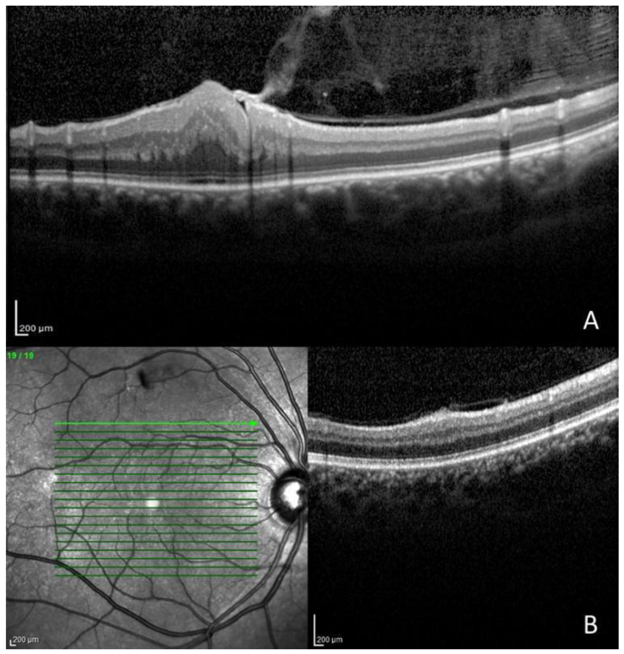
Figure 1. (A): Severe epiretinal membrane in a 16-year-old patient affected by the Wishart variant, OCT image. (B): Retinal tuft, OCT image.
Applsci 13 06304 g001
Figure 2. Optic disc edema associated with intracranial hypertension, fundus photography, and near-infrared image.
Applsci 13 06304 g002
Table 1. Demographic characteristics, BCVA, and main ocular findings. RE: right eye; LE: left eye.
PatientSexAgeAge at DiagnosisVA REVA LENF2-VariantMain Ocular Findings
1F55511.001.00Gardner variantLagophthalmos (LE)
2F58370.900.80Gardner variantEpiretinal membrane grade 0, ptosis (LE), and exotropia
3F68551.000.90Gardner variantPtosis (LE) and exotropia
4F48461.001.00Gardner variantMicrostrabismus
5F63501.001.00Gardner variantLagophthalmos and superficial punctate keratitis (RE)
6F37300.900.90Gardner variantCataract (RE), paramacular epiretinal membrane grade 2 and optic nerve edema (RE and LE), and exotropia
7F62281.001.00Gardner variant-
8F78460.630.32Gardner variantCataract (RE and LE) and retinal hamartoma (LE)
9M5246No light perception0.50Gardner variantCorneal pannus and esotropia (RE) and corneal leukoma (LE)
10M59591.001.00Gardner variantLagophthalmos and superficial punctate keratitis (LE)
11M30241.001.00Gardner variantEpiretinal membrane grade 1 and pseudophakia (RE), corneal leukoma and cataract (LE), and Duane syndrome type 1
12M73711.000.32Gardner variantCataract (LE) and microstrabismus
13F44441.001.00Gardner variantLagophthalmos (RE) and pseudophakia (LE)
14M16101.000.03Wishart variantCataract (LE), epiretinal membrane grade 2 (RE and LE), and microstrabismus
Table 2. Ocular finding after inspection and observation of the anterior segment.
Ocular FindingsNumber of Patients%Number of Eyes%
Lagophthalmos428.6%414.3%
Ptosis of eyelid214.3%27.1%
Duane syndrome type 117.1%
Pseudophakia214.3%27.1%
Cataract535.7%621.4%
Superficial punctate keratitis214.3%27.1%
Leukoma214.3%27.1%
Corneal pannus17.1%13.6%
Table 3. Findings on fundus examination.
Ocular FindingsNumber of Patients%Number of Eyes%
Epiretinal membrane428.6%621.4%
Vitreo retinal Tuft321.4%310.7%
Retinal hamartoma17.1%13.6%
Optic nerve edema17.1%27.1%
Table 4. Quantification of prism diopters in patients presenting strabismus. LCR: light corneal reflexes, CT: cover test, symm: symmetrical, PD: prism diopters.
PatientLCR NearLCR DistanceCT NearCT Distance
2−8°−12°−18 PD−25 PD
3−6°−10°−15 PD−20 PD
4symmsymm+6 PD+6 PD
6−20°−15°−40 DP−35 DP
9+20°+20°+40 PD
(Krimsky test)
+40 PD
(Krimsky test)
12symmsymm+7 PD+5 PD
14symmsymm+5 PD+4 PD
Disclaimer/Publisher’s Note: The statements, opinions and data contained in all publications are solely those of the individual author(s) and contributor(s) and not of MDPI and/or the editor(s). MDPI and/or the editor(s) disclaim responsibility for any injury to people or property resulting from any ideas, methods, instructions or products referred to in the content.