Article

Effect of Different Crestal Sinus Lift Techniques for Implant Placement in the Posterior Maxilla of Deficient Height: A Randomized Clinical Trial

by
Ahmed Halim Hashem
1,*,
Mohamed Fekry Khedr
1,
Mostafa Mohamed Hosny
1,
Mahmoud Taha El-Destawy
1 and
Mohamed Ibrahim Hashem
2
1
Department of Periodontology and Oral Medicine, Faculty of Dental Medicine, Al-Azhar University, Cairo 11651, Egypt
2
Dental Health Department, College of Applied Medical Sciences, King Saud University, Riyadh 11433, Saudi Arabia
*
Author to whom correspondence should be addressed.
Appl. Sci. 2023, 13(11), 6668; https://doi.org/10.3390/app13116668
Submission received: 6 May 2023 / Revised: 29 May 2023 / Accepted: 29 May 2023 / Published: 30 May 2023
(This article belongs to the Section Applied Dentistry and Oral Sciences)

Abstract

:
This study evaluated dental implant stability, vertical bone gain, bone density, and crestal bone loss using different crestal sinus lift techniques (osteotomy, Densah burs, and piezosurgery). A total of 21 patients were randomly divided into three groups: Group 1: patients were treated using a Densah drill crestal sinus lift, Group 2: patients were treated using a piezoelectric crestal sinus lift and Group 3: patients were treated using an osteotome crestal sinus lift. The patients in all three groups underwent bone grafting and implant placement. An Osstell device was used to determine the implant stability by recording the values of the implant stability quotient (ISQ). CBCT was performed before and 6 months after implant placement for radiographic evaluation and comparison among the groups. All dental implants were completely successful, and statistically significant differences from baseline to 6 months were noted in all groups (p < 0.05). The Densah burs technique resulted in the best implant stability of all groups, while the osteotome technique demonstrated better vertical bone augmentation. However, the values for bone density and crestal bone loss showed no significant difference among all treated groups (p > 0.05). All three techniques were successful for crestal sinus lifts with good clinical outcomes at a 6-month follow-up. The Densah group demonstrated better implant stability, shorter surgery time, and fewer complications; however, the vertical bone gain was greater with the osteotome technique.

1. Introduction

Removable complete or partial dentures may replace edentulous areas; however, using removable dentures minimizes taste perception and masticatory capacity [1]. As a result, dental implants have become a reliable treatment option for eligible patients [2]. However, implant success is related to bone quality and quantity [3]; most implant failures occur in the maxillary molar region due to poor bone quality [4,5]. Other factors that could lead to implant failure and complexity in the posterior maxilla include decreased interarch space and gross sinus pneumatization due to alveolar ridge atrophy. In these cases, sinus augmentation is recommended so as to create enough vertical bone volume to allow implant placement with sufficient stability [6].
Several treatment options for a vertically deficient, edentulous posterior maxillary have been postulated. Traditionally, two approaches have been used: direct sinus elevation through a lateral window approach and indirect sinus elevation through a crestal approach [7]. The lateral window technique produces a predictable clinical outcome [8]. However, the procedure’s invasiveness, the patient’s morbidity, the risk of severing the alveolar antral artery, the risk of sinus membrane perforation, the delay in healing, and the enhanced risk of postoperative infection are major disadvantages [9]. Transcrestal indirect sinus elevation procedures, on the other hand, require less time, are less invasive, and have a lower morbidity rate. Yet, the possibility of membrane perforation is almost 24% due to a lack of direct access and visibility [10].
Numerous surgical methods for the indirect lift of the sinus membrane have been developed. Summers described the osteotome sinus floor elevation (OSFE) technique in 1994, which used an osteotome to fracture the sinus floor and lift the sinus membrane, facilitating the placement of graft materials and implants in the subantral space through the osteotomy site [11]. This procedure has some drawbacks, including exposure to the explosive force of the maxilla with inadequate control, inadvertent displacement or fracture, membrane perforation, benign paroxysmal positional vertigo, and patient pain [12].
Using piezoelectric ultrasound was proposed as an efficient alternative for maxillary sinus lift procedures [13]. This device’s ability to selectively cut only mineralized structures without harming soft tissues is an important feature [13,14]. Furthermore, no perforation of the Schneiderian membrane occurs during the lateral antrostomy piezoelectric preparation [15]. Thus, piezosurgery was employed to expose the maxillary sinus mucosa through the alveolar crest while simultaneously elevating the sinus floor with hydraulic pressure for graft and implant insertion. The advantages of this method include reduced trauma, less sinus membrane perforation, the lack of malleting, and shorter surgery times. However, membrane perforation via high hydraulic pressure may occur [16].
As a new bone instrumentation technique, osseous densification, a biomechanical osteotomy method, uses a non-excavating drilling process to preserve bone [17]. It is recommended that practitioners employ drills with tapered geometry and carefully constructed flutes to gradually expand the osteotomy while securing bone to its walls and apex. Since compaction autografting causes an elastic “spring-back” effect in the prepared osteotomy, this bone densification method boosts the implant’s primary stability [17].
It has been acknowledged that CBCT is valuable in implant treatment planning and in postimplant and/or postgrafting evaluation. CBCT is also used to detect bone height, assess marginal bone loss, and determine bone density around implants [18,19].
The present randomized clinical trial (RCT) evaluated dental implant stability, vertical bone gain, bone density, and crestal bone loss after a crestal sinus lift as performed using different techniques (Densah burs, piezosurgery, and osteotome) in the posterior maxilla of deficient height.

2. Materials and Methods

2.1. Ethical Aspect and Patient Induction

The study followed the protocols of the Declaration of Helsinki and was approved by the Ethical Committee (Approval code: 410/293) of Al-Azhar University, Egypt. With the registration number NCT05735613, the current clinical trial is listed on the national clinical trial registry. All study participants gave informed consent after being apprised of the research methods.
A total of 21 participants were enrolled in the RCT between January 2021 and April 2021. The participants were recruited from the patient waiting list at the Periodontology Department, Faculty of Dental Medicine, Al-Azhar University. The inclusion and exclusion criteria for enrolling participants in the study admitted young and adult patients of both sexes, with a residual bone height of ≤5 mm. The remaining natural teeth provided sufficient periodontal tissue support, and the edentulous ridges were covered with mucoperiosteum that showed no ulceration, scarring, or inflammatory symptoms. Occlusion showed sufficient intra-arch and intraarch space for future prosthetic devices. Potential participants were excluded if they had any systemic illnesses that would impair the treatment results or bone quality. These conditions included pregnancy, heavy smoking, active periodontal disease, and neglected oral hygiene. Uncooperative patients, patients with limited mouth-opening ability and unfavorable intermaxillary arch space, and patients with maxillary sinus disease or previous sinus surgery were also excluded.

2.2. Presurgical Therapy and Grouping

Before the implant placement, every patient received thorough oral prophylaxis, including scaling and root planning (SRP), and all were informed that they must maintain acceptable oral hygiene. Cone beam computed tomography (CBCT, Sirona, Orthophos SL3D, Dentsply, Bensheim, Germany) was utilized to estimate the height and width of the bone with the measurement of bone density (HU). Patients were then scheduled for the placement of the dental implants.
The included participants were randomized into three groups (n = 7): Group 1 comprised five females and two males (mean age: 50.7 ± 5.6 years; mean residual bone height: 4.66 mm), and they were treated using the Densah drill (Versah, Jackson, MI, USA) crestal sinus lift technique. Group 2 included three males and four females (mean age: 49.3 ± 7.2 years; mean residual bone height: 4.66 mm), and they were treated using the piezoelectric (Satelec Acteon, Bordeaux-Merignac, Merignac, France) crestal sinus lift technique. Finally, Group 3 included four males and three females (mean age: 50.1 ± 7.7 years; mean residual bone height: 4.68 mm), and they were treated using the osteotome (Summers Osteotomes Kit, SpiralTech, Chicago, IL, USA) crestal sinus lift technique. The patients in all three groups then underwent bone grafting and received the implants.

2.3. Randomization and Allocation

Patients were randomly apportioned to one of the three groups via computer-assessed randomization freeware (http://www.randomizer.org, accessed on 10 January 2023). The participants were given a special identity code and were informed that they should not reveal it to the clinician treating the patient. The allocation process was performed by an external department faculty member who was unaware of the study process.

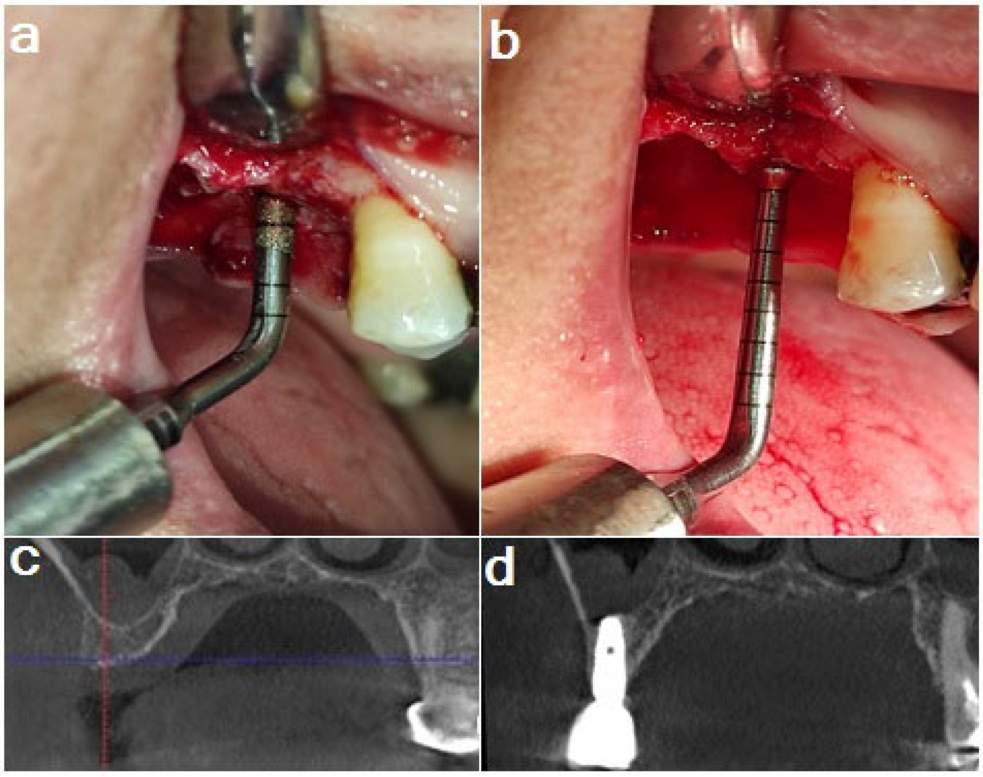
2.4. Surgical Procedures

During the surgical appointment, each patient was asked to rinse with 0.12% chlorhexidine digluconate (Orovex mouth rinse, Macro Group Pharmaceuticals, Cairo, Egypt) followed by administration of local anesthesia (Septanest 1:100,000, Septodont, Inc., Saint-Maur, Cedex, France). A full-thickness flap was raised at the implant site to display the crest of the alveolar ridge. The osteotomy preparation began with a pilot drill that stopped 1 mm short of the sinus floor.
Group 1: Densah sinus lift: The implant motor was switched to reverse-densifying mode (anticlockwise, with a drill speed of 800–1500 rpm, simultaneous with copious irrigation). A 2.2 mm diameter Densah bur was used to make an advanced osteotomy with a wider Densah bur (2.5 mm and 3 mm) by varying the pressure and a pumping motion until it was 1 mm short of the sinus floor. After that, larger Densah drills (3.5 mm and 4 mm) were employed to advance past the sinus floor in increments of 1 mm (Figure 1).
Group 2: Piezoelectric sinus lift: The osteotomy used a 2 mm twist drill to penetrate the cortical bone. Then the intralift tips (TKW 1 to TKW 4) (Acteon, Merignac Cedex, France) were used to progressively widen the entry to the Schneiderian membrane, placing mild pressure on the tips to deepen the pathway and cooling the tips with an 80 mL/min sterile spray to minimize heat damage. The diameter of TKW 1, 2, 3, and 4 tips are 1.35 mm, 2.1 mm, 2.35 mm, and 2.8 mm, respectively. Next, a TKW 5 tip (3 mm) was inserted into the entry canal, and the ultrasonic activation was repeated for 5 s with internal irrigation at 40 mL/min, 50 mL/min, and 60 mL/min. Hydraulic pressure was utilized to push the sinus membrane upward, and then the floating of the sinus membrane was examined. Eventually, implant drills were used successively until the planned implant size was reached (3.0 mm, 3.5 mm, and 4 mm) (Figure 2).
Group 3: Osteotome sinus lift: The drills were used sequentially (3.0 and 3.5 mm) to increase the size of the osteotomy site to the same level (1 mm away from the sinus floor). An osteotome with a diameter of 4 mm, slightly smaller than the planned implant fixture, was placed in the prepared implant site, tapped gently to reach the same level, and then lightly tapped to break up the sinus floor (Figure 3).
Following the completion of the surgical procedure for each patient, a clinical check was performed to ensure that the membrane was still intact by obstructing the patient’s nostrils and requesting that the patient blow through their nose.

2.5. Bone Graft and Implant Placement

A xenograft was added as the grafting material (TutoboneTM, Tutogen Medical GmbH, Neunkirchen am Brand, Germany) in the same amount (0.5 cc for each implant) and pushed to the sinus through the osteotomy site until the required height of sinus elevation was attained, followed by insertion of the implant fixture (NucleOSS T6, Izmir, Turkey). All the implants used in this study were of the same size (10 mm in length × 4.1 mm in diameter). The primary stability was measured, and then the flap was sutured.

2.6. Implant Stability Measurement

Primary stability was checked using the Osstell device (Osstell Inc. W&H Dentalwerk, Salzburg, Austria) (Figure 4) by connecting a smart peg to the implant and measuring the value of the implant stability quotient (ISQ). The Osstell device allows for the evaluation of an implant’s stability by resonance frequency analysis (RFA). Implant stability is measured as the implant stability quotient (ISQ), with values ranging from 1 to 100. An ISQ greater than 70 is regarded as the most favorable for implant stability, whereas ISQ values below 60 indicate low primary stability. The ISQ is related to the lateral stability of the implant, which depends on the rigidity of the bond between the bone and the implant surface [20].

2.7. Postoperative Care

Patients were told to avoid blowing their noses or sneezing for the first 12 h following surgery, to avoid using a straw when drinking for the next 10 days, and to apply cold packs to their cheeks. The following medications were prescribed: ketoprofen 150 mg (Bi-Profenid 150, SANOFI Aventus, Compiegne, France), twice daily for 5 days, and amoxicillin-clavulanate (Hibiotic 1 gm, Amoun, Obour City, Egypt), 1 gm twice daily for 7 days. Metronidazole 250 mg (Flagyl, GlaxoSmithKline, Brentford, UK) was also given thrice daily for 7 days.

2.8. Follow-Up and Procedures

After 10 days, the sutures were removed from the implant site. After 6 months, the prosthetic phase was initiated by physically exposing the implant. The Osstell device was then used to measure secondary stability by recording the ISQ [21]. The gingival former was used for 2 weeks to create the emergence profile; then, the impression was obtained, and the cast was prepared for fabricating the screw-retained restorations. CBCT analysis was performed 6 months after the crestal sinus lift procedure to measure the radiographic changes. The measurement period was adopted from previous studies [22,23].

2.9. Radiographic Evaluation

A routine periapical film was performed initially; if the patient met the inclusion criteria, CBCT was performed to evaluate the bone quality and the width and height of the edentulous site. A 3D imaging system (version 5.3.4., Planmeca ProFace®, Helsinki, Finland) captured the target jaw segment with a 5 × 8 cm2 field of view. The CBCT comparison was performed via rigid image registration through the superimposition of two volumes via a matching tool using the software (high precision matching), followed by minute adjustments for precise matching (Figure 5).

2.9.1. Bone Height

After 6 months, bone height changes were evaluated by measuring preoperative residual bone height and distance from the implant platform to the base of the grafted bone at the middle of the implant (Figure 6).

2.9.2. Bone Density

For measuring the bone density (HU), standard rectangular shapes were drawn buccal and palatal to the simulated future implant area in the preoperative CBCT. After 6 months, rectangular shapes were drawn along the palatal and buccal walls from the implant shoulder to the apex. This was performed using a bone-density measurement tool in the software (Figure 7).

2.9.3. Crestal Bone Loss (CBL)

Marginal bone loss was measured from the implant shoulder to the crest of the alveolar bone, buccally and palatally. Two lines were drawn at the shoulder and apex of the implant, and then the bone between these lines was measured buccally and palatally from the apex of the implant to the crest of the alveolar bone. The mean crestal bone loss was measured by taking the mean value of both the buccal and palatal CBL.

2.10. Primary and Secondary Outcomes

The primary outcome of this study was the evaluation of primary and secondary implant stability and maxillary sinus augmentation (vertical bone gain) 6 months after implant placement.
The secondary outcome was the bone density and crestal bone loss evaluation 6 months after implant placement.

2.11. Statistical Analysis

Statistical analysis was performed using IBM SPSS Statistics for Windows (Version 23.0., IBM Corp, Armonk, NY, USA). Using the Shapiro–Wilk and Kolmogorov–Smirnov tests, numerical data were tested for normality. All data showed a normal (parametric) distribution, except for changes in crestal bone loss and bone density data, which exhibited a non-normal (nonparametric) distribution. Nonparametric data are presented as median and range, whereas parametric data are presented as mean and standard deviation (SD). Two-way repeated measures ANOVA was used to compare the groups for parametric data. The three groups’ mean ages were compared using one-way ANOVA. The Kruskal–Wallis test was applied to the nonparametric data to compare the three groups. The Wilcoxon signed-rank test was used to compare the baseline and post-6-month bone density data. Frequencies and percentages were used to present the data on gender. The three groups were compared using Fisher’s exact test. The significance level was set at p ≤ 0.05.

3. Results

A total of 29 patients were examined for this RCT, and eight were excluded. Of the excluded patients, five did not meet the inclusion prerequisites, while three declined to participate in the study for reasons of privacy. Finally, the remaining 21 patients were allocated at random to three groups (n = 7). All of the study’s participants were recovered for follow-up and were available for the study’s final analysis. The CONSORT flow diagram showing the study method is shown in Figure 8.

3.1. Implant Stability

At baseline, there was a statistically significant difference between mean implant stability measurements among the three groups. The comparisons revealed that the Densah group had the highest mean implant stability; the osteotome group demonstrated the lowest mean value, and the piezosurgery group demonstrated the lowest mean implant stability. After 6 months, a statistically significant difference was observed in the mean implant stability measurements among the groups. The Densah group showed the highest mean implant stability, and no difference between the piezosurgery and osteotome groups was noted; however, both showed the lowest mean implant stability measurement (Table 1).

3.2. Bone Height (mm)

(a) 
Comparisons of bone height measurements among the three groups
At baseline, all three groups demonstrated a statistically significant difference in mean bone height measurements. After 6 months, a significant difference in mean bone height measurements was observed among all three groups. Comparisons among the groups revealed that the osteotome group showed, statistically significantly, the highest mean bone height measurement. There was no significant difference between the Densah and piezosurgery groups; both showed significantly lower mean bone heights (Table 2).
(b) 
Vertical bone gain comparison
A statistically significant difference in the mean bone gain among the three groups was observed after 6 months. The osteotome group demonstrated a considerably larger mean bone gain across the groups. The Densah and piezosurgery groups were characterized by statistically lower mean bone increase values, with no significant difference between them (Table 3).

3.3. Bone Density (HU)

There was no significant difference in the bone density measurements among the three groups, at either baseline or at the 6-month follow-up (Table 4).

3.4. Crestal Bone Loss (mm)

There was no statistically significant difference in crestal bone loss among the three groups at either the buccal or the palatal side. Similarly, for overall crestal bone loss (a value equal to the mean of the buccal and palatal sides), there were no statistically significant differences among the three groups (Table 5).
The posthoc power was calculated in this study to determine whether the sample size would be sufficient to demonstrate a significant difference by using the observed effect size (f = 0.9357406), α = 0.05, and sample size (n = 7, 3 groups). It had high post hoc power values (95%). The effect size was calculated based on the observed means of the post-surgical bone height of the study groups (group 1 = 10.47, group 2 = 10.41, and group 3 = 11.61). The analysis was performed using the G Power freeware (Version 3.1.9., Franz Faul, College of Kiel, Kiel, Germany).

4. Discussion

Dental implants have a favorable success rate, especially when implanted in highly mineralized bone [24,25]. It has been confirmed that the posterior maxilla has the poorest bone quality (D4) [26,27]. Many procedures have been proposed to augment local bone volume to allow for implant placement into the posterior maxilla [28]. Summers pioneered the osteotome technique as a less invasive alternative for sinus floor elevation with simultaneous grafting to increase the primary stability of posterior maxillary implants [11]. Piezoelectric ultrasound was employed as an alternate approach for performing osteotomies in maxillary sinus lift procedures because it reduces trauma and the rate of sinus membrane perforation during surgery [13]. Huwais and Meyer presented a technique using Densah burs for maxillary sinus lifts, providing a safe strategy with fewer complications [29].
It is evident from the obtained results that the Densah group shows the highest mean implant primary stability; the osteotome group shows a lower mean value, and the piezosurgery group shows the lowest mean implant primary stability. This finding is similar to those of other studies [30,31] that demonstrated the highest implant stability to characterize the Densah group. However, our finding contradicts other studies that reported no significant difference concerning primary stability between the Densah and osteotome techniques [32,33]. Secondary stability measured after 6 months shows a statistically significant difference in the mean implant stability measurements among the three groups; again, Densah has the highest mean implant stability, which is in agreement with previous results [32,34]. On the other hand, the results were also compared with those of another study [24,33], which reported no statistically significant difference between the piezosurgery and osteotome techniques; both showed lower mean implant stability measurements that were statistically significant.
Bone density shows a significant difference from baseline to 6 months in all of the groups, but no difference in bone density was observed among the three groups. The increased implant stability in the Densah group can be attributed to the technique. The technology and associated drilling protocol were principally responsible for the considerable variation in stability among the three groups. Compared to the osteotome and piezosurgery groups, the Densah group had greater bone density values around the implant, owing to the osteotomy site’s motorized expansion and the Densah burs’ unique qualities. After implant insertion, the bone’s elastic recoil and spring-back effect on the implant surface improve the mechanical connection between the implant and the surrounding bone. This approach strengthened the implant’s primary stability and encouraged more bone healing throughout the 6-month follow-up period, providing good secondary stability. This effect worked with the intact, well-organized, trabecular pattern of the bone surrounding the implant [17,29,35,36]. The osteotome method increases bone density, enabling greater initial implant stability by compressing the bone laterally, surrounding the implant site with osteotomes of gradually increasing diameter [37]. However, after using an osteotome, the bone quality and even the stability around the implant were worse than those seen in the Densah group [38]. With piezosurgery, soft tissues can be preserved, while mineralized tissues can be removed selectively. Piezosurgery, in particular, permits the surgeon to work directly on the Schneiderian membrane, making it safe for membrane elevation with no impact on implant stability or bone density [39,40].
Bone height measurements in the three groups did not differ at baseline although there were variations in the three groups after 6 months. Densah and piezo revealed the lowest mean bone height, whereas osteotome displayed the greatest mean bone height measurement. The amounts of bone growth in the three groups varied significantly after 6 months. Contrary to a study that found no discernible difference in outcomes between the osteotome and piezosurgery techniques [24]; the osteotome group had the highest mean bone growth. The Densah and piezosurgery groups did not differ in a statistically significant manner; both had the lowest mean bone gain readings.
One criterion for implant success was the result in the marginal bone levels around the implants. For implant success, less than 1.5 mm of bone loss during the first year, less than 0.2 every year, and a maximum of 2 mm over 5 years were recorded [41]. Radiographic evaluation by CBCT for crestal bone loss revealed that, at the buccal and palatal sides, as well as overall, there were no significant differences among the three groups [24].
Autogenous bone has reportedly been found to be the best choice for bone reconstructive therapy [42,43]. Bone substitutes are frequently used to avoid the morbidity brought on by the use of autogenous bone. Therefore, xenograft was used in the current investigation to increase success and predictability [44,45]. In one patient who underwent piezosurgery, just one membrane perforation was clinically seen; however, as previously reported [24], this perforation had no negative effects.
The implant survival rate was 100%, comparable to or greater than that seen in prior investigations involving sinus floor elevation. The residual alveolar ridge’s insufficient height is not a substantial cause of implant failure, but trauma, infection, or contamination during surgery may contribute to this unfavorable outcome [24,30,46,47]. Complications such as trauma, bleeding, sinus membrane perforation, and maxillary sinusitis were seen least often in the osseodensification group. The piezosurgery group suffered from one membrane perforation but did not show any unfavorable consequences, while one osteotome patient showed benign paroxysmal positional vertigo (BPPV). The Densah technique had the shortest surgery time among the three groups.
One of the study’s limitations is the smaller sample size. Although 6 months is considered feasible to demonstrate post-treatment changes, a longer period could provide a more distinct outcome. Therefore, future studies should focus on a larger sample size and longer follow-up period. Furthermore, it would be interesting to see the effectiveness of these techniques at other sites in the oral cavity.

5. Conclusions

Considering the limitations of the current clinical trial, all three techniques were successful for crestal sinus lift, providing excellent clinical results 6 months after implant insertion. The Densah group demonstrated better implant stability, shorter surgical duration, and fewer complications. On the contrary, the vertical bone gain was greater with the osteotome technique.

Author Contributions

Conceptualization: A.H.H., M.F.K. and M.I.H.; data curation: M.M.H. and M.T.E.-D.; formal analysis: M.F.K., M.T.E.-D. and M.I.H.; investigation: A.H.H. and M.M.H.; methodology: A.H.H., M.M.H. and M.T.E.-D.; resources: M.F.K. and M.I.H.; software: M.M.H.; validation: M.T.E.-D.; writing—original draft: A.H.H. and M.T.E.-D.; writing—review and editing: M.F.K. and M.I.H. All authors have read and agreed to the published version of the manuscript.

Funding

This research received no external funding.

Institutional Review Board Statement

The study was approved by the Institutional Review Board of Al-Azhar University, Cairo, Egypt (Approval #410/293).

Informed Consent Statement

Informed consent was obtained from all participants involved in the study.

Data Availability Statement

The data presented in this study are available upon request from the corresponding author.

Acknowledgments

The authors extend their appreciation to the Researchers Supporting Project (No. RSPD2023R680), King Saud University, Riyadh, Saudi Arabia, for supporting this study.

Conflicts of Interest

The authors declare no conflict of interest.

References

  1. Sato, T.; Abe, T.; Nakamoto, N.; Tomaru, Y.; Koshikiya, N.; Nojima, J.; Kokabu, S.; Sakata, Y.; Kobayashi, A.; Yoda, T. Nicotine Induces Cell Proliferation in Association with Cyclin D1 up-Regulation and Inhibits Cell Differentiation in Association with P53 Regulation in a Murine Pre-Osteoblastic Cell Line. Biochem. Biophys. Res. Commun. 2008, 377, 126–130. [Google Scholar] [CrossRef] [PubMed]
  2. Mertens, C.; Meyer-Bäumer, A.; Kappel, H.; Hoffmann, J.; Steveling, H.G. Use of 8-Mm and 9-Mm Implants in Atrophic Alveolar Ridges: 10-Year Results. Int. J. Oral Maxillofac. Implant. 2012, 27, 1501–1508. [Google Scholar]
  3. Sennerby, L.; Roos, J. Surgical Determinants of Clinical Success of Osseointegrated Oral Implants: A Review of the Literature. Int. J. Prosthodont. 1998, 11, 408–420. [Google Scholar]
  4. Kim, B.; Lee, J. The Retrospective Study of Survival Rate of Implants with Maxillary Sinus Floor Elevation. J. Korean Assoc. Oral Maxillofac. Surg. 2010, 36, 108. [Google Scholar] [CrossRef]
  5. Kim, Y.K.; Hwang, J.Y.; Yun, P.Y. Relationship between Prognosis of Dental Implants and Maxillary Sinusitis Associated with the Sinus Elevation Procedure. Int. J. Oral Maxillofac. Implant. 2013, 28, 178–183. [Google Scholar] [CrossRef] [PubMed]
  6. Morand, M.; Irinakis, T. The Challenge of Implant Therapy in the Posterior Maxilla: Providing a Rationale for the Use of Short Implants. J. Oral Implant. 2007, 33, 257–266. [Google Scholar] [CrossRef]
  7. Tatum, H., Jr. Maxillary and Sinus Implant Reconstructions. Dent. Clin. N. Am. 1986, 30, 207–229. [Google Scholar] [CrossRef]
  8. Wallace, S.S.; Froum, S.J. Effect of Maxillary Sinus Augmentation on the Survival of Endosseous Dental Implants. A Systematic Review. Ann. Periodontol. 2003, 8, 328–343. [Google Scholar] [CrossRef]
  9. Wallace, S.S.; Mazor, Z.; Froum, S.J.; Cho, S.C.; Tarnow, D.P. Schneiderian Membrane Perforation Rate During Sinus Elevation Using Piezosurgery: Clinical Results of 100 Consecutive Cases. Int. J. Periodontics Restor. Dent. 2007, 27, 413–419. [Google Scholar]
  10. Reiser, G.M.; Rabinovitz, Z.; Bruno, J.; Damoulis, P.D.; Griffin, T.J. Evaluation of Maxillary Sinus Membrane Response Following Elevation with the Crestal Osteotome Technique in Human Cadavers. Int. J. Oral Maxillofac. Implant. 2001, 16, 833–840. [Google Scholar]
  11. Summers, R.B. A New Concept in Maxillary Implant Surgery: The Osteotome Technique. Compendium 1994, 15, 154–156. [Google Scholar]
  12. Toffler, M.; Rosen, P. Complications with Transcrestal Sinus Floor Elevation: Etiology, Prevention, and Treatment. In Dental Implant Complications; Stuart, J.F., Ed.; Wiley Blackwell: Hoboken, NJ, USA, 2015. [Google Scholar]
  13. Vercellotti, T.; De Paoli, S.; Nevins, M. The Piezoelectric Bony Window Osteotomy and Sinus Membrane Elevation: Introduction of a New Technique for Simplification of the Sinus Augmentation Procedure. Int. J. Periodontics. Restor. Dent 2001, 21, 561–567. [Google Scholar]
  14. Stübinger, S.; Kuttenberger, J.; Filippi, A.; Sader, R.; Zeilhofer, H.F. Intraoral Piezosurgery: Preliminary Results of a New Technique. J. Oral Maxillofac. Surg. 2005, 63, 1283–1287. [Google Scholar] [CrossRef]
  15. Toscano, N.J.; Holtzclaw, D.; Rosen, P.S. The Effect of Piezoelectric Use on Open Sinus Lift Perforation: A Retrospective Evaluation of 56 Consecutively Treated Cases from Private Practices. J. Periodontol. 2010, 81, 167–171. [Google Scholar] [CrossRef]
  16. Li, J.; Lee, K.; Chen, H.; Ou, G. Piezoelectric Surgery in Maxillary Sinus Floor Elevation with Hydraulic Pressure for Xenograft and Simultaneous Implant Placement. J. Prosthet. Dent. 2013, 110, 344–348. [Google Scholar] [CrossRef]
  17. Huwais, S.; Meyer, E.G. A Novel Osseous Densification Approach in Implant Osteotomy Preparation to Increase Biomechanical Primary Stability, Bone Mineral Density, and Bone-to-Implant Contact. Int. J. Oral Maxillofac. Implant. 2017, 32, 27–36. [Google Scholar] [CrossRef]
  18. Sammartino, G.; Prados-Frutos, J.C.; Riccitiello, F.; Felice, P.; Cerone, V.; Gasparro, R.; Wang, H.L. The Relevance of the Use of Radiographic Planning in Order to Avoid Complications in Mandibular Implantology: A Retrospective Study. BioMed Res. Int. 2016, 2016, 8175284. [Google Scholar] [CrossRef]
  19. Shahidi, S.; Zamiri, B.; Abolvardi, M.; Akhlaghian, M.; Paknahad, M. Comparison of Dental Panoramic Radiography and Cbct for Measuring Vertical Bone Height in Different Horizontal Locations of Posterior Mandibular Alveolar Process. J. Dent. 2018, 19, 83–91. [Google Scholar]
  20. Pattijn, V.; Van Lierde, C.; Van Der Perre, G.; Naert, I.; Vander Sloten, J. The Resonance Frequencies and Mode Shapes of Dental Implants: Rigid Body Behaviour Versus Bending Behaviour. A Numerical Approach. J. Biomech. 2006, 39, 939–947. [Google Scholar] [CrossRef]
  21. Huang, H.; Wu, G.; Hunziker, E. The Clinical Significance of Implant Stability Quotient (Isq) Measurements: A Literature Review. J. Oral Biol. Craniofac. Res. 2020, 10, 629–638. [Google Scholar]
  22. El-Destawy, M.T.; Khedr, M.F.; Hosny, M.M.; Bilal, A.M.; Elshamy, A.M.; El Sayed, I.S.; Borhamy, A.E.; Aboamo, A.A.; Gamal, A.Y. A Novel Approach of Periodontal Osseous Wall Piezosplitting and Sequential Bone Expansion in Management of Localized Intra-Bony Defects with Wide Angulation—A Randomized Controlled Trial. Healthcare 2023, 6, 791. [Google Scholar] [CrossRef]
  23. Sculean, A.; Stavropoulos, A.; Windisch, P.; Keglevich, T.; Karring, T.; Gera, I. Healing of Human Intrabony Defects Following Regenerative Periodontal Therapy with a Bovine-Derived Xenograft and Guided Tissue Regeneration. Clin. Oral Investig. 2004, 8, 70–74. [Google Scholar] [CrossRef] [PubMed]
  24. Baldi, D.; Menini, M.; Pera, F.; Ravera, G.; Pera, P. Sinus Floor Elevation Using Osteotomes or Piezoelectric Surgery. Int. J. Oral Maxillofac. Surg. 2011, 40, 497–503. [Google Scholar] [CrossRef] [PubMed]
  25. Simunek, A.; Cierny, M.; Kopecka, D.; Kohout, A.; Bukac, J.; Vahalova, D. The Sinus Lift with Phycogenic Bone Substitute. A Histomorphometric Study. Clin. Oral Implant. Res. 2005, 16, 342–348. [Google Scholar] [CrossRef]
  26. Hieu, P.D.; Chung, J.H.; Yim, S.B.; Hong, K.S. A Radiographical Study on the Changes in Height of Grafting Materials after Sinus Lift: A Comparison between Two Types of Xenogenic Materials. J. Periodontal Implant Sci. 2010, 40, 25–32. [Google Scholar] [CrossRef] [PubMed]
  27. Sorní, M.; Guarinos, J.; Penarrocha, M. Implants in Anatomical Buttresses of the Upper Jaw. Med. Oral Patol. Oral Cir. Bucal 2005, 10, 163–168. [Google Scholar]
  28. Mittal, Y.; Jindal, G.; Garg, S. Bone Manipulation Procedures in Dental Implants. Indian J. Dent. 2016, 7, 86–94. [Google Scholar] [CrossRef]
  29. Huwais, S.; Mazor, Z.; Ioannou, A.L.; Gluckman, H.; Neiva, R. A Multicenter Retrospective Clinical Study with up-to-5-Year Follow-up Utilizing a Method That Enhances Bone Density and Allows for Transcrestal Sinus Augmentation through Compaction Grafting. Int. J. Oral Maxillofac. Implant. 2018, 33, 1305–1311. [Google Scholar] [CrossRef]
  30. Baron, T.K.; Venkatraman, N. Minimally Invasive Crestal Approach Sinus Floor Elevation Using Densah Burs, and Hydraulic Lift Utilising Putty Graft in Cartridge Delivery. Clin. Oral Implant. Res. 2017, 28, 203. [Google Scholar]
  31. El-Ghobashy, M.T.; Shaaban, A.M.; Melek, L.N. Osseodensification by Densah Burs Versus Osteotome for Transcrestal Maxillary Sinus Lifting with Simultaneous Implant Placement. Alex. Dent. J. 2023, 48, 51–58. [Google Scholar] [CrossRef]
  32. Hamdi, D.; Hemd, D. Comparison between Osseo Densification Burs and Osteotome Technique for Closed Sinus Lift in Partially Edentulous Maxilla (Clinical and Radiological Study). J. Univ. Shanghai Sci. Technol. 2021, 23, 392–411. [Google Scholar] [CrossRef]
  33. Amira, K.A. Assessment of Dental Implants Placed in Posterior Maxillary Ridge Using Densah Burs Versus Standard Drills. Master’s Thesis, Cairo University, Cairo, Egypt, 2018. [Google Scholar]
  34. Arafat, S.W.; A Elbaz, M. Clinical and Radiographic Evaluation of Osseodensification Versus Osteotome for Sinus Floor Elevation in Partially Atrophic Maxilla: A Prospective Long Term Study. Egypt. Dent. J. 2019, 65, 189–195. [Google Scholar] [CrossRef]
  35. Ahmed Mohamed, B.; Mohamed Mohamed, B.; Akram Abbass, A.-A.; Mahmoud Taha, E.-D.; Mostafa Mohamed, H. Clinical and Radiographic Evaluation of Crestal Ridge Splitting Versus Osseodensification Techniques for Dental Implant Placement into the Narrow Alveolar Ridge. J. Int. Dent. Med. Res. 2022, 15, 205–211. [Google Scholar]
  36. Lahens, B.; Neiva, R.; Tovar, N.; Alifarag, A.M.; Jimbo, R.; Bonfante, E.A.; Bowers, M.M.; Cuppini, M.; Freitas, H.; Witek, L.; et al. Biomechanical and Histologic Basis of Osseodensification Drilling for Endosteal Implant Placement in Low Density Bone. An Experimental Study in Sheep. J. Mech. Behav. Biomed. Mater. 2016, 63, 56–65. [Google Scholar] [CrossRef]
  37. Beziat, J.L.; Vercellotti, T.; Gleizal, A. What Is Piezosurgery? Two-Years Experience in Craniomaxillofacial Surgery. Rev. Stomatol. Chir. Maxillofac 2007, 108, 101–107. [Google Scholar] [CrossRef]
  38. Slete, F.B.; Olin, P.; Prasad, H. Histomorphometric Comparison of 3 Osteotomy Techniques. Implant. Dent. 2018, 27, 424–428. [Google Scholar] [CrossRef]
  39. Schlee, M.; Steigmann, M.; Bratu, E.; Garg, A.K. Piezosurgery: Basics and Possibilities. Implant. Dent. 2006, 15, 334–340. [Google Scholar] [CrossRef]
  40. Vercellotti, T. Technological Characteristics and Clinical Indications of Piezoelectric Bone Surgery. Minerva Stomatol. 2004, 53, 207–214. [Google Scholar]
  41. Albrektsson, T.; Isidor, F. Proceedings of the First European Workshop on Periodontology; Quintessence Publishing: London, UK, 1994. [Google Scholar]
  42. Cordioli, G.; Mazzocco, C.; Schepers, E.; Brugnolo, E.; Majzoub, Z. Maxillary Sinus Floor Augmentation Using Bioactive Glass Granules and Autogenous Bone with Simultaneous Implant Placement. Clinical and Histological Findings. Clin. Oral Implant. Res. 2001, 12, 270–278. [Google Scholar] [CrossRef]
  43. Moy, P.K.; Lundgren, S.; Holmes, R.E. Maxillary Sinus Augmentation: Histomorphometric Analysis of Graft Materials for Maxillary Sinus Floor Augmentation. J. Oral Maxillofac. Surg. 1993, 51, 857–862. [Google Scholar] [CrossRef]
  44. Boëck-Neto, R.J.; Gabrielli, M.; Lia, R.; Marcantonio, E.; Shibli, J.A.; Marcantonio, E., Jr. Histomorphometrical Analysis of Bone Formed after Maxillary Sinus Floor Augmentation by Grafting with a Combination of Autogenous Bone and Demineralized Freeze-Dried Bone Allograft or Hydroxyapatite. J. Periodontol. 2002, 73, 266–270. [Google Scholar] [CrossRef] [PubMed]
  45. Maiorana, C.; Redemagni, M.; Rabagliati, M.; Salina, S. Treatment of Maxillary Ridge Resorption by Sinus Augmentation with Iliac Cancellous Bone, Anorganic Bovine Bone, and Endosseous Implants: A Clinical and Histologic Report. Int. J. Oral Maxillofac. Implant. 2000, 15, 873–878. [Google Scholar]
  46. Emmerich, D.; Att, W.; Stappert, C. Sinus Floor Elevation Using Osteotomes: A Systematic Review and Meta-Analysis. J. Periodontol. 2005, 76, 1237–1251. [Google Scholar] [CrossRef] [PubMed]
  47. Rodoni, L.R.; Glauser, R.; Feloutzis, A.; Hämmerle, C.H. Implants in the Posterior Maxilla: A Comparative Clinical and Radiologic Study. Int. J. Oral Maxillofac. Implant. 2005, 20, 231–237. [Google Scholar]
Figure 1. (a) Densah sinus floor elevation; (b) CBCT preoperative cross-section and (c) CBCT cross-section for implant showing the sinus floor elevation after 6 months.
Applsci 13 06668 g001
Figure 2. (a) Piezoelectric TKW 1-4 tips for osteotomy preparation and penetration of sinus floor; (b) TKW 5 used for elevation of the sinus membrane; (c) CBCT preoperative cross-section and (d) CBCT cross-section showing the sinus floor elevation and implant in place after 6 months.
Applsci 13 06668 g002
Figure 3. (a) Osteotome sinus floor elevation, (b) CBCT preoperative cross-section and (c) CBCT cross-section of implant showing the sinus floor elevation after 6 months.
Applsci 13 06668 g003
Figure 4. Osstell device measuring implant stability.
Applsci 13 06668 g004
Figure 5. (a1) CBCT volume at 6 months; (a2) preoperative CBCT with identical marks during image registration and (b) superimposition of the two CBCT volumes.
Applsci 13 06668 g005
Figure 6. (a) Pre-operative residual bone height; (b) Six-month postoperative CBCT bone height and (c) sinus radiograph showing difference between pre- and postoperative bone height and sinus augmentation.
Applsci 13 06668 g006
Figure 7. (a) Preoperative bone-density measurement and (b) Six-month postoperative bone-density measurement.
Applsci 13 06668 g007
Figure 8. CONSORT flow diagram.
Applsci 13 06668 g008
Table 1. Mean comparison of implant stability at different measurement periods for each group.
TimeDensah (n = 7)Piezo (n = 7)Osteotome (n = 7)p Value
MeanSDMeanSDMeanSD
Baseline74.2 A3.962.8 C4.768.5 B2.2<0.001 *
At 6-month follow-up87.1 A3.371.8 B4.880.2 B1.6<0.001 *
* Significant at p ≤ 0.05 (two-way repeated measures ANOVA); different superscript letters in the same row indicate significant differences.
Table 2. Mean comparison of bone height measurements (mm) in the three groups.
TimeDensah (n = 7)Piezo (n = 7)Osteotome (n = 7)p Value
MeanSDMeanSDMeanSD
Baseline4.660.374.70.454.680.260.982
At 6-month follow-up10.47 B0.5910.41 B0.4911.61 A0.530.001 *
* Significant at p ≤ 0.05 (two-way repeated measures ANOVA); different superscript letters in the same row indicate significant differences.
Table 3. Mean comparison of bone gain (mm) among the three groups.
Densah (n = 7)Piezo (n = 7)Osteotome (n = 7)p Value
MeanSDMeanSDMeanSD
5.81 B0.515.72 B0.446.93 A0.45<0.001 *
* Significant at p ≤ 0.05 (one-way ANOVA); different superscript letters in the same row indicate significant differences.
Table 4. Median comparison of bone density measurements (HU) among the groups.
TimeDensah (n = 7)Piezo (n = 7)Osteotome (n = 7)p Value *
MedianRangeMedianRangeMedianRange
Baseline266.4106.6–464.4224.8124.9–412.1248.8166.2–523.90.754
At 6-month follow-up660.3350.9–740.2560.4388.1–701530.4444.8–890.70.580
* Kruskal–Wallis test.
Table 5. Median comparison of crestal bone loss (mm) in the three groups.
SideDensah (n = 7)Piezo (n = 7)Osteotome (n = 7)p Value *
MedianRangeMedianRangeMedianRange
Buccal0.240–0.960.70.45–1.20.60–1.140.193
Palatal0.080–0.640.660.3–0.90.40–0.820.071
Overall0.20–0.730.660.4–1.050.430–0.970.073
* Kruskal–Wallis test.
Disclaimer/Publisher’s Note: The statements, opinions and data contained in all publications are solely those of the individual author(s) and contributor(s) and not of MDPI and/or the editor(s). MDPI and/or the editor(s) disclaim responsibility for any injury to people or property resulting from any ideas, methods, instructions or products referred to in the content.

