Article

Insights into the Antimicrobial Potential of Acorn Extracts (Quercus ilex and Quercus suber)

Escola Superior de Biotecnologia, CBQF Centro de Biotecnologia e Química Fina-Laboratório Associado, Universidade Católica Portuguesa, Rua Diogo Botelho 1327, 4169-005 Porto, Portugal
*
Authors to whom correspondence should be addressed.
Appl. Sci. 2023, 13(11), 6820; https://doi.org/10.3390/app13116820
Submission received: 21 April 2023 / Revised: 29 May 2023 / Accepted: 2 June 2023 / Published: 4 June 2023
(This article belongs to the Special Issue Natural Products: Sources and Applications)

Abstract

:
Acorns, frequently left uncollected in the fields, have been a part of the traditional medicine of different cultures. Among the different properties associated with them, their antimicrobial potential is of particular importance. However, this characterization has long been superficial and has not ventured into other topics such as biofilm inhibition. Thus, the current work aimed to characterize the antimicrobial and antibiofilm potential of an array of phenolic rich extracts attained from acorns, two different acorn varieties Q. ilex and Q. suber, considering the fruit and shell separately, fresh and after heat-treating the acorns to aid in the shelling process. To accomplish this, the extracts’ capacity to inhibit an array of different microorganisms was evaluated, the minimum bactericidal concentration (MBC) was determined, time-death curves were drawn whenever an MBC was found and the antibiofilm potential of the most effective extracts was drawn. The overall results showed that Gram-positive microorganisms were the most susceptible out of all the microorganisms tested, with the shell extracts being the most effective overall, exhibiting bactericidal effect against S. aureus, B. cereus and L. monocytogenes as well as being capable of inhibiting biofilm formation via the two S. aureus strains. The attained results demonstrated that acorn extracts, particularly shell extracts, pose an interesting antimicrobial activity which could be exploited in an array of food, cosmetic and pharmaceutical applications.

1. Introduction

Acorns are a fruit produced by trees from the Quercus genus, which are both highly abundant in the Mediterranean area and a resource frequently left, uncollected, in the fields. While its common use nowadays is mostly as animal feed, traditionally acorns have been a part of the medicinal folklore of several cultures, with extracts from different parts being used as astringents, tonics or antiseptics [1,2,3].
The fact that they are used in traditional medicine and that they are a frequently disregarded resource, when coupled with the need to find new sources of bioactive ingredients from, preferably, more sustainable sources, makes acorns a particularly interesting resource to exploit. In fact, in recent decades, the number of studies focusing on the potential health benefits has been rising, with authors reporting that acorns from an array of different Quercus species have exhibited nephron- and hepatoprotective effects, ameliorating an array of diabetes-related metabolic alterations, exhibiting anti-cancer and neuroprotective effects (namely inhibition of angiogenesis), antimicrobial, antifungal and antiviral potentials as well as attenuating inflammation and functioning as antioxidants [1,2,3,4,5,6,7,8,9,10,11,12]. Among the compounds which have been associated with the potential effects observed, those of phenolic nature are among the most frequently mentioned as acorns are not only a good source of these, but the extraction methodologies used also frequently lend themselves to their extraction. In fact, Quercus ilex (Q. ilex) and Quercus suber (Q. suber) have been reported to be good sources of an array of different phenolic compounds such as gallic, chlorogenic, caffeic, syringic, p-coumaric, sinapic and ellagic acids, compounded with an array of different properties associated with them and among which stands their antimicrobial potential [13].
As antimicrobial resistance gains increasing relevance, the need for the development of alternative strategies and the development of new antimicrobial compounds is ever-present. Among the different possibilities contemplated in the literature, the use of phenolic compounds is one of particular relevance as these compounds have been shown to function not only by directly inhibiting the growth/killing an array of several potential pathogens but also as effective co-adjuvants for current antibiotic therapies, attenuating the virulence of microorganisms and hindering infection by interfering with adhesion and subsequent colonization of both cells and surfaces [14].
Considering the arguments above, the current work hypothesized that extracts from two different Quercus species (Q. ilex and Q. suber) had biological potential, namely as antibiofilm. To that end, we aimed to characterize the antimicrobial and, for the first time, the antibiofilm potential of acorn extracts considering the fruit and its shell separately as well as the impact of submitting the acorns to a heat treatment that considerably aids in the shelling process.

2. Materials and Methods

2.1. Acorns

Quercus ilex and Quercus suber acorns were kindly provided by Herdade do Freixo do Meio (Montemor-o-Novo, Portugal) and stored under vacuum at 4 °C until use. Two types of samples were considered: Fresh samples were manually de-shelled and both fractions were powdered using an A327R1 Moulinex mill (Barbastro, Spain) prior to extraction. Heat-treated (HT) samples were prepared by exposing acorns (previously split in half) to 200 °C for 20 min as previously described by Silva and Costa [15], before separating the fruit from the shell and powdering it.

2.2. Extract Production

Extract production was optimized in-house and the two different extraction procedures were adopted depending on the matrix. Shell samples were extracted via decoction using an 8% (w/v) suspension of shells in water with an extraction assembly equipped with a condenser to avoid water loss during the decoction. Fruit samples were suspended at 8% (w/v) in 30% (v/v) ethanol (Panreac, Barcelona, Spain) and stirred for 15 min at room temperature. After each extraction process, the samples were filtered through a 4–7 μm cellulosic filter paper (Prat Dumas, Couze-et-Saint-Front, France), ethanol (when applicable) was removed using a Christ RVC 2-18 Speed-Vaccum (Dail Tech., Seoul, Republic of Korea) and, the samples were freeze-dried using a Heto Drywinner freeze-dryer (Cambridge Biosystems, Devon, UK).

2.3. Total Phenolic Content Determination

The total phenolic content of each extract was determined using the Folin–Ciocalteau assay as described by Gião, González-Sanjosé [16] and Silva, Costa [17]. Briefly, the extracts (diluted as needed) were combined with Folin–Ciocalteu reagent (Merck, Darmstadt, Germany), 75 g/L sodium carbonate solution and deionized water, incubated for 1 h in the absence of light and the optical density (OD; λ = 750 nm) measured using a UV-Vis spectrophotometer (Shimadzu, Kyoto, Japan). The OD of the different samples were then compared against a gallic acid (Sigma, St. Louis, MO, USA) standard curve and the results expressed in gallic acid equivalent. All assays were carried out in triplicate.

2.4. Microorganisms

The antimicrobial and antibiofilm activity of the extracts was evaluated against an array of different, potentially pathogenic/food contaminant microorganisms. Bacillus cereus (B. cereus), Methicilin-resistant Staphylococcus aureus (S. aureus), Salmonella enteritidis (S. enteritidis) and Pseudomonas aeruginosa (P. aeruginosa) were obtained from the American Type Culture Collection (ATCC, USA, ATCC 11778, ATCC 29213, ATCC 13076 and ATCC 10145, respectively). Listeria innocua (L. innocua), Escherichia coli and methicilin-sensitive S. aureus (MRSA) were obtained from the National Collection of Type Cultures (NCTC, UK, NCTC 10528, NCTC 9001 and NCTC 8532, respectively). Candida albicans (C. albicans) was a clinical isolate. MSSA, MRSA, E. coli, B. cereus and L. innocua inoculums were prepared in Muller–Hinton Broth (MHB, Biokar Diagnostics, Allonne, France) for 24 h at 37 °C, while C. albicans inoculums were prepared in Sabouraud Dextrose Broth (SDB; Biokar Diagnostics, Allonne, France) for 24 h at 37 °C.

2.5. Antimicrobial Assays

2.5.1. Well-Diffusion Assay

As an initial screening of the different extract’s capacity to inhibit the growth of the selected microorganisms, a well-diffusion assay was carried out as previously described [18]. To that end, Petri dishes containing 20 mL of Muller–Hinton Agar (MHA; Biokar Diagnostics, Allonne, France) for bacteria or Sabouraud Dextrose Agar (SDA; Biokar Diagnostics, Allonne, France) for C. albicans were inoculated using microorganism suspensions with a turbidity of 0.5 in the McFarland Scale. Afterwards, 4 mm wells were punctured into the inoculated agar and filled, in triplicate, with 50 μL of an aqueous saturated solution of each of the freeze-dried extracts (sterile filtered using 0.22 μm syringe filters). MHA plates were incubated for 24 h at 37 °C, while SDA plates were incubated at 30 °C for 48 h.

2.5.2. Minimum Bactericidal Concentration (MBC)

For all the extract–microorganisms combinations where an inhibition halo was observed, the MBC was determined as previously described [18]. In order to carry this out, solutions of 20, 10, 5 and 2.5 mg/mL for fruit extracts and 10, 5 and 2.5 mg/mL for shell extracts were prepared by dissolving the freeze-dried powder in Muller–Hinton Broth (MHB; Biokar Diagnostics, Allonne, France). The resulting solutions were sterile-filtered using a 0.22 μm syringe filter to sterilize them before being inoculated at 1% (v/v) with an overnight culture of each of the microorganisms and incubated for 24 h at 37 °C. After this period, 100 μL of each solution was plated, in triplicate, into MHA plates and re-incubated under the same conditions as before. The lowest concentration of extract that exhibited no growth was considered the MBC. All assays were performed in triplicate.

2.5.3. Time-Death Curves

Time-death curves were performed by adapting the procedure previously described by Silva, Costa [19]. Briefly, extract solutions at MBC concentration, prepared as described in Section 2.5.2, were inoculated, in triplicate, at 1% (v/v) using an overnight inoculum of each microorganism and incubated at 37 °C. Samples were then collected after 0, 0.25, 0.5, 1, 2, 4, 7, 12 and 24 h for the determination of the total viable counts by plating, in triplicate, in Plate Count Agar (PCA; Biokar Diagnostics, Allonne, France). Plain, inoculated media were used as a positive control. Results were given by plotting the logarithm (log) of the colony forming units (CFU) versus time and whenever a value was below the quantification limit, (log 500) this value was assumed. All individual assays were carried out in quadruplicate.

2.6. Biofilm Formation Inhibition

Inhibition of biofilm formation was evaluated using the crystal violet assay as previously described by Costa, Silva [20]. These assays were performed only for MRSA and MSSA, as out of the bacteria tested, these were the only ones capable of establishing a biofilm under the used conditions. Briefly, the freeze-dried shell extracts (which proved to be more active) were diluted at one-half (1/2 MBC) and one-fourth (1/4 MBC) of the respective MBCs in Tryptic Soy Broth (TSB; Biokar Diagnostics, Allonne, France) supplemented with 1% (w/v) glucose and sterilized via filtration using sterile 0.22 μm syringe filters. These solutions were then inoculated at 2% (v/v) with a 24-hour-old inoculum prepared in the same media. The test solutions were then transferred to flat-bottom, 96-well sterile microplates (Nunclon Delta Surface, ThermoScientific, Roskilde, Denmark) and incubated at 37 °C. After 24 h, the content of each well was carefully discarded, washed 3× to remove non-adherent cells, fixed with absolute ethanol and stained using a crystal violet solution (Sigma, St. Louis, MO, USA). After washing to remove excess stain and air-drying, the content of each well was resuspended in a 0.1 % (v/v) acetic acid solution and the optical density (OD) was measured at 630 nm using a microplate reader. All experiments were carried out in quadruplicate, using inoculated plain media as a positive control. The results are given as biofilm inhibition percentage calculated as described in Equation (1).
%   B i o f i l m   i n h i b i t i o n = 100 ( O D s a m p l e O D p o s i t i v e   c o n t r o l × 100 )

2.7. Statistical Analysis

Statistical analysis of the data was carried out using IBM’s SPSS Statistics’ Software (version 21.0.0; Armonk, NY, USA). When the data followed a normal distribution (evaluated using Shapiro–Wilk’s test), comparisons were carried out using a one-way analysis of Variance test along with Turkey’s test, with differences being considered significant for p-values below 0.05.

3. Results and Discussion

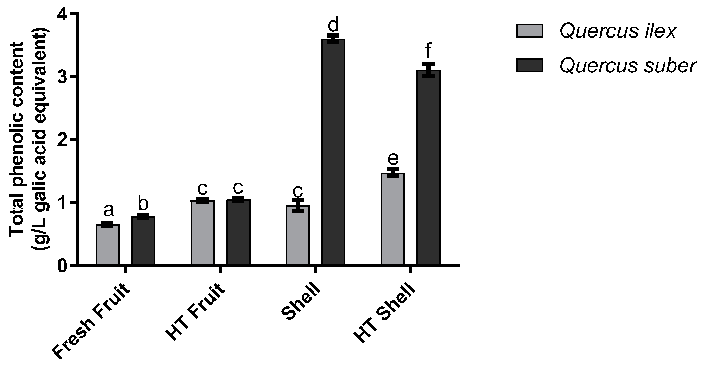
3.1. Total Phenolic Content

The results obtained regarding the total phenolic content of the acorn extracts can be seen in Figure 1. The first major takeaway is the statistically significant (p < 0.05) differences observed between Q. suber and Q. ilex shell and fruit’s phenolic content, with the first presenting values which were 2.1 to 3.7× times superior. This is contrary to what has been previously reported in the work of Cantos, Espín [21] as it showed that acorn’s fruit had a higher phenolic content than that of its shell’s. Similarly, the generally higher phenolic content observed for Q. suber contradicts previous works, where Q. ilex has been reported as possessing a higher content of phenolic compounds [21]. However, the higher phenolic compound content observed here for the roasted samples is in line with previous works, as the thermal treatment of samples has been shown to increase the total phenolic content of acorn extracts due to the degradation at high temperature of hydrolysable tannins present in acorn, originating either ellagic or gallic acids [22,23].

3.2. Well-Diffusion Assay

Phenolic rich extracts, such as the ones used in this work, have been widely described as possessing antimicrobial properties [14,24,25,26,27,28]. The results obtained for the screening of the hereby produced extracts’ antimicrobial activity (Table 1) showed that in the eight microorganisms assayed, acorn extracts were effective in inhibiting the Gram-positive bacteria. Comparing these results with those of previous works, it is interesting to see that the results reported by Güllüce, Adıgüzel [8] were completely opposite to ours as a methanolic Q. ilex extract inhibited the growth of C. albicans, P. fluorescens and E. coli but not that of S. aureus. Similarly, Berahou, Auhmani [6] showed that methanolic-based extracts of Q. ilex bark were capable of inhibiting Gram-positive and Gram-negative bacteria alike. These discrepancies in activity are probably due to the nature of the extracts as these works used methanol as an extraction solvent, while water was used in this work, and different solvents extract different compounds [17]. Moreover, the differences in antimicrobial activity observed between Gram-positive and Gram-negative bacteria may be linked to their phenolic content, as these compounds have been widely described to have an effect that is, mostly, membrane-based. Thus, the outer lipidic membrane characteristic of Gram-negative bacteria presents an additional protection mechanism and explains phenolic compounds’ lack of activity against these bacteria, which is in line with the results observed [29,30,31,32]. In fact, these results are in accordance with both those reported by Sung, Kim [33], who showed that aqueous acorn shell extracts inhibited both MRSA and vancomycin-resistant S. aureus (VRSA), while exhibiting little to no activity against the tested Gram-negative strains; and with those reported by Boulekbache-Makhlouf, Slimani [34] and by Silva, Costa [18], who showed that fruits extracts rich in phenolics and tannins had a strong antibacterial action against Gram-positive bacteria and weak to null activity against Gram-negative bacteria. Furthermore, Coelho, Silva [22] showed that Q. ilex and Q. suber extracts were rich in ellagic acid, gallic acid and (−) epicatechin gallate, all compounds known to inhibit Gram-positive bacteria and S. aureus in particular [35].

3.3. MBC

When regarding the bactericidal activity of the acorn extracts, the MBC values obtained can be seen in Table 2. Considering only the conditions which presented inhibition halos, it is noticeable that shell extracts presented lower MBC values than the fruit ones. When considering the different acorn cultivars assayed, it is interesting to note that while for Q. suber there were no differences between fresh and HT extracts, the same could not be said for Q. ilex, as HT extracts were, in general, more active than fresh extracts. As the antimicrobial activity of plant/fruit extracts is strongly associated with their phenolic compounds content [36], this higher bactericidal activity could be expected as Q. ilex HT extracts presented a higher total phenolic content. Comparing these results with previous works is difficult since, to the best of our knowledge, no previous work has been reported on the MBC values for either Q. ilex or Q. suber acorn extracts. Nevertheless, some comparisons can still be drawn against works using other Quercus cultivars. Khurram, Hameed [9]’s work regarding an aqueous fraction of a methanolic extract of Quercus baloot was contrary to the results obtained here, as it showed that the extracts in question had no bactericidal activity against S. aureus and B. cereus. On the other hand, Basri and Fan [5]’s results are in line with ours, as they showed that an aqueous extract of Q. infectoria had a MBC of 2.5 mg/mL for S. aureus.

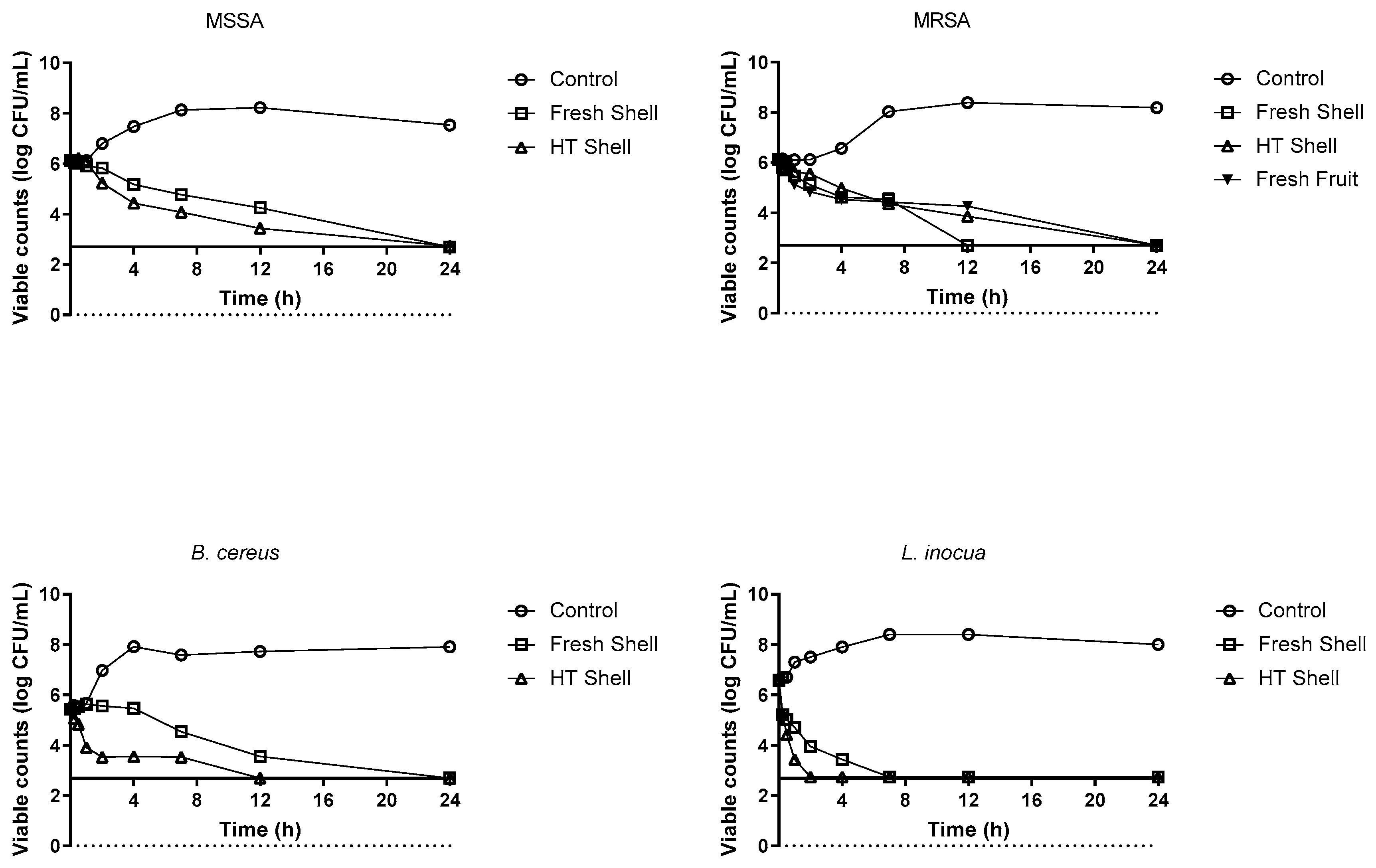
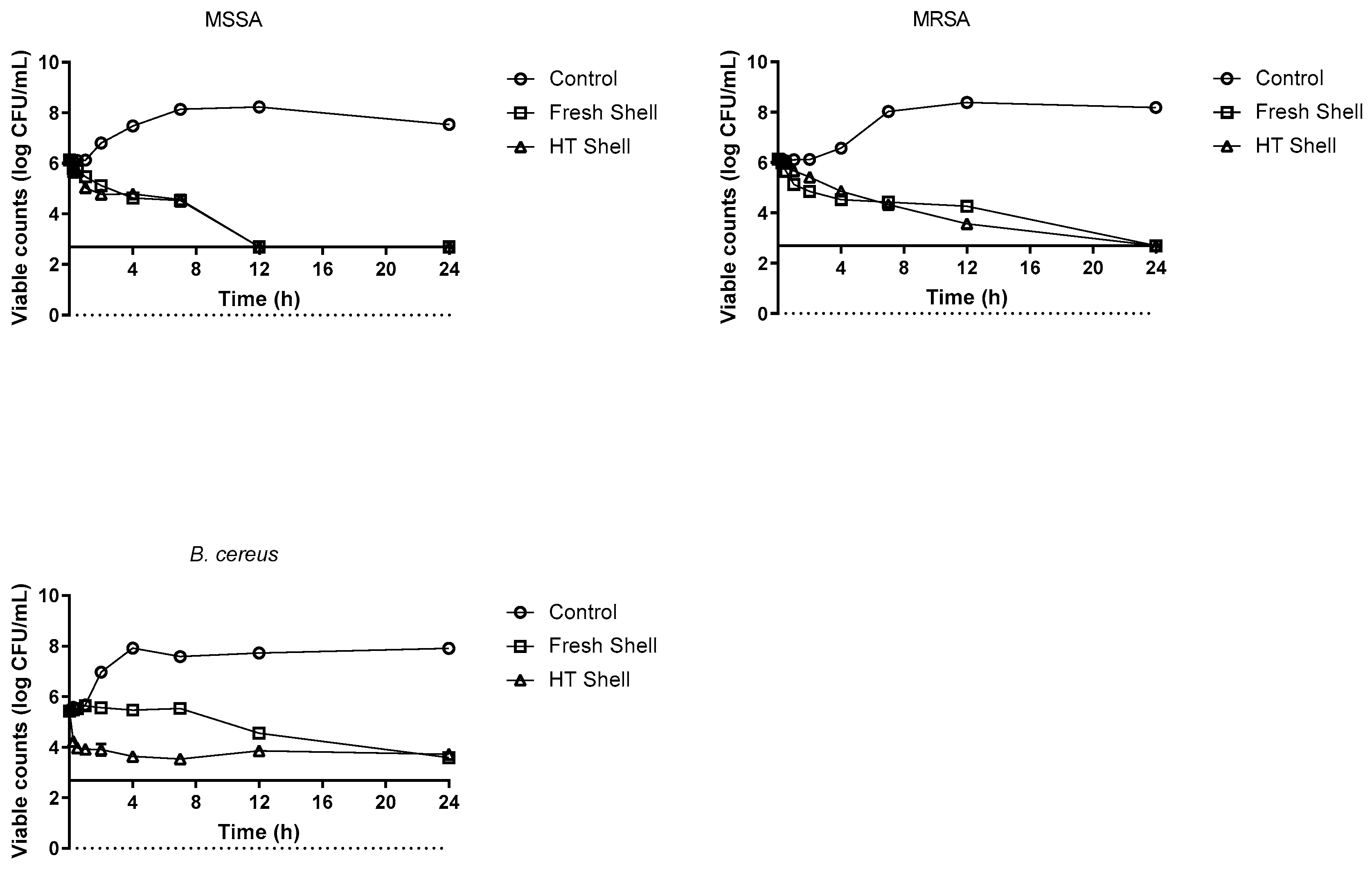
3.4. Time-Death Curves

When regarding the data obtained in these assays (Figure 2), of all of the microorganisms tested, L. inocua appeared to be the most susceptible to Q. ilex’s extracts, with the viable counts dropping below the quantification limit after 2 h in HT shell extracts and 7 h in the fresh shell extract. This higher activity of HT shell extracts was also observed for B. cereus and MSSA. In the first, viable counts dropped below the quantification limit at the 12 h mark for HT shell extracts, while for fresh shell, this only occurred after 24 h. For MSSA, while the viable counts were only below the quantification limit for both shell extracts, the viable counts are systematically lower in the HT shell extract after 24 h. Interestingly, for MRSA, the opposite behaviour was observed, with the fresh shell exhibiting the strongest effect (viable counts dropping below the quantification limit after 12 h for fresh extract and only after 24 in the HT shell extract). This behaviour has two possible explanations: the first is the supposed enhanced antimicrobial activity of phenolic compounds against multidrug-resistant microorganisms, as previously described by Bocquet, Sahpaz [37]; the second possibility is that it be the mechanisms described in Miklasińska-Majdanik, Kępa’s work [38], where compounds such as epicatechin gallate (which is present in accorn) have been reported as causing the formation of pseudomulticellular aggregates of bacteria which are believed to be a single CFU, thus giving a false impression of diminishing CFU levels.
The data obtained regarding the time-death curves produced in the presence of Q. suber extracts at the MBC concentration can be seen in Figure 3. In this case, MSSA appeared to be the most susceptible microorganism, with viable counts dropping below the quantification limit after 12 h regardless of the shell extract, which demonstrates that, at MBC, Q. suber extracts appear to be more effective in inhibiting this microorganism than Q. ilex ones. Interestingly for MRSA, while the HT shell extract exhibited a similar effect that as observed for the HT shell from Q. ilex acorns (values dropped below the quantification limit at the 24 h mark), for the fresh shell extracts, the Q. suber one was less effective than the Q. ilex one, causing a reduction in viable counts below the quantification limit after 24 h instead of after 12 h, as observed for Q. ilex fresh shell. This lower effectiveness of Q. suber extracts was also observed for B. cereus. In fact, when considering the data, it can be observed that, while HT shell extracts appeared to have a stronger impact on the total viable counts, the overall effect does not drop below the quantification limit even after 24 h. As this bacteria species are known to produce spores, a test was carried out by heating the samples to 95 °C before cooling and plating; however, as no growth was observed in these scenarios, the data were not added to the manuscript, although these did show that the viable count levels observed did not originate from the presence of these structures.

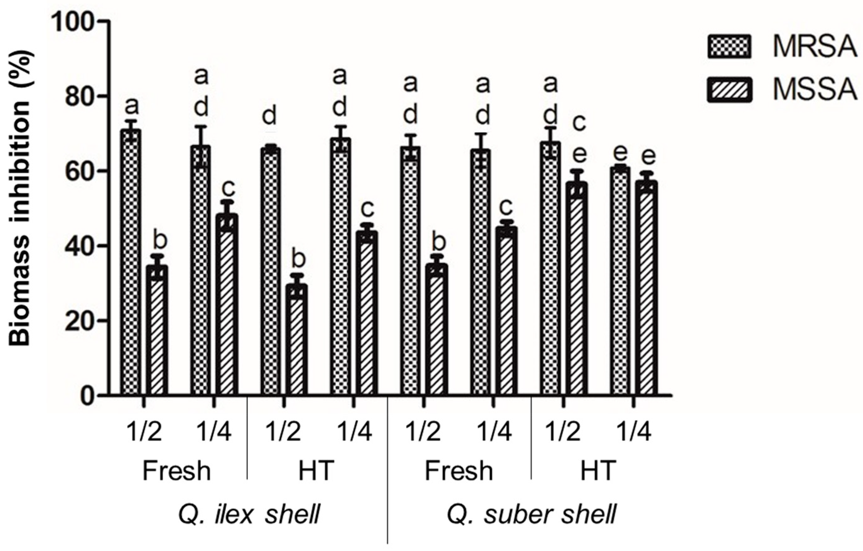
3.5. Antibiofilm Assay

Of all microorganisms that were susceptible to the extract’s effects, S. aureus were the only ones that could establish biofilms. Thus, and considering the higher activities observed, shell extracts were screened for their capacity to inhibit the formation of these structures (Figure 4). Interestingly, MRSA appeared to be more susceptible to extract’s action, with all extracts exhibiting inhibitions around 70% regardless of the heat treatment or the type of Quercus tree considered. For MSSA, while less susceptible to the extracts than MRSA, an interesting trend could be observed. For all extracts, the lower concentration was more effective (statistically significant p < 0.05) than the highest one, with the only exception being for Q. suber HT shell extract (where no significant differences were observed between concentrations; p > 0.05). As biofilms are a recognized protection structure, it has been hypothesized that high concentrations of phenolic compounds could act as a strong stimulus towards the formation of this structure, namely through the stimulation of exopolysaccharides production, which could function as a barrier to protect sessile cells while also increasing the biofilm’s overall biomass [39]. Overall, to the best of our knowledge, no reports have been made on acorns’, and particularly acorn shells’, effects on biofilm establishment by S. aureus, although some reports have been made regarding the effect of acorn extracts from other Quercus species. These include the work of Bahar, Ghotaslou [4], who reported that Q. infectoria subps. persica acorn extracts could inhibit S. aureus biofilm formation at a concentration capable of inhibiting the bacteria’s growth by 57%; and the work of Chusri, Phatthalung [7], who reported that both MRSA and MSSA isolates’ biofilm formation was significantly inhibited when exposed to 0.25 mg/mL of Q. infectioria G. Olivier extract, although these authors use nutgalls, not acorns.

4. Conclusions

Overall, the acorn extracts evaluated exhibited an interesting antimicrobial and antibiofilm potential against an array of pathogenic microorganisms. Of the assayed extracts, those obtained from Q. ilex shells showed the highest potential as they showed the highest antimicrobial activity, with MBCs varying between 10 and 5 mg/mL and strong growth and biofilm inhibitions also being observed. Overall, all extracts showed relevant biological potential and this opens up the possibility of using acorn extracts as natural alternatives to chemical antimicrobials, particularly if one considers that, while the fruits may be used in other applications, namely for food and feed, the most active extracts result from what would be the by-product of this use, which could open interesting valorisation opportunities for this relatively unexploited resource.

Author Contributions

Conceptualization, S.S., E.M.C. and M.P.; methodology, S.S. and M.C.; writing—original draft preparation, S.S.; writing—review and editing, E.M.C. and M.M.; supervision, M.P.; funding acquisition, E.M.C. and M.P. All authors have read and agreed to the published version of the manuscript.

Funding

This research was funded by the Fundação para a Ciência e a Tecnologia through the grant number UIDB/50016/2020. Additionally, author E.M.C. would like to acknowledge Fundação para a Ciência e Tecnologia for his funding under the Scientific Employment Stimulus—Individual Call, number 2022.07206.CEECIND.

Institutional Review Board Statement

Not applicable.

Informed Consent Statement

Not applicable.

Data Availability Statement

The data presented in this study are available on request from the corresponding author. The data are not publicly available due to confidentiality agreements.

Acknowledgments

The authors would like to acknowledge Herdade do Freixo do Meio for kindly providing both acorn types used in the current work.

Conflicts of Interest

The authors declare no conflict of interest.

References

  1. Dogan, A.; Celik, I.; Kaya, M.S. Antidiabetic properties of lyophilized extract of acorn (Quercus brantii Lindl.) on experimentally STZ-induced diabetic rats. J. Ethnopharmacol. 2015, 176, 243–251. [Google Scholar]
  2. Yarani, R.; Mansouri, K.; Mohammadi-Motlagh, H.R.; Mahnam, A.; Emami Aleagha, M.S. In vitro inhibition of angiogenesis by hydroalcoholic extract of oak (Quercus infectoria) acorn shell via suppressing VEGF, MMP-2, and MMP-9 secretion. Pharm. Biol. 2013, 51, 361–368. [Google Scholar] [CrossRef] [Green Version]
  3. Gezici, S.; Sekeroglu, N. Neuroprotective potential and phytochemical composition of acorn fruits. Ind. Crops Prod. 2019, 128, 13–17. [Google Scholar] [CrossRef]
  4. Bahar, Z.; Ghotaslou, R.; Taheri, S. In vitro anti-biofilm activity of Quercus brantii subsp. persica on human pathogenic bacteria. Res. J. Pharm. 2017, 4, 67–73. [Google Scholar]
  5. Basri, D.; Fan, S. The potential of aqueous and acetone extracts of galls of Quercus infectoria as antibacterial agents. Indian J. Pharmacol. 2005, 37, 26–29. [Google Scholar] [CrossRef] [Green Version]
  6. Berahou, A.; Auhmani, A.; Fdil, N.; Benharref, A.; Jana, M.; Gadhi, C.A. Antibacterial activity of Quercus ilex bark’s extracts. J. Ethnopharmacol. 2007, 112, 426–429. [Google Scholar] [CrossRef] [PubMed]
  7. Chusri, S.; Phatthalung, P.N.; Voravuthikunchai, S.P. Anti-biofilm activity of Quercus infectoria G. Olivier against methicillin-resistant Staphylococcus aureus. Lett. Appl. Microbiol. 2012, 54, 511–517. [Google Scholar] [PubMed]
  8. Güllüce, M.; Adıgüzel, A.; Öğütçü, H.; Şengül, M.; Karaman, I.; Şahin, F. Antimicrobial effects of Quercus ilex L. extract. Phytother. Res. 2004, 18, 208–211. [Google Scholar] [CrossRef]
  9. Khurram, M.; Hameed, A.; Khan, M.A.; Amin, M.U.; Hassan, M.; Ullah, N.; Manzoor, W.; Qayum, A.; Bilal, M.; Najeeb, U.; et al. Antibacterial potentials of Quercus baloot Griff. J. Med. Plants Res. 2012, 6, 1244–1249. [Google Scholar]
  10. Soon, L.K.; Hasni, E.; Law, K.S.; Waliullah, S.S.; Farid, C.G.; Mohsin, S.S. Ultrastructural findings and elemental analysis of Quercus infectoria Oliv. Ann. Microsc. 2007, 7, 32–37. [Google Scholar]
  11. Umachigi, S.P.; Jayaveera, K.N.; Kumar, C.A.; Kumar, G.S.; Kumar, D.K. Studies on wound healing properties of Quercus infectoria. Trop. J. Pharm. Res. 2008, 7, 913–919. [Google Scholar] [CrossRef] [Green Version]
  12. Aroonrerk, N.; Kamkaen, N. Anti-inflammatory activity of Quercus infectoria, Glycyrrhiza uralensis, Kaempferia galanga and Coptis chinensis, the main components of Thai herbal remedies for aphthous ulcer. J. Health Res. 2009, 23, 17–22. [Google Scholar]
  13. Mezni, F.; Stiti, B.; Fkiri, S.; Ayari, F.; Slimane, L.B.; Ksouri, R.; Khaldi, A. Phenolic profile and in vitro anti-diabetic activity of acorn from four African Quercus species (Q. suber, Q. canariensis, Q. coccifera and Q. ilex). S. Afr. J. Bot. 2022, 146, 771–775. [Google Scholar] [CrossRef]
  14. Lobiuc, A.; Pavăl, N.E.; Mangalagiu, I.I.; Gheorghiță, R.; Teliban, G.C.; Amăriucăi-Mantu, D.; Stoleru, V. Future antimicrobials: Natural and functionalized phenolics. Molecules 2023, 28, 1114. [Google Scholar] [CrossRef]
  15. Silva, S.; Costa, E.M.; Borges, A.; Carvalho, A.P.; Monteiro, M.J.; Pintado, M.M.E. Nutritional characterization of acorn flour (a traditional component of the Mediterranean gastronomical folklore). J. Food Meas. Charact. 2016, 10, 584–588. [Google Scholar] [CrossRef]
  16. Gião, M.S.; González-Sanjosé, M.L.; Rivero-Pérez, M.D.; Pereira, C.I.; Pintado, M.E.; Malcata, F.X. Infusions of Portuguese medicinal plants: Dependence of final antioxidant capacity and phenol content on extraction features. J. Sci. Food Agric. 2007, 87, 2638–2647. [Google Scholar] [CrossRef]
  17. Silva, S.; Costa, E.M.; Calhau, C.; Morais, R.M.; Pintado, M.M.E. Production of a food grade blueberry extract rich in anthocyanins: Selection of solvents, extraction conditions and purification method. J. Food Meas. Charact. 2017, 11, 1248–1253. [Google Scholar] [CrossRef]
  18. Silva, S.; Costa, E.M.; Pereira, M.F.; Costa, M.R.; Pintado, M.E. Evaluation of the antimicrobial activity of aqueous extracts from dry Vaccinium corymbosum extracts upon food microorganism. Food Control. 2013, 34, 645–650. [Google Scholar] [CrossRef]
  19. Silva, S.; Costa, E.M.; Machado, M.; Morais, R.; Calhau, C.; Pintado, M. Antiadhesive and Antibiofilm Effect of Malvidin-3-Glucoside and Malvidin-3-Glucoside/Neochlorogenic Acid Mixtures upon Staphylococcus. Metabolites 2022, 12, 1062. [Google Scholar] [CrossRef]
  20. Costa, E.M.; Silva, S.; Tavaria, F.K.; Pintado, M. Insights into the Biocompatibility and Biological Potential of a Chitosan Nanoencapsulated Textile Dye. Int. J. Mol. Sci. 2022, 23, 14234. [Google Scholar] [CrossRef]
  21. Cantos, E.; Espín, J.C.; López-Bote, C.; de la Hoz, L.; Ordóñez, J.A.; Tomás-Barberán, F.A. Phenolic Compounds and Fatty Acids from Acorns (Quercus spp.), the Main Dietary Constituent of Free-Ranged Iberian Pigs. J. Agric. Food Chem. 2003, 51, 6248–6255. [Google Scholar] [PubMed]
  22. Coelho, M.; Silva, S.; Rodríguez-Alcalá, L.M.; Oliveira, A.; Costa, E.M.; Borges, A.; Martins, C.; Rodrigues, A.S.; Pintado, M.M.E. Quercus based coffee-like beverage: Effect of roasting process and functional characterization. J. Food Meas. Charact. 2018, 12, 471–479. [Google Scholar] [CrossRef]
  23. Rakić, S.; Petrović, S.; Kukić, J.; Jadranin, M.; Tešević, V.; Povrenović, D.; Šiler-Marinković, S. Influence of thermal treatment on phenolic compounds and antioxidant properties of oak acorns from Serbia. Food Chem. 2007, 104, 830–834. [Google Scholar] [CrossRef]
  24. Silliker, J.H. (Ed.) Microbial Ecology of Foods: Factors Affecting Life and Death of Microorganisms; Academic Press: Cambridge, MA, USA, 1980. [Google Scholar]
  25. Huang, C.B.; Ebersole, J.L. A novel bioactivity of omega-3 polyunsaturated fatty acids and their ester derivatives. Mol. Oral Microbiol. 2010, 25, 75–80. [Google Scholar] [CrossRef] [PubMed]
  26. Cowan, M.M. Plant products as antimicrobial agents. Clin. Microbiol. Rev. 1999, 12, 564. [Google Scholar] [CrossRef] [Green Version]
  27. Huang, C.B.; George, B.; Ebersole, J.L. Antimicrobial activity of n-6, n-7 and n-9 fatty acids and their esters for oral microorganisms. Arch. Oral Biol. 2010, 55, 555–560. [Google Scholar] [CrossRef] [PubMed] [Green Version]
  28. Mandal, S.M.; Dias, R.O.; Franco, O.L. Phenolic Compounds in Antimicrobial Therapy. J. Med. Food 2017, 20, 1031–1038. [Google Scholar] [CrossRef]
  29. Burdulis, D.; Sarkinas, A.; Jasutiene, I.; Stackevicené, E.; Nikolajevas, L.; Janulis, V. Comparative study of anthocyanin composition, antimicrobial and antioxidant activity in bilberry (Vaccinium myrtillus L.) and blueberry (Vaccinium corymbosum L.) fruits. Acta Pol. Pharm. 2009, 66, 399–408. [Google Scholar]
  30. Doyle, M.P.; Beuchat, L.R. Food Microbiology: Fundamentals and Frontiers; ASM Press: Washington, DC, USA, 2007. [Google Scholar]
  31. de Souza, E.L.; de Barros, J.C.; de Oliveira, C.E.V.; da Conceição, M.L. Influence of Origanum vulgare L. essential oil on enterotoxin production, membrane permeability and surface characteristics of Staphylococcus aureus. Int. J. Food Microbiol. 2010, 137, 308–311. [Google Scholar]
  32. Vattem, D.A.; Lin, Y.T.; Labbe, R.G.; Shetty, K. Phenolic antioxidant mobilization in cranberry pomace by solid-state bioprocessing using food grade fungus Lentinus edodes and effect on antimicrobial activity against select food borne pathogens. Innov. Food Sci. Emerg. Technol. 2004, 5, 81–91. [Google Scholar] [CrossRef]
  33. Sung, S.H.; Kim, K.H.; Jeon, B.T.; Cheong, S.H.; Park, J.H.; Kim, D.H.; Kweon, H.J.; Moon, S.H. Antibacterial and antioxidant activities of tannins extracted from agricultural by-products. J. Med. Plants Res. 2012, 6, 3072–3079. [Google Scholar] [CrossRef]
  34. Boulekbache-Makhlouf, L.; Slimani, S.; Madani, K. Total phenolic content, antioxidant and antibacterial activities of fruits of Eucalyptus globulus cultivated in Algeria. Ind. Crops Prod. 2013, 41, 85–89. [Google Scholar] [CrossRef]
  35. Akiyama, H.; Fujii, K.; Yamasaki, O.; Oono, T.; Iwatsuki, K. Antibacterial action of several tannins against Staphylococcus aureus. J. Antimicrob. Chemother. 2001, 48, 487–491. [Google Scholar] [CrossRef] [Green Version]
  36. Silva, S.; Costa, E.M.; Horta, B.; Calhau, C.; Morais, R.M.; Pintado, M.M. Anti-biofilm potential of phenolic acids: The influence of environmental pH and intrinsic physico-chemical properties. Biofouling 2016, 32, 853–860. [Google Scholar] [CrossRef]
  37. Bocquet, L.; Sahpaz, S.; Bonneau, N.; Beaufay, C.; Mahieux, S.; Samaillie, J.; Roumy, V.; Jacquin, J.; Bordage, S.; Hennebelle, T.; et al. Phenolic Compounds from Humulus lupulus as Natural Antimicrobial Products: New Weapons in the Fight against Methicillin Resistant Staphylococcus aureus, Leishmania mexicana and Trypanosoma brucei Strains. Molecules 2019, 24, 1024. [Google Scholar] [CrossRef] [PubMed] [Green Version]
  38. Miklasińska-Majdanik, M.; Kępa, M.; Wojtyczka, R.D.; Idzik, D.; Wąsik, T.J. Phenolic Compounds Diminish Antibiotic Resistance of Staphylococcus Aureus Clinical Strains. Int. J. Environ. Res. Public Health 2018, 15, 2321. [Google Scholar] [CrossRef] [PubMed] [Green Version]
  39. Landini, P. Cross-talk mechanisms in biofilm formation and responses to environmental and physiological stress in Escherichia coli. Res. Microbiol. 2009, 160, 259–266. [Google Scholar] [CrossRef]
Figure 1. Total phenolic content of the different acorn extracts, fruit and shell, fresh and after heat treatment (HT). Different letters mark statistically significant (p < 0.05) differences between the datasets.
Applsci 13 06820 g001
Figure 2. Time-death curves (drawn at MBC concentration) for each of the microorganisms for active Q. ilex extracts. The x-axis crosses the y-axis at the method’s quantification limit (log 500), with points overlapping the axis corresponding to values below it.
Applsci 13 06820 g002
Figure 3. Time-death curves (drawn at MBC concentration) for each of the microorganisms for active Q. ilex extracts. The x-axis crosses the y-axis at the method’s quantification limit (log 500), with points overlapping the axis corresponding to values below it.
Applsci 13 06820 g003
Figure 4. Biofilm inhibition (in percentage of biofilm’s biomass inhibition) by the different shell extracts upon MRSA and MSSA biofilm formation. Different letters above each bar mark statistically significant (p < 0.05) differences between each dataset.
Applsci 13 06820 g004
Table 1. Inhibition halos (in mm) for the extracts observed in the well-diffusion assay.
Q. ilexQ. suber
FruitShellFruitShell
FreshHTFreshHTFreshHTFreshHT
C. albicansNINININININININI
B. cereus9.3 ± 1.09.8 ± 111.5 ± 0.611.8 ± 0.58.8 ± 1.3NI9.5 ± 2.59.8 ± 0.5
E. coliNINININININININI
L. inocuaNINI10.0 ± 0.88.8 ± 1.0NINININI
MRSA10.8 ± 1.911.0 ± 1.411.5 ± 1.210.5 ± 1.3NI10.5 ± 1.39.0 ± 2.010.8 ± 0.5
MSSA10.8 ± 1.313.3 ± 1.013.3 ± 1.011.5 ± 0.6NINI11.3 ± 1.111.3 ± 1.0
P. aeruginosaNINININININININI
S. enteritidisNINININININININI
NI, no inhibition observed.
Table 2. MBC (in mg/mL) for the extract–microorganisms that demonstrated inhibition halos in the well-diffusion assay.
Q. ilexQ. suber
FruitShellFruitShell
FreshHTFreshHTFreshHTFreshHT
B. cereus>20>201010>20NT1010
L. inocuaNTNT1010NTNTNTNT
MRSA10>20105NT>2055
MSSA>20>20105NTNT55
NT, not tested.
Disclaimer/Publisher’s Note: The statements, opinions and data contained in all publications are solely those of the individual author(s) and contributor(s) and not of MDPI and/or the editor(s). MDPI and/or the editor(s) disclaim responsibility for any injury to people or property resulting from any ideas, methods, instructions or products referred to in the content.

