Article

Improvement Effect of Membrane-Free Stem Cell Extract on Atopic Dermatitis in NC/Nga Mice

1
Department of Food Science and Nutrition & Kimchi Research Institute, Pusan National University, Busan 46241, Republic of Korea
2
T-Stem Co., Ltd., Changwon 51573, Republic of Korea
*
Authors to whom correspondence should be addressed.
These authors contributed equally to this work.
Appl. Sci. 2023, 13(7), 4542; https://doi.org/10.3390/app13074542
Submission received: 9 March 2023 / Revised: 28 March 2023 / Accepted: 31 March 2023 / Published: 3 April 2023
(This article belongs to the Special Issue Natural Products: Sources and Applications)

Abstract

:
Membrane-free stem cell extract (MFSCE) derived from adipose tissue has been reported to have anti-inflammatory activity. In the present study, we investigated the effects of MFSCE on atopic dermatitis (AD)-like skin inflammation using house-dust-mite-sensitized NC/Nga mice. Topical application of MFSCE significantly ameliorated AD-like clinical symptoms including erythema, dry skin, edema, excoriation, erosion, lichenification, and scratching. In addition, the levels of serum immunoglobulin E and inflammatory cytokines were decreased by MFSCE treatment. Furthermore, treatment with MFSCE inhibited the increase in epidermal thickness, infiltration of mast cells, expression of interleukin (IL)-4, IL-10, interferon-γ, tumor necrosis factor-α, thymus, and activation-regulated chemokines in the dorsal skin of NC/Nga mice. In conclusion, MFSCE effectively suppressed AD-like manifestations preclinically, systemically, and topically. Our study suggests that MFSCE may be a useful natural product for AD therapeutic strategies.

1. Introduction

Atopic dermatitis (AD) is an inflammatory skin disease that commonly develops early in childhood and persists in adulthood, thereby decreasing the quality of life. AD patients experiencing conditions such as erythema, dry skin, edema, excoriation, erosion, lichenification, and scratching require long-term treatment with antihistamine or immunosuppressive agents [1]. However, the efficacy and safety of these agents need to be verified in further studies. Recently, topical corticosteroids and calcineurin inhibitors have been used for AD treatment; however, side effects, including burning, redness, and pruritus, have been reported [2]. Therefore, the establishment of effective therapeutic agents for AD is of increasing global interest.
The complex etiology of AD has been reported to be cutaneous hyperreactivity caused by interaction mechanisms between genetic susceptibility, skin barrier dysfunction, immune response, and environmental factors [3]. In addition, environmental allergens affect the pathogenic mechanism of AD; in particular, house dust mite (HDM) is a major indoor environmental allergen in many countries. AD patients are predominantly sensitized to HDM allergens, and epicutaneous sensitization with HDM results in atopic flares in AD patients [4]. Bumbacea et al. [4] reported that allergens derived from HDM destroy tight junctions, subsequently eliciting AD and deteriorating the condition of the skin barrier in AD patients. Thus, HDM allergens have been recognized as relevant for causing AD.
The pathogenesis of AD is generally characterized as T-helper (Th)-driven skin inflammation in response to a specific allergen [5]. AD is implicated in pathways modulated by Th, mainly the Th2-associated immune response [6,7]. In AD skin lesions, resident cells, including keratinocytes and macrophages, and recruited cells, such as neutrophils, eosinophils, and lymphocytes, produce Th-associated cytokines and chemokines. Consequently, abnormal immune responses affect the onset, progression, and severity of AD [8]. Therefore, the suppression of impaired immune responses induced by Th-associated inflammatory cytokines is a prominent therapeutic approach for AD.
Adipose tissue-derived stem cells (ADSCs) are pluripotent mesenchymal stem cells that have been widely demonstrated to exhibit remarkable wound healing and regenerative activity via a paracrine mechanism [9,10]. However, sufficient evidence has reported that cell-based therapy has several limitations, as cells have to be grown, preserved, and transported. Additionally, to apply cell-based therapy in clinical trials, side effects such as tumor formation and unwanted immune reactivity must be considered [11]. Hence, accumulated evidence indicates that the application of the secretome from stem cells can be more effective than cell-based therapy and overcome the limitations of cell-based therapy [12,13].
Membrane-free stem cell extract (MFSCE) originating from human adipose tissue, which includes the intracellular components of the cells but lacks cell membranes, is an alternative functional natural material for cell-based therapy [11]. Studies have reported that MFSCE has several biological properties including antioxidant [14] and anti-inflammatory activities [11] and neuroprotective [15] effects. More importantly, our previous study demonstrated the potent anti-inflammatory activities of MFSCE in lipopolysaccharide (LPS) and interferon-gamma (IFNγ)-stimulated RAW 264.7 macrophages [16]. Accordingly, in the present study, we investigated the beneficial effects and mechanisms of MFSCE activity using an HDM-induced, AD-like skin lesion mouse model.

2. Materials and Methods

2.1. Reagents

HDM cream containing Dermatophagoides farinae extract (Dfe) was purchased from Biostir Inc. (Osaka, Japan). Dexamethasone was purchased from Sigma–Aldrich (St. Louis, MO, USA). Protein marker and acrylamide were purchased from GenDEPOT Co. (Katy, TX, USA). Protein assay dye and 2× laemmli sample buffer were purchased from Bio-rad Lab. (Hercules, CA, USA). Protease inhibitor cocktail and polyvinylidene fluoride (PVDF) membranes were purchased from Merck Millipore Co. (Burlington, MA, USA). Radioimmunoprecipitation assay (RIPA) buffer was purchased from iNtRON Bio. (Seongnam, Republic of Korea). Tween-20 was purchased from VWR Life Science Co. (Radnor, PA, USA). Bovine serum albumin was purchased Bioworld Technology, Inc. (St. Louis, MN, USA). Paraformaldehyde solution was purchased from Bio Basic Inc. (Markham, ON, Canada). Sodium dodecyl sulfate (SDS) was purchased from Thermofisher Scientific Co. (Waltham, MA, USA). Beta (β)-actin antibody was purchased from Cell Signaling Technology (Beverly, MA, USA). Tumor necrosis factor (TNF)-α, IFN-γ, and interleukin (IL)-4 antibody was purchased Santa Cruz Biotechnology, Inc. (Dallas, TX, USA). Thymus and activation-regulated chemokine (TARC) and IL-10 antibody was purchased Abcam Inc. (Cambridge, UK). Mouse IgE ELISA kit was purchased Koma Biotech Inc. (Seoul, Republic of Korea). Mouse TNF-α, IFN-γ, and IL-4 ELISA kit was purchased R&D Systems (Minneapolis, MN, USA). Mouse IL-10 ELISA kit was purchased Lifespan Biosciences Inc. (Seattle, WA, USA).

2.2. Preparation of MFSCE

The MFSCE was supplied from T-Stem Co., Ltd. (Changwon, Republic of Korea) and was prepared according to the patented technology as reported in our previous study [16]. Briefly, stem cells purified from female human fat tissue (body mass index, 25–29.9) were cultured at 37 °C and 5% CO2 in serum-free medium. Human fat tissue-derived stem cells were characterized by specific markers on cell membrane (positive, CD105, CD29; negative, CD34). The collected cells were subcultured for 6–8 passages, and the membranes of the collected stem cells were disrupted by ultrasonication. The cells were subjected to centrifugation, followed by filtration. The resulting filtrate was used as the MFSCE in the present study. The MFSCE was approved by the Good Laboratory Practice (GLPs) accreditation authority based on nine safety tests.

2.3. Experimental Animals

NC/Nga mice (male, 7 weeks old, n = 42) were purchased from SLC, Inc. (Shizuoka, Japan). The mice were housed in individual cages under controlled environments, including room temperature (20 ± 2 °C), humidity (50 ± 10%), and a 12 h/12 h light–dark cycle during the entire experimental period. Food and water were provided ad libitum. After acclimatization for one week, the mice were treated and maintained in accordance with the guidelines of the Pusan National University Institutional Animal (PNU-IACIC, approval number: PNU-2022-0104).

2.4. Induction of AD-like Skin Inflammation

AD-like skin inflammation was induced by topical application of an HDM cream containing Dfe to the dorsal skin of NC/Nga mice. Briefly, the back hair of the mice was shaved using a clipper one day before skin inflammation induction. Subsequently, to disrupt the skin dermal barrier, 4% sodium dodecyl sulfate (SDS) was spread on the shaved dorsal skin at a volume of 150 μL. After 3 h, 100 mg of HDM cream containing Dfe was applied to the dried dorsal skin of mice for immune sensitization every two to three days for 3 weeks, totaling six times.

2.5. Design of Experimental Group

The mice were randomly divided into six groups: non-sensitized normal group-applied saline (NOR, n = 7); HDM-sensitized vehicle group-applied saline (VEH, n = 7); HDM-sensitized group treated with 5, 10, and 20 µL (v/v) MFSCE (MFSCE5, MFSCE10, and MFSCE20, n = 7, respectively); and HDM-sensitized group treated with 540 µg (w/v) dexamethasone (DEX, n = 5–7). The concentration of MFSCE groups were determined on the basis of MFSCE product (T-Stem Co., Ltd.), which is consisted of 10% (v/v) MFSCE; thus, experimental groups of MFSCE were three concentrations of 5%, 10%, and 15%. In addition, we calculated the MFSCE requirement by the reference standard weight of humans. Furthermore, the calculated amount was converted again according to animal weight. The concentration of all samples represented the total amount used during the entire treatment period, and the samples were applied to the dorsal skin twice a day for 2 weeks. Figure 1 shows the time schedule used in this study.

2.6. AD Inflammation Score Evaluation

AD-like skin symptoms were evaluated by scoring dermatitis severity and scratching frequency once per week. To evaluate the severity of AD, visual evaluation was performed using Merkmal of symptom score. The severity of dermatitis was assessed based on the following criteria: (1) erythema, (2) dry skin, (3) edema/excoriation, (4) erosion, and (5) lichenification. The final dermatitis severity for each group was defined as the average of individual mouse total scores ranging from 0 to 15 (0, no symptoms; 1, mild; 2, moderate; 3, severe). Thus, the results are presented as the total sum of the scores evaluated for each. The degree of symptoms referred to manufacturer’s dermatitis symptom score sample (Biostir Inc., Okasa, Japan) and Yamamoto’s method [17]. Scratching frequency was measured once a week by recording the number of times the nose, ears, and dorsal skin were rubbed for 10 min to investigate the mouse behavioral changes caused by AD symptoms.

2.7. Evaluation of Serum Immunoglobulin (IgE) and Inflammatory Cytokines

Serum immunoglobulin E (IgE) and serum inflammatory cytokines (IL-4, IL-10, IFN-γ, and TNF-α) were evaluated using an enzyme-linked immunosorbent assay kit according to the manufacturers’ instructions.

2.8. Histological Analysis

The dorsal skin of mice was fixed with 4% formaldehyde solution and embedded in paraffin. Sections were prepared and stained with hematoxylin and eosin staining for the investigation of the epidermal structure and subjected to toluidine blue staining for the detection of the number of mast cells.

2.9. Protein Expression of Inflammatory Cytokines

Western blot analysis was conducted to verify the levels of inflammatory cytokines including IL-4, IL-10, IFN-γ, TNF-α, and TARC in the dorsal skin. Dorsal skin samples were homogenized in RIPA buffer with a protease inhibitor cocktail. The concentrations of proteins extracted from dorsal skin homogenates were investigated using the Bradford assay (Bio-Rad, Hercules, CA, USA), and all samples were produced at the same concentration. Equal amounts of protein were separated on 10–13% polyacrylamide gels and transferred to PVDF membranes. The transferred membrane was blocked in phosphate-buffered saline (PBS) with −0.05% tween 20 (PBS-T) containing 5% skim milk at 25 °C for 1 h. Subsequently, the membrane was incubated with the following primary antibodies: IL-4, IFN-γ, TNF-α, IL-10 and TARC, and β-actin. After washing with PBS-T, the membrane was incubated with horseradish peroxidase-conjugated antimouse, antirabbit, or antigoat IgG secondary antibody (Cell Signaling Technology) for 1 h. The protein bands was visualized using a Davinci-Chemiluminescent imaging system (CoreBio, Seoul, Republic of Korea).

2.10. Statistical Analysis

All results are presented as mean ± standard deviation. Significance among groups was analyzed using SPSS version 23 (IBM Corporation, Armonk, NY, USA) by one-way analysis of variance, followed by Duncan’s multiple range test. p < 0.05 was determined to be statistically significant.

3. Results

3.1. Clinical Severity and Scratching Behavior on Dorsal Skin of HDM-Sensitized NC/Nga Mice

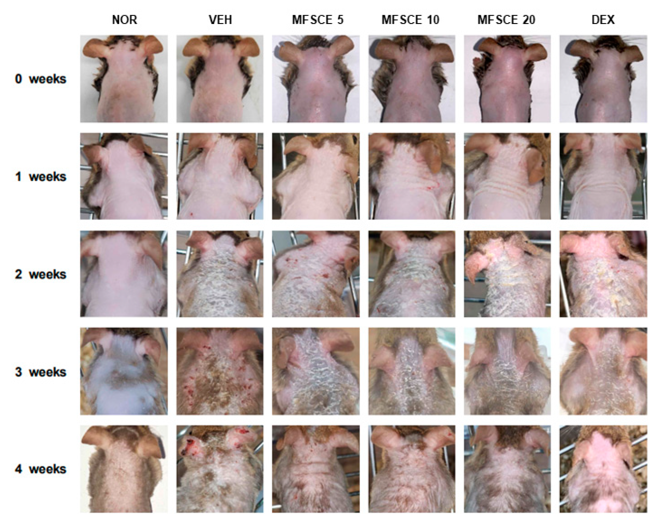
As shown in Figure 2, the dorsal skin of HDM-sensitized VEH mice visibly expressed AD-like symptoms, including erythema, dry skin, edema/excoriation, erosion, and lichenification, beginning at 2 weeks after HDM sensitization. In contrast, MFSCE application to the dorsal skin inhibited these AD-like symptoms. The clinical severity was scored according to the standard for dermatitis symptoms.
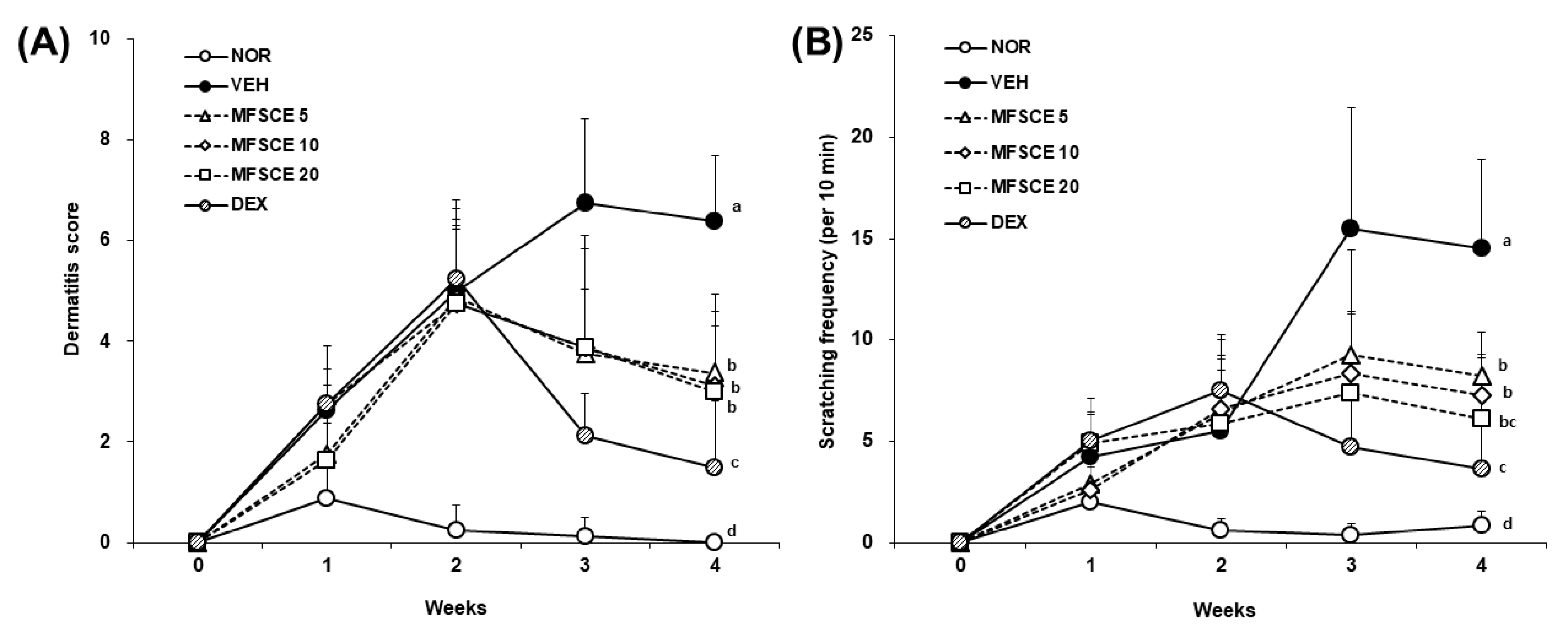
The skin severity scores for each group are shown in Figure 3A. All groups, except the NOR group, exhibited progression of AD-like symptoms for up to 2 weeks. However, starting at 3 weeks, the dermatitis scores in the MFSCE-treated groups showed a downward tendency, similar to those in the DEX group, while the VEH group deteriorated more seriously. Scratching behavior was recorded to assess the effect of spontaneous scratching frequency on pruritus (Figure 3B). Although all VEH- and MFSCE-treated groups showed the highest scratching frequency at 3 weeks, the level of scratching frequency in the MFSCE-treated groups was lower than that in the VEH group. Although the scratching frequency decreased slightly at 4 weeks, that in the VEH and MFSCE-treated groups was significant. These results prove that MFSCE application recovered AD-like clinical symptoms in HDM-sensitized NC/Nga mice.

3.2. Serum Levels of IgE and Inflammatory Cytokines in HDM-Sensitized NC/Nga Mice

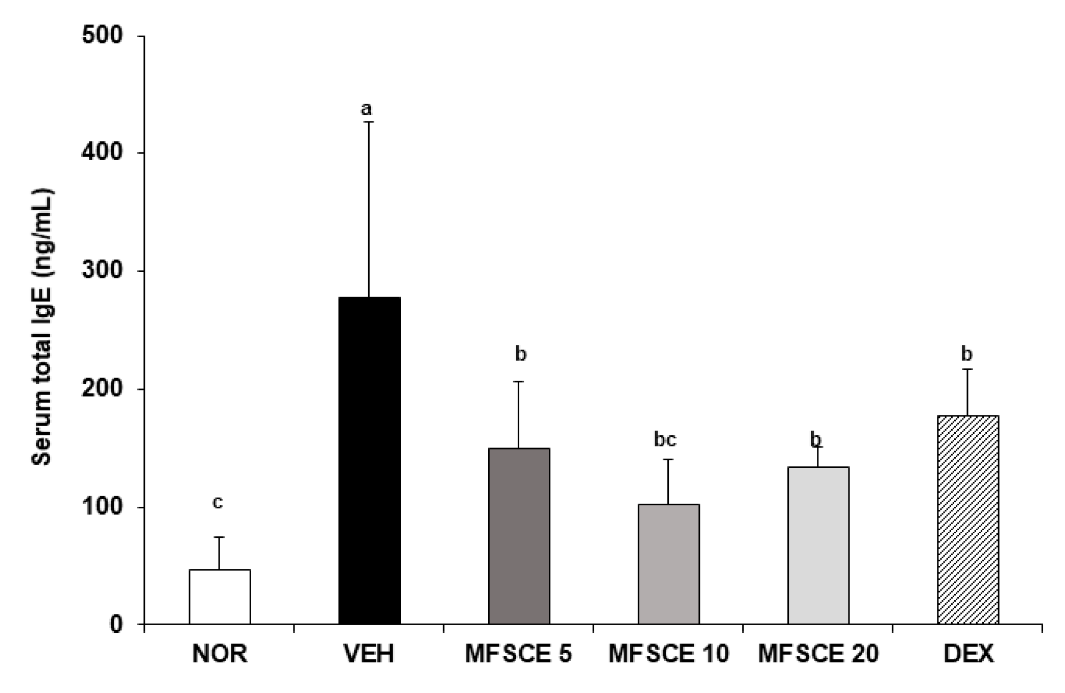
To investigate the effect of MFSCE on systemic allergic response, serum from the cardiac blood of all mice in each group was collected at 4 weeks, which is the day of dissection, and serum IgE (Figure 4) and inflammatory cytokines (Figure 5) were measured. Serum IgE levels in the VEH group (277.59 ± 149.61 ng/mL) were considerably higher (approximately six times) than those in the NOR group (47.18 ± 27.88 ng/mL). This result indicated that HDM sensitization induced AD-like allergic responses in NC/Nga mice; therefore, the AD murine model in the present study was appropriate to verify the effect of MFSCE on AD. In addition, serum IgE in MFSCE-treated groups (MFSCE5, 150.18 ± 55.84 ng/mL; MFSCE10, 101.71 ± 39.39 ng/mL; MFSCE20, 134.07 ± 16.86 ng/mL) was decreased, similar to the serum IgE level of DEX group (177.54 ± 39.41 ng/mL).
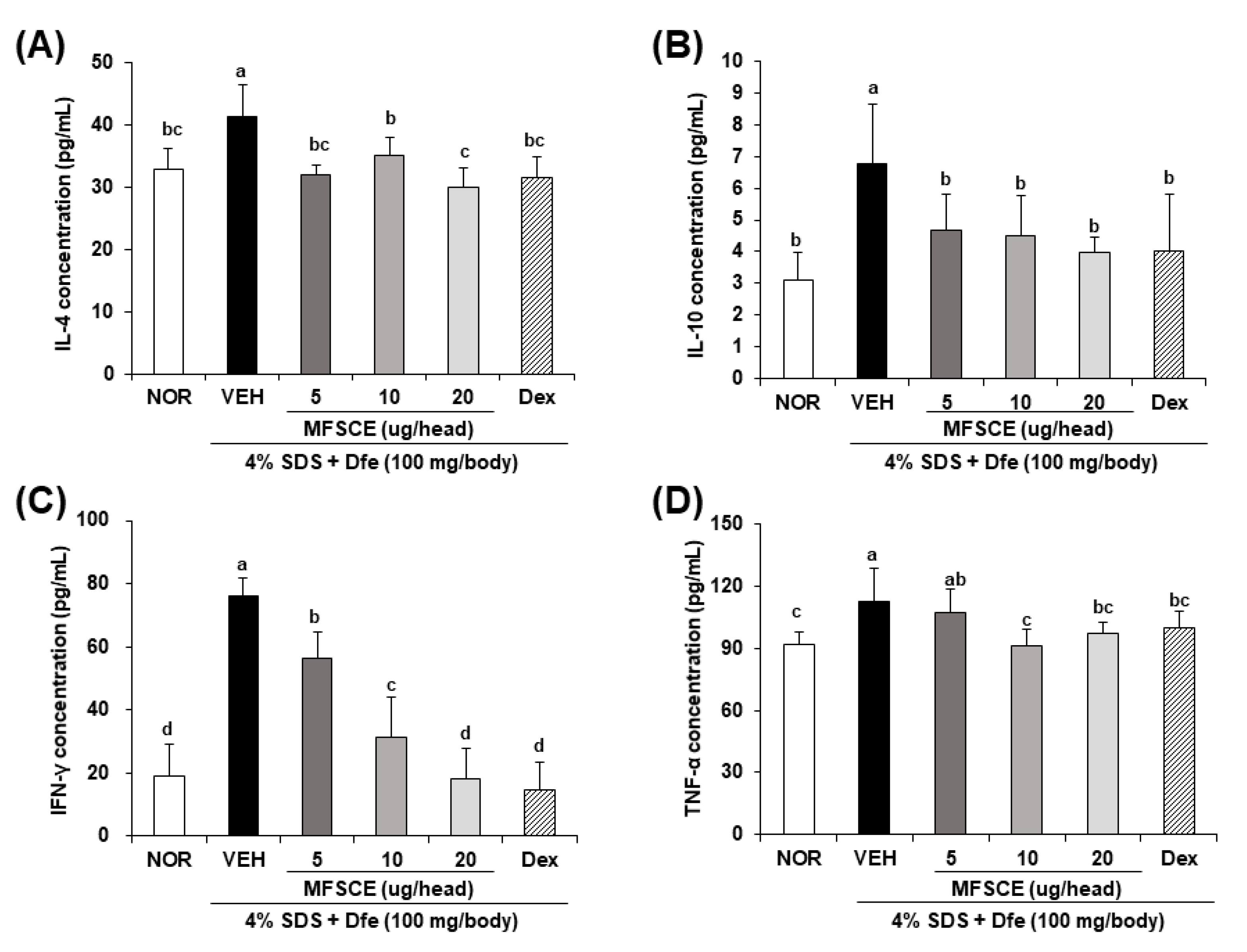
As shown in Figure 5 (A, IL-4; B, IL-10; C, IFN-γ; D, TNF-α), the secretion of inflammatory cytokines, including IL-4 (NOR, 32.93 ± 3.32 pg/mL vs. VEH, 41.28 ± 5.27 pg/mL), IL-10 (NOR, 3.10 ± 0.89 pg/mL vs. VEH, 6.77 ± 1.89 pg/mL), IFN-γ (NOR, 19.16 ± 9.80 pg/mL vs. VEH, 75.97 ± 6.04 pg/mL), and TNF-α (NOR, 92.26 ± 5.57 pg/mL vs. VEH, 112.64 ± 16.40 pg/mL), was stimulated by repeated HDM sensitization; however, MFSCE prevented the production of IL-4 (MFSCE5, 32.01 ± 1.46 pg/mL; MFSCE10, 35.18 ± 2.85 pg/mL; MFSCE20, 30.11 ± 2.90 pg/mL), IL-10 (MFSCE5, 4.67 ± 1.12 pg/mL; MFSCE10, 4.50 ± 1.25 pg/mL; MFSCE20, 134.07 ± 16.86 pg/mL), IFN-γ (MFSCE5, 56.46 ± 8.13 pg/mL; MFSCE10, 31.29 ± 12.71 pg/mL; MFSCE20, 18.26 ± 9.44 pg/mL), and TNF-α (MFSCE5, 150.18 ± 55.84 pg/mL; MFSCE10, 3.96 ± 0.48 pg/mL; MFSCE20, 134.07 ± 16.86 pg/mL). These data suggest that MFSCE application reduced AD-like allergic responses in HDM-sensitized NC/Nga mice.

3.3. Histological Change and Mast Cell Infiltration in Dorsal Skin of HDM-Sensitized NC/Nga Mice

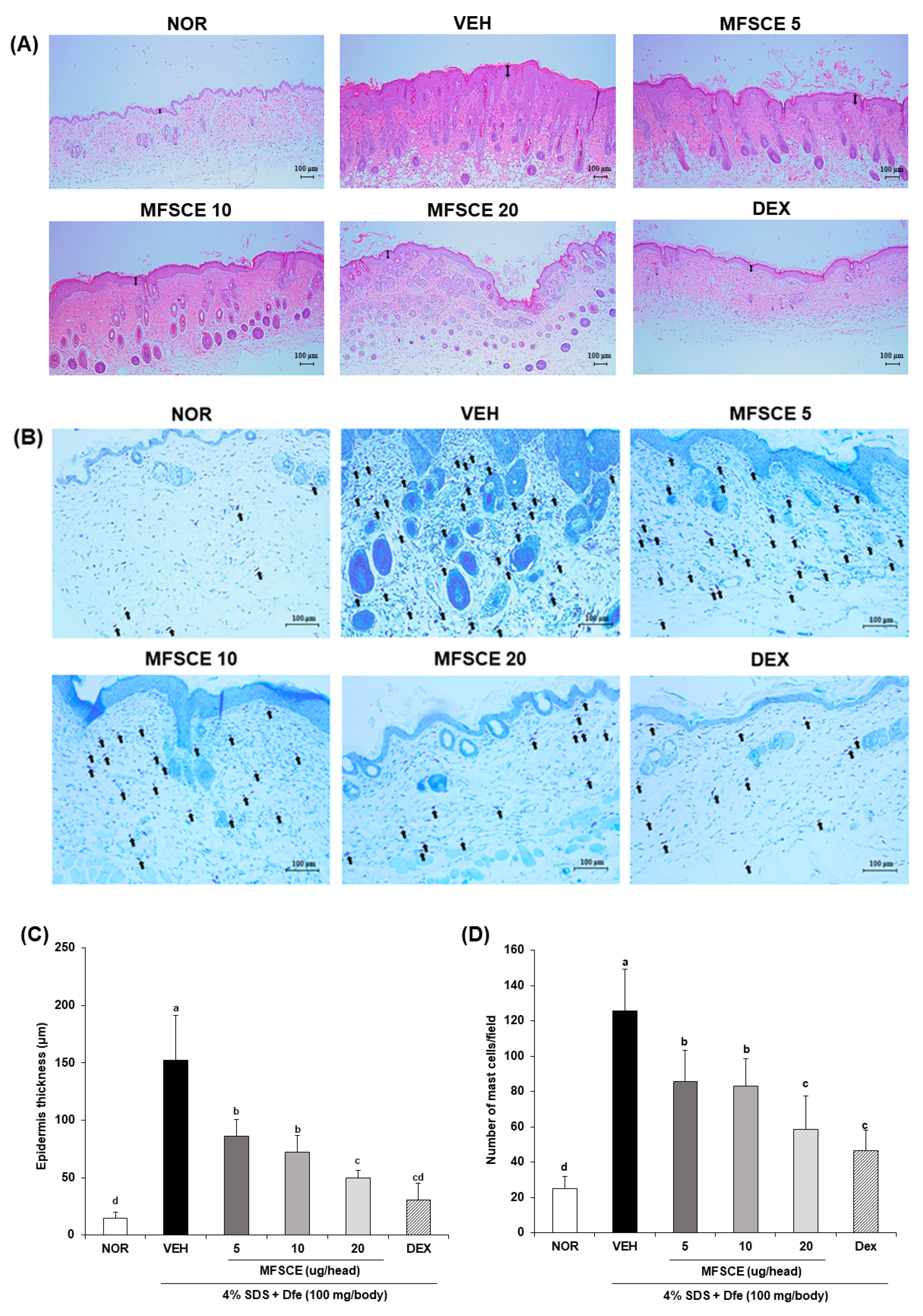
To investigate the effect of MFSCE on HDM-induced AD skin lesions, hematoxylin and eosin staining (Figure 6A) were performed to assess epidermal thickness (Figure 6C) and toluidine blue staining (Figure 6B) was performed to verify mast cell infiltration (Figure 6D) in the dorsal skin of HDM-sensitized NC/Nga mice. HDM-sensitized VEH group exhibited markedly increased epidermal thickness compared to NOR group (NOR, 14.46 ± 5.88 μm vs. VEH, 152.41 ± 38.73 μm); however, epidermal thickness was restored by the application of MFSCE in a dose-dependent manner (MFSCE5, 86.46 ± 14.35 μm; MFSCE10, 72.26 ± 14.72 μm; MFSCE20, 49.68 ± 6.61 μm; DEX, 30.89 ± 13.95 μm). Moreover, the number of mast cells, which are effector cells of allergic responses like AD, was prominently increased in the VEH group (NOR, 25.00 ± 6.80 cells/high field vs. VEH, 125.54 ± 23.92 cells/high field). In contrast, the application of MFSCE inhibited mast cell infiltration in dorsal skin lesions of HDM-sensitized NC/Nga mice (MFSCE5, 85.69 ± 17.42 cells/high field; MFSCE10, 83.09 ± 15.53 cells/high field; MFSCE20, 58.66 ± 18.93 cells/high field; DEX, 46.40 ± 11.88 cells/high field). These results suggest that the skin lesions of the MFSCE-treated groups exhibited suppression of AD via inhibition of epidermal thickening and mast cell infiltration.

3.4. Inflammatory Cytokine Expressions in Dorsal Skin of HDM-Sensitized NC/Nga Mice

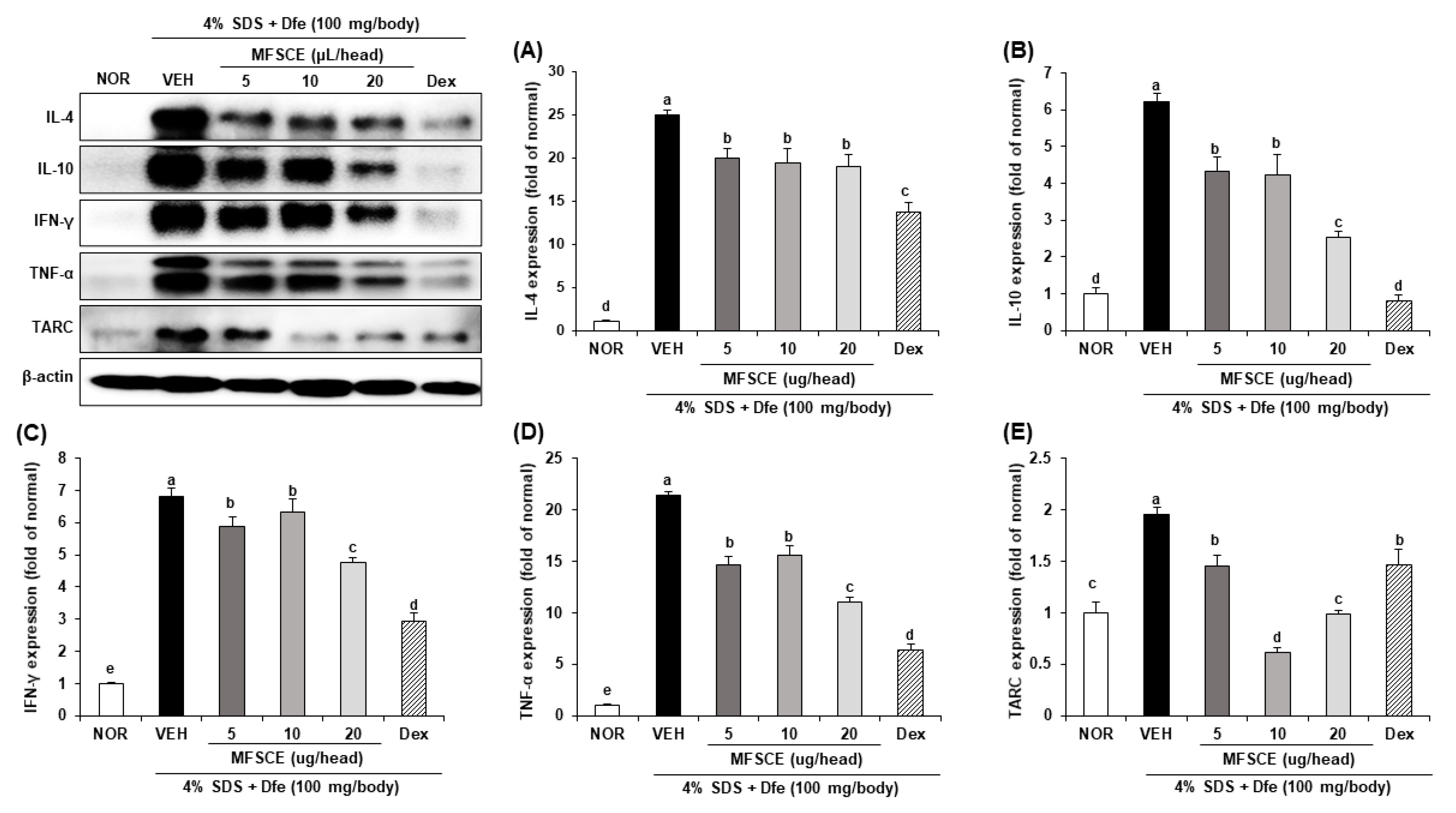
Previous studies have verified the importance of Th1- and Th2-related inflammatory cytokines and chemokines such as TARC. These cytokines and chemokines were overexpressed in the skin of an allergic AD-like murine model and were used as markers for pathogen-stimulated allergic inflammation in an experimental murine model. Thus, we determined the expression of AD-related inflammatory cytokines and chemokines in dorsal skin tissue using western blot analysis (Figure 7). The expression of inflammatory cytokines and chemokines, including IL-4 (Figure 7B), IL-10 (Figure 7C), IFN-γ (Figure 7D), TNF-α (Figure 7E), and TARC (Figure 7A) was remarkably increased in HDM-sensitized VEH mouse skin (IL-4, 24.91 ± 0.64 fold; IL-10, 6.23 ± 0.21 fold; IFN-γ, 6.82 ± 0.23 fold; TNF-α, 21.36 ± 0.34 fold; TARC, 1.95 ± 0.07 fold) than in NOR mouse skin. However, MFSCE application downregulated the expression of HDM-sensitized, AD-related inflammatory cytokines and chemokines in NC/Nga mice. These results demonstrate that MFSCE suppressed HDM-sensitized AD in NC/Nga mice by regulating the Th1- and Th2-related inflammatory response.

4. Discussion

MFSCE derived from ADSCs was found to have more advantages than ADSCs as a novel functional natural product for various biological applications. Venkatarame Gowda Saralamma et al. [11] reported the protein profiling of MFSCE through nanoscale liquid chromatography coupled to tandem mass spectrometry analysis, demonstrating that MFSCE regulated the integrin pathway proteins, inflammation regulatory proteins, and wound healing proteins. In particular, MFSCE-related proteins have been shown to have anti-inflammatory effects. In this regard, our previous study reported that MFSCE attenuated the production of inflammatory mediators including nitric oxide, cyclooxygenase-2, and prostaglandin E2 by modulating the nuclear factor-kappa B (NF-κB) and mitogen-activated protein kinases (MAPKs) pathway in LPS/IFN-γ-induced RAW264.7 macrophages [16]. Moreover, Lee et al. [18] indicated that MFSCE suppressed the production of IL-1α stimulated inflammatory factors, such as inducible nitric oxide synthase (iNOS), cyclooxygenase-2, nitric oxide, and prostaglandin E2, and downregulated the NF-κB and MAPKs signaling pathways in rat chondrocytes. Ha et al. [19] demonstrated that MFSCE-treated keratinocytes and fibroblasts inhibited inflammation and wrinkle formation. AD is a chronic and serious inflammatory skin disease; therefore, in the present study, the beneficial effects of MFSCE with excellent anti-inflammatory effects were investigated in HDM-induced, AD-like NC/Nga mice.
To explore the protective and improvement effects on AD, an animal model of human symptom-like AD is required. Suto et al. [20] demonstrated that the clinical symptoms and histopathological research data in NC/Nga mice were similar to those in human AD. In addition, NC/Nga mice spontaneously hyperproduce serum IgE influenced by environmental factors, particularly HDM allergens, which are highly sensitized as an environmental factor to human AD [21]. These observations are associated with the clinical symptoms of human AD [22]. Here, NC/Nga mice are a relevant model for human AD, as numerous studies have used them to investigate the pathophysiology and treatment of AD using natural products. In addition, a well-known contributor among HDM species is Dermatophagoides farinae (Df). For several decades, it is considered to be a common cause of AD by their body and feces as environmental air-born allergens [23]. Therefore, Dfe used in present study played a role as a representative material of HDM, triggering skin barrier dysfunction in NC/Nga mice. Park et al. [24] demonstrated the ameliorative effects of Gardenia jasminoides extract, a natural herbal medicine, in an Dfe-induced AD model in NC/Nga mice. Kang et al. [25] confirmed that cacao beans from Theobroma cacao, a source of flavonoids, showed strong anti-AD effects on Dfe-stimulated, AD-like symptoms in NC/Nga mice. Therefore, this study used HDM-sensitized NC/Nga mice to demonstrate a clinical, systemic, and topical improvement effect of MFSCE on AD-like skin inflammation.
Clinically, patients with AD experience skin inflammation characterized by erythema, dry skin, edema, excoriation, erosion, lichenification, and scratching [26]. Thus, the alleviation of AD-like symptoms is especially important in improving the quality of life for patients with AD. Our results demonstrate that repeated dermal application of MFSCE ameliorated dermatitis scores and scratching frequency in HDM-stimulated NC/Nga mice. Although DEX treatment also strongly inhibited AD-like symptoms, the body weight of the DEX group mice decreased significantly with repeated application of DEX. Jeong et al. [27] reported that DEX treatment (3 mg/kg body weight) reduced the body weight of 2,4-dinitrochlorobenzene-exposured NC/Nga mice in each experiment group. In addition, Wu et al. [28] found that DEX application (10 μg/ear) significantly decreased the body weight of oxazolone-induced Balb/c mice compared to that of healthy mice. In our study, repeated topical application of MFSCE tends to show a therapeutic effect compared to the VEH group, without any side effects, including a decrease in body weight.
Elevated serum IgE levels are a major characteristic of AD, and approximately 80% of patients with AD showed an increased concentration of IgE [20]. Sung et al. [29] reported that systemic IgE elevation in serum is increased by inflammatory cytokines after repeated application of HDM. In the present study, repeated sensitization by HDM in the dorsal skin of NC/Nga mice stimulated the production of serum IgE, consequently increasing the concentration of serum inflammatory cytokines, including IL-4, IL-10, IFN-γ, and TNF-α. However, treatment of the dorsal skin of NC/Nga mice with MFSCE inhibited both IgE and inflammatory cytokine levels in the serum. Interestingly, we found that the serum concentration of IFN-γ was markedly stimulated by HDM sensitization, and MFSCE downregulated this level in a dose-dependent manner. Evidence has shown that IFN-γ is an important factor in the severity and chronicity of skin inflammation in AD [30]. Moreover, the spontaneous production of IFN-γ is related to AD-like skin lesions in NC/Nga mice [31]. These data suggested that MFSCE suppressed chronic AD in part through systemic downregulation of IFN-γ.
Skin infiltration by mast cells is a common clinical phenomenon associated with AD-like skin lesions [32]. Elevated IgE antibodies in patients with AD have a high affinity for the specific receptor Fc epsilon RI on mast cells and stimulate mast cell degranulation, thereby releasing allergic mediators such as histamine, β-hexosaminidase, prostaglandins, and leukotrienes, which are also associated with an increase in inflammatory cytokines (IL-4, IL-10, IFN-γ, and TNF-α) and chemokines (TARC) [33,34].
TARC is expressed in the skin lesions of patients with AD, and its levels correlate with disease severity [35]. The expression of TARC is regulated by IFN-γ and various cytokines, including IL-4, IL-10, and TNF-α, which are produced by activated immune cells in the skin of AD patients [36,37]. These cytokines bind to their specific receptors on keratinocytes, the predominant cell type in the epidermis, and activate a cascade of intracellular signaling pathways that lead to TARC expression [38]. Previous studies have verified these cytokines and chemokines overexpression in the skin of an allergic AD-like murine model and were used as markers of pathogen-stimulated allergic inflammation in an experimental murine model. In present study, the release and expression of IFN-γ, especially when compared with the other inflammatory cytokines and chemokine, was remarkably inhibited by the MFSCE. These findings suggested that the MFSCE had the regulatory effect on IFN-γ, consequently showing the protective effect on AD-related dysfunctions of HDM-sensitized NC/Nga mice.
This cascade plays a significant role in the pathogenesis and aggravation of AD; therefore, targeting the production of cytokines and chemokines is a potential treatment strategy for AD. In our study, topical application of MFSCE in the dorsal skin decreased the expression levels of IL-4, IL-10, IFN-γ, TNF-α, and TARC, as well as reduced mast cell infiltration and epidermal thickness. Here, we consider that local application of MFSCE in AD-like dorsal skin was effective in reducing mast cell infiltration to AD-like skin lesions, which, in turn, suppressed inflammatory cell recruitment and resulted in improvement of AD-like clinical symptoms in HDM-induced NC/Nga mice. In other words, MFSCE may not only ameliorate AD chronicity preclinically and systemically but also topically improve AD severity.

5. Conclusions

This study provides evidence that MFSCE effectively relieves AD-like skin inflammation in HDM-sensitized NC/Nga mice. With regard to the improvement effects of MFSCE in AD, our results demonstrated that: (1) Preclinically, MFSCE alleviated AD-like symptoms, including erythema, dry skin, edema, excoriation, erosion, lichenification, and scratching. (2) Systemically, the levels of IgE and inflammatory cytokines in serum decreased after MFSCE application in AD-like skin lesions. (3) Topical application of MFSCE inhibited mast cell infiltration, decreased epidermal thickness, and decreased expression of inflammatory cytokines (IL-4, IL-10, IFN-γ, TNF-α) and chemokines (TARC) in AD-like dorsal skin lesions in HDM-sensitized NC/Nga mice. The present study suggests that dermal application of MFSCE to skin lesions of patients with AD might be a possible strategy for ameliorating AD.

Author Contributions

Conceptualization, E.J.C. and Y.S.K.; methodology, H.S.P., Q.Q.P. and B.W.N.; formal analysis, J.-H.K., Q.Q.P. and B.W.N.; investigation, Q.Q.P. and B.W.N.; resources, H.S.P. and Y.S.K.; writing—original draft preparation, J.-H.K.; writing—review and editing, E.J.C.; supervision, E.J.C. All authors have read and agreed to the published version of the manuscript.

Funding

This study received no external funding.

Institutional Review Board Statement

The experimental mice were treated and maintained in accordance with the guidelines of the Pusan National University Institutional Animal (PNU-IACIC, approval number: PNU-2022-0104).

Informed Consent Statement

Not applicable.

Data Availability Statement

The data presented in this study are available upon request from the corresponding author.

Acknowledgments

Membrane-free stem cell extracts were prepared and processed by T-Stem Co., Ltd., Republic of Korea.

Conflicts of Interest

The authors declare no conflict of interest.

References

  1. Suga, H.; Sato, S. Novel topical and systemic therapies in atopic dermatitis. Immunol. Med. 2019, 42, 84–93. [Google Scholar] [CrossRef] [PubMed] [Green Version]
  2. Bratu, D.; Boda, D.; Caruntu, C. Reflectance confocal microscopy in monitoring atopic dermatitis treated with topical calcineurin inhibitors. Healthcare 2023, 11, 152. [Google Scholar] [CrossRef] [PubMed]
  3. Mandlik, D.S.; Mandlik, S.K. Atopic dermatitis: New insight into the etiology, pathogenesis, diagnosis and novel treatment strategies. Immunopharmacol. Immunotoxicol. 2021, 43, 105–125. [Google Scholar] [CrossRef] [PubMed]
  4. Bumbacea, R.S.; Corcea, S.L.; Ali, S.; Dinica, L.C.; Fanfaret, I.S.; Boda, D. Mite allergy and atopic dermatitis: Is there a clear link? (Review). Exp. Ther. Med. 2020, 20, 3554–3560. [Google Scholar] [CrossRef] [PubMed]
  5. Peng, W.; Novak, N. Pathogenesis of atopic dermatitis. Clin. Exp. Allergy 2015, 45, 566–574. [Google Scholar] [CrossRef]
  6. Bieber, T.; Paller, A.S.; Kabashima, K.; Feely, M.; Rueda, M.J.; Ross Terres, J.A.; Wollenberg, A. Atopic dermatitis: Pathomechanisms and lessons learned from novel systemic therapeutic options. J. Eur. Acad. Dermatol. Venereol. 2022, 36, 1432–1449. [Google Scholar] [CrossRef]
  7. Yang, J.; Kuang, H.; Li, N.; Hamdy, A.M.; Song, J. The modulation and mechanism of probiotic-derived polysaccharide capsules on the immune response in allergic diseases. Crit. Rev. Food Sci. Nutr. 2022, 1–13. [Google Scholar] [CrossRef]
  8. Guttman-Yassky, E.; Zhou, L.; Krueger, J.G. The skin as an immune organ: Tolerance versus effector responses and applications to food allergy and hypersensitivity reactions. J. Allergy Clin. Immunol. 2019, 144, 362–374. [Google Scholar] [CrossRef] [Green Version]
  9. Kim, W.-S.; Park, B.-S.; Sung, J.-H. The wound-healing and antioxidant effects of adipose-derived stem cells. Expert Opin. Biol. Ther. 2009, 9, 879–887. [Google Scholar] [CrossRef]
  10. Zhang, W.; Bai, X.; Zhao, B.; Li, Y.; Zhang, Y.; Li, Z.; Wang, X.; Luo, L.; Han, F.; Zhang, J.; et al. Cell-free therapy based on adipose tissue stem cell-derived exosomes promotes wound healing via the PI3K/Akt signaling pathway. Exp. Cell Res. 2018, 370, 333–342. [Google Scholar] [CrossRef]
  11. Venkatarame Gowda Saralamma, V.; Vetrivel, P.; Kim, S.M.; Ha, S.E.; Lee, H.J.; Lee, S.J.; Kim, Y.S.; Pak, J.E.; Lee, H.J.; Heo, J.D. Proteome profiling of membrane-free stem cell components by nano-LS/MS analysis and its anti-inflammatory activity. Evid. Based Complement. Altern. Med. 2019, 2019, 4683272. [Google Scholar] [CrossRef]
  12. Camussi, G.; Deregibus, M.C.; Cantaluppi, V. Role of stem-cell-derived microvesicles in the paracrine action of stem cells. Biochem. Soc. Trans. 2013, 41, 283–287. [Google Scholar] [CrossRef]
  13. Lai, R.C.; Chen, T.S.; Lim, S.K. Mesenchymal stem cell exosome: A novel stem cell-based therapy for cardiovascular disease. Regen. Med. 2011, 6, 481–492. [Google Scholar] [CrossRef] [Green Version]
  14. Kim, M.J.; Kim, J.H.; Park, H.S.; Kim, Y.S.; Cho, E.J. Protective effect of membrane-free stem cell extract against oxidative stress in LLC-PK1 cells. J. Korea Acad-Ind. Coop. Soc. 2019, 20, 303–312. [Google Scholar]
  15. Park, H.S.; Pang, Q.Q.; Kim, Y.S.; Kim, J.H.; Cho, E.J. Neuroprotective effect of membrane-free stem cell extract against amyloid beta25–35-induced neurotoxicity in SH-SY5Y cells. Appl. Sci. 2021, 11, 2219. [Google Scholar] [CrossRef]
  16. He, M.T.; Park, H.S.; Kim, Y.S.; Lee, A.Y.; Cho, E.J. Protective effect of membrane-free stem cells against lipopolysaccharide and interferon-gamma-stimulated inflammatory responses in RAW 264.7 macrophages. Int. J. Mol. Sci. 2021, 22, 6894. [Google Scholar] [CrossRef]
  17. Yamamoto, M.; Haruna, T.; Yasui, K.; Takahashi, H.; Iduhara, M.; Takaki, S.; Deguchi, M.; Arimura, A. A novel atopic dermatitis model induced by topical application with dermatophagoides farinae extract in NC/Nga mice. Allergol. Int. 2007, 56, 139–148. [Google Scholar] [CrossRef] [Green Version]
  18. Lee, H.J.; Lee, S.M.; Moon, Y.G.; Jung, Y.S.; Lee, J.H.; Venkatarame Gowda Saralamma, V.; Kim, Y.S.; Pak, J.E.; Lee, H.J.; Kim, G.S. Membrane-free stem cell components inhibit interleukin-1α-stimulated inflammation and cartilage degradation in vitro and in vivo: A rat model of osteoarthritis. Int. J. Mol. Sci. 2019, 20, 4869. [Google Scholar] [CrossRef] [Green Version]
  19. Ha, S.E.; Vetrivel, P.; Kim, S.M.; Bhosale, P.B.; Kim, H.H.; Pak, J.E.; Heo, J.D.; Kim, Y.S.; Kim, G.S. Inhibitory effect of membrane-free stem cell components derived from adipose tissues on skin inflammation in keratinocytes. Mol. Med. Rep. 2022, 25, 125. [Google Scholar] [CrossRef]
  20. Suto, H.; Matsuda, H.; Mitsuishi, K.; Hira, K.; Uchida, T.; Unno, T.; Ogawa, H.; Ra, C. NC/Nga mice: A mouse model for atopic dermatitis. Int. Arch. Allergy Immunol. 1999, 120, 70–75. [Google Scholar] [CrossRef]
  21. Tanaka, A.; Matsuda, H. Evaluation of itch by using NC/NgaTnd mice: A model of human atopic dermatitis. J. Biomed. Biotechnol. 2011, 2011, 790436. [Google Scholar] [CrossRef] [PubMed]
  22. Matsuda, H.; Watanabe, N.; Geba, G.P.; Sperl, J.; Tsudzuki, M.; Hiroi, J.; Matsumoto, M.; Ushio, H.; Saito, S.; Askenase, P.W.; et al. Development of atopic dermatitis-like skin lesion with IgE hyperproduction in NC/Nga mice. Int. Immunol. 1997, 9, 461–466. [Google Scholar] [CrossRef] [PubMed]
  23. Matsuoka, H.; Maeda, N.; Atsuta, Y.; Ando, K.; Chinzei, Y. Seasonal fluctuations of Dermatophagoides mite population in house dust. Jpn. J. Med. Sci. Biol. 1995, 48, 103–115. [Google Scholar] [CrossRef] [PubMed] [Green Version]
  24. Park, S.H.; Jang, S.; Kim, H.K. Gardenia jasminoides extract ameliorates DfE-induced atopic dermatitis in mice through restoration of barrier function and T-helper 2-mediated immune response. Biomed. Pharmacother. 2022, 145, 112344. [Google Scholar] [CrossRef]
  25. Kang, H.; Lee, C.H.; Kim, J.R.; Kwon, J.Y.; Son, M.-J.; Kim, J.-E.; Lee, K.W. Theobroma cacao extract attenuates the development of Dermatophagoides farinae-induced atopic dermatitis-like symptoms in NC/Nga mice. Food Chem. 2017, 216, 19–26. [Google Scholar] [CrossRef]
  26. Chopra, R.; Silverberg, J.I. Assessing the severity of atopic dermatitis in clinical trials and practice. Clin. Dermatol. 2018, 36, 606–615. [Google Scholar] [CrossRef]
  27. Jeong, D.-Y.; Ryu, M.-S.; Yang, H.-J.; Jeong, S.-Y.; Zhang, T.; Yang, H.J.; Kim, M.J.; Park, S. Pediococcus acidilactici intake decreases the clinical severity of atopic dermatitis along with increasing mucin production and improving the gut microbiome in Nc/Nga mice. Biomed. Pharmacother. 2020, 129, 110488. [Google Scholar] [CrossRef]
  28. Wu, X.X.; Siu, W.S.; Wat, C.L.; Chan, C.L.; Koon, C.M.; Li, X.; Cheng, W.; Ma, H.; Tsang, M.S.M.; Lam, C.W.-K.; et al. Effects of topical application of a tri-herb formula on inflammatory dry-skin condition in mice with oxazolone-induced atopic dermatitis. Phytomedicine 2021, 91, 153691. [Google Scholar] [CrossRef]
  29. Sung, Y.-Y.; Yang, W.-K.; Lee, A.Y.; Kim, D.-S.; Jin Nho, K.; Kim, Y.S.; Kim, H.K. Topical application of an ethanol extract prepared from Illicium verum suppresses atopic dermatitis in NC/Nga mice. J. Ethnopharmacol. 2012, 144, 151–159. [Google Scholar] [CrossRef]
  30. Klunker, S.; Trautmann, A.; Akdis, M.; Verhagen, J.; Schmid-Grendelmeier, P.; Blaser, K.; Akdis, C.A. A second step of chemotaxis after transendothelial migration: Keratinocytes undergoing apoptosis release IFN-γ-inducible protein 10, monokine induced by IFN-γ, and IFN-γ-inducible α-chemoattractant for T cell chemotaxis toward epidermis in atopic dermatitis. J. Immunol. 2003, 171, 1078–1084. [Google Scholar]
  31. Sumiyoshi, K.; Nakao, A.; Ushio, H.; Mitsuishi, K.; Okumura, K.; Tsuboi, R.; Ra, C.; Ogawa, H. Transforming growth factor-β1 suppresses atopic dermatitis-like skin lesions in NC/Nga mice. Clin. Exp. Allergy 2002, 32, 309–314. [Google Scholar] [CrossRef]
  32. Mekori, Y.A.; Metcalfe, D.D. Mast cell–T cell interactions. J. Allergy Clin. Immunol. 1999, 104, 517–523. [Google Scholar] [CrossRef]
  33. Kawakami, T.; Ando, T.; Kimura, M.; Wilson, B.S.; Kawakami, Y. Mast cells in atopic dermatitis. Curr. Opin. Immunol. 2009, 21, 666–678. [Google Scholar] [CrossRef] [Green Version]
  34. Masuda, A.; Yoshikai, Y.; Aiba, K.; Matsuguchi, T. Th2 cytokine production from mast cells is directly induced by lipopolysaccharide and distinctly regulated by c-Jun N-terminal kinase and p38 pathways. J. Immunol. 2002, 169, 3801–3810. [Google Scholar] [CrossRef] [Green Version]
  35. Hijnen, D.; de Bruin-Weller, M.; Oosting, B.; Lebre, C.; de Jong, E.; Bruijnzeel-Koomen, C.; Knol, E. Serum thymus and activation-regulated chemokine (TARC) and cutaneous T cell–attracting chemokine (CTACK) levels in allergic diseases: TARC and CTACK are disease-specific markers for atopic dermatitis. J. Allergy Clin. Immunol. 2004, 113, 334–340. [Google Scholar] [CrossRef]
  36. Saeki, H.; Tamaki, K. Thymus and activation regulated chemokine (TARC)/CCL17 and skin diseases. J. Dermatol. Sci. 2006, 43, 75–84. [Google Scholar] [CrossRef]
  37. Yu, B.; Koga, T.; Urabe, K.; Moroi, Y.; Maeda, S.; Yanagihara, Y.; Furue, M. Differential regulation of thymus- and activation-regulated chemokine induced by IL-4, IL-13, TNF-α and IFN-γ in human keratinocyte and fibroblast. J. Dermatol. Sci. 2002, 30, 29–36. [Google Scholar] [CrossRef]
  38. Jung, M.; Lee, T.H.; Bang, M.-H.; Kim, H.; Son, Y.; Chung, D.K.; Kim, J. Suppression of thymus- and activation-regulated chemokine (TARC/CCL17) production by 3-O-β-d-glucopyanosylspinasterol via blocking NF-κB and STAT1 signaling pathways in TNF-α and IFN-γ-induced HaCaT keratinocytes. Biochem. Biophys. Res. Commun. 2012, 427, 236–241. [Google Scholar] [CrossRef]
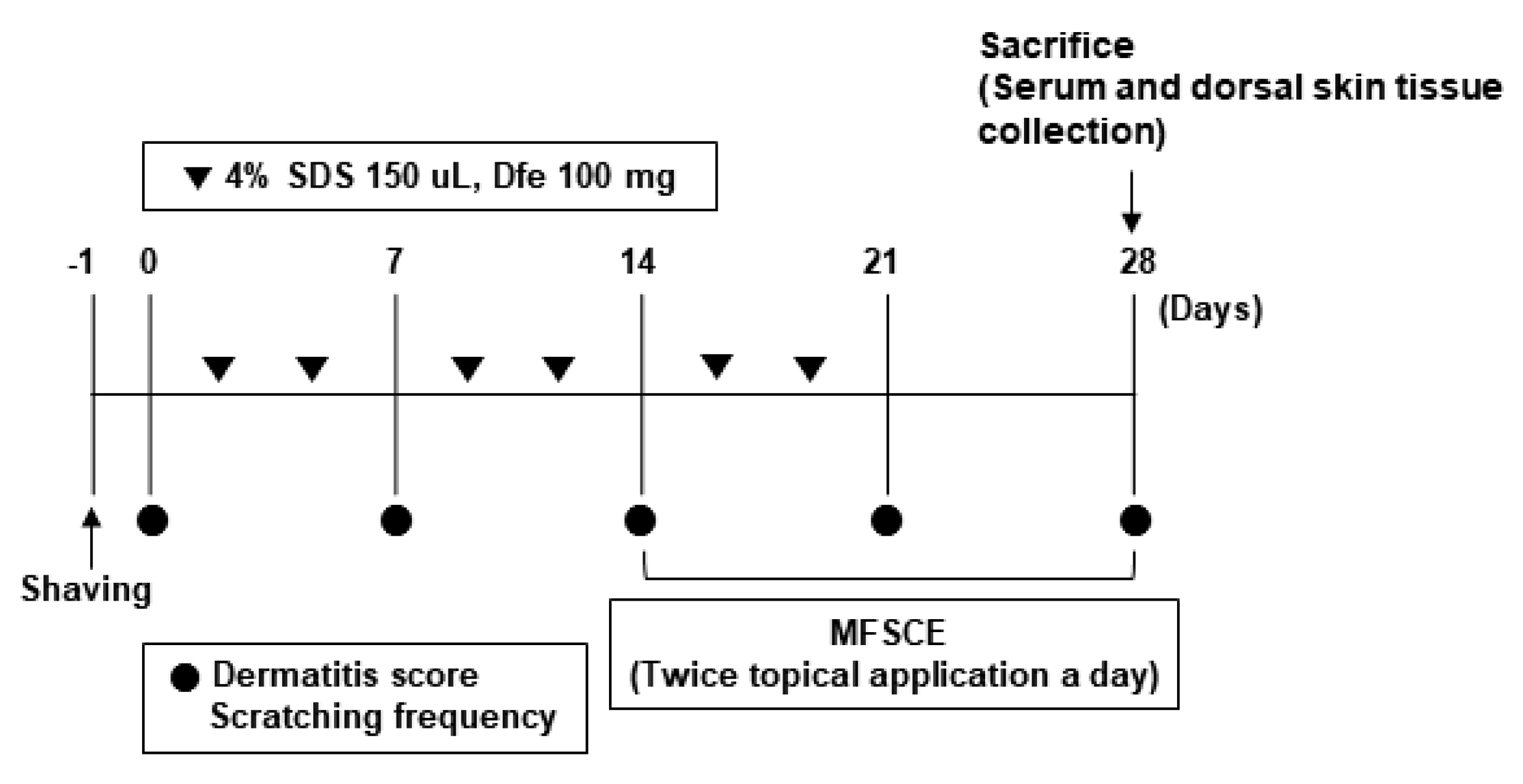
Figure 1. Schedule of induction and treatment for AD-like skin inflammation in NC/Nga mice. SDS: sodium dodecyl sulfate, Dfe: Dermatophagoides farinae extract, MFSCE: membrane-free stem cell extract.
Applsci 13 04542 g001
Figure 2. Clinical pictures of AD-like dorsal skin lesions in HDM-sensitized NC/Nga mice. Six groups: NOR group, VEH group (HDM treatment), MFSCE 5 group (HDM treatment + MFSCE 5 uL/head), MFSCE 10 group (HDM treatment + MFSCE 10 uL/head), MFSCE 20 group (HDM treatment + MFSCE 20 uL/head), and DEX group (HDM treatment + DEX 540 ug/head).
Applsci 13 04542 g002
Figure 3. Effect of MFSCE on AD-like symptoms in HDM-sensitized NC/Nga mice. (A) Dermatitis scores. (B) Scratching frequency. Values are mean ± SD. a–d Means with different letters are significantly different (p < 0.05) as determined by Duncan’s multiple range test. The mice were grouped and treated as described in Figure 2.
Applsci 13 04542 g003
Figure 4. Effect of MFSCE on serum IgE increase in HDM-sensitized NC/Nga mice. Values are mean ± SD. a–c Means with different letters are significantly different (p < 0.05) as determined by Duncan’s multiple range test. The mice were grouped and treated as described in Figure 2.
Applsci 13 04542 g004
Figure 5. Effect of MFSCE on serum inflammatory cytokines in HDM-sensitized NC/Nga mice. The levels of IL-4 (A), IL-10 (B), IFN-γ (C), and TNF-α (D) in serum. Values are mean ± SD. a–d Means with different letters are significantly different (p < 0.05) as determined by Duncan’s multiple range test. The mice were grouped and treated as described in Figure 2.
Applsci 13 04542 g005
Figure 6. Effect of MFSCE on epidermal thickness and mast cell infiltration in AD-like dorsal skin of HDM-sensitized NC/Nga mice. Histological images of dorsal skin lesions for hematoxylin and eosin staining (40× magnification) (A) and toluidine blue staining (100× magnification) (B). Quantification of epidermal thickness (C) and mast cells (D) per field. Values are mean ± SD. a–d Means with different letters are significantly different (p < 0.05) as determined by Duncan’s multiple range test. The mice were grouped and treated as described in Figure 2.
Applsci 13 04542 g006
Figure 7. Effect of MFSCE on IL-4 (A), IL-10 (B), IFN-γ (C), TNF-α (D), and TARC (E) expression in AD-like dorsal skin of HDM-sensitized NC/Nga mice. Values are mean ± SD. a–e Means with different letters are significantly different (p < 0.05) as determined by Duncan’s multiple range test. The mice were grouped and treated as described in Figure 2.
Applsci 13 04542 g007
Disclaimer/Publisher’s Note: The statements, opinions and data contained in all publications are solely those of the individual author(s) and contributor(s) and not of MDPI and/or the editor(s). MDPI and/or the editor(s) disclaim responsibility for any injury to people or property resulting from any ideas, methods, instructions or products referred to in the content.