Communication

Biomechanical Performance of a Novel Implant Design in Simulated Extraction Sites and Sinuslift Procedures

1
Department of Prosthodontics, Saarland University, Kirrberger Strasse 100, 66421 Homburg/Saar, Germany
2
Department of Oral and Maxillofacial Surgery, University of Leipzig, Liebigstrasse 12, 04103 Leipzig, Germany
*
Author to whom correspondence should be addressed.
Appl. Sci. 2023, 13(13), 7541; https://doi.org/10.3390/app13137541
Submission received: 16 May 2023 / Revised: 21 June 2023 / Accepted: 23 June 2023 / Published: 26 June 2023
(This article belongs to the Section Applied Dentistry and Oral Sciences)

Abstract

:
With increasing experience and in an attempt to shorten overall treatment times, implant placement in combination with tooth extractions and sinus lift procedures has become popular. In both cases, primary stability has to be achieved by either engaging apical and oral regions of trabecular bone or by engaging residual host bone beneath the sinus cavity. Extraction sites were formed by pressing a root analog into homogeneous low density polyurethane foam which was used as bone surrogate while a 3 mm thick sheet of medium density foam was used for mimicking a sinus lift situation. Two types (n = 10) of bone level implants with a conventional tapered design and a cervical back taper (NobelActive; control) and a novel design characterized by a shift in core diameter and thread geometry (AlfaGate; test) were placed in these models following conventional osteotomy preparation. Insertion torque was measured using a surgical motor and primary stability was determined by resonance frequency analysis. Statistical analysis was based on Welch two sample t tests with the level of significance set at α = 0.05. In sinuslifting, NobelActive implants required significantly higher insertion torques as compared to AlfaGate (p = 0.000) but did not achieve greater implant stability (p = 0.076). In extraction sites, AlfaGate implants showed both, significantly higher insertion torques (p = 0.004) and significantly greater implant stability (p = 0.000). The novel implant design allowed for greater primary stability when being placed in simulated extraction sockets and sinuslift situations. While in extraction sockets the position of condensing threads in combination with an increase in core diameter is beneficial, the deep cervical threads of the novel implant lead to superior performance in sinuslift situations.

1. Introduction

Attempting to reduce the number of surgical interventions, implant placement simultaneous with sinus lifting [1,2,3] and tooth extractions [4,5] has been advocated. Both procedures are considered being technically sensitive requiring experience from the implant surgeon [4,5,6,7].
Proper positioning seems to be the key factor in immediately placed implants [8] as concerns regarding bone healing leading to greater bone loss still exist [9]. In the anterior maxilla for instance, the implant osteotomy has to leave the alveolus palatally [10,11] resulting in a rather bone driven implant position in order not to perforate the concave alveolar process [12] and for obtaining a stable buccal bone thickness of at least 1 mm [13]. Given the mismatch between socket dimensions and implant diameter, implant selection [14,15] is important. Wider and tapered implants are frequently employed for increasing primary stability [16,17], which often collides with space limitations in single tooth gaps [18,19]. Two main sources for gaining primary implant stability [7] are available in extraction sites. Circumferential support can be achieved by using wider implant diameters or bone apical to the alveolus can be engaged by extending the osteotomy apically [1]. This however may be limited by anatomical factors [20,21]. Despite these approaches, primary implant stability in general has been shown to be lower as compared to healed sites [22,23,24].
In an attempt to optimize the use of the host bone present in extraction sites, a dental implant with an “inverted body shift” has been introduced and described [25]. This implant is characterized by an increase in its outer diameter aimed at engaging the socket walls [3,19,21,26] rendering it unsuitable for healed implant sites.
In sinuslift situations [27] primary stability has to be derived from engaging residual bone underneath the sinus cavity with cervical areas of the implant. Residual bone heights have been reported to reach low values of 2 mm [28], 2.57 mm [29], 3.1 mm [30] and 3.86 mm [31]. Engaging cortical bone has been shown to greatly affect primary stability [32], thereby questioning the benefit of implant designs characterized by a cervical region with a back taper [33].
An implant macrodesign characterized by modulation in core diameter coinciding with a change in thread geometry starting with sharp threads in the apical region, followed by bone condensing threads in the middle portion and sharp threads in the cervical portion of the implant has recently been described [34]. It was the goal of this in vitro experiment to compare this novel implant design (Figure 1a) to an established tapered implant design (Figure 1b; [33]) when being placed in simulated sinus lift situations and extraction sockets.

2. Materials and Methods

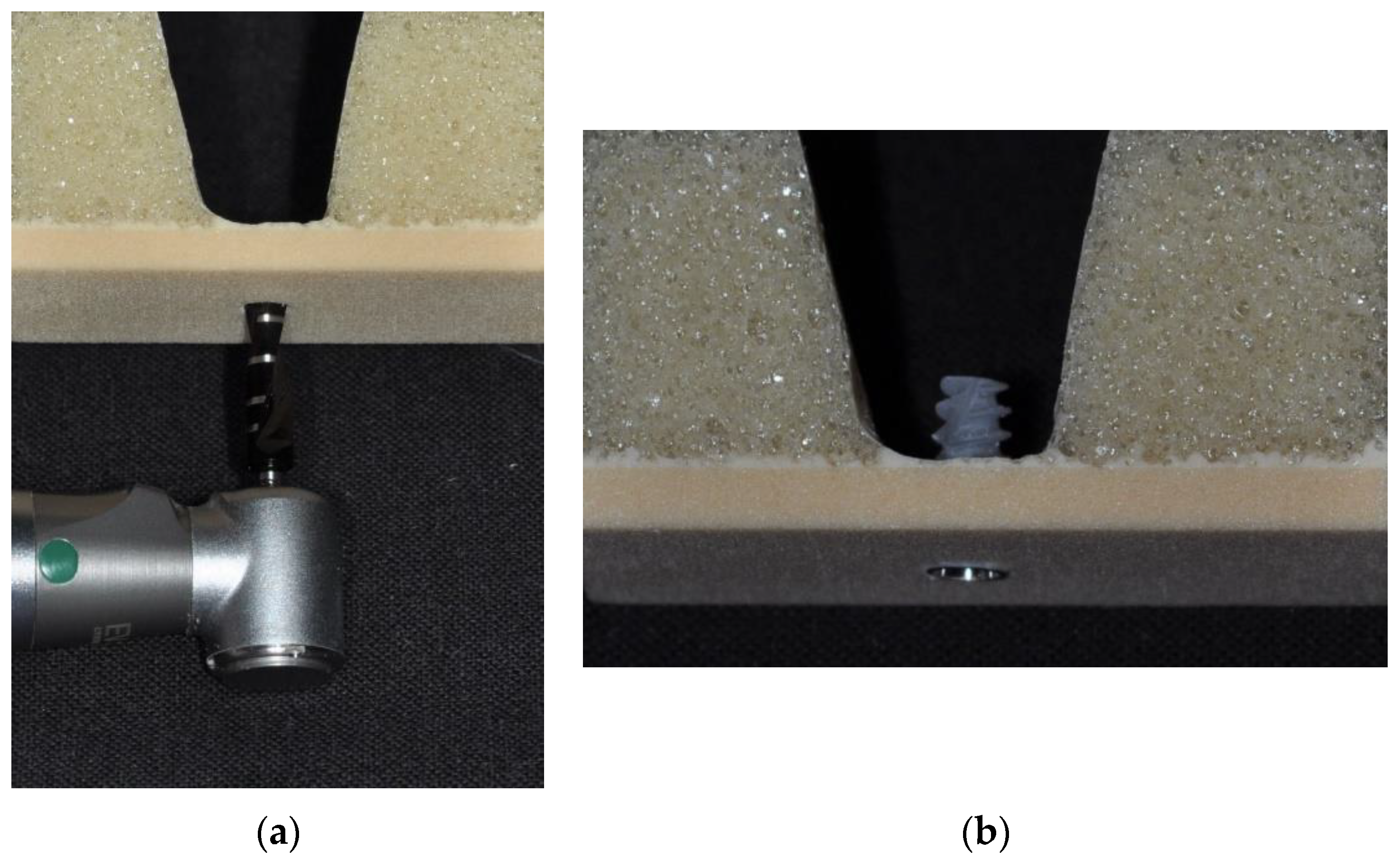
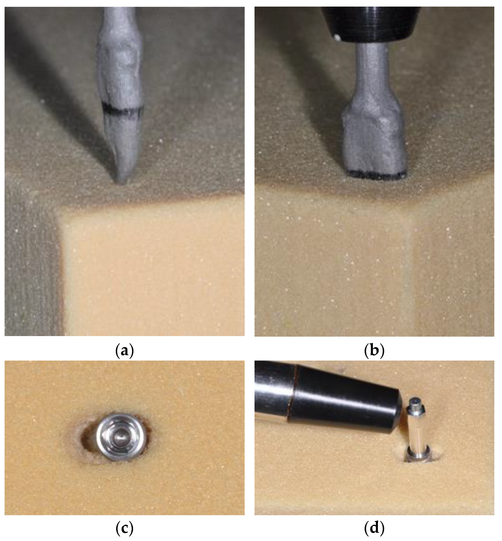
A homogeneous, 3 mm thick [30] sheet of medium density foam (Solid Rigid polyurethane foam 30 pcf, Sawbones Europe AB, Malmö, Sweden) was used for simulating the residual host bone underneath the sinus floor (Figure 2). Mimicking sinus lift situations with simultaneous implant placement, osteotomies were created using the drilling steps described in Table 1. Similarly, low density polyurethane foam material (Solid Rigid polyurethane foam 10 pcf, Sawbones Europe AB) was used as bone surrogate material [16,35,36] for simulating immediate implant placement. To this end, an extracted maxillary premolar was impressed into silicone material in order to achieve a negative form of its root portion which could subsequently be filled with casting wax. The wax pattern and a centrally positioned shaft were then cast in non-noble alloy (Heraenium CE, Kulzer, Hanau, Germany). The root analog was pushed into the bone surrogate material using a surveyor thereby ensuring perpendicular alignment of the extraction sockets to the bone surface (Figure 3). Mimicking clinical practice, apical and palatal preparation of osteotomies was performed using the drilling steps described in Table 1.
In the control group, traditional tapered implants [33] with a cervical back taper (Figure 1a) were placed while in the test group the novel implant design (Figure 1b) described above was utilized. All implants (n = 10) were placed (Figure 2) with a surgical motor capable of recording actual torque over time [16] and which was set at 25 rpm (iChiropro, BienAir, Biel, Switzerland). Following implant placement, specific MulTipegs (MulTipeg #51 for AlfaGate, MulTipeg #29 for NobelActive, Integration Diagnostics, Gothenburg, Sweden) were attached to the implants and resonance frequency analysis (RFA) was performed twice (Osstell ISQ, Osstell, Gothenburg, Sweden) [24].
For statistical analysis, mean values of RFA measurements were calculated and mean maximum values were derived from the curves recorded for insertion torque [16]. Following Shapiro-Wilk-Tests on normality of distribution of measurement values, Welch two sample t tests were applied for comparisons between the two implant types with the level of significance set at α = 0.05.

3. Results

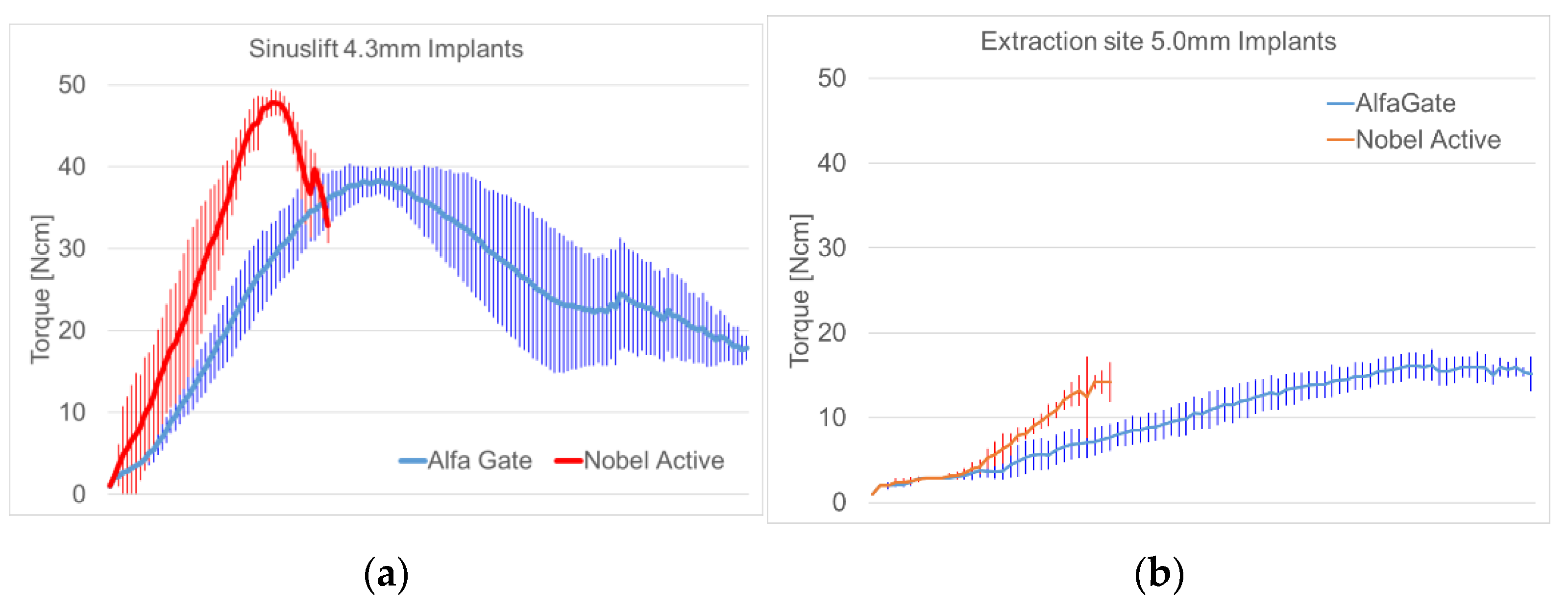
The insertion of AlfaGate implants took up to three times longer as compared to NobelActive implants due to their smaller thread pitch. In sinuslifting, both implant types reached mean insertion torques greater than 40 Ncm and mean primary stability also exceeded ISQ values of 40 (Table 2). In extraction sites, mean insertion torque values were 14.29 Ncm for NobelActive and 16.43 for AlfaGate with latter ones reaching mean primary stability of ISQ 47.80 (Table 2).
The Shapiro-Wilk-Tests (Table 2) could be assumed to be normally distributed measurement values for all parameters with the exception of torque values recorded for NobelActive in sinuslift situations (p = 0.0298). Given the large differences in mean torque values between the two implant types inserted in sinuslift situations, the non-normal distribution could be neglected.
For simulated sinuslift procedures (Figure 4), NobelActive implants required significantly higher insertion torques as compared to AlfaGate (p = 0.000) but did not achieve greater implant stability as measured by resonance frequency analysis (p = 0.076).
In extraction sites, AlfaGate implants showed both, significantly higher insertion torques (p = 0.004) and significantly greater implant stability (p = 0.000).

4. Discussion

Measuring insertion torque and primary implant stability, this experiment attempted to compare a novel implant design characterized by a change in core diameter and thread geometry (AlfaGate) to an existing, well established [33] tapered implant design (NobelActive) when used for immediate placement in simulated extraction sites and sinuslift situations respectively. The methodology applied in this experiment followed comparable previous studies on round [34,37,38] and wedge-shaped [39] dental implants. In order to benefit from standardized conditions, polyurethane foam was used as bone surrogate material instead of using cadaver bone [40], which may be seen as the major limitation of this experiment.
Compared to a recently introduced implant with an “inverted body shift” [25], which is characterized by an increase in its outer diameter for engaging socket walls [3,19,21,26], the experimental implant used here gains stability from an increased core diameter in combination with a condensing thread design in the middle portion of the implant. The outer diameter of the implant of the novel implant does not change in the middle and cervical parts, which allows it to be also placed in healed sites.
An unsolved problem in implant dentistry is the evaluation of alveolar bone quality based on which different drill protocols are recommended [41]. Many conventional screw-type implants tend to induce high levels of stress in the cervical peri-implant bone area, leading to bone loss [42]. In order to reduce this phenomenon, greater drill diameters or countersink drills are recommended thereby removing precious bone quantity. The novel implant design is aimed at simplifying the surgical procedure as osteotomy preparation is mainly governed by the core diameter of the implant (Figure 1b) to be used instead of bone quality judgement.
In a previous experiment [34], the novel implant was tested in homogeneous bone surrogates while the situations simulated here are more critical as primary stability has been shown to be lower in immediate implant cases as compared to healed sites [22,23,24]. Questioning two clinical studies on immediate implant placement [14,15] where no difference between implant design has been described, greater primary stability was observed here with the novel implant design. The general notion that implant design in fact impacts stability in extraction sites is supported by two previous reports [38,43]. Deviating from clinical practice where immediate implants in the anterior maxilla are placed such way that the implant leaves the alveolus palatally [10,11] and engages bone areas apical to the socket, socket depth and implants had a length of 10 mm for creating a worst case scenario. In contrast to the sinuslifting procedure, 5.0 mm implants had to be chosen here in order to engage lateral walls which seems to be in line with clinical practice [14,15].
Similar to extraction sites, a wide variety of clinical situations exists in sinuslifting which can not be standardized [28,30,31] and it may be argued whether or not a residual bone height of 3 mm underneath the sinus floor is sufficient for placing implants. However, different clinical studies reported residual bone heights in the range of 3.1 to 9.6 mm [30], 3.86 mm [31] and even reaching a minimum of 2 mm of residual bone height [28]. As such the situation simulated here with a layer of residual bone 3 mm in thickness may not even be considered a worst-case scenario. Besides bone quantity, bone quality has also been reported to be compromised in sinuslift procedures with soft bone being present [29] as compared to the rather compact foam material used here.
In this in vitro study standardized bone models made from polyurethane foam material have been used as a bone surrogate [36,44,45,46]. Consequently, the absolute values measured here cannot be directly transferred to clinical reality where changes in bone quality may occur over the length of an osteotomy. Additionally, these materials are not able to fully mimic the elastic and anisotropic properties of alveolar bone, but in turn, allow for reproducible measurements [47].

5. Conclusions

The novel implant design allowed for greater primary stability in both, extraction sites and sinuslifting situations. This is due to the synergistic effect of changes in thread design and core diameter of the implant along its axis. While the back taper of the outer implant shape incorporated in the control implant used here is supposed to avoid over-compression of cortical bone in healed sites, it causes a loss in stability when the implant is fully seated in a sinuslift situation. The novel implant instead derives stability from condensing trabecular areas of bone while engaging cortical bone with sharp threads. Despite the seemingly advantageous behavior of the novel implant design, clinical studies will be required for verifying a potentially positive effect on treatment outcomes.

Author Contributions

Conceptualization, B.L.; methodology, V.K.; software, A.S.; formal analysis, V.K.; investigation, R.Z.; data curation, A.S.; writing—original draft preparation, R.Z.; writing—review and editing, V.K. All authors have read and agreed to the published version of the manuscript.

Funding

This research received no external funding.

Data Availability Statement

Data is available from the corresponding author upon reasonable request.

Acknowledgments

The authors wish to thank Friedrich Graef, Department of Mathematics, University of Erlangen-Nuremberg for statistical data analysis. Implants were kindly provided by AlfaGate, Kfar Qara, Israel. We acknowledge support by the Deutsche Forschungsgemeinschaft (DFG, German Research Foundation) and Saarland University within the funding programme Open Access Publishing.

Conflicts of Interest

The authors declare no conflict of interest.

References

  1. Gamborena, I.; Sasaki, Y.; Blatz, M.B. Predictable immediate implant placement and restoration in the esthetic zone. J. Esthet. Restor. Dent. 2021, 33, 158–172. [Google Scholar] [CrossRef]
  2. Ekhlasmandkermani, M.; Amid, R.; Kadkhodazadeh, M.; Hajizadeh, F.; Abed, P.F.; Kheiri, L.; Kheiri, A. Sinus floor elevation and simultaneous implant placement in fresh extraction sockets: A systematic review of clinical data. J. Korean Assoc. Oral Maxillofac. Surg. 2021, 47, 411–426. [Google Scholar] [CrossRef] [PubMed]
  3. Christiaens, V.; Pitman, J.; Glibert, M.; Hommez, G.; Atashkadeh, M.; De Bruyn, H. Rationale for a reverse tapered body shift implant for immediate placement. Int. J. Oral Maxillofac. Surg. 2020, 49, 1630–1636. [Google Scholar] [CrossRef]
  4. Canellas, J.V.D.S.; Medeiros, P.J.D.; Figueredo, C.M.D.S.; Fischer, R.G.; Ritto, F.G. Which is the best choice after tooth extraction, immediate implant placement or delayed placement with alveolar ridge preservation? A systematic review and meta-analysis. J. Craniomaxillofac. Surg. 2019, 47, 1793–1802. [Google Scholar] [CrossRef]
  5. Garcia-Sanchez, R.; Dopico, J.; Kalemaj, Z.; Buti, J.; Pardo Zamora, G.; Mardas, N. Comparison of clinical outcomes of immediate versus delayed placement of dental implants: A systematic review and meta-analysis. Clin. Oral. Implants. Res. 2022, 33, 231–277. [Google Scholar] [CrossRef]
  6. Maddalone, M.; Mirabelli, L.; Venino, P.M.; Karanxha, L.; Porcaro, G.; Del Fabbro, M. Long-term stability of autologous bone graft of intraoral origin after lateral sinus floor elevation with simultaneous implant placement. Clin. Implant. Dent. Relat. Res. 2018, 20, 713–721. [Google Scholar] [CrossRef] [PubMed]
  7. Rues, S.; Schmitter, M.; Kappel, S.; Sonntag, R.; Kretzer, J.P.; Nadorf, J. Effect of bone quality and quantity on the primary stability of dental implants in a simulated bicortical placement. Clin. Oral Investig. 2021, 25, 1265–1272. [Google Scholar] [CrossRef] [PubMed]
  8. Kuo, P.J.; Lin, C.Y.; Hung, T.F.; Chiu, H.C.; Kuo, H.Y. A novel application of dynamic guided navigation system in immediate implant placement. J. Dent. Sci. 2022, 17, 354–360. [Google Scholar] [CrossRef] [PubMed]
  9. Singh, G.; Pareek, R.; Rajawat, G.S.; Kadam, A.; Al Abdulsalam, M.; Al Abdulathim, A. Comparison of Bone Healing in Immediate Implant Placement versus Delayed Implant Placement. J. Pharm. Bioallied. Sci. 2021, 13, S1309–S1314. [Google Scholar]
  10. Weigl, P.; Strangio, A. The impact of immediately placed and restored single-tooth implants on hard and soft tissues in the anterior maxilla. Eur. J. Oral Implantol. 2016, 9, S89–S106. [Google Scholar]
  11. Rodrigues, D.M.; Petersen, R.L.; Montez, C.; de Moraes, J.R.; Ferreira, V.; Barboza, E.P. The relationship between tomographic sagittal root position of maxillary anterior teeth and the bone housing. J. Prosthet. Dent. 2022, ahead of print. [Google Scholar] [CrossRef]
  12. Botermans, A.; Lidén, A.; de Carvalho Machado, V.; Chrcanovic, B.R. Immediate Implant Placement in the Maxillary Aesthetic Zone: A Cone Beam Computed Tomography Study. J Clin. Med. 2021, 10, 5853. [Google Scholar] [CrossRef] [PubMed]
  13. Heimes, D.; Schiegnitz, E.; Kuchen, R.; Kämmerer, P.W.; Al-Nawas, B. Buccal Bone Thickness in Anterior and Posterior Teeth-A Systematic Review. Healthcare 2021, 9, 1663. [Google Scholar] [CrossRef] [PubMed]
  14. Wei, S.M.; Shi, J.Y.; Qiao, S.C.; Zhang, X.; Lai, H.C.; Zhang, X.M. Accuracy and primary stability of tapered or straight implants placed into fresh extraction socket using dynamic navigation: A randomized controlled clinical trial. Clin. Oral Investig 2021, ahead of print. [Google Scholar] [CrossRef]
  15. Sanchez-Perez, A.; Nicolas-Silvente, A.I.; Sanchez-Matas, C.; Molina-García, S.; Navarro-Cuellar, C.; Romanos, G.E. Primary stability and PES/WES evaluation for immediate implants in the aesthetic zone: A pilot clinical double-blind randomized study. Sci. Rep. 2021, 11, 20024. [Google Scholar] [CrossRef]
  16. Yang, B.; Irastorza-Landa, A.; Heuberger, P.; Ploeg, H.L. Effect of insertion factors on dental implant insertion torque/energy-experimental results. J. Mech. Behav. Biomed. Mater. 2020, 112, 103995. [Google Scholar] [CrossRef]
  17. Noaman, A.T.; Bede, S.Y. The Effect of Bone Density Measured by Cone Beam Computed Tomography and Implant Dimensions on the Stability of Dental Implants. J. Craniofac. Surg. 2021, 33, e553–e557. [Google Scholar] [CrossRef]
  18. Herrero-Climent, M.; López-Jarana, P.; Lemos, B.F.; Gil, F.J.; Falcão, C.; Ríos-Santos, J.V.; Ríos-Carrasco, B. Relevant Design Aspects to Improve the Stability of Titanium Dental Implants. Materials 2020, 13, 1910. [Google Scholar] [CrossRef]
  19. Chu, S.J.; Levin, B.P.; Egbert, N.; Saito, H.; Nevins, M. Use of a Novel Implant with an Inverted Body-Shift and Prosthetic Angle Correction Design for Immediate Tooth Replacement in the Esthetic Zone: A Clinical Case Series. Int. J. Periodontics Restor. Dent. 2021, 41, 195–204. [Google Scholar] [CrossRef]
  20. Hamzani, Y.; Yassien, E.; Moskovich, L.; Becker, T.; Chaushu, G.; Haj Yahya, B. Potential Circumferential Bone Engagement following Tooth Extraction in the Posterior Mandible: Computed Tomography Assessment. Medicina 2021, 57, 874. [Google Scholar] [CrossRef] [PubMed]
  21. Levin, B.P.; Chu, S.J.; Saito, H.; Nevins, M.; Levin, J.P. A Novel Implant Design for Immediate Extraction Sites: Determining Primary Stability. Int. J. Periodontics Restor. Dent. 2021, 41, 357–364. [Google Scholar] [CrossRef]
  22. Rowan, M.; Lee, D.; Pi-Anfruns, J.; Shiffler, P.; Aghaloo, T.; Moy, P.K. Mechanical versus biological stability of immediate and delayed implant placement using resonance frequency analysis. J. Oral Maxillofac. Surg. 2015, 73, 253–257. [Google Scholar] [CrossRef]
  23. Levin, B.P. The Correlation Between Immediate Implant Insertion Torque and Implant Stability Quotient. Int. J. Periodontics Restor. Dent. 2016, 36, 833–840. [Google Scholar] [CrossRef] [PubMed]
  24. Bavetta, G.; Bavetta, G.; Randazzo, V.; Cavataio, A.; Paderni, C.; Grassia, V.; Dipalma, G.; Gargiulo Isacco, C.; Scarano, A.; De Vito, D.; et al. A Retrospective Study on Insertion Torque and Implant Stability Quotient (ISQ) as Stability Parameters for Immediate Loading of Implants in Fresh Extraction Sockets. Biomed. Res. Int. 2019, 2019, 9720419. [Google Scholar] [CrossRef] [PubMed]
  25. Chu, S.J.; Saito, H.; Levin, B.P.; Baumgarten, H.; Egbert, N.; Wills, M.J.; Del Castillo, R.A.; Tarnow, D.P.; Nevins, M. Outcomes of a 1-Year Prospective Single-Arm Cohort Study Using a Novel Macro-Hybrid Implant Design in Extraction Sockets: Part 1. Int. J. Periodontics Restor. Dent. 2021, 41, 499–508. [Google Scholar] [CrossRef]
  26. Saito, H.; Chu, S.J.; Tarnow, D.P. The effect of implant macrogeometry in immediate tooth replacement therapy: A case series. J. Esthet. Restor. Dent. 2021, ahead of print. [Google Scholar] [CrossRef]
  27. Kwon, J.J.; Hwang, J.; Kim, Y.D.; Shin, S.H.; Cho, B.H.; Lee, J.Y. Automatic three-dimensional analysis of bone volume and quality change after maxillary sinus augmentation. Clin. Implant Dent. Relat. Res. 2019, 21, 1148–1155. [Google Scholar] [CrossRef]
  28. Andrés-García, R.; Ríos-Santos, J.V.; Herrero-Climent, M.; Bullón, P.; Fernández-Farhall, J.; Gómez-Menchero, A.; Fernández-Palacín, A.; Ríos-Carrasco, B. Sinus Floor Elevation via an Osteotome Technique without Biomaterials. Int. J. Environ. Res. Public Health 2021, 18, 1103. [Google Scholar] [CrossRef]
  29. Park, Y.H.; Jung, U.W.; Kim, C.S.; Choi, S.H.; Cho, K.S.; Lee, J.S. Resonance Frequency Analysis of Tapered Implants Placed at Maxillary Posterior Sites After Lateral Sinus Augmentation: A 1.5-year Follow-Up Prospective Study. Implant Dent. 2019, 28, 62–67. [Google Scholar] [CrossRef] [PubMed]
  30. Kim, B.J.; Kim, C.H.; Kim, J.H. Immediate implant placement following tooth extraction with simultaneous lateral sinus augmentation: A retrospective clinical study after at least 1 year of loading. Int. J. Implant Dent. 2021, 7, 96. [Google Scholar] [CrossRef]
  31. Bhandari, S.; Thomas, R.; Kumar, T.; Shah, R.; Mehta, D.S. Maxillary Sinus Augmentation Using Hydraulic Pressure by Lateral Approach and Simultaneous Implant Placement: Clinicoradiographic Study. Implant Dent. 2019, 28, 514–519. [Google Scholar] [CrossRef]
  32. De Oliveira Nicolau Mantovani, A.K.; de Mattias Sartori, I.A.; Azevedo-Alanis, L.R.; Tiossi, R.; Fontão, F.N.G.K. Influence of cortical bone anchorage on the primary stability of dental implants. Oral Maxillofac. Surg. 2018, 22, 297–301. [Google Scholar] [CrossRef] [PubMed]
  33. Irinakis, T.; Wiebe, C. Clinical evaluation of the NobelActive implant system: A case series of 107 consecutively placed implants and a review of the implant features. J. Oral. Implantol. 2009, 35, 283–288. [Google Scholar] [CrossRef] [PubMed]
  34. Schulz, A.; Klär, V.; Grobecker-Karl, T.; Karl, M. Biomechanical rationale for a novel implant design reducing stress on buccal bone. Appl. Sci. 2023, 13, 666. [Google Scholar] [CrossRef]
  35. Dard, M.; Kuehne, S.; Obrecht, M.; Grandin, M.; Helfenstein, J.; Pippenger, B.E. Integrative Performance Analysis of a Novel Bone Level Tapered Implant. Adv. Dent. Res. 2016, 28, 28–33. [Google Scholar] [CrossRef]
  36. Wang, R.; Eppell, S.J.; Nguyen, C.; Morris, N. Relative Contribution of Trabecular and Cortical Bone to Primary Implant Stability: An In Vitro Model Study. J. Oral Implantol. 2016, 42, 145–152. [Google Scholar] [CrossRef]
  37. Steiner, C.; Karl, M.; Grobecker-Karl, T. Insertion and loading characteristics of three different bone-level implants. Int. J. Oral Maxillofac. Implants. 2020, 35, 560–565. [Google Scholar] [CrossRef]
  38. Karl, M.; Irastorza-Landa, A. Does implant design affect primary stability in extraction sites? Quintessence Int. 2017, 48, 219–224. [Google Scholar]
  39. Steiner, C.; Karl, M.; Grobecker-Karl, T. Wedge shaped vs. round implants: Bone strain during the insertion process. J. Oral Implantol. 2022, 48, 557–561. [Google Scholar] [CrossRef]
  40. Klär, V.; Karl, M.; Grobecker-Karl, T. Bone damage during dental implant insertion: A pilot study combining strain gauge and histologic analysis. Appl. Sci. 2022, 12, 291. [Google Scholar] [CrossRef]
  41. Velikov, S.; Susin, C.; Heuberger, P.; Irastorza-Landa, A. A New Site Preparation Protocol That Supports Bone Quality Evaluation and Provides Predictable Implant Insertion Torque. J. Clin. Med. 2020, 9, 494. [Google Scholar] [CrossRef]
  42. Dantas, T.A.; Carneiro Neto, J.P.; Alves, J.L.; Vaz, P.C.S.; Silva, F.S. In silico evaluation of the stress fields on the cortical bone surrounding dental implants: Comparing root-analogue and screwed implants. J. Mech. Behav. Biomed. Mater. 2020, 104, 103667. [Google Scholar] [CrossRef] [PubMed]
  43. Hsieh, M.C.; Huang, C.H.; Hsu, M.L. Influences of screw design features on initial stability in immediate implant placement and restoration. Clin. Biomech. 2021, 89, 105453. [Google Scholar] [CrossRef]
  44. Ueno, D.; Nakamura, K.; Kojima, K.; Toyoshima, T.; Tanaka, H.; Ueda, K.; Koyano, K.; Kodama, T. A stepwise under-prepared osteotomy technique improves primary stability in shallow-placed implants: A preliminary study for simultaneous vertical ridge augmentation. Odontology 2018, 106, 187–193. [Google Scholar] [CrossRef]
  45. Sierra-Rebolledo, A.; Allais-Leon, M.; Maurette-O’Brien, P.; Gay-Escoda, C. Primary Apical Stability of Tapered Implants Through Reduction of Final Drilling Dimensions in Different Bone Density Models: A Biomechanical Study. Implant. Dent. 2016, 25, 775–782. [Google Scholar] [CrossRef] [PubMed]
  46. Di Stefano, D.A.; Arosio, P.; Gastaldi, G.; Gherlone, E. The insertion torque-depth curve integral as a measure of implant primary stability: An in vitro study on polyurethane foam blocks. J. Prosthet. Dent. 2018, 120, 706–714. [Google Scholar] [CrossRef]
  47. Elias, C.N.; Rocha, F.A.; Nascimento, A.L.; Coelho, P.G. Influence of implant shape, surface morphology, surgical technique and bone quality on the primary stability of dental implants. J. Mech. Behav. Biomed. Mater. 2012, 16, 169–180. [Google Scholar] [CrossRef]
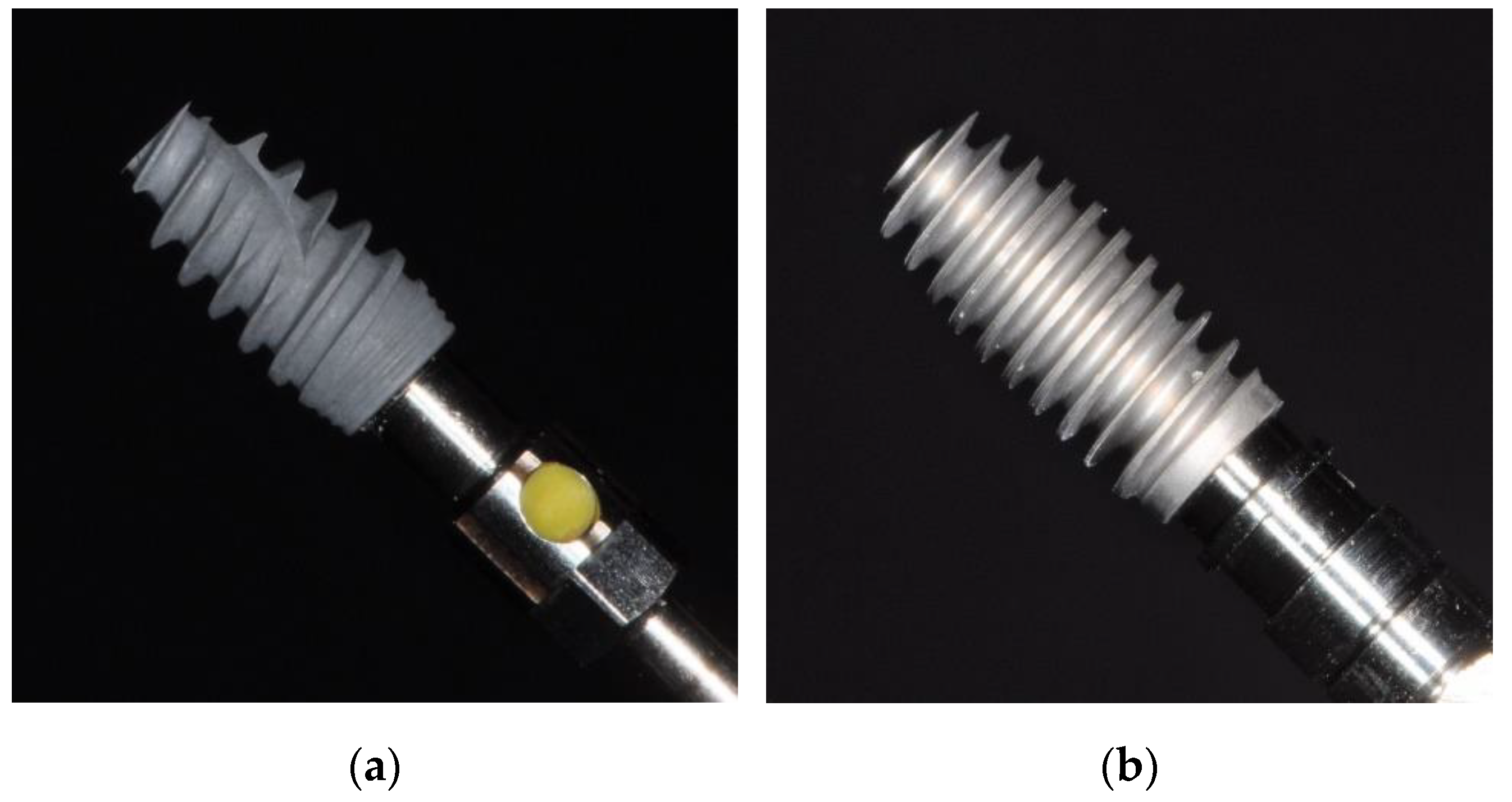
Figure 1. Bone level implant types used in this study comprising NobelActive 4.3 × 10 mm (a) and AlfaGate 4.3 × 10 mm (b).
Applsci 13 07541 g001
Figure 2. Simulation of sinuslift procedure with simultaneous implant placement following osteotomy preparation with twist drills (a) and anchorage of implants in residual bone (b).
Applsci 13 07541 g002
Figure 3. Simulation of immediate implant placement in an extraction site obtained through pressing a root analog (a) into polyurethane foam material to a depth of 10 mm (b) and following osteotomy preparation using a twist drill (c). Implant stability was assessed by means of resonance frequency analysis (d).
Applsci 13 07541 g003
Figure 4. Torque development over time during implant insertion for both implant types investigated in simulated sinuslift (a) and extraction site (b).
Applsci 13 07541 g004
Table 1. Description of the implant systems and drill sequences used in this study for creating osteotomies in simulated sinuslift situations and extraction sites. All implants were inserted with a surgical motor set at 25 rpm.
SinusliftExtraction Site
ImplantsNobelActive Internal RP 4.3 × 10 mm
REF 34131 LOT 13125039
NobelActive Internal RP 5.0 × 10 mm
REF 34137 LOT 12168347
AlfaGate Novel Design 4.3 × 10 mmAlfaGate Novel Design 5.0 × 10 mm
Drill sequence2.0
2.4/2.8
3.2/3.6
Preformed Socket Depth: 10 mm
2.4/2.8
Table 2. Mean values and standard deviations recorded for insertion torque and primary stability for both implant types. Results of Shapiro-Wilk-Tests on normality of distribution of measurement values are given as p-values; significant differences (p < 0.05) are written in bold.
ParameterNobelActiveShapiro-Wilk-Test
(p-Value)
AlfaGateShapiro-Wilk-Test
(p-Value)
MeanSDMeanSD
SinusliftMaximum insertion torque [Ncm]48.491.9650.029840.421.4600.1877
Osstell [ISQ]40.655.8740.849844.703.1640.6327
Extraction siteMaximum insertion torque [Ncm]14.291.2440.460516.431.5780.2829
Osstell [ISQ]41.803.2250.925247.802.5410.5593
Disclaimer/Publisher’s Note: The statements, opinions and data contained in all publications are solely those of the individual author(s) and contributor(s) and not of MDPI and/or the editor(s). MDPI and/or the editor(s) disclaim responsibility for any injury to people or property resulting from any ideas, methods, instructions or products referred to in the content.