Article

A Three-Year Prospective Study Comparing Stereolithography Printed Models to Classical Impression and Plaster Cast Models in Orthodontic Therapy: A 3D Objectification Approach

1
Department of Stomatology, 2nd Medical Faculty, Charles University, 15006 Prague, Czech Republic
2
Department of Mathematics, Informatics and Cybernetics, University of Chemistry and Technology in Prague, 16628 Prague, Czech Republic
3
Czech Institute of Informatics, Robotics and Cybernetics, Czech Technical University in Prague, 16000 Prague, Czech Republic
4
Department of Stomatology, Military University Hospital, 16902 Prague, Czech Republic
*
Author to whom correspondence should be addressed.
Appl. Sci. 2023, 13(13), 7542; https://doi.org/10.3390/app13137542
Submission received: 3 June 2023 / Revised: 24 June 2023 / Accepted: 25 June 2023 / Published: 26 June 2023
(This article belongs to the Special Issue Present and Future of Orthodontics)

Abstract

:
The integration of computational intelligence and augmented reality has become increasingly prevalent in dental practices. Three-dimensional (3D) printing techniques have now become routine in orthodontics, prosthetics, and maxillofacial surgery. The objective of this study is to assess the effectiveness of stereolithography (SLA) printed models compared to traditional plaster casts over a three-year period. The experimental dataset consisted of 36 orthodontic patients, each with SLA printed models and plaster casts for both the upper and lower jaws, resulting in a total of 72 scans in the form of Standard Template Library (STL) files and 72 traditional impressions. The upper dental arch models were constructed using an SLA 3D printer, employing a blue 405 nm laser beam to solidify a liquid polymer. The classical plaster casts were prepared in a dental laboratory. The models were retained for long-term orthodontic therapy control. To evaluate the differences between the dental models, a laboratory scanner was used to generate virtual casts. The meshes obtained were adjusted and pre-aligned using the best-fit algorithm. Subsequently, registration of the models was performed using the iterative closest point (ICP) algorithm. Distances between the point clouds and meshes for each point of the printed model were calculated by determining the nearest triangle on the reference mesh (cast). Additionally, the model surfaces were assessed using a scanning electron microscope and a stereomicroscope. The results from 22 experimental datasets indicated a high level of agreement between the plaster casts and virtual surfaces, with a mean absolute difference of 0.018 mm and a standard deviation of 0.17 mm. These values were obtained by averaging 231,178 data points for each comparison. Overall, this study demonstrates the comparability and accuracy of SLA printed models in relation to traditional plaster casts, supporting their potential as reliable alternatives in dental practice.

1. Introduction

Dental anatomy, including the alveolar bone and mucosa, as well as the shape of the dental arch, is traditionally recorded using dental impressions in various branches of stomatology, including dental prosthetics, orthodontics, dental implantology, and maxillofacial surgery.
Most conventional dental technologies and procedures involve the production of plaster models. However, these classical methods have certain disadvantages for both patients (discomfort during impression-taking, gagging) and dentists (bubble formation in the material, mechanical damage during transport or storage, errors caused by difficult repeated use). Moreover, plaster models lack mechanical resistance, are easily destroyed, and are difficult to reuse. Long-term storage of models for archival reasons also requires significant space.
To address these issues, computer-aided design (CAD) and computer-aided manufacturing (CAM) techniques have emerged as alternatives to conventional dental technology. These new procedures start with a digital model, which is obtained by digitizing a plaster cast or, in some cases, using intra-oral scanners. Most commercially available scanners are based on triangulation technology or parallel confocal microscopy, although wavefront sampling and interferometry are not yet widely used [1,2,3,4,5]. The standard tessellation language (STL) has become the standard format for digital models in dentistry [6].
The main advantages of digital impressions include clear visualization of defective areas for dentists and the ability to rescan specific areas without the need to repeat the entire impression [7]. However, a major drawback of digital three-dimensional casts is the so-called merging error, which can deform the entire digital cast. This error is caused by the principle of intra-oral scanners, which create small 3D models due to the size of the scanning head and then merge them into a larger model using software tools.
Currently, intra-oral scanners are commonly used to capture data for 3D printing. These scanners consist of a computer with specific software and a handheld camera. The software is responsible for creating the three-dimensional geometry of the dental arch, and the resulting STL format is used as a source for 3D printing [6]. In dentistry and orthodontics, four types of printers are particularly interesting:
  • Fused-deposition modeling (FDM): This 3D printing method still has relatively low resolution and is primarily used as an auxiliary method. FDM 3D printers heat a thermoplastic filament to create each layer;
  • Stereolithography (SLA) or digital light processing (DLP): These technologies use similar principles but differ in the wavelength of light used;
  • PolyJet 3D printing: This method involves the layering of liquid polymers;
  • Selective laser sintering/selective laser melting (SLS/SLM): These techniques are used for metal printing [4,8]. The main difference between SLS and SLM is the laser energy used.
The thickness of the layers formed by 3D printers depends on the technology used and the desired print quality. The layers are gradually applied, cured, and ultimately result in a complete model [9].
Stereolithography (SLA) was the first commercially available 3D printing technology, which utilizes polymerization to construct layered structures using different polymers. The process involves three important steps: light/laser exposure, platform movement, and resin refilling. Compared to gypsum models, 3D SLA prints are more accurate [10]. Scanning electron microscopy has shown that the surface structure of plaster models exhibits grainy texture with sharp edges of orthorhombic crystals, while SLA surfaces are more homogeneous, smoother, and only show traces of the layering polymer [11]. The 3D bioprinting technology forms an emerging area of research enabling high printing resolution and stability [12,13,14,15]. It allows the fabrication of accurate and cost effective 3D models for implant placement and maxillofacial prostheses. Associated software tools [16] can be used to align SLA dental models and point clouds. The comparison of the complete arch or selected regions is often used in assessing the clinical validity of model differences.
In orthodontics, models and their analysis play a crucial role as they accurately represent malocclusion. Traditional plaster models, however, are susceptible to damage, require significant storage space, are challenging to transport, and offer low replication accuracy. Furthermore, taking impressions can cause discomfort for patients, and fear and resistance among children can affect the accuracy of the resulting models [17].
The advent of intra-oral scanning technology has revolutionized examination methods in dentistry. Compared to traditional plaster casts, intra-oral scanning enables more precise digital analysis of dental arch components during the treatment of dental disorders. The acquired data can be utilized to create three-dimensional (3D) models using 3D printers [9].
The digitalization process provides several advantages and enables the utilization of various signal and image processing tools. These tools include magnification, detection of regions of interest, capturing of missing or defective areas during scanning, and the ability to immediately rescan important areas within a single treatment session [7]. However, it is important to note that a suboptimal scanning technique can lead to greater distortion of the final digital model compared to elastomeric impression materials. Additionally, each scanning technique has its specific limitations.
Data acquisition for mathematical analysis, validity of dental measurements [18,19], and digital processing includes the use of dental panoramic X-ray images [20], spatial modeling by camera systems [21,22], and intra-oral scanning [11,23] for the detection of dental disorders and the printing of three-dimensional models [17,24]. The 3D printing in dentistry and maxillofacial surgery is also an important area related to printing techniques and materials. These methods involve general digital signal and image processing techniques [25] and include the methodology of registering dental images [26], implementation of augmented reality [27,28], feature extraction, and machine learning for classification [22]. The main goals of these methods are evaluating treatment progress and early diagnosis of dental problems. Evaluation of digital implants, comparison of scanning technologies [15,29], and three-dimensional digital modeling [16,30,31] are also important areas of research. Storage time has also been studied in other works [32,33]. The ultimate aim of this research is to completely replace physical models with virtual digital structures.
The aim of this work is to assess the efficiency of stereolithography printed models compared to classical plaster casts in dental practice, specifically in the fields of orthodontics and prosthodontics. It emphasizes the integration role of computational intelligence and the increasing significance of 3D printing techniques in these areas.
The study presents the utilization of a 3D scanner to acquire precise data of the patient’s dental arch, including detailed information about dental anatomy and surrounding structures. The primary objective is to evaluate how these data can be translated into a 3D model and compare it with a plaster cast model of the patient’s dental arch. The evaluation focuses on various aspects, such as the accuracy of geometric details, the precision of anatomical features, and the long-term surface quality.

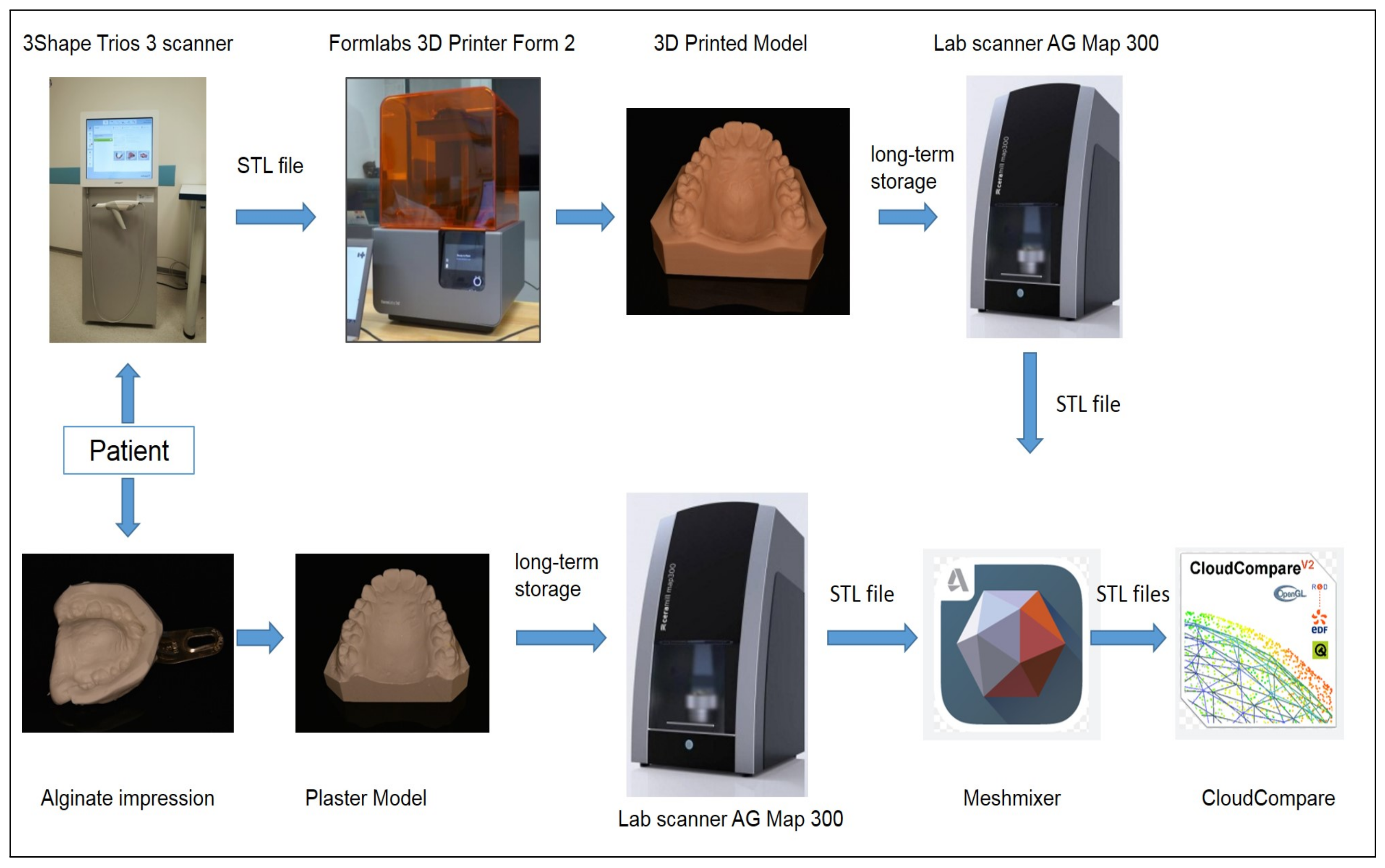
2. Materials and Methods

2.1. Subject

Our study group comprised 36 orthodontic patients, from whom we collected a total of 72 datasets of classical impressions and plaster casts, as well as 72 datasets of 3D scans and stereolithography (SLA) printed models of their upper and lower dental arches. The data were obtained during the course of orthodontic therapy using the 3Shape TRIOS 3 scanner. Prior to participating in the study, selected patients provided informed consent by signing a consent form. All participants were in good general health, and the study was conducted in accordance with the recommendations of the American Dental Association (ADA) and the principles outlined in the Declaration of Helsinki.
To ensure compliance with ethical standards, patients were required to provide informed consent for the clinical examination, as indicated by the informed consent form. The confidentiality and anonymity of the collected data were strictly maintained throughout the study. Ethical approval for the research was obtained from the Ethics Committee (EK-973IGA 1.12/11). The inclusion criteria for the study were as follows: absence of carious lesions and periodontal diseases in the dental arches.

2.2. Analyzing Methods and Measuring Instruments

The construction of 3D dental models involves two main principles: (i) utilizing a printed 3D model generated from data acquired by a 3D scanner and (ii) employing a plaster cast model. These models are then compared using a laboratory scanner and appropriate software tools as presented in Figure 1. Table 1 includes the list of experiments for the comparison between the plaster cast and virtual surfaces with the average number of 231,178 points for each dataset couple. Statistical evaluations were carried out in the MATLAB 2022b (MathWorks, Boston, MA, USA) computational environment.
Firstly, the process begins by acquiring data of the patient’s dental arch using a 3D scanner. This scanner captures the precise details of the dental anatomy and creates a digital representation of the patient’s teeth and surrounding structures. These data are then used to generate a 3D model, which can be printed using a 3D printer. Secondly, a traditional method involves creating a plaster cast model of the patient’s dental arch. This involves taking an impression of the patient’s teeth using dental materials and then pouring plaster into the impression to create a physical model.
Once both the printed 3D model and plaster cast model are available, they are scanned using a laboratory scanner. This scanner captures the geometric details and characteristics of each model and converts them into digital format.
Finally, the digital representations of the printed 3D model and plaster cast model are compared using appropriate software tools. These tools enable the evaluation of various aspects, such as accuracy, precision, and anatomical details. The comparison helps to assess the quality and reliability of the printed 3D model in relation to the traditional plaster cast model.
To align the virtual casts, modifications were made using the best-fit algorithm in Autodesk Meshmixer 3.5 (Autodesk, Inc., San Rafael, Kaliforni, USA). The reference mesh (plaster model) was adjusted to be clearly larger than the compared mesh. Subsequently, the models were imported into CloudCompare 2.13, where the final fine registration was conducted using the iterative closest point (ICP) algorithm. The inaccuracy of the virtual casts was determined by calculating the cloud–mesh distances using the root mean square method:
R M S = 1 N i = 1 N D i 2
for N cloud–cloud differences { D i } i = 1 N , evaluated for each point of the compared cloud, searching the nearest triangle in the reference mesh. The presentation of the cloud–cloud and cloud–mesh distances is shown in Figure 2.
The dental models were stored for long-term orthodontic therapy control. In order to assess the changes between the dental models, a laboratory scanner (AG Map 300; Amann Girrbach AG) was utilized to obtain a virtual cast. For the printed models, two color variants of the material were used. However, the white variant exhibited excessive reflections that hindered direct processing by the laboratory scanner. As a result, any damaged plaster models or printed models requiring powdering prior to scanning were excluded from the study. The final dataset for comparison consisted of 22 pairs of printed and plaster models.
By employing this process, the advantages and limitations of the printed 3D model can be analyzed and evaluated in comparison to the conventional plaster cast model, providing valuable insights for dental practitioners and researchers.

2.3. The Stereolithography Printer

In our study, we utilized the Formlabs 3D PrinterForm 2, which employs stereolithography (SLA) technology. This printer utilizes a violet laser (405 nm) with a power output of 250 mW. The printer operates on the low force stereolithography (LFS) technology, which involves a focused laser beam, lenses, and movable parabolic mirrors to cure the resin point by point in a flexible tank. The laser spot size used was 140 microns. For our study, we employed Dental SG Resin, a Class 1 biocompatible resin. Each layer of the printed model had a height of 50 μm and was continuously exposed to the laser light for 5 s, with data points recorded every 0.3 s. The laser was precisely regulated by two galvanometers. As the laser light traveled along the optical pathway, it was deflected by two rapidly oscillating, finely-tuned mirrors, which accurately positioned the laser. This control hardware enabled the laser to sweep across the build platform hundreds to thousands of times per second with submillimeter accuracy. The layer height could vary between 25 and 100 μm.

2.4. Surface Profile and Structure Analysis

Detailed surface analysis of plaster casts and models was performed using a scanning electron microscope JSM 5500 LV (Jeol, Tokyo, Japan) at a secondary electron mode after sputter-coating the surface with gold (JFC-1200 Fine Coater, Jeol, Tokyo, Japan). Differences between the SLA and plaster casts were also evaluated with a NIKON SMZ-2T stereomicroscope connected to the Mintron color video camera (MTV-73X11P-R). The compared model (printed) of the upper and lower jaws was trimmed to contain the teeth and about 5 mm of soft tissue.
The roughness measurement took place on an Alpha Step IQ profilometer with a scan length of 5 mm, stylus force of 14.5 μm, and scan speed of 20 μms−1. The stylus diamond tip has a 5 μm radius and 60°. Roughness average (Ra) and root mean square (Rq) values were calculated in accordance with ISO 4288 with Gaussian filters of different cut-off values (25/80/250/800 μm). To obtain reproducible results, the scans were made in two places in each of the two perpendicular directions (in the tooth growth/print direction and perpendicular to that), and each line was measured three times.

3. Results

The evaluation of the comparison between the plaster cast and virtual surfaces was conducted based on the number of data points, as indicated in Table 1. Each experiment yielded an average of 231,178 points for each pair of datasets. The results for the upper and lower jaws of the 22 individuals are depicted in Figure 3, showing the median as well as the 25th and 75th percentiles.

3.1. Evaluation of Classical and 3D Model Precision

Table 1 provides a detailed comparison of the plaster cast and virtual model surfaces for 22 individuals. It includes the number of data points, absolute values of mean differences, and standard deviations for the upper and lower jaws. The results of this comparison, after the removal of gross errors, are depicted in Figure 4. The analysis was conducted using an average of 231,178 data points.
The findings indicate a higher level of agreement for the bottom jaw, with a mean difference of 0.0152 mm, compared to the upper jaw, which had a mean difference of 0.0211 mm. The standard deviations presented in Table 1, with an average value of 0.17 mm, are similar for both the upper and lower jaws.
Figure 5 displays the color difference map for the upper and lower jaws. The saturation range of the scalar field is defined as the value of the point represented by blue or red, which falls within the range of 0.3 , 0.3 . This interval is divided into 256 steps along the blue–green–yellow–red axis.

3.2. Surface Profile and Structure Characterization: An Analysis of Dental Models

Scanning electron microscopy revealed a grainy surface structure with sharp edges of orthorhombic crystals in the plaster models (Figure 6). The imperfect surface is clearly visible, with the most significant changes observed at higher magnification. After three years of storage, the plaster models showed partial surface destruction, making it challenging to reuse them for producing new retention splints. Bubble formation, surface cavitation, and smooth depolymerization of the plastic surface are among the main disadvantages. No significant differences were found between the upper and lower jaw models.
On the other hand, the SLA surface appeared more homogeneous and smoother, and only residual layering polymer was visible. We were able to repeatedly use the model for retainer fabrication (Figure 7).

4. Discussion

This study aimed to assess the effectiveness of stereolithography (SLA) printed models compared to traditional plaster casts in a dental practice setting. The use of computational intelligence, augmented reality, and 3D printing techniques has gained popularity in orthodontics, prosthetics, and maxillofacial surgery.
The dataset for this experiment included 22 orthodontic patients with both upper and lower jaw SLA printed models and plaster casts. The SLA 3D printer utilized a blue 405 nm laser beam to harden a liquid polymer and create the upper dental arch model, while the plaster casts were prepared in a dental lab. These models were stored for long-term orthodontic therapy control.
To evaluate the differences between the dental models, a laboratory scanner was employed to obtain casts, which were subsequently modified and pre-aligned using appropriate software. The final fine registration was conducted using an iterative closest point algorithm to facilitate the assessment of distances between the cloud and the mesh of the printed model. It was observed that SLA prints exhibited a smoother surface compared to plaster casts, although at higher resolutions, the layer thicknesses became visible [11]. This phenomenon can be attributed to the non-homogeneous nature of plaster casts, characterized by a grainy surface structure with sharp edges of orthorhombic crystals [6]. The findings are consistent with [34], which noted that laser printer intensity is influenced by color (gray and white resin) and layer thickness (optimal at approximately 100 μm). In our study, it was confirmed that, after a three-year period, the plaster cast surface became smoother, with broken-off crystals and evident signs of long-term abrasion. On the other hand, stereolithography produced a different surface for the model, featuring a smooth depolymerization of the plastic surface. Moreover, this model could be used multiple times without any loss of precision. The results from the 22 experimental sets demonstrated a close agreement between the plaster cast and virtual surfaces, with a mean absolute difference of 0.018 mm and a standard deviation of 0.17 mm. These measurements were based on an average of 231,178 data points for each comparison.
Significantly, the model of the lower jaw demonstrated greater accuracy attributed to the utilization of less complex scanning technology and simpler anatomical structure. The mean absolute difference between the the lower jaw models was 0.016 mm, whereas the upper jaw models exhibited a slightly higher difference of 0.021 mm. These findings highlight the remarkable efficiency of stereolithography (SLA) printed models when compared to traditional plaster casts.

5. Conclusions

Digital technology has become an integral part of dentistry, with three-dimensional (3D) printing technology, also known as additive manufacturing, making significant advancements since the late 20th century. Three-dimensional printing offers numerous advantages in terms of simplified production, reduced staff involvement, and improved workflow efficiency. Within the medical field, 3D printing has revolutionized medical model manufacturing, implant placement, prosthodontics, and orthodontics.
This study highlights the exceptional accuracy and efficiency of SLA printed models when compared to classical plaster casts. By integrating computational intelligence, augmented reality, and 3D printing techniques into dental practices, clinicians can achieve enhanced precision, faster production times, and improved patient experiences. These findings align with the increasing trend of adopting advanced technologies to optimize treatment planning and outcomes in orthodontics, prosthetics, and maxillofacial surgery. Further research and implementation of these technologies hold promising potential for the future of dental care.
This paper presents a comprehensive review of a three-year prospective study that applies a 3D objectification approach to compare stereolithography printed models with impression and plaster cast models in orthodontic therapy. An important advantage of these new methods is in the possibility of the use of an advanced scanning technology and mathematical modeling only for treatment planning, measurements during therapy, and its evaluation. It also reduces the problem of plastic and polymer waste storage.
The integration of computational intelligence and augmented reality has become increasingly prevalent in clinical practice within orthodontics, dentistry, and maxillofacial surgery. The present study aims to evaluate the efficiency of stereolithography (SLA) printed models in comparison to classical plaster casts.

Author Contributions

Conceptualization: A.N., A.P., M.K., P.H. and T.D.; Methodology: A.N., A.P., M.K., P.H., M.S. and T.D.; Software: P.H.; Validation: T.D.; Formal analysis: A.N., A.P., M.K., P.H., M.S. and T.D.; Investigation: A.N., M.K., P.H. and T.D.; Resources: P.H.; Visualization: A.P. and M.S.; Project administration: T.D. All authors have read and agreed to the published version of the manuscript.

Funding

This research received no external funding.

Institutional Review Board Statement

Ethical approval for the study was obtained from the Motol University Hospital and 2nd Medical Faculty of Charles University Ethics Committees, Prague, Czech Republic (EK-973IGA 1.12/11).

Informed Consent Statement

The prospective study was conducted according to the recommendations of the American Dental Association (ADA). Patients were requested to provide informed consent to the clinical examination and regular follow-ups by means of the informed consent form in accordance with the Declaration of Helsinki. The anonymity of the data obtained was strictly respected.

Data Availability Statement

Datasets are available from the first author on request.

Acknowledgments

This research has been supported by project 00064203 (FN Motol).

Conflicts of Interest

The authors declare no conflict of interest.

References

  1. Grunheid, T.; Patel, N.; De Felippe, N.; Wey, A.; Gaillard, P.; Larson, B.E. Accuracy, reproducibility, and time efficiency of dental measurements using different technologies. Am. J. Orthod. Dentofac. Orthop. 2014, 145, 157–164. [Google Scholar] [CrossRef]
  2. Stevens, D.; Flores-Mir, C.; Nebbe, B.; Raboud, D.; Heo, G.; Major, P. Validity, reliability, and reproducibility of plaster vs digital study models: Comparison of peer assessment rating and Bolton analysis and their constituent measurements. Am. J. Orthod. Dentofac. Orthop. 2006, 129, 794–803. [Google Scholar] [CrossRef] [PubMed]
  3. Hernandez-Alfaro, F.; Guijarro-Martinez, R. On a definition of the appropriate timing for surgical intervention in orthognathic surgery. Int. J. Oral Maxillofac. Surg. 2014, 43, 846–855. [Google Scholar] [CrossRef] [PubMed]
  4. Ahlholm, P.; Sipila, K.; Vallittu, P.; Jakonen, M.; Kotiranta, U. Digital Versus Conventional Impressions in Fixed Prosthodontics: A Review. J. Prosthodont. 2018, 27, 35–41. [Google Scholar] [CrossRef] [PubMed] [Green Version]
  5. Kašparová, M.; Gráfová, L.; Dvořák, P.; Dostálová, T.; Procházka, A.; Eliášová, H.; Pruša, J.; Kakawand, S. Possibility of reconstruction of dental plaster cast from 3D digital study models. Biomed. Eng. Online 2013, 12, 49:1–49:11. [Google Scholar]
  6. Dostalova, T.; Prochazka, A.; Urbanova, P.; Eliasova, H. 3D Stereolithography Print (SLA) in Clinical Orthodontic and Dental Applications; Lasers in Dentistry XXVIII; Rechmann, P., Fried, F., Eds.; International Society for Optics and Photonics, SPIE: Bellingham, WA, USA, 2022; Volume 11942, p. 1194207. [Google Scholar]
  7. Khorsandi, D.; Fahimipour, A.; Abasian, P.; Saber, S.; Seyedi, M.; Ghanavati, S.; Ahmad, A.; Amoretti, A.; De Stephanis, A.; Taghavinezhaddilami, F.; et al. 3D and 4D printing in dentistry and maxillofacial surgery: Printing techniques, materials, and applications. Acta Biomater. 2021, 122, 26–49. [Google Scholar] [CrossRef]
  8. Eliášová, H.; Dostálová, T.; Jelínek, M.; Remsa, J.; Bradna, P.; Procházka, A.; Kloubcová, M. Surface Morphology of Three-Dimensionally Printed Replicas of Upper Dental Arches. Appl. Sci. 2020, 10, 5708. [Google Scholar] [CrossRef]
  9. Dostalova, T.; Kasparova, M.; Chleborad, K.; Jelínek, M.; Bradna, P.; Mendricky, R. Intraoral Scanner and Stereographic 3D Print in Orthodontics; Lasers in Dentistry XXV; Rechmann, P., Fried, F., Eds.; International Society for Optics and Photonics, SPIE: Bellingham, WA, USA, 2019; Volume 10857, p. 1085706. [Google Scholar]
  10. Dawood, A.; Marti, B.; Suaret-Jackson, V.; Darwood, A. 3D printing in dentistry. Br. Dent. J. 2015, 219, 521–529. [Google Scholar] [CrossRef]
  11. Dostálova, T.; Kašparová, M.; Kříž, P.; Halamová, S.; Jelínek, M.; Bradna, P.; Medřický, R. Intraoral scanner and stereographic 3D print in dentistry—Quality and accuracy of model—New laser application in clinical practice. Laser Phys. 2018, 28, 125602. [Google Scholar] [CrossRef]
  12. Reddy, M.; Shetty, S.; Shetty, R.; Vannala, V.; Shakeei, S.; Rajasekar, S. Focus on periodontal engineering by 3D printing technology—A systematic review. J. Oral Res. 2020, 9, 522–531. [Google Scholar] [CrossRef]
  13. Turkyilmaz, I.; Wilkins, N. 3D printing in dentistry—Exploring the new horizons. J. Dent. Sci. 2021, 16, 1037–1038. [Google Scholar] [CrossRef]
  14. Piedra-Cascón, W.; Methani, M.; Quesada-Olmo, N.; Jiménez-Martínez, M.; Revilla-León, M. Scanning Accuracy of Nondental Structured Light Extraoral Scanners Compared with That of a Dental-Specific Scanner. J. Prosthet. Dent. 2021, 126, 110–114. [Google Scholar] [CrossRef]
  15. Nulty, A. A Comparison of Full Arch Trueness and Precision of Nine Intra-Oral Digital Scanners and Four Lab Digital Scanners. Dentistry 2021, 9, 75. [Google Scholar] [CrossRef] [PubMed]
  16. Son, K.; Lee, W.; Lee, K. Effect of Different Software Programs on the Accuracy of Dental Scanner Using Three-Dimensional Analysis. Int. J. Environ. Res. Public Health 2021, 18, 8449. [Google Scholar] [CrossRef] [PubMed]
  17. Czajkowska, M.; Walejewska, E.; Zadrozny, L.; Wieczorek, M.; Swieszkowski, W.; Wagner, L.; Mijiritsky, E.; Markowski, J. Comparison of Dental Stone Models and Their 3D Printed Acrylic Replicas for the Accuracy and Mechanical Properties. Materials 2020, 13, 4066. [Google Scholar] [CrossRef]
  18. Gholinia, F.; Dalili, Z.; Nahvi, A.; Sigaroudi, A. Reliability and validity of dental measurements made on digital and stone orthodontic models. J. Dentomaxillofac. Radiol. Pathol. Surg. 2012, 1, 26–33. [Google Scholar] [CrossRef] [Green Version]
  19. Yuzbasioglu, E.; Kurt, H.; Turunc, R.; Bilir, H. Comparison of digital and conventional impression techniques: Evaluation of patients’ perception, treatment comfort, effectiveness and clinical outcomes. BMC Oral Health 2014, 14, 1–7. [Google Scholar] [CrossRef] [PubMed] [Green Version]
  20. Gráfová, L.; Kašparová, M.; Kakawand, S.; Procházka, A.; Dostálová, T. Study of Edge Detection Task in Dental Panoramic X-ray Images. Dentomaxillofac. Radiol. 2013, 42, 20120391:1–20120391:12. [Google Scholar] [CrossRef]
  21. Procházka, A.; Kašparová, M.; Yadollahi, M.; Vyšata, O.; Grajciarová, L. Multi-Camera Systems Use for Dental Arch Shape Measurement. Visual Comput. 2015, 31, 1501–1509. [Google Scholar] [CrossRef]
  22. Yadollahi, M.; Procházka, A.; Kašparová, M.; Vyšata, O. The Use of Combined Illumination in Segmentation of Orthodontic Bodies. Signal Image Video Process. 2015, 9, 243–250. [Google Scholar] [CrossRef]
  23. Kašparová, M.; Halamová, S.; Dostálová, T.; Procházka, A. Intra-Oral 3D Scanning for the Digital Evaluation of Dental Arch Parameters. Appl. Sci. 2018, 8, 1838. [Google Scholar] [CrossRef] [Green Version]
  24. Park, M.; Shin, S. Three-dimensional comparative study on the accuracy and reproducibility of dental casts fabricated by 3D printers. J. Prosthet. Dent. 2018, 119, 861.e1–861.e7. [Google Scholar] [CrossRef] [PubMed]
  25. Procházka, A.; Vyšata, O.; Mařík, V. Integrating the Role of Computational Intelligence and Digital Signal Processing in Education. IEEE Signal Process. Mag. 2021, 38, 154–162. [Google Scholar] [CrossRef]
  26. Kašparová, M.; Procházka, A.; Gráfová, L.; Yadollahi, M.; Vyšata, O.; Dostálová, T. Evaluation of Dental Morphometrics During the Orthodontic Treatment. Biomed. Eng. Online 2014, 14, 68:1–68:13. [Google Scholar] [CrossRef] [Green Version]
  27. Procházka, A.; Dostálová, T.; Kašparová, M.; Vyšata, O.; Charvátová, H.; Sanei, S.; Marík, V. Augmented Reality Implementations in Stomatology. Appl. Sci. 2019, 9, 2929. [Google Scholar] [CrossRef] [Green Version]
  28. Eliášová, H.; Dostálová, T.; Procházka, A.; Šedivá, E.; Horáček, M.; Urbanová, P.; Hlinakova, P. Comparison of 2D OPG Image versus Orthopantomogram from 3D CBCT from the Forensic Point of View. J. Leg. Med. 2021, 48, 101802. [Google Scholar] [CrossRef] [PubMed]
  29. Sanda, M.; Miyoshi, K.; Baba, K. Trueness and precision of digital implant impressions by intraoral scanners: A literature review. Int. J. Implant. Dent. 2021, 7, 2–25. [Google Scholar] [CrossRef]
  30. Ender, A.; Zimmermann, M.; Mehl, A. Accuracy of complete- and partial-arch impressions of actual intraoral scanning systems in vitro. Int. J. Comput. Dent. 2019, 22, 11–19. [Google Scholar]
  31. Ender, A.; Attin, T.; Mehl, A. In vivo precision of conventional and digital methods of obtaining complete-arch dental impressions. J. Prosthet. Dent. 2016, 115, 313–320. [Google Scholar] [CrossRef] [Green Version]
  32. Joda, T.; Matthisson, L.; Zitzmann, U. Impact of Aging on the Accuracy of 3D-Printed Dental Models: An In Vitro Investigation. J. Clin. Med. 2020, 9, 1436. [Google Scholar] [CrossRef]
  33. Sabbah, A.; Romanos, G.; Delgado-Ruiz, R. Impact of Layer Thickness and Storage Time on the Properties of 3D-Printed Dental Dies. Materials 2021, 14, 509. [Google Scholar] [CrossRef] [PubMed]
  34. Tahayeri, A.; Morgan, M.; Fugolin, A.; Bompolaki, D.; Athirasala, A.; Pfeifer, C.; Ferracane, J.; Bertassoni, L. 3D printed versus conventionally cured provisional crown and bridge dental materials. Dent. Mater. 2018, 34, 192–200. [Google Scholar] [CrossRef] [PubMed]
Figure 1. Principle of the 3D dental models construction using the printed 3D model based on data acquired by the 3D scanner and the plaster cast model and their comparison by the laboratory scanner and the appropriate software tools.
Applsci 13 07542 g001
Figure 2. Comparison of the cloud–cloud distance and cloud–mesh distance to evaluate the inaccuracy of the virtual casts.
Applsci 13 07542 g002
Figure 3. The median and the 25th and 75th percentiles, respectively, of the (a) upper and (b) bottom jaws for 22 individuals with white arrows pointing to mean values of difference for separate individuals.
Applsci 13 07542 g003
Figure 4. Comparison of absolute differences between the plaster cast and virtual surfaces of the (a) upper and (b) bottom jaws for 22 individuals with their mean values after the removal of gross errors.
Applsci 13 07542 g004
Figure 5. Upper (A) and lower (B) jaw difference map with the scalar field saturation in the range 0.3 , 0.3 (displayed in blue or red) and division of this range by 256 steps with the colormap covering blue–green–yellow–red values.
Applsci 13 07542 g005
Figure 6. Surface degradation after 3 years of storage for long-term orthodontic therapy control. A: upper printed model (magnification 100×); B: upper printed model (magnification 2000×); C: lower printed model (magnification 100×); D: lower printed model (magnification 2000×); E: upper plaster cast (magnification 100×); F: upper plaster cast (magnification 2000×); G: lower plaster cast (magnification 100×); H: lower plaster cast (magnification 2000×).
Applsci 13 07542 g006
Figure 7. Example of upper and lower SLA model and vacuum formed retainer.
Applsci 13 07542 g007
Table 1. Comparison of the plaster cast and virtual model surface for 22 individuals with number of data points, medians, means, and standard deviations of their difference for the upper and bottom jaws.
Surface Difference Comparison
IndividualUpper JawBottom Jaw
PointsMedianMeanStdPointsMedianMeanStd
05239,3400.00600.01280.1317222,6900.01680.01630.1282
08181,522−0.0122−0.01210.2163187,267−0.0041−0.00810.1684
09234,314−0.0166−0.02090.1212209,0480.04430.03300.1433
12197,424−0.0172−0.02610.2311208,4710.01140.00570.1748
15228,273−0.0047−0.02920.2155220,984−0.0353−0.08450.3617
16236,978−0.0424−0.03170.2742232,0640.01280.00510.1504
17278,309−0.0316−0.04700.3280256,598−0.0213−0.03150.2336
18206,480−0.0198−0.02280.1032195,6610.02270.03450.3595
19275,752−0.0349−0.02800.2214245,8810.0008−0.00090.1412
20253,589−0.0162−0.01890.1149230,604−0.0220−0.00920.1402
21254,839−0.0280−0.02640.1180209,657−0.0028−0.02630.1248
22269,743−0.0142−0.01900.1340229,829−0.0060−0.01000.2710
23280,864−0.0404−0.01740.2135268,421−0.0018−0.00700.1566
25227,458−0.00660.00320.0969199,842−0.0018−0.00280.1550
28242,926−0.0180−0.02480.1581256,832−0.0018−0.00970.2219
29235,302−0.0091−0.00460.2089207,3560.00360.00140.1285
30198,720−0.0035−0.00800.1305172,267−0.0059−0.01950.1338
32253,200−0.0338−0.04660.1790235,288−0.0309−0.02860.1612
34243,778−0.01290.09780.5841230,922−0.0163−0.00970.1345
36237,367−0.0017−0.00950.1283224,377−0.0131−0.02280.1982
37237,520−0.0295−0.02680.0951199,934−0.0031−0.01210.1382
40268,848−0.0052−0.00830.1454245,2850.02160.02470.0954
Mean240,116−0.0178−0.01430.1886222,240−0.00130.01520.1695
Disclaimer/Publisher’s Note: The statements, opinions and data contained in all publications are solely those of the individual author(s) and contributor(s) and not of MDPI and/or the editor(s). MDPI and/or the editor(s) disclaim responsibility for any injury to people or property resulting from any ideas, methods, instructions or products referred to in the content.

