Article

Optimization of Image Capture Distance for Facial Thermograms in Dentistry

Clinic of General-, Special Care- and Geriatric Dentistry, Center of Dental Medicine, University of Zurich, Plattenstrasse 11, 8032 Zurich, Switzerland
*
Author to whom correspondence should be addressed.
Appl. Sci. 2023, 13(15), 8851; https://doi.org/10.3390/app13158851
Submission received: 25 April 2023 / Revised: 11 July 2023 / Accepted: 25 July 2023 / Published: 31 July 2023
(This article belongs to the Topic State-of-the-Art Dentistry and Oral Health)

Abstract

:
Thermography has not yet been validated for the screening of oral disorders and no clear guidelines or methodology for this purpose have been defined in the literature. The current pilot study was aimed as an initial step to evaluate the influence of shooting distances on the detection of the temperature asymmetries on the face for the purpose of dental thermography. Facial thermograms were taken in three views at three shooting distances for each participant. The mean temperature of the overall image and at the region of interest (ROI) were measured from the analysis software. Thirty adult volunteers participated in this study [n = 30, mean age: 25.8 ± 6.0]. The differences between the mean temperature values at the ROI for all different shooting distances within each profile view were statistically significant [Front: H(2) = 80.176, p < 0.001; Left: H(2) = 21.399, p < 0.001; Right: H(2) = 49.451, p < 0.001]. The mean ROI temperature was influenced by personal effects (p < 0.001), medications (p < 0.001), undergoing dental treatments (p < 0.001) as well as the consumption of food (p < 0.001). This study concludes that the detection of temperature asymmetries on the face in adult volunteers are affected by the capture distance as well as factors such as medication, oral-care, and alimentation. Therefore, it is advised that personal as well as medical histories are obtained in detail prior to making the facial thermograms.

1. Introduction

Population demographics reveal that the number of elderly persons is increasing all over the world. Aging has certain implications on society on the health, economic, and psychosocial fronts [1,2,3]. An aging society implies that older adults will become the major stake holders in the community and will be more frequent in all interactions in everyday life. Older adults frequently present with a set of health problems that are unique to this segment in the population, collectively known as “geriatric syndromes” [4]. Often, they present with a multimorbid status including polypharmacy, cognitive impairment, possible physical handicaps, frailty, and most importantly, are usually dependent for their activities of daily living (ADLs) [5,6,7,8]. Another pertinent demographic aspect that needs to be kept in mind is that natural teeth are being retained to an advanced age [9,10,11]. Although natural teeth retention in old age is beneficial, there may be certain issues because of it in this frail cohort [12,13,14].
Maintaining oral health is complex in elders with severe cognitive and/or physical impairments. Bad oral hygiene is a common finding in care-dependent, institutionalized older adults [15]. Low plaque control increases the oral bacterial loads, augmenting the incidence of coronal/root caries and/or periodontal problems [16,17,18,19,20,21,22]. Untreated caries and periodontal conditions result in oral infections causing pain, and may eventually result in tooth loss, thus compromising the Oral Health-Related Quality of Life (OHRQoL) [23,24,25]. It is difficult to screen for oral health problems early in cognitively impaired elders because they cannot communicate properly about their pain/discomfort, or they may exhibit care-resistant behavior. Frequently, the oral condition is evident only after it has progressed to a visible and/or a palpable swelling. This could be avoided if an efficient, easy, innovative tool system for screening oral conditions in non-communicative dependent frail older adults existed. This might assist in the identification of acute oral health problems appropriately and result in the provision of the necessary dental care, preventing any complications.
Changes in body temperature are considered as reliable indicators for diagnosing medical conditions. Thermograms are images that capture the heat distributions in tissues, structures, and regions using an infrared camera. Medical infrared thermography (MIT) is a non-invasive test that is helpful in making medical diagnoses by detecting heat patterns. These heat patterns are converted into electrical signals and visualized as an image. MIT is employed in medicine mostly for diagnostic purposes, observing treatment progress, for research, and in veterinary medicine [26,27,28,29,30,31,32,33,34,35,36,37,38,39,40,41,42,43,44].
The application of thermography in dentistry has been limited and has been generally employed primarily for the diagnosis of chronic orofacial pain and temporomandibular disorders [45,46,47,48,49], for studying inferior alveolar nerve deficits [50,51,52,53], and also as a complimentary tool detecting patients at risk for stroke [54]. Recently, other applications of thermography in dentistry include diagnosing burning mouth syndrome [55,56,57] and periapical lesions [58], as well as the assessment of the surgical removal of retained roots [59] have been cited in the literature. MIT might be a novel innovative tool that could be of interest in the screening of oral diseases in cognitively declined elders. Its advantage lies in the fact that it is a non-invasive, painless, procedure that can be performed without contact, therefore limiting all factors that may result in precipitating noncompliant behavior from a care-resistant cognitively declined patient. Thermography has not yet been validated for the screening of oral disorders in cognitively declined individuals and no clear guidelines or methodology for this purpose have yet been defined in the literature.
Therefore, the aim of the current pilot study was to evaluate the influence of the shooting distance on the detection of temperature asymmetries on the face to identify ideal shooting distances for capturing extraoral image views for the purpose of dental thermography. The study further aimed to identify whether the temperatures in the captured images were affected by the demographic factors and/or other miscellaneous influencing factors (such as use of personal effects, medications, treatments, consumption of food and beverages). Based on the study aims, the null hypothesis (H0) set for the current pilot study is that that there will be no effect of the different shooting distances on the mean overall temperature and the mean temperature at the defined region of interest (ROI) in the extraoral thermographic images of the face when the image capturing views are kept constant. The secondary hypothesis (H1) set was that the mean overall temperature and the mean temperature at the defined region of interest (ROI) in the extraoral thermographic images of the face are not influenced by demographic and other external factors.

2. Materials and Methods

This pilot study had ethical authorization (Basec Nr. 2021-00231) from the relevant ethics commission in Kantonale Ethikkommission Zürich (KEK-Zürich).

2.1. Study Design and Setting

This was designed as a single-center pilot study. It was conducted at the Clinic of General-, Special Care- and Geriatric Dentistry (ABS) in the Center of Dental Medicine (ZZM) at the University of Zurich (UZH), in Zurich, Switzerland. No external funds were received for the conception, execution, and completion of the current study.

2.2. Study Population

This pilot study recruited adult volunteers who were studying at the ZZM at the UZH and were in their third year of the 5-year dental curriculum. All participants were informed in detail about the study and the procedure and were included in the study if they were willing to participate and sign an informed consent form. They were excluded from the study if they withdrew their consent after recruitment.

2.3. Thermography Procedure and the Thermograms

Standardized thermographic images for each participant were captured using a professional thermal camera that was attached to a mobile phone.

2.3.1. Thermal Camera

A smartphone attachment thermal imaging camera (FLIR One Gen 3—IOS, Teledyne FLIR LLC, Wilsonville, OR, USA) was used in this study. The camera was equipped with multi-spectral dynamic imaging (MSX®) technology, which embosses details from the visual camera onto the thermal images. The camera has a thermal resolution of 80 × 60 and detects readings from an object temperature range between −20 °C and 120 °C at an operating temperature between 0 °C and 35 °C. The reported accuracy of the camera is a ±3 °C (±5%) difference between the ambient and scene temperature (applicable 60 s after starting the unit and lies in the range of 15 °C–35 °C with the scene temperature within 5 °C–120 °C). The optical specifications of the camera include a fixed focus at 15 cm till infinity, a frame rate of 8.7 Hz, horizontal (HFOV) and vertical/diagonal (VFOV) field of views of 50° ± 1° and 38° ± 1°, respectively, with an automatic/manual shutter. The camera is equipped with a thermal sensor with a pixel size of 17 µm, and a 150 mK (0.15 K) thermal sensitivity, capable of capturing video as well as still images with a visual resolution of 1440 × 1080.
The camera was attached to an iOS smartphone (Apple Inc., Cupertino, CA, USA). The captured images were analyzed using an analysis software (FLIR Research Studio for Windows, Ver 1.1.3, FLIR systems Inc., Wilsonville, OR, USA).

2.3.2. Imaging Procedure

Participant Positioning

All of the participants’ thermograms were taken in a photography room situated in the ABS. The images were shot with the participant seated on a fixed chair in front of a black background, seated at a distance of 4 cm from the black background (back of the chair). The temperature (°C) and relative humidity (%) of the photo room were measured at each session for each participant using a digital hygrometer (Model No. E0119 TH, Intertronic Hygrometer Thermo, Interdiscount AG, Jegenstorf, Switzerland). These measurements were inserted in the object parameter settings of the software during image analysis. All personal effects including jewelry, glasses, masks, hearing aids, scarves, hair clips, or similar items were asked to be removed (when possible) before the imaging. The participant was instructed to remain calm and sit on the chair for 3 min before the thermographic pictures were taken. The participants were asked to look straight at eye-level with their chin parallel to the floor. Prior to the imaging, the participants’ temperatures were measured using an ear thermometer (Braun Thermoscan, Kaz Europe Sàrl, Lausanne, Switzerland).

Camera Positioning

A mobile phone with the attached thermography camera was fixed on a tripod for image capture. The tripod could be adjusted for height and comprised a swivel arm that could be adjusted to set the camera distance from the subject.

Image Views

Three views were captured for each participant: frontal, left profile, and right profile.
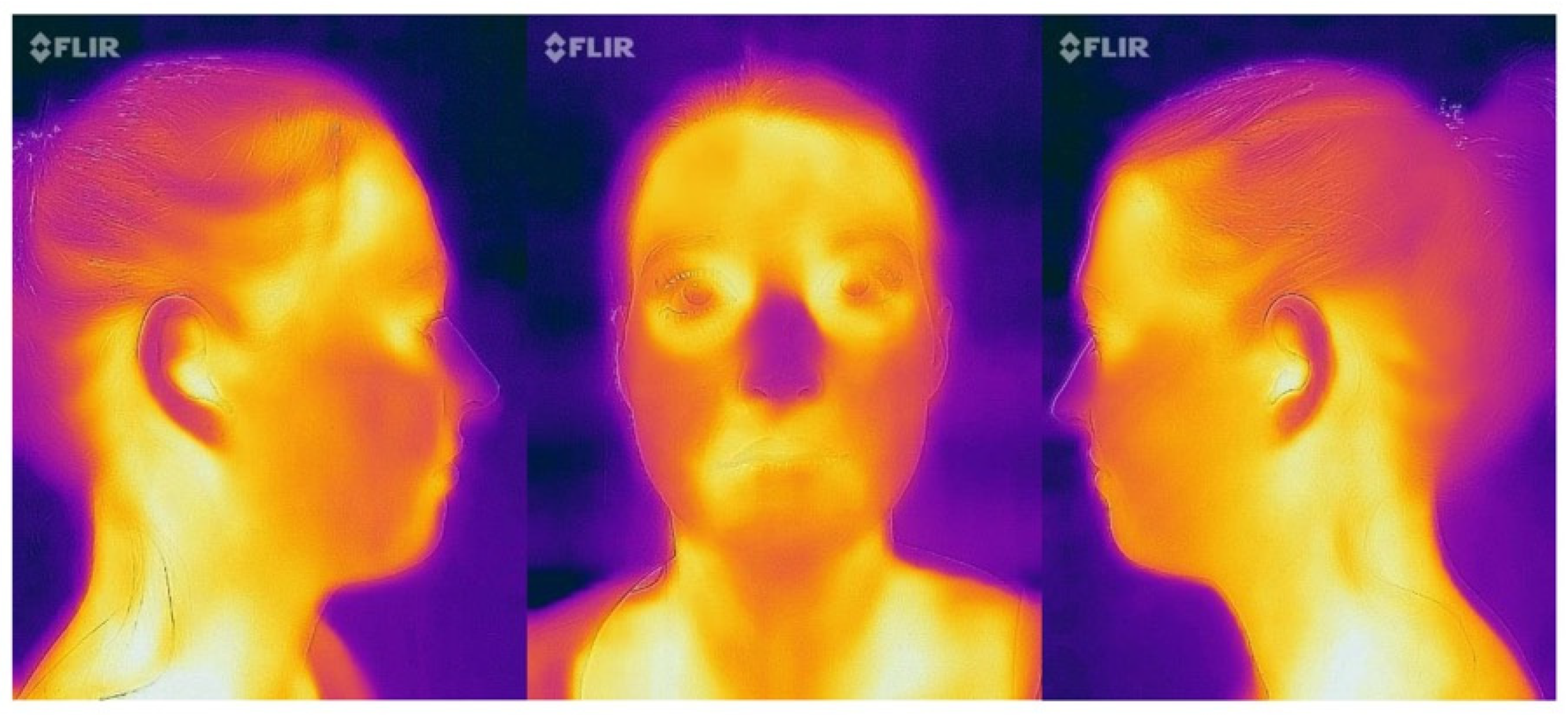
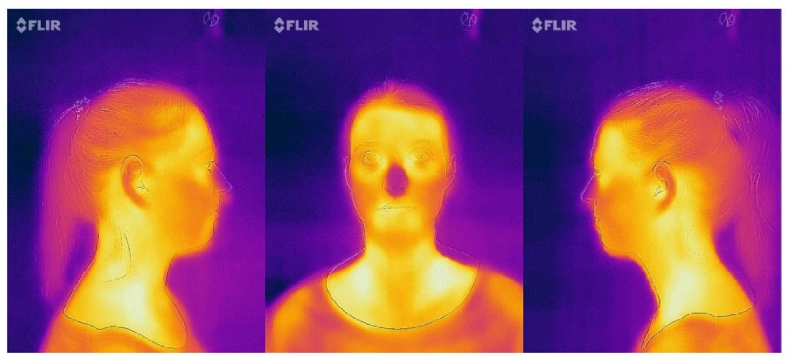
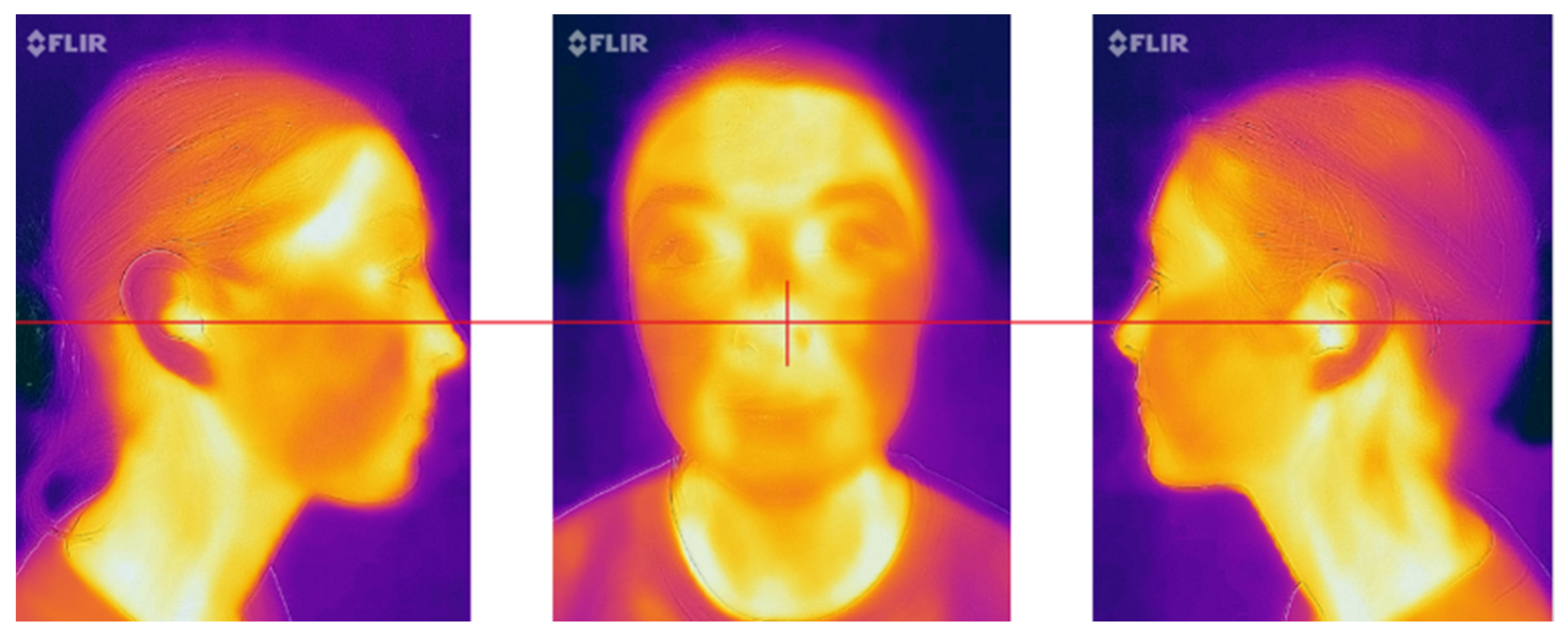
Each of the views were shot at a distance of 15 cm, 30 cm, and 50 cm (Figure 1, Figure 2 and Figure 3).
Each view at each distance was repeated five times for each participant. Therefore, a total of 45 images were taken for each participant, and a total of 1350 thermal images were taken for the 30 participants.

Composing the Frame

To ensure that all the images were standardized and captured in the exact same manner, the subject was composed in the camera frame using the following criteria:
  • Frontal view: the nose was placed in the center of the frame (Figure 4);
  • Profile images (15 cm and 30 cm): the tip of the nose was positioned at the border of the frame with the ears maintained at the middle of the picture (Figure 4);
  • Profile image (50 cm): the ears were positioned at the center of the image (Figure 5).
No magnification was applied during the imaging process for any of the images and views.

Object Parameters

Emissivity was set to 0.98, with the reflected temperature set at 22 °C and atmospheric temperature of 21.4 °C. The relative humidity percentage during the imaging sessions was 27.0%. The external optics temperature and external optics transmission were fixed at 25 °C and 0.800, respectively. The distances of the captures were fixed at 0.15 m, 0.30 m, and 0.50 m, as per the study protocol.

2.3.3. Region of Interest

A region could be selected in the frame and the software was able to provide the temperature within the selected region of interest (ROI).
The required ROI (region of interest) was drawn on all images for the different views. The images were converted to black and white mode, as a first step, to eliminate any influence of the color on the box drawing. A rectangular shape was used to mark the ROI. For the frontal view, the superior borders of the rectangle were placed at the lower borders of the nose and laterally limited to the perceivable outer edge of the nostrils, with the “cross” positioned at the center of the lips (in closed position), as shown in Figure 6. For the profile pictures, the superior borders on the inferior border of the ala of the nose edge of the nose until the imaginary extended line from the corner of the eye, and the cross was placed in the center of the lips. This is shown in Figure 7.

2.4. Endpoints/Outcome Measures

The outcome measure for this pilot study was the temperature observed in the thermograms. The software measured the temperature from the observed heat patterns. The software was able to measure the minimum, maximum, and mean temperatures from the provided frame. In this pilot experiment, we decided to use two measured temperatures provided by the software for the various views and shooting distances for each participant.
Two temperature measurements were used for the analyses in this study:
  • Mean overall temperature observed in the thermogram;
  • Mean temperature observed in the region of interest (ROI).

2.5. Sample Size

A previously published study that analyzed the visualization of temperature patterns in the oral cavity used a sample size of 20 [60]. Based on this number, and taking Cohen’s medium effect size of 0.5 with an ⍺ err prob = 0.05 and Power (1 − β err prob) = 0.80, the total sample size was calculated as 27 (critical t = 1.706, Df = 26, actual power = 0.8118) [61]. Assuming a dropout rate of 10%, a total number of 30 participants were from the student pool of the ZZM at the UZH. The sample size calculation was performed using free software (G*Power, version 3.1.9.6 for Mac OS X, Dusseldorf, Germany) [62,63].

2.6. Statistical Methods

The means and standard deviations were calculated for the mean overall temperature and the mean temperature at the ROI for the various views and regions. Data were verified for a Gaussian distribution using the Kolmogorov–Smirnov (K–S test) with significance set at p < 0.05 (95% CI). The mean overall and the mean ROI temperatures were not normally distributed (p-value < 0.001: K–S test). Non-parametric Kruskal–Wallis tests and Spearman’s rank order correlations were used to analyze the temperature differences between the different views and the relationship with the demographic/influencing factors (⍺ = 0.05). All statistical analyses were performed using statistical software (IBM SPSS Statistics, version 28.0.1.1, IBM Corp., Armank, New York, NY, USA).

2.7. Study Procedure

All participants were informed about the study and were recruited after receiving their informed signed consent. Participants were brought to the study room and asked to fill out a questionnaire evaluating their exposure to the possible temperature influencing factors relevant to this current study.
The humidity, room temperature, and participants’ body temperature (ear) were measured. The thermographic images were taken in a standardized manner, as described in the above sections. After all the required thermographic images and information on the influencing factors were collected, the images were uploaded in the FLIR software, and the ROIs (regions of interest) were plotted. The software program then calculated the mean overall temperature in the entire image and the mean temperature in the ROI. The required thermographic data were exported and used for statistical analysis.

3. Results

A total of thirty students in their third year of dental school volunteered and participated in this study [n = 30, women: 21 (70%), men: 9 (30%); mean age: 25.8 ± 6.0]. The mean room temperature recorded at the study site was 22.07 ± 0.36 °C (min: 21.3 °C, max: 22.6 °C). The relative humidity (%) was 0.36 ± 0.05 (min: 0.3%, max: 0.5%). All participants were healthy and did not have any general or intraoral pathology that required any immediate care. None of the participants reported any pain or symptoms at the time of the thermographic imaging. The mean body temperature calculated as an average of the temperatures recorded from the two ears was 37.03 ± 0.28 °C (median: 37.05 °C; min: 36.55 °C, max: 37.50 °C, range: 0.95).
The average number of teeth present in the participants was 28.5 ± 1.2 (min: 27, max: 32), and their mean DMF (T) score was 2.97 ± 2.86 (min: 0.0, max: 11). The mean gingival and plaque indices were 0.004 ± 0.01 and 0.16 ± 0.52, respectively. The demographic information of the participants is given in Table 1.
The distribution of participants with/without the presence of influencing factors/components is shown in Table 2. Four participants (13.3%) wore facial jewelry. The majority of the participants (n = 23, 76.7%) had some kind of cosmetic product applied on the face. Eighteen participants (60.0%) reported wearing spectacles in the last four hours, and 26 participants reported wearing either tight clothes or pressure stockings, or scarfs, headphones, caps, hats, and protection for the neck in the previous 1–4 h before the thermography photo session. Six participants reported being on medications that included sympathetic blockers, steroids, and vasoactive drugs. The detailed history for this consumption was not requested as it was beyond the scope of this study. Seven participants (23.3%) had undergone a professional dental hygiene procedure, and one of the participants (3.3%) had a physiotherapy session in the preceding 24 h. Twenty-one participants (70.0%) had reported brushing their teeth four hours prior, and six (20.0%) had also used a mouthwash, while four participants (13.3%) reported using an interdental device. Nineteen (63.3%) and seven (23.3%) participants reported drinking a beverage and eating something, respectively, one hour prior to the session. The other details of the food and beverage consumed by the participants as well as other miscellaneous factors are listed in Table 2.
A total of 1350 images were taken for the 30 participants in the various distances and views. An example of the typical thermographic images captured in the various views and distances for a randomized study participant is shown in Figure 3, Figure 4 and Figure 5.

3.1. Overall Temperature of the Image

The differences in the mean overall temperatures measured by the software in the thermographic images of the participants for the front view were 31.80 ± 1.43 °C, 29.62 ± 0.73 °C, 27.25 ± 0.74 °C at 15 cm, 30 cm, and 50 cm, respectively (Table 3). For the left profile view at 15 cm, 30 cm, and 50 cm, the mean overall temperature of the image was 31.90 ± 0.85 °C, 29.72 ± 0.93° C, 26.86 ± 0.71 °C, respectively (Table 3). The mean overall temperature measured for the right profile view was 31.67 ± 0.79 °C, 29.86 ± 1.08 °C, 26.67 ± 0.70 °C for 15 cm, 30 cm, and 50 cm, respectively (Table 3).
The differences in the mean overall temperatures between the different shooting distances within each profile view were statistically significant [Front: H(2) = 368.018, p < 0.001; Left: H(2) = 384.353, p < 0.001; Right: H(2) = 364.811, p < 0.001; Table 4]. Pairwise comparisons revealed significant differences in the mean overall temperature between all of the distance comparisons within all profile views (p < 0.001, p-value: Bonferroni; Table 5).

3.2. Temperature of the Region of Interest (ROI) in the Image

The differences in the mean temperatures recorded in the ROI for the front view were 32.93 ± 1.05 °C, 34.07 ± 0.86 °C, and 33.62 ± 1.06 °C at 15 cm, 30 cm, and 50 cm, respectively (Table 3). The mean ROI measured in the left profile view was 32.83 ± 2.55 °C, 32.83 ± 1.15 °C, and 32.57 ± 0.87 °C for the different shooting distances of 15 cm, 30 cm, and 50 cm, respectively (Table 3). In the right view, the mean ROI recorded was 33.06 ± 0.80 °C, 32.85 ± 0.96 °C, and 32.35 ± 0.93 °C at 15 cm, 30 cm, and 50 cm, respectively (Table 3).
The differences in the mean temperatures at the ROI for all different shooting distances within in each profile view were statistically significant [Front: H(2) = 80.176, p < 0.001; Left: H(2) = 21.399, p < 0.001; Right: H(2) = 49.451, p < 0.001; Table 4]. Pairwise comparisons revealed significant differences in the mean temperature at ROI between all of the distance comparisons within all profile views (p < 0.001, p-value: Bonferroni; Table 5), except in the 30 cm–15 cm comparisons in both the left (p = 0.789, p-value: Bonferroni; Table 5) and right (p = 0.132, p-value: Bonferroni; Table 5) views.

3.3. Correlations of the Mean Overall Temperature

The Spearman’s Rho correlation statistics (Table 6) revealed that the mean overall temperature correlated to the presence of personal effects such as spectacles [rs (1348) = 0.070, p = 0.011], scarfs/headphones/cap/hat/shawls [rs (1348) = −0.084, p = 0.002], and protective wear for neck protection [rs (1348) = −0.099, p < 0.001]. The mean overall temperature was positively influenced by having undergone dental treatments [rs (1348) = 0.079, p = 0.004] and negatively correlated to having undergone physiotherapy sessions [rs (1348) = −0.071, p = 0.009] in the last 24 h, respectively. The mean overall temperature was also positively correlated to interdental cleaning [rs (1348) = 0.095, p < 0.001] and hairdryer use [rs (1348) = 0.082, p = 0.003]. No demographic or other influencing factors had any correlation with the mean overall temperature in the thermograms (Table 6).

3.4. Correlations of the Mean Temperature at ROI

The mean temperature in the ROI was negatively correlated to the room temperature [rs (1348) = −0.150, p < 0.001] and room humidity [rs (1348) = −0.058, p = 0.033]. Wearing tight clothing or spectacles positively influenced the mean temperature in the ROI [tight clothes: rs (1348) = −0.206, p < 0.001; spectacles: rs (1348) = 0.136, p < 0.001; Table 6] while wearing a scarf/headphones/cap/hat/shawl on the head or wearing an object on the neck negatively influenced the mean temperature in the ROI (p < 0.001; Table 6).
The mean temperatures in the ROI of the participants under medications were negatively correlated [sympathetic blockers: rs (1348) = −0.058, p = 0.034; steroids: rs (1348) = −0.123, p < 0.001; vasoactive drugs: rs (1348) = −0.058, p = 0.034; Table 6]. The mean temperature in the ROI was positively influenced by having undergone dental treatments [rs (1348) = 0.160, p < 0.001] and negatively correlated to having undergone physiotherapy sessions [rs (1348) = −0.179, p < 0.001] in the last 24 h, respectively (Table 6). Procedures in oral care influenced the mean temperature in the ROI [tooth brushing: rs (1348) = −0.183, p < 0.001; inter-dental cleaning: rs (1348) = 0.214, p < 0.001; mouthwash use: rs (1348) = 0.228, p < 0.001; Table 6]. Consuming coffee as well as smoking negatively influenced the mean temperature in the ROI [coffee: rs (1348) = −0.119, p < 0.001; smoking: rs (1348) = −0.219, p < 0.001; Table 6], while all other food and beverage consumption were positively correlated to the mean temperature at the ROI [bonbons: rs (1348) = 0.090, p < 0.001; eating: rs (1348) = 0.056, p = 0.040; drinking: rs (1348) = 0.056, p = 0.041; energy drinks: rs (1348) = −0.183, p < 0.001; Table 6]. Hair dryer use was positively correlated (p < 0.001) while the use of hot water on the face (p < 0.001) and shaving (p = 0.005) were negatively correlated to the mean temperature at the ROI (Table 6).

4. Discussion

The current study was undertaken to standardize and optimize the shooting distances when capturing facial thermographic images for screening oral health problems. For this purpose, normal adult volunteers who were considered generally healthy were recruited and facial thermograms were taken in three profile views (front, left, and right) with three shooting distances (15 cm, 30 cm, and 50 cm) for each view. The findings of this study revealed that there was a decrease in the temperature measured as the distances were increased, which was highly significant (p < 0.001). This trend was observed in both the evaluated temperatures (i.e., in the mean overall temperature of the entire image as well as in mean temperature in the selected ROI). Therefore, the primary null hypothesis, that there is no effect of different shooting distances on the mean overall temperature and the mean temperature at the ROI in the extraoral thermographic images of the face when the image capturing views are kept constant, is thus rejected. Furthermore, this study further confirmed that the mean overall temperature and the mean temperature at the ROI were influenced by demographic and other external factors. Therefore, the secondary hypothesis was also rejected.
This trend for the temperature to vary at different shooting distances was consistent with the findings observed in previously published studies where an acceptable temperature difference of 0.2 °C was seen when the measurements were performed at 0.2 m and 2.5 m distances from the ROI [64,65,66]. However, the magnitude of the measured differences in this study, in comparison to the previously published studies, varied. The temperature differences observed in the ROI for these three distances ranged between 0.00 and 0.26 °C for the left profile; a difference of 0.21 and 0.71 °C was calculated for the right view, respectively. The temperature differences for the various shooting distances for the front view were between 0.45 and −1.14 °C. In the present study, the distances chosen were between 15 cm (0.15 m), which was the camera’s minimum focusing, and two other convenient distances of 30 cm (0.3 m) and 50 cm (0.5 m). These were selected to remain within realistic clinical norms, as distances beyond these may not be practically possible in a normal dental clinic setting because of a lack of space. Furthermore, a large distance would involve the inclusion of various elements in the clinic environment within the frame, which may cause interferences in the thermal imaging. To avoid these influences, the shooting distances were kept small but at the same time viable. Currently, there exists no consensus for a recommended shooting distance for taking facial thermograms. In dental applications, for monitoring post-surgical healing and inflammatory sites, the camera distance used was limited to 0.9 ± 0.1 m [59]. However, it has been reported that measurement distances between 0.2 m and 2.5 m produced small differences in temperature variations (0.2 °C), however, this recommendation was for the thermography of musculoskeletal imaging of the lower extremities [64]. In bench experiments, an image capture distance between 0.7 m and 1.5 m produced the highest inter-camera reproducibility, and shorter capture distances and cameras with superior hardware were preferable [67].
In the current study, the temperature difference observed for the different distances for the left view was in an acceptable range of less than 0.2 °C, thus indicating that perhaps all three chosen distances for the facial thermograms were acceptable. However, the results of the temperature differences obtained for the front and right views for the different shooting distances were higher (>0.2 °C), thus indicating that the different distances selected in this study impacted the measurements, which is in agreement with a former published study where the experimental setup was comprised of focusing distances of 0.05 m and 0.15 m with a camera distance of 0.8 m [66]. It is important to bear in mind that acceptable temperature variations of less than 0.2 °C, as described in previous studies, used cameras with an advanced hardware system [64,65,66,67]. The camera used in the current study was a portable device that could be attached to a mobile phone and had a sensitivity measurement uncertainty of ±5 °C, therefore, the differences observed in the current study for the right side may also be considered as acceptable. In this study, the temperature differences in the front view were greater this 5 °C, hence it must be acknowledged that the three distances chosen did influence the measurements.
This variation in the temperature differences measured at different distances for the various views could perhaps have been due to a number of factors including the examination room factors, camera accuracy, and analysis methods [68]. This study was conducted in strict adherence to follow the recommended guidelines for thermographic imaging. The ambient room temperature and the humidity of the room in this study was maintained at a mean temperature of 22.07 ± 0.36 °C [min: 21.30 C, max: 22.6] and 36 ± 5% [min: 30%, max: 50%] [66]. It has been recommended that for better stability, drift, and uniformity, cameras with sturdy hardware should be used [68]. The camera used in this study was from a reputed manufacturer that has previously been used for dental applications and is especially recommended for expected differences in temperature measurement greater than 0.05 °C and for its feature of portability [59,67]. The analysis software employed in this study was manufacturer recommended and therefore, concerns arising because of compatibility/sensitivity issues could be ruled out.
Although this study was conducted adhering to the recommended guidelines for thermography and with sound methodology, certain limitations may have existed. First, as this study only included healthy volunteers, the true effect of screening an actual oral health condition was not possible. However, it must be kept in mind that the scope of this study was to standardize shooting distances and the effect of influencing factors on the temperature measurements. A further critique that can be made is that only one portable thermography camera was employed in this study. This aspect was designed with intent. In the domain of geriatric dentistry, older adults with complex health and cognitive statuses are treated. These adults are often care-resistant, non-cooperative, aggressive, or immobile, or present with severe physical handicaps. Therefore, the concept of using thermography to identify and screen oral diseases in non-communicative individuals is novel and could greatly ease the entire process of screening and diagnosis as well as avoid cumbersome procedures like employing general anesthesia just for examination and diagnosis. When considering these aspects, it is equally important that the thermography technique employed does not necessitate elaborate equipment or extensive preparations. Therefore, the camera used in this study was selected while considering its features of portability, ease of use as well as its compatibility with a mobile smartphone device. Moreover, its technical specifications recommend it for use in situations where a temperature variation of more than 0.05 °C is expected. The facial surface temperature variations during the post-surgical healing phase following a dental treatment have been documented to be in the range of 2.1 °C and 2.8 °C, and therefore the camera employed was sufficient to perform the required task [59,67]. Nevertheless, future studies are deemed necessary to compare the present findings using cameras with superior hardware.

5. Conclusions

The findings of this study conclude that the detection of temperature asymmetries on the face in adult volunteers is affected by the distance between the camera and the subject. The findings of this study further confirm that the temperature asymmetries of the face are influenced by medication, oral-care as well as alimentation. Therefore, it is advised that personal as well as medical histories are obtained in detail prior to taking the facial thermograms. Further studies are required to make clear recommendations on the ideal shooting distance for facial thermograms.

Author Contributions

Conceptualization, M.S. (Murali Srinivasan) and A.S.; methodology, M.S. (Mona Schöffauer), L.A., A.S. and M.S. (Murali Srinivasan); validation, M.S. (Mona Schöffauer), A.S. and M.S. (Murali Srinivasan); formal analysis, M.S. (Mona Schöffauer) and M.S. (Murali Srinivasan); investigation, M.S. (Mona Schöffauer), L.A. and M.S. (Murali Srinivasan); resources, M.S. (Murali Srinivasan); data curation, M.S. (Mona Schöffauer), L.A. and M.S. (Murali Srinivasan); writing—original draft preparation, M.S. (Mona Schöffauer), and M.S. (Murali Srinivasan); writing—review and editing, M.S. (Mona Schöffauer), and M.S. (Murali Srinivasan); visualization, M.S. (Murali Srinivasan); supervision, M.S. (Murali Srinivasan); project administration, M.S. (Mona Schöffauer), L.A. and M.S. (Murali Srinivasan) All authors have read and agreed to the published version of the manuscript.

Funding

This research received no external funding.

Institutional Review Board Statement

The study was conducted in accordance with the Declaration of Helsinki, and approved by the Kantonale Ethikkommission Zürich (Basec Nr. 2021-00231).

Informed Consent Statement

Informed consent was obtained from all subjects involved in the study.

Data Availability Statement

The data presented in this study are available on request from the corresponding author.

Acknowledgments

The authors would like to thank Elena van der Heijden for her support and aid in the administrative work and project coordination.

Conflicts of Interest

The authors declare no conflict of interest.

References

  1. UNDESA (United Nations Department of Economic and Social Affairs, Population Division). World Population Ageing 2020 Highlights: Living Arrangements of Older Persons (ST/ESA/SER.A/451); United Nations: New York, NY, USA, 2020. [Google Scholar]
  2. UNDESA. Ageing Populations: We Are Living Longer Lives, but Are We Healthier? UNDESA/POP/2021/TP/NO. 2; Department of Economics and Social Affairs, Population Division, United Nations: New York, NY, USA, 2021; p. 116. [Google Scholar]
  3. United-Nations; World Health Organization. Aging and Health. 2021. Available online: https://www.who.int/news-room/fact-sheets/detail/ageing-and-health (accessed on 7 January 2022).
  4. Inouye, S.K.; Studenski, S.; Tinetti, M.E.; Kuchel, G.A. Geriatric syndromes: Clinical, research, and policy implications of a core geriatric concept. J. Am. Geriatr. Soc. 2007, 55, 780–791. [Google Scholar] [CrossRef] [Green Version]
  5. Jaul, E.; Barron, J. Age-Related Diseases and Clinical and Public Health Implications for the 85 Years Old and Over Population. Front. Public Health 2017, 5, 335. [Google Scholar] [CrossRef] [Green Version]
  6. Wallace, E.; Salisbury, C.; Guthrie, B.; Lewis, C.; Fahey, T.; Smith, S.M. Managing patients with multimorbidity in primary care. BMJ 2015, 350, h176. [Google Scholar] [CrossRef] [Green Version]
  7. Fortin, M.; Bravo, G.; Hudon, C.; Vanasse, A.; Lapointe, L. Prevalence of multimorbidity among adults seen in family practice. Ann. Fam. Med. 2005, 3, 223–228. [Google Scholar] [CrossRef] [PubMed] [Green Version]
  8. Cockburn, N.; Pradhan, A.; Taing, M.W.; Kisely, S.; Ford, P.J. Oral health impacts of medications used to treat mental illness. J. Affect. Disord. 2017, 223, 184–193. [Google Scholar] [CrossRef]
  9. Schneider, C.; Zemp, E.; Zitzmann, N.U. Oral health improvements in Switzerland over 20 years. Eur. J. Oral Sci. 2017, 125, 55–62. [Google Scholar] [CrossRef]
  10. Slade, G.D.; Akinkugbe, A.A.; Sanders, A.E. Projections of U.S. edentulism prevalence following 5 decades of decline. J. Dent. Res. 2014, 93, 959–965. [Google Scholar] [CrossRef] [PubMed] [Green Version]
  11. Peltola, P.; Vehkalahti, M.M.; Wuolijoki-Saaristo, K. Oral health and treatment needs of the long-term hospitalised elderly. Gerodontology 2004, 21, 93–99. [Google Scholar] [CrossRef]
  12. Holmen, A.; Stromberg, E.; Hagman-Gustafsson, M.L.; Wardh, I.; Gabre, P. Oral status in home-dwelling elderly dependent on moderate or substantial supportive care for daily living: Prevalence of edentulous subjects, caries and periodontal disease. Gerodontology 2012, 29, e503–e511. [Google Scholar] [CrossRef] [Green Version]
  13. Imsand, M.; Janssens, J.P.; Auckenthaler, R.; Mojon, P.; Budtz-Jorgensen, E. Bronchopneumonia and oral health in hospitalized older patients. A pilot study. Gerodontology 2002, 19, 66–72. [Google Scholar] [CrossRef] [PubMed]
  14. Stromberg, E.; Hagman-Gustafsson, M.L.; Holmen, A.; Wardh, I.; Gabre, P. Oral status, oral hygiene habits and caries risk factors in home-dwelling elderly dependent on moderate or substantial supportive care for daily living. Community Dent. Oral Epidemiol. 2012, 40, 221–229. [Google Scholar] [CrossRef] [PubMed]
  15. Chalmers, J.M.; Carter, K.D.; Spencer, A.J. Oral diseases and conditions in community-living older adults with and without dementia. Spec. Care Dent. 2003, 23, 7–17. [Google Scholar] [CrossRef] [PubMed]
  16. Schwendicke, F.; Krois, J.; Kocher, T.; Hoffmann, T.; Micheelis, W.; Jordan, R.A. More teeth in more elderly: Periodontal treatment needs in Germany 1997–2030. J. Clin. Periodontol. 2018, 45, 1400–1407. [Google Scholar] [CrossRef] [PubMed] [Green Version]
  17. Scannapieco, F.A.; Cantos, A. Oral inflammation and infection, and chronic medical diseases: Implications for the elderly. Periodontology 2000 2016, 72, 153–175. [Google Scholar] [CrossRef]
  18. Loesche, W.J.; Lopatin, D.E. Interactions between periodontal disease, medical diseases and immunity in the older individual. Periodontology 2000 1998, 16, 80–105. [Google Scholar] [CrossRef]
  19. Hayes, M.; Brady, P.; Burke, F.M.; Allen, P.F. Failure rates of class V restorations in the management of root caries in adults—A systematic review. Gerodontology 2016, 33, 299–307. [Google Scholar] [CrossRef]
  20. Zhang, J.; Leung, K.C.M.; Sardana, D.; Wong, M.C.M.; Lo, E.C.M. Risk predictors of dental root caries: A systematic review. J. Dent. 2019, 89, 103166. [Google Scholar] [CrossRef]
  21. Tonetti, M.S.; Bottenberg, P.; Conrads, G.; Eickholz, P.; Heasman, P.; Huysmans, M.C.; Lopez, R.; Madianos, P.; Muller, F.; Needleman, I.; et al. Dental caries and periodontal diseases in the ageing population: Call to action to protect and enhance oral health and well-being as an essential component of healthy ageing—Consensus report of group 4 of the joint EFP/ORCA workshop on the boundaries between caries and periodontal diseases. J Clin. Periodontol. 2017, 44 (Suppl. S18), S135–S144. [Google Scholar] [CrossRef] [Green Version]
  22. Tonetti, M.S.; Eickholz, P.; Loos, B.G.; Papapanou, P.; van der Velden, U.; Armitage, G.; Bouchard, P.; Deinzer, R.; Dietrich, T.; Hughes, F.; et al. Principles in prevention of periodontal diseases: Consensus report of group 1 of the 11th European Workshop on Periodontology on effective prevention of periodontal and peri-implant diseases. J. Clin. Periodontol. 2015, 42 (Suppl. S16), S5–S11. [Google Scholar] [CrossRef]
  23. Ervin, R.B.; Dye, B.A. Number of natural and prosthetic teeth impact nutrient intakes of older adults in the United States. Gerodontology 2012, 29, e693–e702. [Google Scholar] [CrossRef]
  24. Gil-Montoya, J.A.; de Mello, A.L.; Barrios, R.; Gonzalez-Moles, M.A.; Bravo, M. Oral health in the elderly patient and its impact on general well-being: A nonsystematic review. Clin. Interv. Aging 2015, 10, 461–467. [Google Scholar] [CrossRef] [Green Version]
  25. Stenman, U.; Ahlqwist, M.; Bjorkelund, C.; Hakeberg, M. Oral health-related quality of life—Associations with oral health and conditions in Swedish 70-year-old individuals. Gerodontology 2012, 29, e440–e446. [Google Scholar] [CrossRef]
  26. Wright, T.; McGechan, A. Breast cancer: New technologies for risk assessment and diagnosis. Mol. Diagn. 2003, 7, 49–55. [Google Scholar] [CrossRef]
  27. Pawl, R.P. Thermography in the diagnosis of low back pain. Neurosurg. Clin. N. Am. 1991, 2, 839–850. [Google Scholar] [CrossRef]
  28. Chiu, W.T.; Lin, P.W.; Chiou, H.Y.; Lee, W.S.; Lee, C.N.; Yang, Y.Y.; Lee, H.M.; Hsieh, M.S.; Hu, C.J.; Ho, Y.S.; et al. Infrared thermography to mass-screen suspected SARS patients with fever. Asia Pac. J. Public Health 2005, 17, 26–28. [Google Scholar] [CrossRef]
  29. Spiegel, S.; Spiegel, F.; Luepke, M.; Wendt, M.; von Altrock, A. Ultrasonography and Infrared Thermography as a Comparative Diagnostic Tool to Clinical Examination to Determine Udder Health in Sows. Animals 2022, 12, 2713. [Google Scholar] [CrossRef]
  30. Arora, N.; Martins, D.; Ruggerio, D.; Tousimis, E.; Swistel, A.J.; Osborne, M.P.; Simmons, R.M. Effectiveness of a noninvasive digital infrared thermal imaging system in the detection of breast cancer. Am. J. Surg. 2008, 196, 523–526. [Google Scholar] [CrossRef]
  31. Bagavathiappan, S.; Saravanan, T.; Philip, J.; Jayakumar, T.; Raj, B.; Karunanithi, R.; Panicker, T.M.; Korath, M.P.; Jagadeesan, K. Infrared thermal imaging for detection of peripheral vascular disorders. J. Med. Phys. 2009, 34, 43–47. [Google Scholar] [CrossRef] [PubMed]
  32. Cherkas, L.F.; Carter, L.; Spector, T.D.; Howell, K.J.; Black, C.M.; MacGregor, A.J. Use of thermographic criteria to identify Raynaud’s phenomenon in a population setting. J. Rheumatol. 2003, 30, 720–722. [Google Scholar] [PubMed]
  33. Hewlett, A.L.; Kalil, A.C.; Strum, R.A.; Zeger, W.G.; Smith, P.W. Evaluation of an infrared thermal detection system for fever recognition during the H1N1 influenza pandemic. Infect. Control Hosp. Epidemiol. 2011, 32, 504–506. [Google Scholar] [CrossRef] [PubMed]
  34. Kosus, N.; Kosus, A.; Duran, M.; Simavli, S.; Turhan, N. Comparison of standard mammography with digital mammography and digital infrared thermal imaging for breast cancer screening. J. Turk. Ger. Gynecol. Assoc. 2010, 11, 152–157. [Google Scholar] [CrossRef]
  35. Lee, C.H.; Dershaw, D.D.; Kopans, D.; Evans, P.; Monsees, B.; Monticciolo, D.; Brenner, R.J.; Bassett, L.; Berg, W.; Feig, S.; et al. Breast cancer screening with imaging: Recommendations from the Society of Breast Imaging and the ACR on the use of mammography, breast MRI, breast ultrasound, and other technologies for the detection of clinically occult breast cancer. J. Am. Coll. Radiol. 2010, 7, 18–27. [Google Scholar] [CrossRef] [PubMed]
  36. Liu, C.C.; Chang, R.E.; Chang, W.C. Limitations of forehead infrared body temperature detection for fever screening for severe acute respiratory syndrome. Infect. Control Hosp. Epidemiol. 2004, 25, 1109–1111. [Google Scholar] [CrossRef] [PubMed]
  37. Ng, E.Y. Is thermal scanner losing its bite in mass screening of fever due to SARS? Med. Phys. 2005, 32, 93–97. [Google Scholar] [CrossRef] [PubMed]
  38. Ng, E.Y.; Acharya, R.U. Remote-sensing infrared thermography. IEEE Eng. Med. Biol. Mag. 2009, 28, 76–83. [Google Scholar] [CrossRef] [PubMed]
  39. Ng, E.Y.; Kaw, G.J.; Chang, W.M. Analysis of IR thermal imager for mass blind fever screening. Microvasc. Res. 2004, 68, 104–109. [Google Scholar] [CrossRef] [PubMed]
  40. Ng, E.Y.; Muljo, W.; Wong, B.S. Study of facial skin and aural temperature. IEEE Eng. Med. Biol. Mag. 2006, 25, 68–74. [Google Scholar] [CrossRef]
  41. Nguyen, A.V.; Cohen, N.J.; Lipman, H.; Brown, C.M.; Molinari, N.A.; Jackson, W.L.; Kirking, H.; Szymanowski, P.; Wilson, T.W.; Salhi, B.A.; et al. Comparison of 3 infrared thermal detection systems and self-report for mass fever screening. Emerg. Infect. Dis. 2010, 16, 1710–1717. [Google Scholar] [CrossRef]
  42. Renkielska, A.; Kaczmarek, M.; Nowakowski, A.; Grudzinski, J.; Czapiewski, P.; Krajewski, A.; Grobelny, I. Active dynamic infrared thermal imaging in burn depth evaluation. J. Burn. Care Res. 2014, 35, e294–e303. [Google Scholar] [CrossRef]
  43. Ring, F. Thermal imaging today and its relevance to diabetes. J. Diabetes Sci. Technol. 2010, 4, 857–862. [Google Scholar] [CrossRef] [Green Version]
  44. Wishart, G.C.; Campisi, M.; Boswell, M.; Chapman, D.; Shackleton, V.; Iddles, S.; Hallett, A.; Britton, P.D. The accuracy of digital infrared imaging for breast cancer detection in women undergoing breast biopsy. Eur. J. Surg. Oncol. 2010, 36, 535–540. [Google Scholar] [CrossRef] [PubMed] [Green Version]
  45. Biagioni, P.A.; Longmore, R.B.; McGimpsey, J.G.; Lamey, P.J. Infrared thermography. Its role in dental research with particular reference to craniomandibular disorders. Dento Maxillo Facial Radiol. 1996, 25, 119–124. [Google Scholar] [CrossRef]
  46. Clemente, M.; Coimbra, D.; Silva, A.; Aguiar Branco, C.; Pinho, J.C. Application of Infrared Thermal Imaging in a Violinist with Temporomandibular Disorder. Med. Probl. Perform. Art 2015, 30, 251–254. [Google Scholar] [CrossRef] [Green Version]
  47. Dibai Filho, A.V.; Packer, A.C.; Costa, A.C.; Rodrigues-Bigaton, D. Accuracy of infrared thermography of the masticatory muscles for the diagnosis of myogenous temporomandibular disorder. J. Manip. Physiol. Ther. 2013, 36, 245–252. [Google Scholar] [CrossRef]
  48. Rodrigues-Bigaton, D.; Dibai-Filho, A.V.; Packer, A.C.; Costa, A.C.; de Castro, E.M. Accuracy of two forms of infrared image analysis of the masticatory muscles in the diagnosis of myogenous temporomandibular disorder. J. Bodyw. Mov. Ther. 2014, 18, 49–55. [Google Scholar] [CrossRef]
  49. Gratt, B.M.; Sickles, E.A. Electronic facial thermography: An analysis of asymptomatic adult subjects. J. Orofac. Pain 1995, 9, 255–265. [Google Scholar]
  50. Gratt, B.M.; Shetty, V.; Saiar, M.; Sickles, E.A. Electronic thermography for the assessment of inferior alveolar nerve deficit. Oral Surg. Oral Med. Oral Pathol. Oral Radiol. Endod. 1995, 80, 153–160. [Google Scholar] [CrossRef] [PubMed]
  51. Gratt, B.M.; Anbar, M. Thermology and facial telethermography: Part II. Current and future clinical applications in dentistry. Dento Maxillo Facial Radiol. 1998, 27, 68–74. [Google Scholar] [CrossRef] [PubMed]
  52. Gratt, B.M.; Sickles, E.A.; Shetty, V. Thermography for the clinical assessment of inferior alveolar nerve deficit: A pilot study. J. Orofac. Pain 1994, 8, 369–374. [Google Scholar]
  53. Shetty, V.; Gratt, B.M.; Flack, V. Thermographic assessment of reversible inferior alveolar nerve deficit. J. Orofac. Pain 1994, 8, 375–383. [Google Scholar] [PubMed]
  54. Friedlander, A.H.; Gratt, B.M. Panoramic dental radiography as an aid in detecting patients at risk for stroke. J. Oral Maxillofac. Surg. 1994, 52, 1257–1262. [Google Scholar] [CrossRef]
  55. Nicolas-Rodriguez, E.; Garcia-Martinez, A.; Molino-Pagan, D.; Marin-Martinez, L.; Pons-Fuster, E.; Lopez-Jornet, P. Thermography as a Non-Ionizing Quantitative Tool for Diagnosing Burning Mouth Syndrome: Case-Control Study. Int. J. Environ. Res. Public Health 2022, 19, 8903. [Google Scholar] [CrossRef] [PubMed]
  56. Wolowski, A.; Schwarzbach, N.; Horning, H. Thermal quantitative sensory testing in burning mouth syndrome. Clin. Oral Investig. 2021, 25, 3059–3066. [Google Scholar] [CrossRef]
  57. Mo, X.; Zhang, J.; Fan, Y.; Svensson, P.; Wang, K. Thermal and mechanical quantitative sensory testing in Chinese patients with burning mouth syndrome—A probable neuropathic pain condition? J. Headache Pain 2015, 16, 84. [Google Scholar] [CrossRef] [PubMed] [Green Version]
  58. Aboushady, M.A.; Talaat, W.; Hamdoon, Z.; Elshazly, T.E.; Ragy, N.; Bourauel, C.; Talaat, S. Thermography as a non-ionizing quantitative tool for diagnosing periapical inflammatory lesions. BMC Oral Health 2021, 21, 260. [Google Scholar] [CrossRef]
  59. Kasprzyk-Kucewicz, T.; Cholewka, A.; Bałamut, K.; Kownacki, P.; Kaszuba, N.; Kaszuba, M.; Stanek, A.; Sieroń, K.; Stransky, J.; Pasz, A.; et al. The applications of infrared thermography in surgical removal of retained teeth effects assessment. J. Therm. Anal. Calorim. 2021, 144, 139–144. [Google Scholar] [CrossRef] [Green Version]
  60. Komoriyama, M.; Nomoto, R.; Tanaka, R.; Hosoya, N.; Gomi, K.; Iino, F.; Yashima, A.; Takayama, Y.; Tsuruta, M.; Tokiwa, H.; et al. Application of thermography in dentistry—Visualization of temperature distribution on oral tissues. Dent. Mater. J. 2003, 22, 436–443. [Google Scholar] [CrossRef] [Green Version]
  61. Sullivan, G.M.; Feinn, R. Using Effect Size-or Why the P Value Is Not Enough. J. Grad. Med. Educ. 2012, 4, 279–282. [Google Scholar] [CrossRef] [Green Version]
  62. Faul, F.; Erdfelder, E.; Buchner, A.; Lang, A.G. Statistical power analyses using G*Power 3.1: Tests for correlation and regression analyses. Behav. Res. Methods 2009, 41, 1149–1160. [Google Scholar] [CrossRef] [PubMed] [Green Version]
  63. Faul, F.; Erdfelder, E.; Lang, A.G.; Buchner, A. G*Power 3: A flexible statistical power analysis program for the social, behavioral, and biomedical sciences. Behav. Res. Methods 2007, 39, 175–191. [Google Scholar] [CrossRef]
  64. Tkáčova, M.; Hudak, R.; Foffová, P.; Živčák, J. An importance of camera—Subject distance and angle in musculoskeletal application of medical thermography. Acta Electrotech. Et Inform. 2010, 10, 57–60. [Google Scholar]
  65. Liu, G.; Xia, Y.; Yang, C.; Zhang, L. The review of the major entropy methods and applications in biomedical signal research. In Bioinformatics Research and Applications; Zhang, F., Cai, Z., Skums, P., Zhang, S., Eds.; Springer International Publishing: Cham, Switzerland; University of Hong Kong: Hong Kong, China, 2018; pp. 87–100. [Google Scholar]
  66. Ghassemi, P.; Pfefer, T.J.; Casamento, J.P.; Simpson, R.; Wang, Q. Best practices for standardized performance testing of infrared thermographs intended for fever screening. PLoS ONE 2018, 13, e0203302. [Google Scholar] [CrossRef] [PubMed] [Green Version]
  67. Machado, A.S.; Cañada-Soriano, M.; Jimenez-Perez, I.; Gil-Calvo, M.; Carpes, F.P.; Perez-Soriano, P.; Priego-Quesada, J.I. Distance and camera features measurements affect the detection of temperature asymmetries using infrared thermography. Quant. Infrared Thermogr. J. 2022, 2022, 2143227. [Google Scholar] [CrossRef]
  68. Clark, R.P.; de Calcina-Goff, M.L. International standardisation in medical thermography-draft proposals. In Proceedings of the 18th Annual International Conference of the IEEE Engineering in Medicine and Biology Society, Amsterdam, The Netherlands, 31 October–3 November 1996; Volume 5, pp. 2089–2090. [Google Scholar]
Figure 1. Thermograms taken at a 15 cm distance (right profile, front, left profile).
Applsci 13 08851 g001
Figure 2. Thermograms taken at a 30 cm distance (right profile, front, left profile).
Applsci 13 08851 g002
Figure 3. Thermograms taken at a 50 cm distance (right profile, front, left profile).
Applsci 13 08851 g003
Figure 4. Head positioning at a 15 cm and 30 cm distance.
Applsci 13 08851 g004
Figure 5. Head positioning for profile images at a 50 cm distance.
Applsci 13 08851 g005
Figure 6. Placement of the ROI in the frontal images.
Applsci 13 08851 g006
Figure 7. Placement of the ROI in the profile images.
Applsci 13 08851 g007
Table 1. Participant demographics.
n (%)MeanSDMinimumMaximum
Participants30 (100.0)
Age [in years] 25.86.021.045.0
Sex (n)
Men9 (30.0)
Women21 (70.0)
Body Temperature
Right ear 37.000.2736.537.5
Left ear 37.050.3236.537.6
Average of right and left ear 37.030.2836.637.5
No. of teeth present 28.531.2027.032.0
DMF (T) 2.972.860.011.0
D 0.030.180.01.0
F 2.932.850.011.0
M 0.000.000.00.0
Gingival index 0.0040.010.00.1
Plaque index 0.160.520.02.9
Presence of pain
No0 (0.0)
Yes30 (100.0)
Presence of any intra-oral findings
No29 (96.7)
Yes1 (3.3)
Room temperature 22.070.3621.322.6
Room relative humidity (%) 3653050
n—number; %—percentage; SD—standard deviation.
Table 2. Distribution of the participants (total n = 30) present with various influencing factors.
Influencing factorsNoYes
n (%)n (%)
Personal effects
Jewelry26 (86.7)4 (13.3)
Facial cosmetics present7 (23.3)23 (76.7)
Spectacles *12 (40.0)18 (60.0)
Tight clothes *27 (90.0)3 (10.0)
Pressure stockings *28 (93.3)2 (6.7)
Scarf over head, neck and mouth *28 (93.3)2 (6.7)
Use of scarf/headphones/cap/hat/shawl ***22 (73.3)8 (26.7)
Object around the neck ***19 (63.3)11 (36.7)
Mask ***0 (0.0)30 (100.0)
Medications
Sympathetic blockers **29 (96.7)1 (3.3)
Neurological Blockers **30 (100)0 (0.0)
Steroids **26 (86.7)4 (13.3)
Vasoactive drugs **29 (96.7)1 (3.3)
Treatments
Dental treatments **23 (76.7)7 (23.3)
Physiotherapy **29 (96.7)1 (3.3)
Oral care
Tooth brushing *9 (30.0)21 (70.0)
Inter-dental cleaning *26 (86.7)4 (13.3)
Mouthwash use *24 (80.0)6 (20.0)
Food & beverages
Bonbons *28 (93.3)2 (6.7)
Eating *14 (46.7)16 (53.3)
Drinking *7 (23.3)23 (76.7)
Coffee *27 (90.0)3 (10.0)
Smoking *28 (93.3)2 (6.7)
Energy drinks **28 (93.3)2 (6.7)
Eating ***23 (76.7)7 (23.3)
Drinking ***11 (36.7)19 (63.3)
Miscellaneous
Hot-water *20 (66.7)10 (33.3)
Sauna **29 (96.7)1 (3.3)
Shower ****10 (33.3)20 (66.7)
Shaving ****27 (90.0)3 (10.0)
Hairdryer use *27 (90.0)3 (10.0)
n—number; %—percentage; * 4 h; ** 24 h; *** last 1 h; **** Same day.
Table 3. The mean overall temperature and the mean temperature at the region of interest (ROI) measured from the thermographic images.
ViewsTemperature
(in °C)
Measured Temperatures at Different Shooting Distances for the Different Profiles (in °C)Temperature Differences between Shooting Distances (in °C)
15 cm30 cm50 cm15–30 cm30–50 cm15–50 cm
Front NMeanSD (SE)MeanSD (SE)MeanSD (SE)Mean (SE)Mean (SE)Mean (SE)
Overall15031.801.43 (0.12)29.620.73 (0.06)27.250.74 (0.06)2.19 (0.12)2.36 (0.12)4.55 (0.12)
ROI15032.931.05 (0.09)34.070.86 (0.07)33.621.06 (0.09)−1.14 (0.11)0.45 (0.11)−0.69 (0.11)
Left
Overall15031.900.85 (0.07)29.720.93 (0.08)26.860.71 (0.06)2.18 (0.10)2.86 (0.10)5.04 (0.10)
ROI15032.832.55 (0.21)32.831.15 (0.09)32.570.87 (0.07)0.00 (0.20)0.26 (0.20)0.26 (0.20)
Right
Overall15031.670.79 (0.06)29.861.08 (0.09)26.670.70 (0.06)1.81 (0.10)3.19 (0.10)5.00 (0.10)
ROI15033.060.80 (0.07)32.850.96 (0.08)32.350.93 (0.04)0.21 (0.10)0.50 (0.10)0.71 (0.10)
°C—Celsius; cm—centimeter; n—number, SD—standard deviation; SE—standard error; ROI—region of interest.
Table 4. Analysis of the mean overall temperature and the mean temperature at the region of interest (ROI) at different shooting distances.
Profile Views Mean Overall TemperatureMean ROI
FrontTotal n450.00450.00
Test Statistic368.018 a80.176 a
df2.002.00
p-value<0.001<0.001
LeftTotal n450.00450.00
Test Statistic384.353 a21.399 a
df2.002.00
p-value<0.001<0.001
RightTotal n450.00450.00
Test Statistic364.811 a49.451 a
df2.002.00
p-value<0.001<0.001
ROI—region of interest; n—number; df—degree of freedom; a The test statistic was adjusted for ties; p-value: Kruskal–Wallis; significance: p = 0.05.
Table 5. Pairwise comparisons of the profile views at different distances for the mean overall and mean temperature in the region of interest (ROI).
Front
Mean overall temp.Test StatisticStd. ErrorStd. Test StatisticSig.p-value
50 cm–30 cm152.3815.0210.15<0.001<0.001
50 cm–15 cm287.9115.0219.17<0.001<0.001
30 cm–15 cm135.5315.029.03<0.001<0.001
Mean ROI
50 cm–30 cm−78.5515.02−5.23<0.001<0.001
50 cm–15 cm−133.7815.02−8.91<0.001<0.001
30 cm–15 cm55.2415.023.68<0.001<0.001
Left
Mean overall temp
50 cm–30 cm155.5615.0210.36<0.001<0.001
50 cm–15 cm294.2415.0219.59<0.001<0.001
30 cm–15 cm138.6715.029.24<0.001<0.001
Mean ROI
50 cm–30 cm49.9715.023.33<0.001<0.001
50 cm–15 cm66.7715.024.45<0.001<0.001
30 cm–15 cm16.8115.021.120.260.789
Right
Mean overall temp
50 cm–30 cm161.3215.0210.74<0.001<0.001
50 cm–15 cm286.0415.0219.05<0.001<0.001
30 cm–15 cm124.7115.028.31<0.001<0.001
Mean ROI
50 cm–30 cm72.5015.024.83<0.001<0.001
50 cm–15 cm102.7415.026.84<0.001<0.001
30 cm–15 cm30.2415.022.010.040.132
Temp.—temperature; ROI—region of interest; p-value: adjusted Bonferroni for multiple comparisons; significance: p = 0.05.
Table 6. Correlation of the measured mean overall temperature and the mean temperature at the region of interest (ROI) with the various parameters.
Parameters Mean Overall Temp.Mean ROI
Demographic factorsnCCp-valueCCp-value
Sex1350.00−0.0330.223−0.0400.146
Age1350.000.0310.2490.0090.744
Room temperature1350.00−0.0140.603−0.150 **<0.001
Relative humidity1350.00−0.0120.670−0.058 *0.033
Personal effects
Jewelry1350.00−0.0420.127−0.0010.979
Facial cosmetics present1350.000.0420.1260.206 **<0.001
Spectacles *1350.000.070 *0.0110.136 **<0.001
Tight clothes *1350.00−0.0010.971−0.0130.639
Pressure stockings *1350.000.0060.8390.0230.406
Scarf over head, neck, and mouth *1350.00−0.0430.1180.0220.424
Use of scarf/headphones/cap/hat/shawl ***1350.00−0.084 **0.002−0.163 **<0.001
Object around the neck ***1350.00−0.099 **<0.001−0.243 **<0.001
Mask ***1350.00....
Medications
Sympathetic blockers **1350.000.0100.711−0.058 *0.034
Neurological Blockers **1350.00....
Steroids **1350.00−0.0310.259−0.123 **<0.001
Vasoactive drugs **1350.000.0100.711−0.058 *0.034
Treatments
Dental treatments **1350.000.079 **0.0040.160 **<0.001
Physiotherapy **1350.00−0.071 **0.009−0.179 **<0.001
Oral care
Tooth brushing *1350.00−0.0410.134−0.183 **<0.001
Inter-dental cleaning *1350.000.095 **<0.0010.214 **<0.001
Mouthwash use *1350.000.077 **0.0050.228 **<0.001
Food & beverages
Bonbons *1350.00−0.0180.5190.090 **<0.001
Eating *1350.000.0110.6850.056 *0.040
Drinking *1350.000.0020.9560.056 *0.041
Coffee *1350.000.0390.147−0.119 **<0.001
Smoking *1350.00−0.0220.411−0.219 **<0.001
Energy drinks **1350.000.0430.1130.132 **<0.001
Eating ***1350.000.0210.4360.0250.352
Drinking ***1350.000.0080.783−0.0050.845
Miscellaneous
Hot-water use *1350.000.0430.115−0.098 **<0.001
Sauna **1350.00−0.0360.181−0.0090.749
Shower ****1350.000.0400.1380.0340.209
Shaving ****1350.00−0.0340.208−0.076 **0.005
Hairdryer use *1350.000.082 **0.0030.132 **<0.001
n—number; CC—correlation constant; significance—p < 0.05 (p-value: Spearman’s Rho); * 4 h; ** 24 h; *** last 1 h; **** Same day.
Disclaimer/Publisher’s Note: The statements, opinions and data contained in all publications are solely those of the individual author(s) and contributor(s) and not of MDPI and/or the editor(s). MDPI and/or the editor(s) disclaim responsibility for any injury to people or property resulting from any ideas, methods, instructions or products referred to in the content.