Abstract
The industrial use of nanoparticles is rapidly increasing in agricultural products, and it causes numerous effects on plant growth and seed germination. There are limited studies about the uptake, accumulation, and effect of nanoparticles on plant growth. Moreover, there are no studies that have been able to measure the biological activity of seeds’ interiors under nanoparticle treatment prior to germination. Hence, in this study, the possible size-defendant effect of alumina nanoparticles on lentil (Lens culinaris) seed germination was monitored using biospeckle Optical Coherence Tomography (bOCT) at an early stage before germination. bOCT is a non-contact, non-destructive, and non-invasive image modality developed by researchers to visualize internal biological activities in vivo. Previous studies by researchers using the technique have demonstrated its potential to observe the effect of different stimuli on seed germination at an early stage before germination occurs. In the present study, the possible size-defendant effect of 200 nm and 2000 nm alumina nanoparticles (AlNPs) on lentil seed germination was examined at a concentration of 100 mg/L. The results imply that the AlNPs could enhance lentil seed germination and seedling growth compared to control treatments, and the effect is more significant as the particle size decreases. This is believed to be due to the enhancement of energy metabolism under exposure to AlNPs.
1. Introduction
The widespread use of AlNPs leads to their possible escape into the environment [1] while enhancing the need for immediate attention to be directed to seed germination and plant growth. The industrial use of nanoparticles has rapidly increased in the fields of cosmetics, pharmaceuticals, textiles, electronics, biosensors, catalysts, etc. Ref. [2] owing to their special physicochemical characteristics, such as small size, large specific surface area, and surface energy compared to bulk materials. Aluminium oxide or AlNPs are frequently utilized in explosives and rocket propellants, catalysis, electrosensors and electroanalysis, coatings, high-performance ceramics, sunscreens, etc. [1]. In addition to their use as fillers and packaging materials [3], they find unique uses in wastewater treatment technology through the creation of ultrafiltration membranes with smaller pore sizes [4] that contribute to a better performance index. AlNPs also serve as a solid phase extraction material to remove heavy metal and other pollutants from wastewater through the processes of absorption and adsorption [5].
There are limited investigations about the impact of alumina nanoparticles on the growth of seedlings and seed germination. Some studies have concluded by identifying negative effects [6], positive effects [7], or no effect for different plant species [1]. The effect of aluminium ions (Al3+) and alumina nanoparticles on cabbage seedlings were compared [8]. The results of that study imply the strong adverse effect of aluminium ions and the positive effect of a low concentration of alumina nanoparticles. However, the biochemical parameters and nitrate reductase activities of the treated seedlings were negatively impacted by the greater dosages of AlNPs. Moreover, the effect of AlNPs and aluminium ions on plant growth were further studied using lettuce (Lactuca sativa L.). In that study, AlCl3 was used to observe the effect of aluminium ions. Interestingly, a positive effect of AlNPs on root elongation was observed, whereas a low concentration of alumina was able to enhance the biomass compared to control conditions. The results of that study emphasize that the AlNPs can absorb root hair and promote the uptake of macronutrients, but the micronutrient uptake was reduced by AlNPs [9]. Moreover, the effects of alumina nanoparticles and macroparticles on Trigonella foenum-graceum L. were observed with the help of growth parameters and antioxidative stress parameters [10]. The results of that study emphasize that low concentrations of alumina nanoparticles do not induce a negative effect on plant growth, even at those concentrations for which alumina microparticles could induce negative effects. Therefore, the results obtained by this study provide a clear idea of the allowed amount of alumina NPs that can be exposed to the terrestrial environment without causing any detrimental effects is higher than that of alumina microparticles. Nevertheless, the high concentrations of alumina nanoparticles could alter the soybean growth by affecting the cell wall structure and lignin composition [11]. However, the methods used in most of the studies are destructive, highly time-consuming, and do not enable the observation of the effect at an early stage. Therefore, in this study, we propose the biospeckle optical coherence tomography (bOCT) developed by researchers to study the germination of lentil seeds under the exposure of AlNPs.
Optical coherence tomography (OCT) is a non-destructive, in vivo, image modality that maps 2D or 3D depth-resolved tomographic images of internal tissue structures with high speed, quality, and spatial resolution when compared to existing image modalities. Moreover, our group developed bOCT technology by integrating OCT technology with biospeckle phenomena, and demonstrated the micronutrient and toxic effects of increasing Zn concentrations (0, 5, 10 and 100 mg/L) on seed germination and seedling growth [12], the individual effect of four different PEMPs concentrations (0, 10, 50 and 100 mg/L) on lentil seed germination [13], the synergic effect of 100 m/L PEMPs and Zn on seed germination [14] and the impact of Acid Mine Drainage (AMD) on seed germination and seedling growth [15]. In these studies, the observed bOCT results were compared with some conventional measurements to observe the reliability and observation speed of the bOCT technique. Interestingly, the conventional measurements are in agreement with the bOCT results. However, the effect of an external agent was successfully observed within 6 h of exposure using the proposed bOCT technique, in contrast to conventional measurements which took a minimum of 40 h to exhibit significant effects, emphasizing the relative reliability and observation speed of the bOCT technique.
In this study, a novel optical technology called bOCT was deployed to examine the possible size-dependent effect of AlNPs on lentils (Lens culinaris), a popular leguminous crop consumed and cultivated around the world. It has been estimated that the demand for lentils will increase from 6.3 million tons in 2018 to grow up to 8.4 million tons in 2024 globally, owing to the important properties of lentils, such as possessing a substantial amount of protein and fiber, low calories, essential amino acids, fatty acids, and trace minerals [16].
2. Materials and Methods
2.1. Alumina Nanoparticles and Characterization
In order to observe the effect of nanoparticles on seed germination, alumina (Al2O3) nanoparticles (AlNPs) were used at a nominal size of 200 nm (1 × 10¹⁰ AlNPs per seed) and 2000 nm (1 × 10⁷ AlNPs per seed) under the concentration of 100 mg/L. This concentration was selected based on available research studies [1]. The following protocol was used to obtain high emulsion dispersity and homogeneity. The solutions were first centrifuged for five minutes, vortexed for one minute, and then sonicated at 28 °C for ten minutes. Six repetitions of the same procedure were carried out to ensure that all of the particles were evenly distributed and that there were no AlNP clusters in the solutions. Thereafter, 10 mL of the emulsion was decanted into Petri dishes for germination. An OCT observation was performed after 6 h and 24 h of exposure to AlNPs and compared with conventional measurements. Three replicates were used for each of the experimental calculations to acquire bOCT images and conventional parameters.
2.2. Treatments and Seed Breeding
To observe the effect of AlNPs, three treatment groups were prepared: a control, and two concentrations of 100 mg/L AlNPs at sizes of 2000 nm and 200 nm. The lentil seeds were bought from a company (Greenfield Project Co., Ltd., Kumamoto, Japan) that sells organic seeds, and stored in a dry location until use. In the bOCT experiments, six seeds were selected for each treatment and 12 seeds were selected for each conventional measurement. In general, the seed coat of many seeds consists of numerous unwanted surfactants which can directly affect the reliability of the results. Therefore, it is critically important to remove unwanted surfactants from the seed coat to obtain reliable results. Hence, in this study, the selected seeds (approximate weight of 40 mg) were sterilized by immersing them in 2.5% H2O2 solution for 15 min, followed by thoroughly rinsing them three times with distilled water to remove unnecessary surfactants. Following that, the seeds were put into each Petri dish and kept in a growth chamber (Conviron, Controlled Environmental Ltd., Winnipeg, MB, Canada) that was kept at an air temperature of 25 °C/20 °C, a light intensity of 260–350 μmol m−2 s−1/0 μmol m−2 s−1, and relative humidity of 55–65% over the course of a 12 h/12 h cycle.
2.3. Biospeckle Contrast and Optical Coherent Tomography (OCT) Experimental Setup
In this study, biospeckle is a very important phenomenon. When a biological object is exposed to coherent light, such as a laser light, the light that is scattered by the object interferes to create a phenomenon known as biospeckles [17]. Due to the continual movement within a biological object caused by biological processes, such as cytoplasmic streaming, organelle migration, cell development, cell division, and biochemical reactions, a dynamic speckle pattern can be seen [18]. However, the intensity of the speckle pattern remains constant throughout time for a static object. As a result, the magnitude of change in a speckle pattern can be used to observe interior biological activity.
The temporal variation of the obtained OCT structural images under the exposure of different alumina exposure during different time intervals were evaluated using a measure called biospeckle contrast (γ). The biospeckle contrast was defined as the ratio of the standard deviation of the intensity to the mean value at each pixel along the temporal axis. Equation (1) can be used to calculate this biospeckle contrast:
where j is the scan number, x and y are the coordinates of the pixels, and N is the total number of scans. It can be seen that the seeds have undergone considerable variations over time by looking for greater temporal variation or speckle contrast. As a result, alterations in speckle contrast magnitude may be a useful measure of seed biological activity when exposed to external stimuli [12,13].
A schematic diagram of the customized, fiber-based spectral domain OCT system developed in an optical bench is shown in Figure 1. This system was used to observe the internal biological activity of biological objects after just a few hours of exposure to varying treatments and environmental conditions. For the scope of this paper, a brief description of the system is given, and a detailed explanation of the experimental system and procedures can be found in our previous studies [12,13,15].
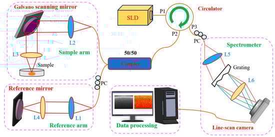
Figure 1.
A schematic representation of the experimental biospeckle Optical Coherence Tomography system. SLD: Superluminescent diode, PC: Polarization controller, L1–L6: Objective and collimating lenses, P1–P3: Circulator terminals.
2.4. Conventional Measurements
In order to make a comparison with the observed bOCT results, some conventional measurements, such as germination rate, root length, shoot length, root fresh weight and shoot fresh weight were observed. A total of twelve seeds were used per treatment to observe the germination rate. The number of germinated seeds in each treatment was recorded from 6 h to 40 h of exposures. The seeds were deemed to have germinated when their radical extensions reached 2 mm [19]. The seeds were taken out of the Petri dishes if fungus developed on their seed coats, indicating that they were dead.
Seedlings were collected after 7 days of exposure to observe the root and shoot length. Six seedlings from both the AlNPs treated samples and the control samples were taken, and they were adequately cleaned five times with distilled water to get rid of extra surfactants. Then, using ImageJ software, the shoot and root lengths of the seedlings were measured at the junction of the root-shoot.
In order to observe the root and shoot fresh weight, 7-day-old seedlings were collected and washed five times with distilled water. Thereafter, the seedlings were dried properly with filter papers and weighed on an electronic balance with an accuracy of 0.1 mg.
2.5. Statistical Analysis
The results of each triplicate sample are shown as the mean ± standard deviation of the mean. By using a one-way analysis of variance, significant differences between the treatments and the control were found (ANOVA, p < 0.05), followed by Tukey’s post hoc test using SPSS 16.0. The graphs and histograms were generated using Origin 9.5. The OCT image data were obtained using LabVIEW (13.0.1f5), and MATLAB (R2016b) software was used to obtain and analyze the bOCT image.
3. Results and Discussion
3.1. Comparison of the Ability of OCT and bOCT Images to Observe Internal Biological Activity
After the proper sterilization process, the lentil seeds were exposed to 200 nm and 2000 nm alumina at a 100 mg/L concentration for 24 h to activate internal biological activities. Thereafter, OCT images were acquired, and bOCT images were calculated and examined to observe the internal biological activities and compare them with the conventional OCT images. To observe the clear changes in biological activities, the longest exposure time of 24 h was selected for the comparison. Figure 2a–c corresponds to the bOCT images, and Figure 2d–f corresponds to the conventional OCT images. In the biospeckle pseudo color images, red regions indicate higher temporal fluctuations or higher activities, whereas blue regions indicate lower temporal variations or lower activities. Compared to the bOCT image selected as a control, both of those taken under AlNPs exposure display increments of red color regions, and these increments were more significant for the smallest particle size of 200 nm AlNPs. This kind of change in internal biological activities depending upon the exposure time, concentration, and particle size could only be visualized by bOCT images. However, in OCT structural images, the internal laminar structure of the seed can be visualized where bright regions represent a strong reflectivity of tissue, while dark regions represent a weak reflectivity of tissue. The seed coat and epidermis can be seen at the top of the structural image with strong reflectivity. However, any changes in internal biological activities cannot be monitored in OCT images like they can in bOCT images.
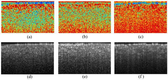
Figure 2.
bOCT contrast images (top row) and OCT structural images (bottom row) after 24 h exposure of the control (a,d), 100 mg/L alumina (2000 nm) (b,e), 100 mg/L alumina (200 nm) (c,f). A clear increment in biospeckle contrast could be seen for both alumina exposures. However, the contrast was higher for the 200 nm alumina exposure compared to the other treatment. This clear change of contrast, which cannot be observed in OCT structural images, can only be observed in bOCT biospeckle images.
OCT measures the variation in optical reflectivity, which we see as structures. The OCT images consist of not only laminar organization, but also fine granular structures called speckles, which were produced by the random interference between the scattered lights produced due to finer internal microstructures, such as mitochondria, Golgi bodies, and possibly chloroplasts within the seed. Cell growth, cell division, and cytoplasmic flow may cause these structures to move, and these movements can alter the biospeckles. A clear increment of biospeckle contrast can be seen for both AlNPs exposures compared to that of the control. However, the effect was more significant for the smaller particle size. Thus, AlNPs have a possible size-dependent positive effect. This clear change of contrast, which cannot be observed from OCT structural images, can only be observed in bOCT biospeckle images.
3.2. Biospeckle Contrast and bOCT Results after 24 h of Exposure
Biospeckle images were calculated at 0 h, 6 h, and 24 h after exposure to 200 nm and 2000 nm AlNPs. Figure 3a shows the bOCT images obtained after 24 h exposure along with that of the control at 0 h. The results clearly show that there was increased internal activity within the seed 24 h after its exposures to AlNPs, in comparison to that of the control. In order to attain a qualitative comparison, the average bOCT image contrast of six ROIs, as indicated by the rectangles, was calculated using bOCT images obtained at 24 h after exposure, and was further averaged over a total of three seeds, as shown in Figure 3b. A significant difference in the average bOCT contrast can be seen for the 200 nm and 2000 nm AlNPs compared to that of the control. The results of the study imply that the positive effect of AlNPs on seed germination can be visualized at a very early stage of germination. The addition of AlNPs is believed to enhance energy metabolism, which in turn would lead to increased dynamical variation or cellular movements within the seed, and thus result in the increased temporal variation or increased bOCT contrast. The effect was more significant as the particle size decreased.
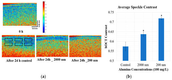
Figure 3.
Region of interest (ROI) and comparison of bOCT images of lentil seed under exposure to AlNPs of two sizes, 200 nm and 2000 nm, at 24 h (a). The horizontal and vertical scale bars represent 0.4 mm and 0.06 mm, respectively, and average speckle contrast within ROIs of bOCT images (b). Data reflect mean ± SD (n = 3), * denotes a significant increment of average bOCT contrast compared to the control (* p < 0.05).
3.3. Quantitative Analysis and Normalized bOCT Contrast
Figure 4 shows a quantitative comparison of bOCT contrast images over the treatments. The quantitative analysis was performed using six ROIs to clearly observe the possible size-dependent effect of alumina as shown in Figure 3a (Figure S1). The ROIs were selected close to the seed coat since bOCT data are more reliable for a few micron depths, and seed biological activities are also dominant close to the seed coat. Then, the average normalized contrast was calculated using the contrast of each ROI as a function of time to compare the effect of AlNPs quantitatively among the treatments. For reasons related to brevity, a brief description of the ANC and the calculation procedure are given in this paper, and a detailed explanation of the calculation can be found in previous publications [12,13]. In this study, a normalization process was used to eliminate the individual variation across the samples. First, the average contrasts across the six different ROIs were calculated before and after 24 h of exposure. Then, the normalized contrasts were calculated by taking the ratio of the above two steps. Finally, the Averaged Normalized Contrast (ANC) was calculated by taking an average of six different seed samples. Interestingly, a significant difference from the average bOCT contrast was observed after only 6 h of exposure. During the experiment, the bOCT observations were taken after 0 h, 6 h, and 24 h following the exposures, since the average germination rate of the lentil seed was estimated as 24 h. Therefore, the bOCT observations were taken before the germination to observe the significant effect at an early stage before the germination. A clear significant increment (p < 0.05) of ANC could be seen for both alumina treatments after only 6 h of exposure, compared to the control. For the 2000 nm AlNPs exposures, the increment rates of ANCs compared to that of the control were 4.7% and 9.2% at 6 h and 24 h, respectively. The same tendency could be observed for the 200 nm AlNPs exposures, for which the increment rates compared to the control were 9.3% and 17.9% at 6 h and 24 h, respectively.
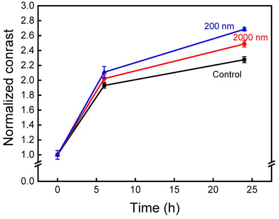
Figure 4.
Averaged normalized bOCT contrast of lentil seeds under control, 200 nm, and 2000 nm alumina exposures throughout a 24 h period.
This result clearly emphasizes the positive impact of AlNPs on lentil seed germination and seedling growth. Interestingly, the positive effect was enhanced at smaller particle sizes. Therefore, alumina has a possible size-dependent positive effect on lentil seed germination. This is believed to be due to the enhancement of energy metabolism while under exposure to AlNPs. The results of this study agree with the research conducted by [7,8], where they observed a positive effect after 2 and 7 days. However, using the bOCT technique, the effect on lentil seed germination was observed after only 6 h of exposure.
Moreover, the internal activity of bOCT contrast of seeds for all the tested concentrations were gradually enhanced over time, as shown in Figure 4. However, the ANC at 0 h to 6 h rapidly increased compared to the time interval of 6 h to 24 h. In general, when seeds are exposed to water or any other treatments, in the first few hours, the seeds start to absorb water in order to activate basic metabolic processes, thus enhancing their internal activity. Furthermore, the diffusion of water within the lentil seed reached a maximum at around 6 h, and then plateaued [20]. The enlargement of the seed could be observed due to the enlargement of internal cells that occurred as a result of rapid water absorption. Therefore, during this process, the rapid increment of internal activity could be observed compared to the subsequent time intervals. This internal structural change within the seed might possibly contribute to the increase in the bOCT contrast observed at 0 h to 6 h after exposure, for all alumina treatments.
3.4. Comparison of bOCT Results with Conventional Measurements to Understand the Reliability and Observation Speed of the bOCT Technique
To compare and verify the reliability of the obtained bOCT results, a few conventional measurements, such as germination rate, shoot length, root length, shoot fresh weight, and root fresh weight were obtained after the exposure of three different treatments including a control, 100 mg/L 2000 nm, and 200 nm AlNPs.
The percentage of seed germination is one of the common, traditional physiological indicators that is widely used to observe the effect of external agents [21]. Lentil seed germination rates under different alumina exposures are shown in Figure 5. Increments of seed germination were observed for both alumina treatments compared to the control. The germination rates after 40 h of exposures were recorded as 75%, 91%, and 100% for the control, the 2000 nm, and the 200 nm AlNPs, respectively. Hence, the highest germination rate was observed for the 200 nm AlNPs exposure, exhibiting a possible size-dependent positive effect. The results were in agreement with those observed in the bOCT results. However, it takes 40 h to observe a significant difference from seed germination, whereas bOCT takes 6 h to exhibit a significant increment.
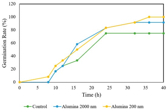
Figure 5.
Percentage of seed germination under control and exposures of 200 and 2000 nm.
In this study, to make a comparison with those results obtained by bOCT, the effects of AlNPs on root length, shoot length, root fresh weight, and shoot fresh weight were measured, as shown in Figure 6. The same tendency could be observed for all of the parameters, as seen in the seed germination percentage (Figures S2 and S3). Significant increments (p < 0.01) were observed for 200 nm AlNPs exposure compared to the control for both root length and root fresh weight. Furthermore, shoot length and shoot fresh weight also had significant increments (p < 0.05) for 200 nm AlNPs exposure compared to those obtained from the control. The highest significant increment was observed for the smaller particle size, thus emphasizing a possible size-dependent positive effect. The results were in agreement with the observed bOCT results.
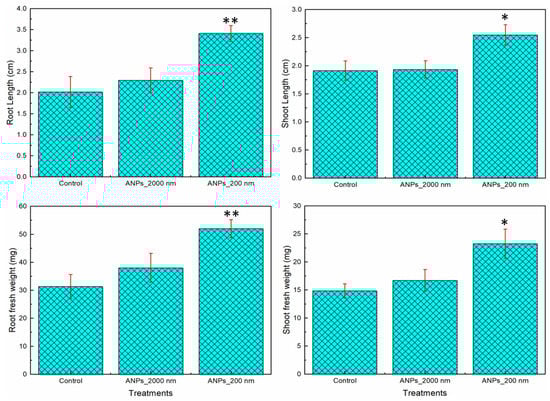
Figure 6.
Effects of different alumina treatments (control, 2000 nm, and 200 nm alumina nanoparticles) on different conventional growth parameters in 7day old lentil seedlings. Data reflect mean ± SD (n = 3), * denotes a significant increment of growth parameters compared to the control (* p < 0.05, ** p < 0.01).
The conventional measurements follow the same tendency as the bOCT results. Increments of seed germination percentage, root length, shoot length, root fresh weight, and shoot fresh weight were observed for all AlNPs exposures compared to those of the control. A similar positive effect of AlNPs on root elongation was observed for lettuce (Lactuca sativa L.), whereas a low concentration of alumina was able to enhance biomass when compared to the control condition. Moreover, the results of that study emphasize that the AlNPs can absorb root hair and promote the uptake of macronutrients [9]. The effect of alumina on lentil seed germination and growth is inversely proportional to the particle size, which is the same phenomenon as that seen in the bOCT experiments. However, conventional measurements took a minimum of 40 h to exhibit a significant increment, whereas bOCT took only 6 h, thereby emphasizing the relative observation speed and reliability.
4. Conclusions
In this study, for the first time, the possible size-dependent effects of 100 mg/L AlNPs (2000 nm and 200 nm) on seed germination were observed using bOCT. The higher internal activity within the seed was observed in biospeckle OCT images for AlNPs compared to the control after 24 h following exposure. Moreover, the highest increment of internal biological activity was observed for the 200 nm AlNPs exposure group compared to the 2000 nm AlNPs exposure and the control group, thus emphasizing a possible size-dependent positive effect. In addition, seed germination percentage, root length, shoot length, root fresh weight, and shoot fresh weight were also observed to compare with the bOCT results. The results are in agreement with the bOCT results. However, the conventional measurements took a minimum of 40 h to exhibit a significant effect, while the same effect was observed within 6 h of exposure using bOCT, thus emphasizing its greater reliability and observation speed. Hence, our results imply the possible size-dependent positive impact of AlNPs on seed germination due to the enhancement of energy metabolism. The findings presented in this paper reveal the positive effect of AlNPs on lentils. The effect is more significant when the particle size decreases. This is believed to be due to the enhancement of energy metabolism under the exposure to AlNPs. Further investigations are required on the biospeckles to determine the effect of AlNPs on seed germination.
Supplementary Materials
The following supporting information can be downloaded at: https://www.mdpi.com/article/10.3390/app13169203/s1, Figure S1: The average OCT structural image obtained by averaging over 100 OCT (x–z) scans with six regions of interest (ROI) indicated by rectangles. Each rectangle corresponds to 512 × 25 pixels; Figure S2: Effects of different alumina treatments (control, 2000 nm, and 200 nm alumina nanoparticles) on root dry weight of 7-day-old lentil seedlings; Figure S3: Effects of different alumina treatments (control, 2000 nm, and 200 nm alumina nanoparticles) on shoot dry weight of 7-day-old lentil seedlings.
Author Contributions
Conceptualization, Y.S.K.D.S. and H.K.; methodology, H.K. and U.M.R.; software, H.K.; validation, Y.S.K.D.S.; formal analysis, Y.S.K.D.S.; investigation, Y.S.K.D.S.; resources, Y.S.K.D.S.; data curation, Y.S.K.D.S.; writing—original draft preparation, Y.S.K.D.S.; writing—review and editing, Y.S.K.D.S., U.M.R. and H.K.; visualization, Y.S.K.D.S.; supervision, H.K.; project administration, H.K.; funding acquisition, H.K. All authors have read and agreed to the published version of the manuscript.
Funding
This research was funded by the JSPS Grant-in-Aid for Scientific Research (B) 19H04289 of the Ministry of Education, Culture, Sports, Science, and Technology in Japan.
Institutional Review Board Statement
Not applicable.
Informed Consent Statement
Not applicable.
Data Availability Statement
The data presented in this study are available on request from the corresponding author.
Conflicts of Interest
The authors declare no conflict of interest.
Nomenclature
| OCT | Optical Coherence Tomography |
| bOCT | biospeckle Optical Coherence Tomography |
| AlNPs | Alumina Nanoparticles |
| GA3 | |
| PEMPs | polyethylene microplastics |
| Zn | Zinc |
| AMD | Acid Mine Drainage |
| Al2O3 | alumina |
| H2O2 | Hydrogen peroxide |
| γ | biospeckle contrast |
| j | scan number |
| x, y | coordinates of the pixels |
| N | total number of scans |
| SLD | superluminescent diode |
| PC | polarization controller |
| ROIs | region of interests |
| ANC | average normalized contrast |
References
- Shabnam, N.; Kim, H. Non-toxicity of nano alumina: A case on mung bean seedlings. Ecotoxicol. Environ. Saf. 2018, 165, 423–433. [Google Scholar] [CrossRef] [PubMed]
- Handy, R.D.; Owen, R.; Valsami-Jones, E. The ecotoxicology of nanoparticles and nanomaterials: Current status, knowledge gaps, challenges, and future needs. Ecotoxicology 2008, 17, 315–325. [Google Scholar] [CrossRef] [PubMed]
- Said, S.; Mikhail, S.; Riad, M. Materials Science for Energy Technologies Recent processes for the production of alumina nano-particles. Mater. Sci. Energy Technol. 2020, 3, 344–363. [Google Scholar] [CrossRef]
- Kim, J.; Bruggen, B.V.D. The use of nanoparticles in polymeric and ceramic membrane structures: Review of manufacturing procedures and performance improvement for water treatment. Environ. Pollut. 2010, 158, 2335–2349. [Google Scholar] [CrossRef] [PubMed]
- Kumari, V.; Tripathi, A.K. Remediation of heavy metals in pharmaceutical effluent with the help of Bacillus cereus—Based green—Synthesized silver nanoparticles supported on alumina. Appl. Nanosci. 2020, 10, 1709–1719. [Google Scholar] [CrossRef]
- Ogolo, N.; Onyekonwu, M.O. Effect of aluminum oxide nanoparticles on biotic factors of the environment—A Review. Arab. J. Chem. Environ. Res. 2021, 8, 247–258. [Google Scholar]
- Juhel, G.; Batisse, E.; Hugues, Q.; Daly, D.; van Pelt, F.N.; O’Halloran, J.; Jansen, M.A. Alumina nanoparticles enhance growth of Lemna minor. Aquat. Toxicol. 2011, 105, 328–336. [Google Scholar] [CrossRef]
- Amist, N.; Singh, N.B.; Yadav, K.; Singh, S.C.; Pandey, J.K. Comparative studies of Al3+ ions and Al2O3 nanoparticles on growth and metabolism of cabbage seedlings. J. Biotechnol. 2017, 254, 1–8. [Google Scholar] [CrossRef]
- Hayes, K.L.; Mui, J.; Song, B.; Sani, E.S.; Eisenman, S.W.; Sheffield, J.B.; Kim, B. Effects, uptake, and translocation of aluminum oxide nanoparticles in lettuce: A comparison study to phytotoxic aluminum ions. Sci. Total Environ. 2020, 719, 137393. [Google Scholar] [CrossRef]
- Owji, H.; Hemmati, S.; Heidari, R.; Hakimzadeh, M. Effect of alumina (Al2O3) nanoparticles and macroparticles on Trigonella foenum-graceum L. in vitro cultures: Assessment of growth parameters and oxidative stress-related responses. 3 Biotech 2019, 9, 419. [Google Scholar] [CrossRef] [PubMed]
- de Almeida, G.H.; de Cássia Siqueira-Soares, R.; Mota, T.R.; de Oliveira, D.M.; Abrahão, J.; de Paiva Foletto-Felipe, M.; Dos Santos, W.D.; Ferrarese-Filho, O.; Marchiosi, R. Plant Physiology and Biochemistry Aluminum oxide nanoparticles affect the cell wall structure and lignin composition slightly altering the soybean growth. Plant Physiol. Biochem. 2021, 159, 335–346. [Google Scholar] [CrossRef] [PubMed]
- De Silva, Y.S.K.; Rajagopalan, U.M.; Kadono, H.; Li, D. Positive and negative phenotyping of increasing Zn concentrations by Biospeckle Optical Coherence Tomography in speedy monitoring on lentil (Lens culinaris) seed germination and seedling growth. Plant Stress 2021, 2, 100041. [Google Scholar] [CrossRef]
- De Silva, Y.S.K.; Rajagopalan, U.M.; Kadono, H.; Li, D. Effects of microplastics on lentil (Lens culinaris) seed germination and seedling growth. Chemosphere 2022, 303, 135162. [Google Scholar] [CrossRef] [PubMed]
- De Silva, Y.S.K.; Rajagopalan, U.M.; Kadono, H. Biospeckle optical coherence tomography reveals the mitigation of the harmful effects of heavy metal zinc in combination with polyethylene microplastics in lentil seeds. SPIE Future Sens. Technol. 2023, 33, 135–142. [Google Scholar] [CrossRef]
- Li, D.; Rajagopalan, U.M.; De Silva, Y.S.K.; Liu, F.; Kadono, H. Biospeckle Optical Coherence Tomography (bOCT) in the Speedy Assessment of the Responses of the Seeds of Raphanus sativus L. (Kaiware daikon) to Acid Mine Drainage (AMD). Appl. Sci. 2022, 12, 355. [Google Scholar] [CrossRef]
- Alam, M.Z.M.; Hoque, A.; Ahammed, G.J.; McGee, R.; Carpenter-Boggs, L. Arsenic accumulation in lentil (Lens culinaris) genotypes and risk associated with the consumption of grains. Sci. Rep. 2019, 9, 9431. [Google Scholar] [CrossRef] [PubMed]
- Aizu, Y.; Asakura, T. Bio-speckles. In Trends in Optics. Research, Developments, and Applications; Consortini, A., Ed.; Academic Press: San Diego, CA, USA, 1996; pp. 27–49. [Google Scholar] [CrossRef]
- Braga, R.A.; Dupuy, L.; Pasqual, M.; Cardoso, R.R. Live biospeckle laser imaging of root tissues. Eur. Biophys. J. 2009, 38, 679–686. [Google Scholar] [CrossRef] [PubMed]
- Turk, M.A.; Tawaha, A.R.M.; Lee, K.D. Seedling growth of three lentil cultivars under moisture stress. Asian J. Plant Sci. 2004, 3, 394–397. [Google Scholar] [CrossRef]
- Seyhan-Gürtas, F.; Mehmet, A.K.; Evranuz, Ö.E. Water diffusion coefficients of selected legumes grown in Turkey as affected by temperature and variety. Turk. J. Agric. 2001, 25, 297–304. [Google Scholar]
- Lin, D.; Xing, B. Phytotoxicity of nanoparticles: Inhibition of seed germination and root growth. Environ. Pollut. 2007, 150, 243–250. [Google Scholar] [CrossRef] [PubMed]


Disclaimer/Publisher’s Note: The statements, opinions and data contained in all publications are solely those of the individual author(s) and contributor(s) and not of MDPI and/or the editor(s). MDPI and/or the editor(s) disclaim responsibility for any injury to people or property resulting from any ideas, methods, instructions or products referred to in the content. |
© 2023 by the authors. Licensee MDPI, Basel, Switzerland. This article is an open access article distributed under the terms and conditions of the Creative Commons Attribution (CC BY) license (https://creativecommons.org/licenses/by/4.0/).