Review

Clinical Approaches to the Three-Dimensional Endodontic Obturation Protocol for Teeth with Periapical Bone Lesions

Department of Conservative Dentistry, Medical University of Sofia, 1431 Sofia, Bulgaria
*
Author to whom correspondence should be addressed.
Appl. Sci. 2023, 13(17), 9755; https://doi.org/10.3390/app13179755
Submission received: 27 December 2022 / Revised: 8 August 2023 / Accepted: 9 August 2023 / Published: 29 August 2023

Abstract

:
Knowledge of dental anatomy and the three-dimensional principles of debridement, sterilization and obturation is necessary for effective root canal treatment. The chemo–mechanical preparation of the root canal biomechanically significantly reduces the number of microorganisms present. However, research has demonstrated that microorganisms frequently persist. The antimicrobial effect of the irrigants is essential to their biological function. Several obturation techniques are currently available, the selection of which depends on the canal’s morphology and the individual therapeutic goals of each instance. New sealing materials and methods for regenerative root canal obturation are enhancing and improving the predictability of three-dimensional (3D) sealing procedures. Epoxy sealers and gutta-percha are currently employed by clinicians as the gold standard in obturation procedures. The most recent developments in endodontic sealing materials are related to bioceramic sealers. Reports indicate that bioceramic sealers are biocompatible, harmless, hydrophilic, expandable and antibacterial. In the therapy of teeth with periapical lesions, not only the effective treatment of the root canal system but also the quality of the used sealer are of particular importance. Regenerative processes in the bone lesion are potentiated by the action of the sealer due to its highly biocompatible, non-toxic, aluminum-free and antibacterial capabilities. This paper discusses sophisticated root canal sealing materials and contemporary filling techniques in the regenerative therapy protocol for teeth with periapical bone lesions.

1. Introduction

The main objective of endodontic treatment is to perform maximum decontamination of the root canal system and prevent any future propensity for reinfection. In order to achieve these aims, proper three-dimensional sealing of the endodontic procedure is essential to reduce the probability of microbial proliferation and the future occurrence of pathological processes. The obturation process is the third main stage of endodontic treatment, following the procedures of cleaning and shaping the root canal system [1].
A variety of essential factors, including maximal instrumentation, biomechanical preparation, obturation, and ultimately post-endodontic restoration, impact the success of root canal therapy. To achieve fluid-tight sealing of the apical foramina and portals, if present, one of the criteria for successful endodontic treatment is to obturate the root canal system in three dimensions. The antimicrobial properties of sealing and obturating materials can combat microorganisms in the dentin’s tubules [1].
After that, filling the root canal system is the most critical step in successful root canal treatment. It is described as “the three-dimensional” filling of the root canal system with a biocompatible and inert material as close to the cemento–dental junction as possible [1]. For that reason, successful root canal therapy depends on the critical related root canal treatment phases: cleaning, shaping, microbial control, and obturation. Therefore, the goal of root canal filling techniques should be to obtain a three-dimensional seal of the entire complex canal system with an inert material that prohibits connection between the root canal and periapical tissues. Thus, root canal obturation aims to fill the canal in all dimensions to form a fluid-tight seal that blocks microorganisms and their toxins from invading and migrating into the periapical tissues [2,3]. In addition, it should provide the ideal conditions for repair, stimulating the process of bio-mineralization.
A lot of complex factors may render endodontic treatment unsuccessful. The presence of remaining contaminated pulpal tissue, chronic caries, dental fractures, or abortive coronal restoration may facilitate the re-establishment of pathogenic bacteria biofilms and obstruct the survival of the tooth [3]. Hermetic obturation of the endodontic space is one of the goals of root canal treatment. The lack of a satisfied apical barrier, which results in apical discharge, is one of the most prevalent causes of endodontic treatment failure [4]. Sealer in combination with optimal gutta conicity had the greatest sealing performance, particularly in the apical area [5]. Therefore, failure to eliminate microleakage and prevent further irritation due to continued contamination is the prime reason for the failure of nonsurgical and surgical root canal endodontic therapy [6]. In multiplication, obturation has been considered one of the critical steps in the three principles of root canal treatment (enlargement, cleaning, and filling). These principles were understood more than a century ago and represent the basis of modern endodontics.
However, most root canal fillings do not adequately seal the canal system [7]. Before definitive coronal restoration, teeth with inadequate obturation, unfilled root canals, or under-extended root fillings may need retreatment [8]. Possessing a preoperative periapical bone lesion was substantially related to a 49% reduced likelihood of success compared to not having a lesion [9]. These voids may be problematic because they may contain bacteria that can proliferate when exposed to nutrients through the periapical region or lateral canals [8,9]. Some experts say that inadequate sealing of the radicular system causes 60% of endodontic failures. Root canal obturation improves apical healing. This step’s mechanical techniques, notably the sealing material, should be biocompatible with periapical tissues. Endodontic fillings shield root canals against infiltration. All stages of endodontic treatment must not damage periapical tissues [10]. Effectively filling the prepared root canal space is one of the secrets to satisfactory root canal therapy. Using gutta-percha and sealer, the canal space can be obturated. Epley et al. and Schilder proposed that the optimal root canal obturating material should be well-adapted to the canal walls and their irregularities and that the entire length of the canal should be densely packed with a homogenous mass of gutta-percha (GP). This is because, over the past 20 years, root canal filling materials and procedures have greatly improved [11].
Today, most filling methods utilize different formulations of gutta-percha in conjunction with a certain amount of sealer. Consequently, the preferred obturation method historically has been gutta-percha points with a sealer. Bioceramic sealers are gaining more and more indications in the treatment of teeth with a diagnosed periapical lesion, which in recent years have been thoroughly studied and justifiably advocated in daily clinical practice due to their various degrees of antimicrobial activity [12,13,14].

2. Requirement Properties for Root Canal Sealer in the Treatment of Teeth with Periapical Lesions

Biocompatibility is the response of a material to live cells; it encompasses cytotoxicity, cytogenicity, genotoxicity, and carcinogenicity. One of the primary requirements for an endodontic root canal sealer is that it be non-toxic and immunologically suitable for the tissue. Due to the fact that they may come into close contact with tissues, the biocompatibility of root canal sealers is crucial for the clinical efficacy of endodontic therapy, especially when the sealer extends the apical foramen and comes into contact with surrounding periapical tissues for a prolonged period [15].
In other words, a material is biocompatible when in contact with a tissue and does not induce undesirable reactions such as toxicity, irritation, inflammation, or hypersensitivity [16]. The majority of bioceramic-based root canal sealers are biocompatible. Their biocompatibility is due to the calcium phosphate present in the sealer itself. Calcium phosphate is also the principal inorganic constituent of hard tissues (teeth and bone). Many bioceramic sealers have the ability to stimulate bone regeneration if accidentally extruded through the apical foramen during root canal filling or root perforation repairs [17]. The biological compatibility of sealers is typically determined by evaluating fibroblasts’ vitality in the material’s presence [17].
In a perfect situation, one would have some leeway in one’s schedule while the sealer in root canals sets. However, a slow setting period might cause tissue irritation, as the majority of root canal sealers are poisonous until they are entirely set. According to the producers of bioceramic sealers, the setting reaction is catalyzed by the presence of moisture in the dentinal tubules, which is particularly significant when treating teeth with apical resorption. The normal setting time is four hours, although patients with extremely dry canals may require a significantly longer setting time [18].
Flow is a feature that allows the sealer to enter inaccessible places such as the thin irregularity of the dentin, the isthmus, accessory canals, and the gaps between the master and accessory cones of gutta-percha [19]. According to the International Standards Organization (ISO 6786/2001), a root canal sealer should have a flow rate of no less than 20 mm [20]. The flow rate of the sealer is affected by particle size, temperature, shear rate, and time spent forming the mix. When calculating the flow rate using a rheometer, the tubes’ inner diameter and insertion rate are considered [15].
Material mass loss for a predetermined amount of time immersed in water is known as solubility. According to ISO 6876 and American Dental Association (ADA) specification No. 7, the sealer’s solubility should not be greater than 3%. Sealers with a high solubility always permit the formation of space between the interfaces—sealer, dentin and GP points. This provides avenues for leakage from the oral cavity and periapical tissues [15]. The sealer used in root canals needs to be sufficiently radiopaque and distinct from surrounding anatomical structures [21]. This enables the radiographic inspection of the root canal filling to assess its quality. In accordance with ISO 6876/2001, the minimum radiopacity for root canal sealers is based on a reference standard of 3 mm aluminum.
According to the literature, root canal sealers’ basic antibacterial effects are linked to their alkalinity and calcium ion release, which promote healing by encouraging the formation of mineralized tissue [22]. Root canal sealers are evaluated for antimicrobial activity using two tests—agar diffusion and direct contact [23].
Enterococcus faecalis is a particularly widespread intra-radicular microbe isolated from teeth with periapical changes and is used as a modified contact test to test the antimicrobial activity of endodontic sealers [24,25].
The efficiency of a sealer’s sealing ability is dependent on its solubility and its capability to adhere to the radicular walls and GP cones [26]. In this context, adhesion is defined as the ability to stick to the root canal dentin and increase GP cone adherence to one another and to the dentin [27]. Bonding should be used in place of adhesion to describe the attachment between root canal sealer and interfaces employing mechanical forces as opposed to molecular attraction. There is no standard method for measuring dentin adhesion. As a result, microleakage and binding strength tests are widely used to evaluate the adhesion potential of root-filling materials [28]. Leakage is a major factor in root canal failure therapy. The optimal endodontic sealer would establish a barrier that prevents oral germs from entering the root canal. The ideal root canal sealer would be capable of forming a strong link between the root canal’s pulp material and dentine, thereby avoiding leakage. Additionally, it should be non-toxic and ideally promote the healing of periapical lesions [29]. In pursuing “clinically relevant” laboratory testing for the clinical efficacy of root-filling materials, leakage tests have become both the most popular and the most controversial tests. For the conventional leakage test, radioisotopes coupled to soluble markers were given time to permeate into the root canal system of root-filled teeth in vitro [15].

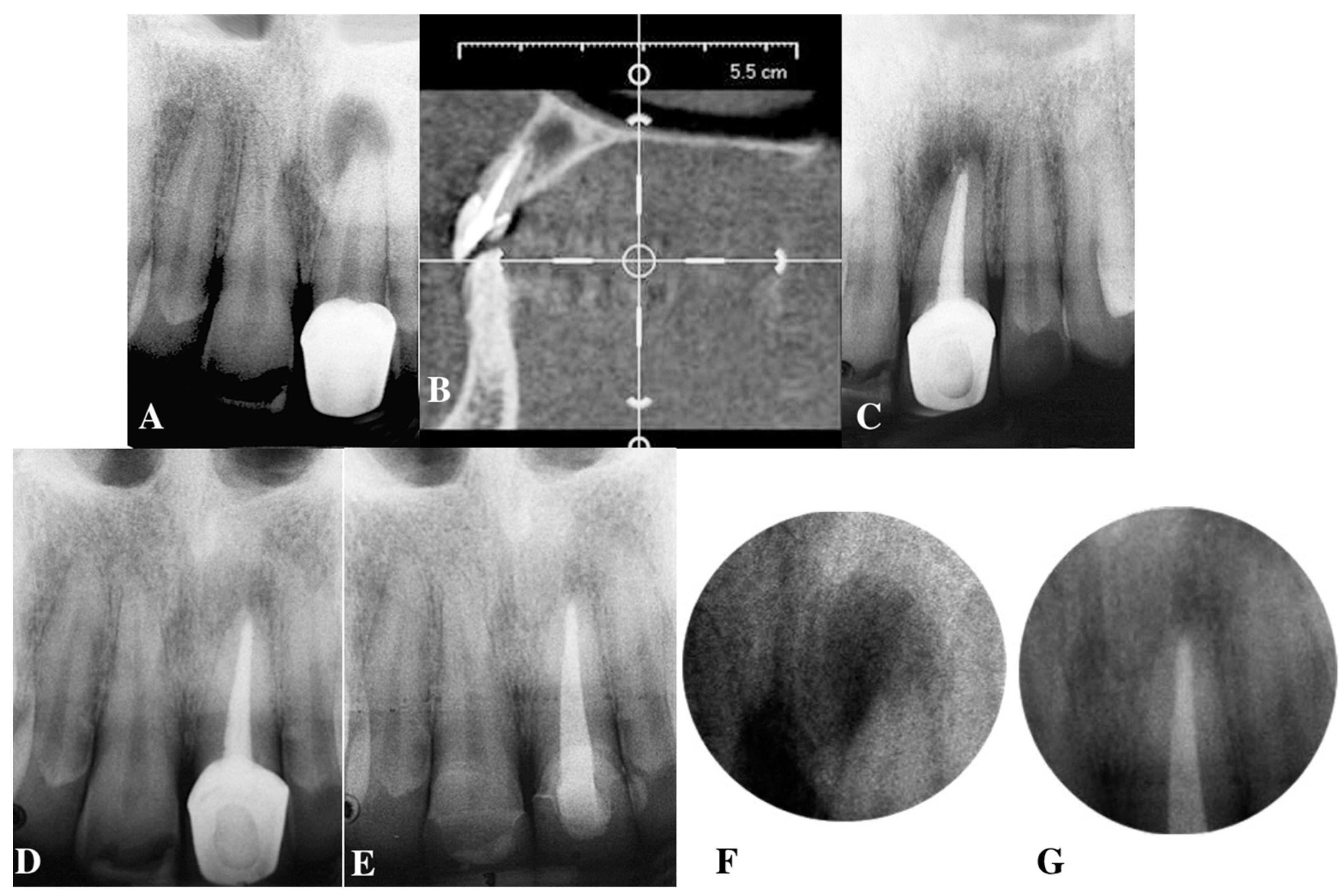
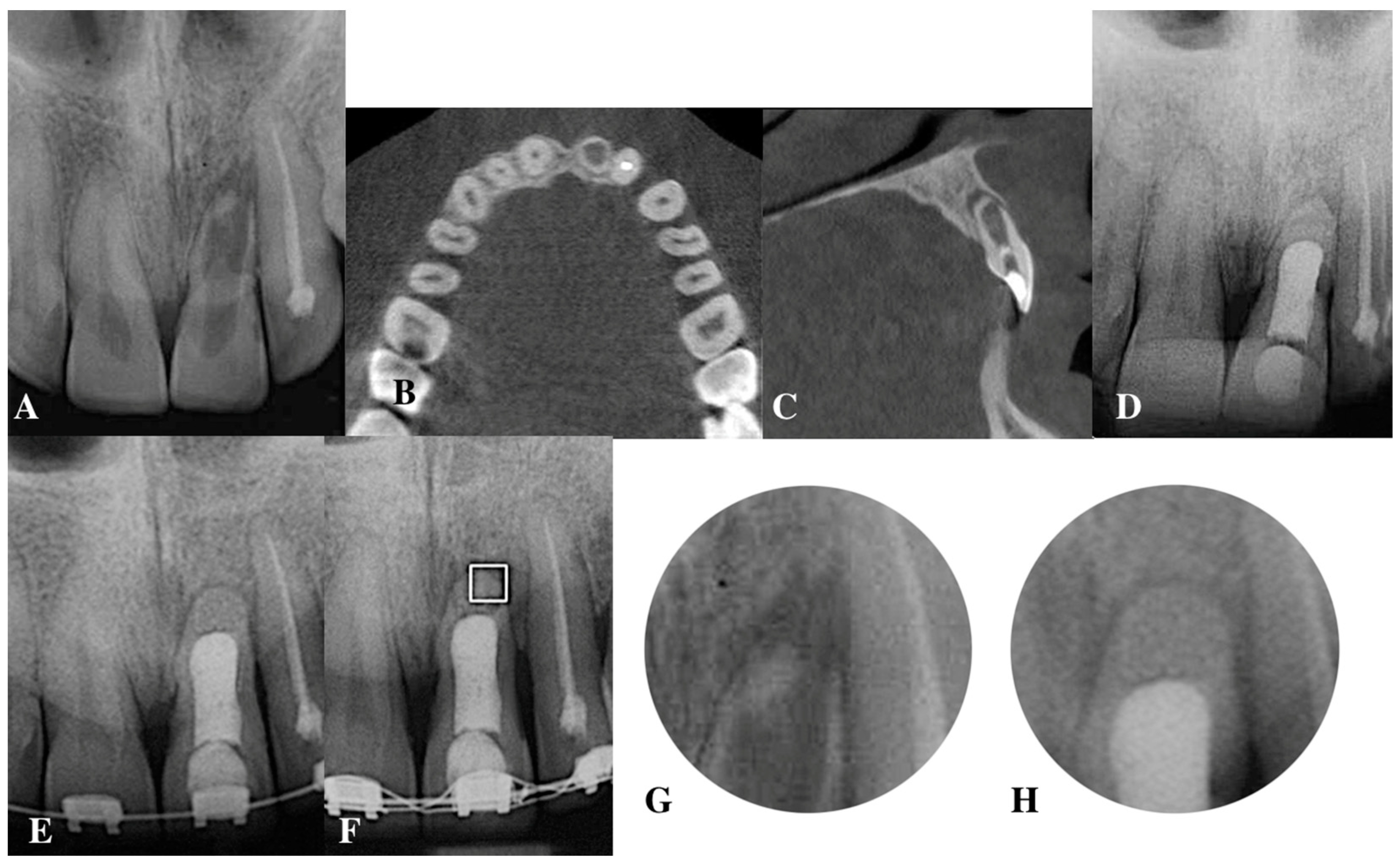
3. Factors Affecting Obturation Protocol in Teeth with Periapical Bone Lesions

In the best situation, teeth with periapical lesions should be observed for 12 months as the minimum period after the therapy before assessing success and functional tooth stability for the subsequent prosthetic treatment [30]. First, one should treat any inflammatory periapical lesions conservatively and plan apical surgery if the lesion persists or worsens after the treatment [31]. In the absence of anatomical–physiological constriction, chronic apical periodontitis can be healed by decontaminating the endodontic space and creating biomimetic environments for appropriate sealing [32].
In order to analyze the factors that influence the quality of the three-dimensional obturation of root canals, they must be divided into two main groups. The first group of factors are the pre-operative ones, which include points such as adequate access to the apical area; the possibility of effective isolation with a rubber dam; the elimination of cases with common cracks and fractures, where the complications are foreseen even before the treatment is carried out; and the possibility of precise postoperative recovery, i.e., the presence of a ferrule and tissues that can bear an effective load in the chewing process.
The second group of factors are the operative factors that directly affect the treatment result. These include the effective and comprehensive treatment of the endodontic space, both mechanically and chemically, using an evidence-based clinical protocol [33,34], the selection of the obturation technique according to the condition of the apical third, the presence or absence of resorption processes, as well as the selection of an appropriate sealer with optimal maximum sealing qualities and the subsequent impact on the periapical zone by releasing ions, which relies on the possibility of isolating the periodontium from the oral environment. Chemomechanical preparation should sterilize the endodontic space and condition the radicular dentin walls for good adhesion of the sealer to them. Preparing the apical part and activating the irrigant’s penetration into the dentin tubules with ultrasound are crucial to achieving satisfactory postoperative treatment results [35].
The quality of three-dimensional obturation depends on two closely related points: the effective elimination of the smear layer and the effective adaptation of the sealer chosen for obturation. This provokes more in-depth scientific research to ascertain the exact irrigation protocol for the selected sealer with which the root canals will be obturated [36].

4. Contemporary Sealers with Indications for the Treatment of Teeth with Periapical Lesions

4.1. Epoxy Resin-Based Sealers: AH 26, AH Plus

The first resin sealer, AH 26 (De Trey, Dentsply, Baligues, Switzerland), consists of an epoxy resin base that sets slowly when mixed with an activator. It has good sealing, adhesive and antimicrobial properties. It is a two-paste system administered in two tubes on a newly designed double syringe. The epoxide paste contains radiopaque fillers and aerosol. Aerosol, radiopaque fillers, and three types of amines are included in the amine paste. Because of its long setting time, AH Plus has better penetration into micro-irregularities and improves mechanical interlocking between the sealer and root dentin. It has outstanding solubility, expansion, adherence to dentin, and sealing characteristics. AH Plus is regarded as the “Gold Standard” [37].
Resilon high-performance industrial polyurethane is another resin-based material adopted for root canal treatment applications. It resembles and can be modified equally to gutta-percha. It is composed of a plastic core material, available in conventional cones or granules and a resin sealer. Resilon is safe to use, non-mutagenic, and biologically compatible [37].

4.2. Calcium Hydroxide-Based Canal Sealers

Calcium oxide integrates with water in the tooth and becomes calcium hydroxide, which is a well-known and long-used root canal material with excellent reported performance. Its seal is similar to that of zinc oxide–eugenol, but its solubility raises questions regarding its long-term integrity during exposure to tissue fluids [38]. The setting reactions of calcium hydroxide-containing sealers are complex. The theory for the addition of calcium hydroxide to root canal sealers is based on the observation of its antimicrobial and tissue repair abilities. After 3 days, calcium hydroxide sealers were shown to be substantially more biotoxic than resin-based sealers [39].

4.3. Bioceramic Sealers

Bioceramics are inorganic materials that have been designed specifically for dental and medical applications. The classification of bioceramic materials as bioactive or bioinert, is determined by their interaction with the surrounding body tissue [40]. Glass and calcium phosphate contain bioactive materials that interact with the surrounding tissue to enhance the formation of more resistant tissue. Bioinert substances, such as zirconia and alumina, elicit a minor response from the surrounding tissue and have no biological or physiological impact [40]. Bioactive materials are further categorized as degradable or non-degradable based on their stability. Bioceramics applied as obturation materials in current endodontics often include alumina, zirconia, bioactive glass, glass ceramics, hydroxyapatite, and various calcium phosphates [41]. Recently introduced bioceramic materials are used, such as root repair cement [42] and root canal sealers [43,44]. These materials are generally biologically compatible, harmless, without shrinkage, and chemically resistant to change. At this stage, bioceramic sealers exhibit the necessary physical qualities for effective root canal sealing in a variety of clinical situations. Nevertheless, their high solubility remains a challenge. Despite the range of sealers used in the last phase of endodontic therapy—the obturation of the tooth’s root canal system—none meet all needs [45]. They are also suited to producing hydroxyapatite during the setting process, thereby forming an interaction between dentin and the sealer [43,44]. It has been reported that bioceramic byproducts have alkaline activity, which facilitates the penetration of the sealers into dentin tubules [46].

4.3.1. Calcium Silicate-Based Sealers

The most recent innovations in bioceramic sealers have come with the introduction of calcium silicate or MTA-based sealers. These materials have been characterized as biocompatible and promote the deposition of hydroxyapatite crystalline deposits [47]. Two different types of calcium silicate-based sealers have been introduced: one-component (iRoot SP, Endosequence BC, Total Fill BC, Endoseal, Well-Root ST) and two-component (MTA Fillapex, NeoMTAPlus, BioRoot RCS, ProRoot ES) materials.
  • iRootSP sealer
iRoot SP sealer (Innovative Bioceramix, Vancouver, BC, Canada) is another newly introduced calcium silicate-based sealer with desirable properties such as apatite formation. Injectable Root Canal Sealer (iRoot® SP) was introduced in 2007 as a white, injectable, premixed, and ready-to-use hydraulic substance for permanent root canal closure. iRoot SP is a revolutionary next-generation bioceramic material that is user-friendly, non-toxic, aluminum-free, antibacterial, hydrophilic, and has exceptional sealing properties. The sealer is highly biocompatible even in cases of overfilling and an effective filling material for periapical bone regeneration [48,49]. The composition of the sealer is as follows: zirconium oxide, dicalcium silicate, tricalcium silicate, calcium phosphate monobasic, calcium hydroxide, filler, and thickening agents. “Bioceramic” is a new term for sealers classified as bioactive, bioinert, or biodegradable, depending on their interactions with surrounding tissues. iRoot® SP is also available as the private-label Endosequence BC Sealer, distributed by Brasseler USA [49].
The mechanism by which bioceramic sealers adhere to root dentin interfaces is as yet unknown. Nevertheless, the following mechanisms have been proposed for calcium silicate-based sealers: (i) the diffusion of sealer particles into dentin tubules; (ii) the infiltration of sealer particles into dentin; and (iii) the chemical reaction of the sealant with the dentin’s moisture, leading to the formation of hydroxyapatite [42,43,44,45,46]. In their formulation, bioceramic products may contain alumina and zirconia, bioactive glass, calcium silicates, hydroxyapatite, and resorbable calcium phosphates [50]. These materials are generally biocompatible, innocuous, nonshrinking, and chemically stable within the biological environment. They can also form hydroxyapatite during the setting process, forming a bond between dentin and the filling material [51,52].
  • Total Fill BC sealer
Total Fill® (FKG Dentaire, La Chaux-de-Fonds, Switzerland) is a pre-mixed bioceramic obturation material that contains calcium silicates, zirconium oxide, tantalum oxide, calcium phosphate monobasic, and thickening agents. It is administered with a syringe for root canal sealing and either a syringe or a plaster for root restoration and retrograde obturations [53].
The setting time is 4 h. In very dry root canals, however, the setting time can exceed 10 h. The TotalFill setting time is contingent on moisture in the dentin tubules. Dentin inherently contains the quantity of moisture required to complete the setting reaction. Before situating the material, it is therefore optional to introduce moisture to the root canal [53].
  • MTA Fillapex
MTA Fillapex is a hybrid bioceramic sealer created by Angelus Londrina/Parana/Brazil, in 2010 based on MTA. The multimodal application of MTA required the incorporation of material additives into the original cement/radiopacifier composition in order to generate superior formulations [54,55]. Supposedly, these additions improve the material’s characteristics and functionality. In addition, the original Portland cement formulation was replaced with tricalcium silicate to eliminate the presence of an aluminum phase and trace elements. It combines the established benefits of MTA with the exceptional qualities of root canal obturation products. Its formulation in the paste/paste system contains salicylate resin, diluting resin, natural resin, nanoparticulated silica, and bismuth trioxide and permits complete filling of the root canal, including accessory and lateral canals. It offers a steady supply of calcium ions to the tissues and maintains an antimicrobial pH. Additionally, it does not discolor the tooth structure. It is made chiefly of MTA, salicylic resin, natural resin, bismuth oxide, and silica after being mixed [56]. In several studies, it was shown that this sealer strengthened the root against fracture. MTA, which is contained in MTA-Fillapex, is more stable than calcium hydroxide, allowing for a steady release of calcium ions into the tissues and the maintenance of an antibacterial pH. Applying MTA and salicylate resin optimizes tissue regeneration and prevents inflammatory reactions [57,58].

4.3.2. Calcium Phosphate-Based Sealer

Extensive experimental and some clinical studies show that calcium phosphate bio-ceramic materials can be used as filling materials. LeGeros et al. were the first to use calcium phosphate as a bioceramic restorative dental cement [59]. Krell and Wefel compared the effectiveness of experimental calcium phosphate cement to Grossman’s sealer in extracted teeth and found no significant differences in apical occlusion, adaptation, dentinal tubule occlusion, or adhesion in their investigation [60]. Subsequently, calcium phosphate cement has been successfully implemented in endodontic treatments such as pulp capping, apical barrier construction, periapical defect repairs, and bifurcation perforation repairs. The bioactive potential of calcium silicate-based sealants is due to the minor solubility of these materials after they have hardened; however, the sealant’s solubility could affect its ability to seal a root canal against regrowth and reinfection. To identify the therapeutic significance of the gap between bioactivity and solubility, additional clinical research is necessary [61,62].

4.4. Nanomaterials of Hydroxyapatite as Root Canal Filling Materials

Nanostructured materials composed of hydroxyapatite (HA) are biocompatible, more reactive, and capable of adhering to dentinal tubules. Nano-HA is identical to the structure of dentine and/or enamel, and the nanometer-sized particles have been shown to increase osteoblast adhesion, proliferation, and mineralization and speed up bone repair processes [63,64]. Tooth #21 was prepared for endodontic treatment with preoperative radiographic observation—two- and three-dimensional images. The patient’s informed consent was provided before performing the endodontic therapy. The preoperative radiographs on tooth #21 present extensive resorption, both intraforaminal and internal radicular. The CBCT images confirmed this radiographic analysis. The apical zone is obturated with nanometer-sized bioceramics (β-TCP) as a barrier, and the root canal space is sealed with a bioceramic sealer (iRoot SP, Innovative Bioceramix, Vancouver, BC, Canada) and injected with thermoplastic gutta-percha (Bee Fill, VDW, Munich, Germany) after the completed preparation. The control radiographs were obtained during the 1st and 4th years of treatment. A successful result of the treatment was represented by enlarged apical areas (Figure 1A–H) [64].
Nano-HA is biocompatible, dynamic, and capable of adhering to the tubules of the dentin. In addition, it stimulates apical healing and is capable of producing an apical hermetic seal. Various filling procedures based on heated or preheated gutta-percha have been developed to improve the root canal’s three-dimensional filling. Little is known about the apical sealing capabilities of the novel experimental nano-HA sealer when used in conjunction with various occlusion procedures [65]. In the field of nano-calcium phosphate-based sealers, we synthesized a biphasic filling material characterized using X-ray diffraction, tests for biotoxicity and microbiologic studies [64,66], which can be applied for the treatment of apical root resorption in long-standing periapical diseases. It is widely known that the apices of teeth with necrotic pulps exhibit moderate to severe resorption of the apical foramen and peri-apical bone zone. We used an orthograde filling method to seal these zones with nano-biphasic material containing a physiological solution or 0.1% hyaluronan [67]. Water and hydroxyapatite are natural molecular ingredients in the life of biomineralized structures and represent a great biomimetic challenge in root canal-filling materials.

5. Apical Plug in Teeth with Periapical Lesions

5.1. Apical Plug in Teeth with Apical Resorption and “Open Apex”

The major challenge in endodontic treatment is to achieve complete debridement, canal disinfection, and the sealing of the root canal space in teeth with open apices and teeth with apical resorption resembling an open apex (Figure 2A–E) [64]. An open apex is a clinical situation that is a result of the iatrogenic enlargement of apical constriction with both hand and rotary files. Years ago, we treated these teeth with immature apical formation by generating mineralized formations at the apical zone as a barrier via the repeated placement of calcium hydroxide paste over many months. Incomplete root development often arises secondary to pulpal necrosis as a result of caries or trauma.
Nowadays, such clinical situations are treated with an apical plug with MTA and an apical barrier made from collagen [68]. Chronic lesions alter the adjacent bone structure, periodontal ligaments, and cementum and dentine in the periapical region. In the majority of instances, the apical physiological constriction is either absent or enlarged. In the absence of a restriction, it is difficult to attain satisfactory treatment outcomes. MTA was used for an apical 5 mm plug to prevent sealer overfilling when the root canal was definitively obturated. MTA has been demonstrated to be a suitable material for apical obturation in cases of an “open apex” in teeth with embryonic root formation, apical root resorption, and iatrogenic enlargement, as it avoids apical surgical procedures with a similar prognosis. Apical periodontitis is a destructive process in the bone around the root apex that also affects the cement and dentin. The radiographic image replicates the apical image of immature teeth, which are characterized by large, exposed apices and thin, fracture-prone dentinal walls. The apical width is measured using an instrument between #80 and #140. The presence of a periapical lesion may also result in foraminal and peri-foraminal resorption of the root apex.
The clinical case presents the placement of an MTA apical plug into the distal root canal, with the apical foramen’s width being ISO #60. The patient’s informed consent was obtained before the endodontic therapy. From the preoperative radiograph, a periapical index of PAI1 was determined with suspicion of apical resorption, and the tooth was planned for retreatment. The control radiograph after separating instrument (lentulo) removal displays the complete elimination of the fragment. The observation of root canal filling from the postoperative X-ray shows that the mesial canals were filled with a bioceramic sealer (TotalFill, FKG, Switzerland) and gutta-percha, and the apical 5 mm of the distal canal was obturated with MTA due to the width of the foramen (ISO #60). The apical barrier formed from collagen is applied before the MTA plug. The control X-ray and follow-ups after 1 month, 6 months, 1 year and 4 years demonstrate the successful endodontic outcome (Figure 3A–G).

5.2. Apical Barrier

Calcium hydroxide has for many years been the material of choice for tooth apexification in teeth with an open apex or pathological resorption. The purpose of the application was to cause the formation of a calcium barrier over a period of 5–20 months. This long period of treatment requires a high level of patient compliance. For these reasons, the method of treatment for these clinical cases has been changed. The contemporary materials of choice are MTA and bioceramic root repair materials (putty material), which have the characteristics of a fast-setting material. Thus, clinicians have the ability to complete the treatment/retreatment with an apical plug or perforation seal in one visit.
In the past few years, new CSCs have been released with similar characteristics to MTA. Five mineral oxide CSC (5MO) is derived from Portland cement and was designed to treat multiple dental problems and endodontic complications. It is regarded as an efficient pulp closure material, apicoectomies filling material, and perforation sealer [69,70]. It is very important to use an apical barrier (collagen piece or calcium phosphate bioceramic), which has the potential to stop bleeding and accelerate repairing processes in periapical/lateral bone lesions [71,72] (Figure 4).

6. Conclusions

Success in the treatment of teeth with periapical lesions is a multifactorial process that depends on the precision of each of the clinical stages. The follow-up (prospective and retrospective) of clinical cases is extremely important for the possibility of formulating dependencies and statistically significant results between the individual stages of diagnosis and treatment and the long-term success rate of the treatment. Clinicians are required to be aware of anatomical variations in canal configuration, and tactile or microscopic examination is crucial for locating extra canals. In addition to sealing the apical third, three-dimensional obturation also seals the additional “portals of exit” if present, as well as accessory canals and furcal canals.

Author Contributions

A.G. and E.D. planned, wrote and revised the article. All authors have read and agreed to the published version of the manuscript.

Funding

This research received no external funding.

Institutional Review Board Statement

This study was conducted in accordance with the Declaration of Helsinki, and the protocol was approved by the Ethics Committee of CENIMUS (for Grant Project 39/2009).

Informed Consent Statement

Informed consent was obtained from all subjects involved in the study. Written informed consent has been obtained from the patient(s) to publish this paper.

Data Availability Statement

The data presented in this study are available on request from the corresponding author.

Acknowledgments

The authors wish to thank Filip Ivanov for drawing Figure 4 to illustrate the clinical protocol of apical barrier and apical plug placement.

Conflicts of Interest

The authors declare no conflict of interest.

References

  1. Tomson, R.M.E.; Polycarpou, N.; Tomson, P.L. Contemporary obturation of the root canal system. Br. Dent. J. 2014, 216, 315–322. [Google Scholar] [CrossRef]
  2. Ozok, A.; van der Sluis, L.; Wu, M.; Wesselink, P. Sealing ability of a new polydimethylsiloxane based root canal filling material. J. Endod. 2008, 34, 204–207. [Google Scholar] [CrossRef]
  3. Haapasalo, M.; Shen, Y.; Qian, W.; Gao, Y. Irrigation in Endodontics. Dent. Clin. N. Am. 2010, 54, 291–312. [Google Scholar] [CrossRef] [PubMed]
  4. Goldberg, F.; Massone, E.J.; Artaza, L.P. Comparison of the sealing capacity of three endodontic filling technique. J. Endod. 1995, 21, 1–3. [Google Scholar] [CrossRef]
  5. Kikly, A.; Jaâfoura, S.; Kammoun, D.; Sahtout, S. Sealing Ability of Endodontic Cements: An In Vitro Study. Int. J. Dent. 2020, 2020, 5862598. [Google Scholar] [CrossRef] [PubMed]
  6. Basmadjian-Charles, C.; Farge, P.; Bourgeois, D.; Lebrun, T. Factors influencing the long-term results of endodontic treatment: A review of the literature. Int. Dent. J. 2002, 52, 81–86. [Google Scholar] [CrossRef]
  7. Lin, Z.-M.; Jhugroo, A.; Ling, J.-Q. An evaluation of the sealing ability of a polycaprolactone-based root canal filling material (Resilon) after retreatment. Oral. Surg. Oral. Med. Oral. Pathol. Oral. Radiol. Endod. 2007, 104, 846–851. [Google Scholar] [CrossRef] [PubMed]
  8. Van der Sluis, L.; Wu, M.; Wesselink, P. An evaluation of the quality of root fillings in mandibular incisors and maxillary and mandibular canines using different methodologies. J. Dent. 2005, 33, 683–688. [Google Scholar] [CrossRef]
  9. Ng, Y.L.; Mann, V.; Gulabivala, K. A prospective study of the factors affecting outcomes of nonsurgical root canal treatment: Part 1: Periapical health. Int. Endod. J. 2011, 44, 583–609. [Google Scholar] [CrossRef] [PubMed]
  10. Karabucak, B.; Kim, A.; Chen, V.; Iqbal, M.K. The comparison of Gutta-Percha and Resilon penetration into lateral canals with different thermoplastic delivery systems. J. Endod. 2008, 34, 847–849. [Google Scholar] [CrossRef]
  11. Schilder, H. Filling root canals in three dimensions. J. Endod. 2006, 32, 281–290. [Google Scholar] [CrossRef]
  12. Šimundić Munitić, M.; Poklepović Peričić, T.; Utrobičić, A.; Bago, I.; Puljak, L. Antimicrobial efficacy of commercially available endodontic bioceramic root canal sealers: A systematic review. PLoS ONE 2019, 14, e0223575. [Google Scholar] [CrossRef]
  13. Dagna, A.; Colombo, M.; Poggio, C.; Russo, G.; Pellegrini, M.; Pietrocola, G.; Beltrami, R. In Vitro Antibacterial Activity of Different Bioceramic Root Canal Sealers. Ceramics 2022, 5, 901–907. [Google Scholar] [CrossRef]
  14. Mangat, P.; Dhingra, A.; Muni, S.; Bhullar, H.K. To compare and evaluate the antimicrobial activity of three different root canal sealers: An In Vitro Study. J. Conserv. Dent. 2020, 23, 571–576. [Google Scholar] [CrossRef]
  15. Ørstavik, D. Materials used for root canal obturation: Technical, biological and clinical testing. Endod. Top. 2005, 12, 25–38. [Google Scholar] [CrossRef]
  16. Williams, D. Definitions in Biomaterials. In Progress in Biomedical Engineering, Proceedings of the Consensus Conference of the European Society for Biomaterials, Chester, England, 3–5 March 1986; Elsevier: Amsterdam, The Netherlands, 1987; Volume 4. [Google Scholar]
  17. Bryan, T.E.; Khechen, K.; Brackett, M.; Messer, R.L.; El-Awady, A.; Primus, C.M.; Gutmann, J.L.; Tay, F.R. In vitro osteogenic potential of an experimental calcium silicate-based root canal sealer. J. Endod. 2010, 36, 1163–1169. [Google Scholar] [CrossRef]
  18. Yang, Q.; Lu, D. Premixed Biological Hydraulic Cement Paste Composition and Using the Same. Google Patents EP2142225A1, 13 January 2010. [Google Scholar]
  19. De Miranda Candeiro, G.T.; Correia, F.C.; Duarte, M.A.H.; Ribeiro-Siqueira, D.C.; Gavini, G. Evaluation of radiopacity, pH, release of calcium ions, and flow of a bioceramic root canal sealer. J. Endod. 2012, 38, 842–845. [Google Scholar] [CrossRef]
  20. ISO 6876/2001; Dental Root Canal Sealing Materials. International Organization for Standardization: Geneva, Switzerland, 2001.
  21. Tanomaru, J.M.G.; Duarte, M.A.H.; Gonçalves, M.; Tanomaru-Filho, M. Radiopacity evaluation of root canal sealers containing calcium hydroxide and MTA. Braz. Oral. Res. 2009, 23, 119–123. [Google Scholar] [CrossRef] [PubMed]
  22. Okabe, T.; Sakamoto, M.; Takeuchi, H.; Matsushima, K. Effects of pH on mineralization ability of human dental pulp cells. J. Endod. 2006, 32, 198–201. [Google Scholar] [CrossRef]
  23. Zhang, H.; Shen, Y.; Ruse, N.D.; Haapasalo, M. Antibacterial activity of endodontic sealers by modified direct contact test against Enterococcus faecalis. J. Endod. 2009, 35, 1051–1055. [Google Scholar] [CrossRef] [PubMed]
  24. Pinheiro, E.T.; Gomes, B.P.F.A.; Ferraz, C.C.R.; Sousa, E.L.R.; Teixeira, F.B.; Souza-Filho, F.J. Microorganisms from canals of root-filled teeth with periapical lesions. Int. Endod. J. 2003, 36, 1–11. [Google Scholar] [CrossRef]
  25. Stuart, C.H.; Schwartz, S.A.; Beeson, T.J.; Owatz, C.B. Enterococcus faecalis: Its role in root canal treatment failure and current concepts in retreatment. J. Endod. 2006, 32, 93–98. [Google Scholar] [CrossRef] [PubMed]
  26. Desai, S.; Chandler, N. Calcium hydroxide-based root canal sealers: A review. J. Endod. 2009, 35, 475–480. [Google Scholar] [CrossRef]
  27. Sousa-Neto, M.D.; Coelho, F.I.S.; Marchesan, M.A.; Alfredo, E.; Silva-Sousa, Y.T.C. Ex vivo study of the adhesion of an epoxy-based sealer to human dentine submitted to irradiation with Er: YAG and Nd: YAG lasers. Int. Endod. J. 2005, 38, 866–870. [Google Scholar] [CrossRef]
  28. Schwartz, R. Adhesive dentistry and endodontics. Part 2: Bonding in the root canal system—The promise and the problems: A review. J. Endod. 2006, 32, 1125–1134. [Google Scholar] [CrossRef]
  29. Kumar, S.A.; Shivanna, V.; Naian, M.T.; Shivamurthy, G. Comparative evaluation of the apical sealing ability and adaptation to dentine of three resin-based sealers: An in vitro study. J. Conserv. Dent. 2011, 14, 16–20. [Google Scholar] [CrossRef]
  30. Friedman, S. Considerations and concepts of case selection in the management of post-treatment endodontic disease (treatment failures). Endod. Top. 2002, 1, 54–78. [Google Scholar] [CrossRef]
  31. Gulabivala, K.; Ng, Y.L. Factors that affect the outcomes of root canal treatment and retreatment—A reframing of the principles. Int. Endod. J. 2023, 56, 82–115. [Google Scholar] [CrossRef]
  32. Gusiyska, A. Factors affecting the prognosis of endodontic retreatment of teeth with bone lesions. Int. J. Sci. Res. 2022, 11, 386–392. [Google Scholar]
  33. Basrani, B.; Malkhassian, G. Update on Irrigation Disinfection. Available online: https://www.aae.org/specialty/update-on-irrigation-disinfection/ (accessed on 1 May 2023).
  34. Gyulbenkiyan, E.; Gusiyska, A.; Vassileva, R.; Dyulgerova, E. Scanning electron microscopic evaluation of the sealer/dentine interface of two sealers using two protocols of irrigation. J. IMAB 2020, 26, 2887–2891. [Google Scholar] [CrossRef]
  35. Li, Q.; Zhang, Q.; Zou, X.; Yue, L. Evaluation of four final irrigation protocols for cleaning root canal walls. Int. J. Oral. Sci. 2020, 12, 29. [Google Scholar] [CrossRef]
  36. Zancan, R.F.; Hadis, M.; Burgess, D.; Zhang, Z.J.; Di Maio, A.; Tomson, P.; Duarte, M.A.H.; Camilleri, J. A matched irrigation and obturation strategy for root canal therapy. Sci. Rep. 2021, 11, 4666. [Google Scholar] [CrossRef]
  37. Lim, M.; Jung, C.; Shin, D.-H.; Cho, Y.-B.; Song, M. Calcium silicate-based root canal sealers: A literature review. Restor. Dent. Endod. 2020, 45, e35. [Google Scholar] [CrossRef]
  38. Mohammadi, Z.; Soltani, M.K.; Shalavi, S.; Yazdizadeh, M.; Jafarzadeh, M. Calcium hydroxide-based root canal sealers: An updated literature review. Compend. Contin. Educ. Dent. 2014, 35, 334–340. [Google Scholar] [PubMed]
  39. Shah, N.; Logani, A.; Mishra, N.; Kaur, A. Biotoxicity of commonly used root canal sealers: A meta-analysis. J. Conserv. Dent. 2015, 18, 83–88. [Google Scholar] [CrossRef] [PubMed]
  40. Best, S.; Porter, A.; Thian, E.; Huang, J. Bioceramics: Past, present and for the future. J. Eur. Ceram. Soc. 2008, 28, 1319–1327. [Google Scholar] [CrossRef]
  41. Kock, K.; Brave, D. Bioceramics, part I: The clinician’s viewpoint. Dent. Today 2012, 31, 130–135. [Google Scholar]
  42. Damas, B.; Wheater, M.; Bringas, J.; Hoen, M. Cytotoxicity comparison of mineral trioxide aggregates and EndoSequence bioceramic root repair materials. J. Endod. 2011, 37, 372–375. [Google Scholar] [CrossRef]
  43. Zhang, W.; Li, Z.; Peng, B. Assessment of a new root canal sealer’s apical sealing ability. Oral. Surg. Oral. Med. Oral. Pathol. Oral. Radiol. Endod. 2009, 107, 79–82. [Google Scholar] [CrossRef] [PubMed]
  44. Loushine, B.A.; Bryan, T.E.; Looney, S.W.; Gillen, B.M.; Loushine, R.J.; Weller, R.N.; Pashley, D.H.; Tay, F.R. Setting properties and cytotoxicity evaluation of a premixed bioceramic root canal sealer. J. Endod. 2011, 37, 673–677. [Google Scholar] [CrossRef]
  45. Borisova-Papancheva, T.; Svetlozazrova, S.; Vasileva, V. Comparative characteristics of some sealer types used in root canal obturation. Var. Med. For. 2022, 11, 179–185. [Google Scholar]
  46. Balgerie, E.; van der Sluis, L.; Vallaeys, K.; Gurgel-gergelin, M.; Dimer, F. Sealer penetration and adaptation in the dentinal tubules: A scanning electron microscopic study. J. Endod. 2013, 37, 1567–1569. [Google Scholar] [CrossRef] [PubMed]
  47. Camilleri, J.; Gandolfi, M.G.; Siboni, F.; Prati, C. Dynamic sealing ability of MTA root canal sealer. Int. Endod. J. 2011, 44, 9–20. [Google Scholar] [CrossRef]
  48. Li, J.; Chen, L.; Zeng, C.; Liu, Y.; Gong, Q.; Jiang, H. Clinical outcome of bioceramic sealer iRoot SP extrusion in root canal treatment: A retrospective analysis. Head. Face Med. 2022, 18, 28. [Google Scholar] [CrossRef]
  49. Wu, L.; Xue, K.; Hu, G.; Du, H.; Gan, K.; Zhu, J.; Du, T. Effects of iRoot SP on osteogenic differentiation of human stem cells from apical papilla. BMC Oral. Health 2021, 21, 407. [Google Scholar] [CrossRef] [PubMed]
  50. Koch, K.; Brave, D. Bioceramic technology: The game changer in endodontics. Endod. Pract. 2009, 2, 17–21. [Google Scholar]
  51. Zhang, W.; Li, Z.; Peng, B. Ex vivo cytotoxicity of a new calcium silicate-based canal filling material. Int. Endod. J. 2010, 43, 769–774. [Google Scholar] [CrossRef]
  52. Zhou, H.-M.; Shen, Y.; Zheng, W.; Li, L.; Zheng, Y.F.; Haapasalo, M. Physical properties of 5 root canal sealers. J. Endod. 2013, 39, 1281–1286. [Google Scholar] [CrossRef] [PubMed]
  53. Premixed Bioceramic Endodontic Materials, TotalFill. Available online: https://www.fkg.ch/products/endodontics/obturation/totalfill (accessed on 1 May 2023).
  54. Available online: https://angelus.ind.br/produto/mta-fillapex (accessed on 1 May 2023).
  55. Wang, Z.; Shen, Y.; Haapasalo, M. Antimicrobial and Antibiofilm Properties of Bioceramic Materials in Endodontics. Materials 2021, 14, 7594. [Google Scholar] [CrossRef]
  56. Silva, E.J.; Accorsi-Mendonça, T.; Pedrosa, A.C.; Granjeiro, J.M.; Zaia, A.A. Long-Term Cytotoxicity, pH and Dissolution Rate of AH Plus and MTA Fillapex. Braz. Dent. J. 2016, 27, 419–423. [Google Scholar] [CrossRef]
  57. Andreasen, J.O.; Munksgaard, E.C.; Bakland, L.K. Comparison of fracture resistance in root canals of immature sheep teeth after filling with calcium hydroxide or MTA. Dent. Traumatol. 2006, 22, 154–156. [Google Scholar] [CrossRef]
  58. Massi, S.; Tanomaru-Filho, M.; Silva, G.F.; Duarte, M.A.H.; Grizzo, L.T.; Buzalaf, M.A.R.; Guerreiro-Tanomaru, J.M. pH, calcium ion release, and setting time of an experimental mineral trioxide aggregate-based root canal sealer. J. Endod. 2011, 37, 844–846. [Google Scholar] [CrossRef]
  59. LeGeros, R.; Chohayeb, A.; Shulman, A. Apatitic calcium phosphates: Possible dental restorative materials. J. Dent. Res. 1982, 61, 343. [Google Scholar]
  60. Krell, K.; Wefel, J. A calcium phosphate cement root canal sealer—Scanning electron microscopic analysis. J. Endod. 1984, 10, 571–576. [Google Scholar] [CrossRef]
  61. Seo, D.-G.; Lee, D.; Kim, Y.-M.; Song, D.; Kim, S.-Y. Biocompatibility and Mineralization Activity of Three Calcium Silicate-Based Root Canal Sealers Compared to Conventional Resin-Based Sealer in Human Dental Pulp Stem Cells. Materials 2019, 12, 2482. [Google Scholar] [CrossRef] [PubMed]
  62. Donnermeyer, D.; Bürklein, S.; Dammaschke, T.; Schäfer, E. Endodontic sealers based on calcium silicates: A systematic review. Odontology 2019, 107, 421–436. [Google Scholar] [CrossRef] [PubMed]
  63. Dolci, G.; Mongiorgi, R.; Prati, C.; Valdre, G. Novel apatites designed for dentinal hypersensitivity therapy. J. Dent. Univ. Ital. Dent. Industr. Assoc. 2001, 2, 9–21. [Google Scholar]
  64. Gusiyska, A. Orthograde Treatment of Chronic Apical Periodontitis—Biological Approaches. Ph.D. Thesis, Medical University of Sofia, Sofia, Bulgaria, 2012. [Google Scholar]
  65. Farea, M.; Masudi, S.M.; Wan Bakar, W.Z.; Pameijer, C.H. Apical sealing of a nano-hydroxyapatite sealer using cold lateral and continuous wave condensation techniques: An in vitro study. Endod. Pract. 2009, 11, 18–22. [Google Scholar]
  66. Ilieva, R.; Dyulgerova, E.; Petrov, O.; Aleksandrova, R.; Titorenkova, R. Effects of high energy dry milling on biphase calcium phosphates. Adv. Appl. Ceram. 2013, 112, 219–226. [Google Scholar] [CrossRef]
  67. Ilieva, R.; Dyulgerova, E.; Petrov, O.; Tarassov, M.; Gusiyska, A.; Vasileva, R. Preparation of hydroxyapatite/hyaluronan biomimetic nano-hybrid material for reconstruction of critical size bone defects. Bulg. Chem. Commun. 2018, 50, 93–101. [Google Scholar]
  68. Van Pham, K.; Tran, T.A. Effectiveness of MTA apical plug in dens evaginatus with open apices. BMC Oral. Health 2021, 21, 566. [Google Scholar] [CrossRef]
  69. Abu Hasna, A.; de Paula Ramos, L.; Campos, T.M.B.; de Castro Lopes, S.L.P.; Rachi, M.A.; de Oliveira, L.D.; Carvalho, C.A.T. Biological and chemical properties of five mineral oxides and of mineral trioxide aggregate repair high plasticity: An in vitro study. Sci. Rep. 2022, 12, 14123. [Google Scholar] [CrossRef] [PubMed]
  70. Abu Hasna, A.; Pereira Santos, D.; de Oliveira, T.R.G.; Pinto, A.B.A.; Pucci, C.R.; Lage-Marques, J.L. Apicoectomy of Perforated Root Canal Using Bioceramic Cement and Photodynamic Therapy. Int. J. Dent. 2020, 9, 6677588. [Google Scholar] [CrossRef] [PubMed]
  71. Pace, R.; Giuliani, V.; Prato, L.P.; Baccetti, T.; Pagavino, G. Apical plug technique using mineral trioxide aggregate: Results from a case series. Int. Endod. J. 2007, 40, 478–484. [Google Scholar] [CrossRef]
  72. Gusiyska, A.; Ilieva, R. Nanosize Biphasic Calcium Phosphate used for Treatment of Periapical Lesions. Int. J. Curr. Res. 2015, 7, 11564–11567. [Google Scholar]
Figure 1. (A) Preoperative radiograph on tooth #21 with extensive resorption; (B,C) CBCT images; (DF) the apical zone is obturated with nanometer-sized bioceramics (β-TCP) as a barrier, and the root canal space is sealed with a bioceramic sealer (iRoot SP) and injected with thermoplastic gutta-percha (Bee Fill, VDW, Germany). The control radiographs are obtained during the 1st and 4th years of treatment; (G,H) the result of the treatment is shown in the enlarged apical areas (With permission of A. Gusiyska).
Applsci 13 09755 g001
Figure 2. (A) Preoperative X-ray on tooth #21, which is planned for primary endodontic treatment—PAI5; (B) CBCT image to control the condition of the corticalis. When the periapical bone lesion is closed, orthograde treatment is recommended; (C) Postoperative X-ray—the canal is filled with an apical barrier of nanometer-sized bioceramics (β-TCP), which stops the bleeding and realizes drying at the apical third. Then, the whole canal is filled with a bioceramic sealer and injectable thermoplasticized gutta-percha. The apical width is ISO #80; (D) Control X-ray after 6 months; (E) after 1 year—after repair at the periapical bone lesion, the old crown is replaced with an all-ceramic one; (F,G) magnifications at the apical zone—from PAI5 to PAI1 (with the permission of A. Gusiyska).
Applsci 13 09755 g002
Figure 3. (A) Preoperative X-ray on tooth #46, which is planned for retreatment; (B) Control X-ray after the separating instrument (lentulo) has been totally removed; (C) Postoperative X-ray—mesial canals are filled with a bioceramic sealer (TotalFill) and gutta-percha, and the apical 5 mm of the distal canal is obturated with MTA due to the width of the foramen (ISO #60). An apical barrier made from collagen is applied before the MTA plug; (D) Control X-ray after 1 month; (E) at the 6-month follow-up; (F) at the 1-year follow-up; (G) at the 4-year follow-up (with permission of A. Gusiyska).
Applsci 13 09755 g003
Figure 4. Techniques of apical obturation of teeth with an open apex: (A) typical characteristics of an apical foramen; (B) the periapical lesion—in this case, PAI5; (C) determination of working length; (D) application of apical barrier material over the working length determination—collagen (with plugger) or calcium phosphate bioceramic (with MAP System Syringe and plugger). The amount of apical barrier material depends on the size of the bone lesion; (E) after placement of the apical barrier material, the canal is sufficiently dry and can be continued with MTA or bioceramic root repair materials; (F) after the apical plug, the canal has to be filled with sealer and gutta-percha according to the technique chosen.
Applsci 13 09755 g004
Disclaimer/Publisher’s Note: The statements, opinions and data contained in all publications are solely those of the individual author(s) and contributor(s) and not of MDPI and/or the editor(s). MDPI and/or the editor(s) disclaim responsibility for any injury to people or property resulting from any ideas, methods, instructions or products referred to in the content.

