Review

Osteoarthritis—The Role of Mesenchymal Stem Cells in Cartilage Regeneration

by
Robert Gherghel
1,
Luana Andreea Macovei
2,*,
Maria-Alexandra Burlui
2,
Anca Cardoneanu
2,
Ioana-Irina Rezus
3,
Ioana Ruxandra Mihai
2 and
Elena Rezus
2
1
Department of Orthopedics and Trauma Surgery, Piatra Neamt Emergency Hospital, 700115 Piatra Neamt, Romania
2
Department of Rheumatology and Rehabilitation, Faculty of Medicine, “Grigore T. Popa” University of Medicine and Pharmacy Iasi, 700115 Iasi, Romania
3
Department of Dermatology, Faculty of Medicine, “Grigore T. Popa” University of Medicine and Pharmacy Iasi, 700115 Iasi, Romania
*
Author to whom correspondence should be addressed.
Appl. Sci. 2023, 13(19), 10617; https://doi.org/10.3390/app131910617
Submission received: 17 August 2023 / Revised: 20 September 2023 / Accepted: 22 September 2023 / Published: 23 September 2023
(This article belongs to the Special Issue Advanced Stem Cell Technology and Regenerative Medicine)

Abstract

:
Osteoarthritis (OA) is a condition that can cause substantial pain, loss of joint function, and a decline in quality of life in patients. Numerous risk factors, including aging, genetics, and injury, have a role in the onset of OA, characterized by structural changes within the joints. Most therapeutic approaches focus on the symptoms and try to change or improve the structure of the joint tissues. Even so, no treatments have been able to stop or slow the progression of OA or give effective and long-lasting relief of symptoms. In the absence of disease-modifying drugs, regenerative medicine is being investigated as a possible treatment that can change the course of OA by changing the structure of damaged articular cartilage. In regenerative therapy for OA, mesenchymal stem cells (MSCs) have been the mainstay of translational investigations and clinical applications. In recent years, MSCs have been discovered to be an appropriate cell source for treating OA due to their ability to expand rapidly in culture, their nontumorigenic nature, and their ease of collection. MSCs’ anti-inflammatory and immunomodulatory capabilities may provide a more favorable local environment for the regeneration of injured articular cartilage, which was thought to be one of the reasons why they were seen as more suited for OA. In addition to bone marrow, MSCs have also been isolated from adipose tissue, synovium, umbilical cord, cord blood, dental pulp, placenta, periosteum, and skeletal muscle. Adipose tissue and bone marrow are two of the most essential tissues for therapeutic MSCs. Positive preclinical and clinical trial results have shown that, despite current limitations and risks, MSC-based therapy is becoming a promising approach to regenerative medicine in treating OA.

1. Introduction

Osteoarthritis (OA) is the most prevalent form of chronic joint disease and the leading cause of joint pain, function loss, and disability. In 2020, it was anticipated to be the fourth leading cause of invalidity worldwide. OA is a common degenerative joint condition characterized by cartilage loss and bone proliferation [1,2]. The extended duration of damage experienced by individuals with osteoarthritis (OA) makes it a prominent contributor to disability. Those with severe OA often suffer from persistent pain and functional limitations in their limbs, leading to a diminished quality of life. Although OA is commonly associated with aging, there are several controllable and non-modifiable risk factors for its occurrence [3].
Over the last decade, the frequency of OA has reportedly quadrupled in men and doubled in women [4]. The prevalence is estimated to be between 10 and 15 percent worldwide, with greater rates among women. Recent estimates indicate that by 2050, over 20% of the global population will be over 60 years old. By 2050, 130 million individuals worldwide will suffer from OA, and 40 million will be severely disabled [5,6].
OA primarily affects the knee joint, making it the predominant location of pathology with a prevalence of roughly 85 percent worldwide. The hand and hip are the following most common sites afflicted [7,8,9].
OA is characterized by structural alterations in several components of the joint, including joint cartilage, subchondral bone, ligaments, synovium, and periarticular muscles. Degeneration of the extracellular matrix (ECM) is responsible for the clearest arthritic condition, joint cartilage deficiency. Individuals with OA experience physical weakness, mental stress, and a decline in quality of life [5,10,11].
The frequency of OA varies geographically and ethnically. Asian and Eastern groups have a low frequency of hip OA, but African Americans are more prone to developing symptomatic OA of the knee [1,10].
According to etiology, OA may be classified into two distinct categories: primary and secondary. The etiology of primary OA remains elusive. Secondary OA can arise due to several factors, including an underlying illness, inflammation, infection, deformity, or injury [12]. Primary OA is related to aging and typically affects older adults; it is an idiopathic phenomenon that affects previously healthy joints and has no known cause. Secondary OA is a disease of the synovial joints caused by a predisposing condition that negatively impacts the joint tissues. Moreover, secondary OA can occur in relatively youthful individuals [13]. Secondary OA may arise from several causes, such as congenital disabilities affecting the joints at birth, recurrent joint surgeries or trauma causing damage to joint structures, metabolic problems (ochronosis, microcrystalline arthritis, Wilson’s disease), endocrine diseases (acromegaly, diabetes mellitus, hyperparathyroidism, hypothyroidism), and inflammatory arthritis, such as rheumatoid arthritis (RA), ankylosing spondylosis, reactive arthritis, psoriasis, gout, pseudogout, and ulcerative colitis [12,14,15].
Mesenchymal stem cells (MSCs), a distinct category of adult stem cells, show significant promise in bone tissue engineering and regenerative treatment, mainly attributed to their inherent ability to undergo self-renewal and differentiation. Intra-articular injections of MSCs have shown improvements in the knee joint’s structure, pain, and function, thereby establishing them as a potentially practical and innovative therapeutic approach for OA [16].
MSCs, widely dispersed in many tissues such as the bone marrow, periosteum, trabecular bone, fat pad tissue, and synovial membrane, have significant capabilities in facilitating chondrocyte regeneration and undergoing cartilage differentiation [16]. Autologous cells may be obtained from patients via two methods: liposuction or aspiration from bone marrow [17].
MSCs and their exosomes have been found to perform various functions in treating OA. These functions include promoting chondrogenesis, enhancing the proliferation of chondrocytes, reducing apoptosis, maintaining autophagy, regulating the synthesis and breakdown of the extracellular matrix (ECM), modulating the immune response, suppressing inflammation, monitoring mitochondrial dysfunction, and exerting a paracrine effect [16].
MSCs have shown disease-modifying characteristics in bone and cartilage defects. The multipotent features of MSCs have garnered considerable attention in the field of clinical research, particularly in the areas of cardiovascular, neurological, and orthopedic therapeutics. Furthermore, the anti-inflammatory and antifibrotic characteristics shown by MSCs render them very suitable for use in regenerative medicine. The cells mentioned above can impede the growth of activated T-cells and aid in regulating regulatory T-cell (Treg) production [17].
The aim of the article, based on the provided abstract, is to discuss the potential of regenerative medicine, specifically MSCs, as a promising treatment approach for OA. The paper will highlight the limitations of current therapeutic approaches in treating OA and emphasize the unique properties of MSCs that make them suitable for regenerative therapy. It will also explore the different sources of MSCs, focusing on adipose tissue and bone marrow, and discuss the positive outcomes of preclinical and clinical trials investigating MSC-based therapy for OA.

2. Etiopathogenesis

OA is distinguished by the degradation of cartilage in the joints and the thickening of subchondral bone, accompanied by the formation of osteophytes, inflammation of the synovial membrane, degeneration of ligaments, and enlargement of the joint capsule [7]. OA is not a dormant degenerative disease but rather a dynamic disease resulting from an imbalance between joint healing and destruction [18].
The most prevalent type of joint afflicted by OA [19] is the synovial joint (diarthrosis), often known as “freely moveable joints”. As a non-neuronal, non-lymphatic, and non-vascular connective tissue, joint cartilage is incapable of self-healing. The primary function of shock absorption is to mitigate the impact of bone collisions by distributing stress uniformly throughout the joints during weight-bearing activities, including walking, weightlifting, and high-intensity workouts such as sprinting and leaping. Two to four millimeters of hyaline cartilage on healthy human joint surfaces are present. A microgram of wet cartilage typically consists of around 65–85% water, 12–24% collagen, 3–6% glycosaminoglycans (GAG), and a population of 16,000–90,000 chondrocytes. The regulation of the biomechanical characteristics of joint cartilage is influenced by the content and integrity of the extracellular fluid (ECF) [20].
Deterioration of the articular cartilage and the bones that support it is what leads to the onset of OA. Cartilage is an important structural element found throughout the human body. It is made up of cells that are referred to as chondrocytes, cells that regulate, produce, and repair articular cartilage [8,19,21].
ECM is composed of collagen, elastin, and proteoglyph fibers. These chondrocytes produce a great deal of ECM. Under typical situations, this matrix is constantly evolving. This process is governed by matrix metalloproteinases (MMPs), which activate latent enzymes. These MMPs are released as inactive proenzymes, which another enzyme must cleave in order to become active. Alpha-2-macroglobulin, derived from blood, and MMPs tissue inhibitors (TIMPs), which are obtained from synovial and chondrocyte cells, can block active MMPs [22,23].
OA is caused by overproducing the MMPs that break down collagen and gelatin. Because of this, the equilibrium is tipped in favor of the matrix being broken down, which ultimately leads to the loss of collagen and proteoglycan. In response, chondrocytes increase their production of proteoglycan and collagen when they detect a deficiency. Irritants, such as interleukin-1 beta (IL-1β) and tumor necrosis factor-alpha (TNF-α), are found in the body. They stimulate catabolic pathways and keep OA from getting better. However, as the disease progresses, remedial measures are overtaken by the gradual deterioration of cartilage. This leads to the progression of the disease, which makes the cartilage on the ends of the bones soft, prickly, and rough, and it breaks down over time. Joint linings may develop inflammation and thickening. Because the muscles around the joint become weak, the nerves become more sensitive. Such changes can make it hard to move and cause pain [23,24].
Synovial fluid and subchondral bone nourish the joint via diffusion during normal articular mobility. As a result of joint movement, synovial fluid enters and exits the synovial joint cartilage, delivering nutrients and eliminating waste [25]. The proximal subchondral bone provides the cartilage with glucose, oxygen, and water via thick capillaries in the region [26]. Hence, the interaction and involvement of cartilage, subchondral bone, and synovium are of utmost importance in developing osteoarthritis, particularly in aberrant joint mechanics [19]. Although the etiology is not entirely understood, genetic, constitutional, and environmental factors are considered [27]. Age, gender, a previous joint injury, overweight, genetic weakness, and mechanical factors are the most common risk factors [9].
OA has been linked to an elevated risk of cardiovascular and atherosclerotic diseases [28,29,30]. Furthermore, people with lower-limb OA are more likely to feel depressed because of the pain, the most common and severe complication of this disease [15].
OA has become increasingly prevalent over the past few decades, with environmental change being the primary culprit. Four critical environmental factors are linked to its etiology [31,32]: obesity, metabolic syndrome, dietary alterations, and physical inactivity.
A genetic component has long been known, especially in OA involving several joints. Numerous genes have been explicitly linked to OA, while many others have been identified as connected with contributory variables, such as heightened inflammation and obesity [13]. Genetic susceptibility is an important risk factor for primary OA, which accounts for around 30% to 65% of the risk [15,33,34]. The genetic risk factors have been significantly enhanced in our comprehension by recent genome-wide association studies (GWAS). As of 2019, 90 genetic risk loci associated with osteoarthritis (OA) have been identified. Among this set of hypothetical genes, no genes associated with inflammation were identified; instead, there is a notable abundance of growth factor clusters. The genetic differences discussed above include TGF-family genes, which consist of ligands (TGFB1, GDF5), latent binding proteins (LTBP1, LTBP3), and signaling molecules (SMAD3). The FGF family is also present. The findings together emphasize the significance of the loss of reparative characteristics inside the joint in the progression of osteoarthritis [35]. However, in order to understand the molecular mechanism by which these polymorphisms raise the risk of OA, functional studies are necessary. Functional investigations of these locations will be influenced by incorporating genetic variations with genome-wide datasets of cartilage allelic imbalance, chromatin states, and open chromatin areas. The characterization of new chondrocyte subtypes and the systematic identification of all microRNAs (miRNAs), long noncoding RNAs (lncRNAs), and circular RNAs (circRNAs) present in OA cartilage have been accomplished by RNA sequencing investigations [36,37].
Early-onset OA may be caused by infrequent mutations in monogenetic illnesses linked to OA. On the other hand, late-onset OA is often distinguished by a multifaceted clinical presentation consisting of prevalent DNA variations and other risk factors [35].
The involvement of genes within the BMP (bone morphogenetic protein) and WNT (wingless type) signaling pathways has been associated with the pathogenesis of OA. In animal research, a robust association has been shown between OA and two specific genes, GDF5 (growth and differentiation factor 5) and FRZB (frizzled-related protein), wound in the articular cartilage [13].
Genetic factors have a significant role in heritable developmental abnormalities and skeletal anomalies, which may lead to congenital misalignment of joints. These factors have the potential to cause harm to the cartilage and integrity of the joint [13].
Age is commonly considered to be the most important independent risk factor for OA, although the underlying processes and related variables remain unknown [14,15,33]. An imbalance in the synthesis and activity of catabolic mediators leads to the breakdown and loss of joint cartilage in aged cells. People’s cartilage and other joint tissues, such as the synovium, subchondral bone, and muscles, all change as they get older [36,37].
Obesity is a significant risk factor, especially for knee OA, because it puts more pressure on the joints that hold up the body [32]. It is a prevalent illness projected to impact 20% of the global population by 2030 [38,39]. It is a well-established risk factor for the progression and development of OA. Overweight people are more likely to get knee OA [40], because their weight puts mechanical stress on the joints that support them and because adipokines cause low-grade systemic inflammation.
Metabolic syndrome is a cluster of cardiometabolic factors linked to cardiovascular disease and type 2 diabetes, including central obesity, dyslipidemia, impaired fasting glucose levels, and high blood pressure [41,42]. Numerous studies have demonstrated that type 2 diabetes can alter the structure of the ECM, which subsequently sends a signal to chondrocytes, resulting in the degradation of the cartilage matrix [42].
Inactivity is a significant risk factor for OA that inhibits individuals from maintaining normal joint function [43]. Researchers between a sedentary lifestyle, obesity, a chronic inflammatory state, and the manifestation of osteoarthritis symptoms have identified a correlation. Maintaining cartilage and muscle mass requires regular mechanical stress, such as moderate to vigorous adapted physical activity (APA). The goal of APA is to reduce the associated risk factors (such as metabolic syndrome, obesity, sarcopenia, and chronic inflammation), as well as the most bothersome symptoms to the patient (e.g., pain, joint stiffness, and activity limitations). Being more active and less sedentary is a big part of reducing OA pain, physical function loss, and other symptoms, and it can also improve the quality of life (QOL). Numerous studies and meta-analyses have shown APA’s effectiveness in OA [44,45].
Over the past few decades, dietary changes have led to an increase in the consumption of oxidizing foods and omega-6 fatty acids that promote inflammation [46]. Even though the direct link between dietary changes and OA is still up for debate, there is evidence that increasing fiber intake reduces knee pain in people with this condition [47].
Knee injuries considerably increase the likelihood of developing OA in athletically active young individuals. During the year, 41–51 percent of patients with a history of knee injury develop knee OA [48].
Inflammation is an important factor in cartilage breakdown [49]. Proinflammatory cytokines, which are significant mediators in the physiopathology of OA, alter the quantity and quality of cartilaginous ECM. Its pathophysiology has been linked primarily to IL-1β and TNF-α; other cytokines, such as IL-2, IL-6, IL-15, IL-17, and IL-21, as well as several chemokines, have been linked to the development of OA [50]. Researchers between a sedentary lifestyle, obesity, a chronic inflammatory state, and the manifestation of osteoarthritis symptoms have identified a correlation [50,51].
IL-1β and TNF-α were discovered in increased amounts in OA patients’ synovial fluid, cartilage, synovial membrane, and subchondral bone layer [50,52]. IL-1β causes joint cartilage to degrade and become inflammatory, resulting in cartilage loss. Many studies have demonstrated that IL-1β and TNF-α have a direct effect on the formation of type II collagen and aggrecan in chondrocytes, resulting in the release of MMP-1, 3, and -13 [53,54].
TNF-α and IL-1β also release proinflammatory cytokines such as IL-6, IL-17A, the C-X-X motif ligand 8 (CXXL8), vascular endothelial growth factor (VEGF) [55], and Regulated upon Activation, Normal T Cell Expressed and Presumably Secreted (RANTES) [56], when they are present in the bloodstream [57]. Anti-inflammatory chemicals TNF-α and IL-1β are known to increase the production of A Disintegrin and Metalloproteinase with Thrombospondin motifs (ADAMTS), which break down aggregating molecules. The primary function of IL-1 and TNF-α in the pathogenesis of OA is to maintain the inflammatory response by activating cascades that control the expression of essential pathway genes, including nuclear factor kappa-light-chain-enhancer of activated B cells (NF-kB), Extracellular signal-Regulated Kinase (ERK), c-Jun N-terminal kinases (JNK), and p38 kinase. Additionally, they induce the production of ADAMTS and prostaglandin E2 (PGE2) in chondrocytes. The primary genetic regulatory genetic mechanism for catabolism in chondrocytes is NF-κB activation. Hypoxia-inducible factor 2 (HIF2), nitric oxide synthase (NOS2), cyclooxygenase-2 (COX2), and IL-1 are produced when NF-κB is activated. These proteins stimulate inflammation, produce MMPs and ADAMTS, and self-perpetuate this pathway by producing IL-1 [58,59].
IL-6 has a significant effect on bone by regulating osteoclasts’ activity, which are cells that degrade bone. IL-6 stimulates the synthesis of MMPs responsible for cartilage degradation. Additionally, it modifies the synthesis of type II collagen [60]. During the course of OA, IL-6 production increases, resulting in subchondral bone layer alterations [61].
The role of IL-17A in OA has only been studied in a few studies [62]. According to Askari, a high level in the blood may be linked to pathophysiology. Another study found more significant amounts of an IL-17-related protein in the synovial fluid of those with advanced OA of the knees and hips.
It has also been established that Toll-like Receptors (TLRs) play a role in the development of OA. The stimulation of catabolic processes and subsequent inflammatory responses in articular cartilage have been previously connected with damage-associated molecular patterns (DAMPs) or alarmins originating from the ECM [63].
Much of the damage caused by DAMPs (Damage-Associated Molecular Patterns) to cartilage is related to the disintegration of the extracellular matrix (ECM). Included among these are fibronection [64,65], biglycan [65], tenetin C [66], and hyaluronic acid (HA) [67].
DAMPs are produced by the release of ECM breakdown products and intracellular alarmins after joint damage. These substances induce an inflammatory response, which can result in pain and edema. These activated cells generate inflammatory substances, such as cytokines, chemokines, and catabolic enzymes, either directly or by eliciting proteolytic enzymes, which accelerate cartilage degradation in OA [24].
OA can change cellular metabolism by increasing the production of antianabolic, procatabolic, and proinflammatory chemicals [37,68,69]. The source of chondrocyte energy metabolism in OA changes from oxidative phosphorylation to anaerobic glycolysis. By altering the structure and dynamics of mitochondria in chondrocytes, the mitochondrial metabolism is altered, causing oxidative injury [70]. Mitochondrial damage affects AMP-Activated Protein Kinase (AMPK) activity, sirtuin-1 (SIRT1), and the production of peroxisome proliferator-activated receptor γ (PPARγ) coactivator-1 (PGC1), the master regulator of mitochondrial biosynthesis [71].

3. Treatment of OA

People with OA should educate themselves on disease management, and obese and overweight individuals should lose weight. To shed and maintain a healthy weight, nutrition and exercise are required; however, due to the discomfort and physical limitations associated with OA, exercise is difficult to perform and maintain. It has been established that focused joint physiotherapy improves pain and function, but there is no long-term benefit [9,72].
Despite its high frequency and morbidity, OA remains plagued by a lack of effective treatments. The need to use a multifaceted strategy is indisputable when addressing a complicated situation [8]. The management of osteoarthritis involves many strategies aimed at impeding the advancement of initial manifestations, mitigating discomfort, and maintaining optimal joint mobility and functionality at advanced phases [73].
Current OA treatment focuses primarily on pain reduction, function enhancement, and OA process management [72]. Numerous non-pharmacological and pharmacological treatments are currently utilized for the treatment of OA. In addition to existing medications, other novel therapy techniques are being explored and developed. To attain the best possible results, patients are typically treated with a mix of drugs [74].
Cell-based therapies targeting the regenerative capacity and immunomodulatory effects of mesenchymal stem cells (MSCs) in osteoarthritic joints have emerged as a potential adjunct to conventional treatment [1].
The Osteoarthritis Research Society International (OARSI), the American College of Rheumatology (ACR), and the American Academy of Orthopedic Surgeons (AAOS) have identified three distinct treatment modalities for OA: physical measures, pharmaceutical therapy, and surgery [75].

3.1. Physical Measures

As a supplemental therapy for OA, adopting a healthier lifestyle consisting of a balanced diet and physical activity may be utilized. Overweight or obese people should lose weight because doing so can reduce mechanical stress, joint pain, and the likelihood of developing OA [76]. For pain alleviation and enhancement of joint mobility function, self-management, regular exercise, strength training, and weight control are strongly recommended [77]. However, the disease’s pain and physical restrictions sometimes make it difficult to do and sustain such activities [78,79].
Using braces aims to provide support, alignment, and stabilization to the afflicted joint area [80]. They prevent and treat abnormalities, hence boosting function and slowing the progression of the disease [5]. Despite their benefits, these gadgets are only beneficial for short-term pain relief and do not affect chronic pain [81].

3.2. Drug Therapy

Pharmaceutical approaches are routinely employed when non-pharmacological measures fail to alleviate pain and reduce disability [82]. The focus of pharmacological treatment for OA is pain management. Medications such as acetaminophen (paracetamol), non-steroidal anti-inflammatory drugs (NSAIDs), opioids, and corticosteroids are often used to relieve pain. However, these drugs do not work long-term and have harmful side effects [83].
Acetaminophen is considered the first line of therapy for mild to moderate OA by OARSI and AAOS [84]. In addition, the use of acetaminophen has been connected to gastrointestinal (GI) adverse effects and multi-organ insufficiency [85], with minimal short-term benefit [86].
NSAIDs have been used for an extended period in the treatment of moderate to severe OA owing to their therapeutic effects in reducing inflammation and alleviating pain. NSAIDs function by inhibiting the enzymes COX-2 and COX-1, both involved in prostaglandin production [87]. Because NSAIDs produce GI side effects, they should be taken with a GI protectant [88]. NSAIDs have also been linked to an increased risk of cardiovascular disease [89]. Although topical NSAIDs prevent gastrointestinal side effects, they are less effective and have been linked to dermatological side effects [90,91].
In cases where NSAIDs and acetaminophen were ineffective or were deemed unsuitable due to contraindications, opioids such as oxycodone, morphine, or tramadol were used as a means of managing pain of moderate to severe intensity [87]. On the other hand, their long-term efficacy is limited [91], and they have been linked to side effects, including respiratory depression, opioid use disorder, and overdose [91,92].
As an alternative treatment method for OA of the knee and hip, intra-articular injections of glucocorticoid and hyaluronic acid (HA) are recommended by OARSI [87]. When oral analgesics and anti-inflammatory drugs have not been effective in treating moderate to severe pain in a patient, an intra-articular injection may be considered a potential alternative treatment option. It appears that intra-articular injections of steroids are only successful in studies that last for a limited period, typically no longer than two weeks [91,93]. Supplementing with HA has been shown to reduce the symptoms of OA [94] by enhancing the function of synovial fluid [95].
The administration of platelet-rich plasma (PRP) through intra-articular injection is a viable and potentially efficacious therapeutic approach for OA. PRP consists of a diverse array of tissue growth factors, such as transforming growth factor (TGF), platelet-derived growth factor (PDGF), insulin-like growth factor-1 (IGF-1), and hepatocyte growth factor (HGF). These growth factors promote various biological processes, including chondrogenesis, angiogenesis, epithelial cell proliferation, osteoblast proliferation, and fibroblast proliferation. PRP was used to heal tissue, resulting in permanent pain alleviation and functional improvement [96].
Duloxetine is a Food and Drug Administration (FDA)-approved serotonin–norepinephrine reuptake inhibitor to decrease pain and improve function in osteoarthritic knee patients [94]. Capsaicin, a naturally occurring molecule found in chili peppers, is a topical anesthetic [95].
By enhancing proteoglycan formation in joint cartilage, glucosamine and chondroitin are useful in lowering joint swelling and discomfort, increasing joint flexibility, and improving function [97].
Because existing treatments for OA are not very effective and come with a wide range of unwanted side effects. There is a significant demand for innovative drugs to treat this condition. Inducers of chondrogenesis, inhibitors of matrix breakdown, apoptotic inhibitors, osteogenesis inhibitors, and anti-inflammatory cytokines are some of the therapeutic targets that new drugs aim for [98,99].
In addition to the traditional medicines already available, researchers are investigating and evaluating a wide range of biological therapeutic agents as potential treatments for OA. On the other hand, the therapies mentioned above are more focused on relieving pain and improving function than on reducing inflammation or regenerating damaged joint cartilage [2].
According to studies, adherence to plant-based dietary patterns may reduce the risk of osteoarthritis. A higher phytochemical intake may be associated with a slower progression of OA, although the exact mechanism is unknown. According to the available information, dietary polyphenols, which constitute the most extensive category of phytochemicals, might provide advantageous outcomes in the therapy of individuals with osteoarthritis [100,101].

3.3. Surgical Intervention

Individuals with severe OA (significant joint degradation, extreme discomfort, and function impairment), who have failed to improve with pharmacological and non-pharmacological therapies, are candidates for surgery [102,103].
The first surgical methods to restore structural stability include joint debridement using arthrotomy or arthroscopy. These procedures aim to eliminate loose cartilage, meniscus fragments, cartilage shaving, and osteophytes. It has been shown that these approaches provide specific improvements in pain reduction and functional capacity [103]. Arthroscopy is still the most commonly performed operation globally to aid in the mechanical mobility of the afflicted stiff knee [103,104,105].
For those with OA who have reached the end stage of the disease, joint replacement is considered the last resort. Hip and knee replacement surgeries are excruciatingly painful and necessitate a lengthy recovery period. Furthermore, complete knee replacement produced negative consequences such as pulmonary embolism, surgical-associated infections, and mortality in some cases [87,102,105].
Marrow stimulation methods, such as subchondral drilling, include the penetration of subchondral bone, which results in bleeding, migration of bone marrow stem cells to the site of damage, and the formation of a blood clot [106].
The addition or removal of a tiny amount of bone above or below the joint to reduce pain and slow the degenerative process is known as osteotomy [102,105].
Conservative procedures, frequently reserved for young patients with early-stage lesions, leave damaged tissue behind. The anticipated outcome of arthroscopic lavage and cartilage debridement is alleviating symptoms by eliminating inflammatory debris and cytokines [102,105]. Arthrodesis, or articulation-joining surgery, can be performed to boost strength and decrease pain.
Cellular therapy has been utilized to treat cartilage since the 1980s. Brittberg and colleagues first described an autologous chondrocyte implant (ACI) technique in 1994 [107]. Because it combines surgical and cell culture procedures, this procedure necessitates two rounds of surgery. Despite promising therapeutic outcomes, ACI has a few downsides. The principal disadvantages of periosteal flap surgery include delamination and periosteal hypertrophy. Another disadvantage of ACI, caused by fibrocartilage disorganization, is structural variability.
Scientists are exploring strategies to make the existing chondrocyte phenotype more stable in vitro to find novel cells for cartilage tissue engineering. These methods are intended to restore joint tissue and reduce inflammation. MSCs are currently the most promising cell source for cartilage tissue development [108,109].
Table 1 lists the treatment methods of OA, the mode of administration, and their main therapeutic effects in degenerative disease.

4. MSC-Based Therapy for OA

4.1. General Characteristics

The origins of MSCs date to 1960 when Friedenstein revealed that those cells, isolated from bone marrow (BM), could produce ectopic bone in vivo. It was revealed that fibroblastic non-hematopoietic colony cells maintained hematopoietic stem cells in the perivascular niche [110]. Multipotent plasticity was demonstrated by the cells’ ability to differentiate in vitro into osteocytes, chondrocytes, and adipocytes [111]. After successfully detecting MSCs grown from human bone marrow, Caplan developed the term “mesenchymal stem cells” in 1991 [112].
MSCs are pluripotent cells that can differentiate into a wide variety of cell types [111,112,113,114,115]. These cell types include chondrocytes, adipocytes, osteoblasts, myogenic, and neuronal cells [116,117]. MSCs have been discovered in a variety of tissues, including BM [118], umbilical cord (UC) blood [119], UC tissue [120], placenta [121,122], periosteum [123,124], trabecular bone [124,125], adipose tissue [126], synovium [127], and skeletal muscle [128]. According to [127], the principal sources of therapeutic MSCs are found in adipose tissue and bone marrow. They are distinguished from other cells by their fibroblastic appearance and the particular markers, CD11b+, CD14+, CD34+, CD45+, HLA-DR+, CD73+, CD90+, and CD105+, that they express [128].
In contrast to chondrocytes, MSCs are easier to cultivate and have the ability to develop into cartilage [129]. Important cartilage-specific markers, such as type II collagen, aggregate proteoglycans, and sulfate proteoglycans, are expressed by differentiated cells. Inducers, similar to those employed in other tissue engineering methods, may be put to use to bring about chondrogenic growth. Bone morphogenetic proteins (BMP) [130] and IGF-1 [131] are two of the different inducers that have been discovered, although TGF-β [129] is the most well-known inducer.
Figure 1 illustrates the MSCs’ sources and differentiation possibilities.
There are several concerns that need to be addressed regarding the quantity and quality of MSCs, even though an OA treatment based on MSC shows promise [132]. To be effective, a therapy based on MSCs must fulfil at least these three essential requirements: an acceptable cell count, the survival of the joint, and the capacity for chondrocyte development.
MSCs are distinguished by the following characteristics:
Plasticity: because of their ability to self-renew, maintain stemness, and maintain cell potency, MSCs may aid tissue healing. They can differentiate into a range of mesodermal tissues and migrate to sites of injury where they demonstrate tropic effects [133].
Tropic effects: MSCs govern the synthesis of proliferative, angiogenesis-promoting, and regenerative chemicals [133,134]: granulocyte-colony stimulating factor (G-CSF), stem cell factor (SCF), macrophage colony-stimulating factor (M-CSF), IL-6, and decreased serum TNF-α levels.
MSCs have immunosuppressive and immunomodulatory effects on lymphocytes [135], B cells [136], dendritic cells (DC) [137], and natural killer cells (NKC) [135,136,137,138]. MSCs respond to inflammation by migrating into wounded tissue, where they regulate immune and inflammatory responses and promote tissue healing.
It has been demonstrated that MSCs can influence innate and adaptive immune responses. MSCs govern the production of cytokines by DC and Th1/Th2 cells [139], restrict the maturation and activation of cells antigen presentation (APCs), and regulate the development of CD4+CD25+ regulatory cells [140].
Therapeutic effects: owing to their ability to differentiate into numerous cell types, including myocytes, tenocytes, and ligament cells, MSCs appear to be a viable therapy option for knee OA [111,112].
MSCs were initially used to treat knee osteoarthritis in a carbine meniscectomy model 2003. The therapy group demonstrated significant medial meniscus regeneration. Degeneration of articular cartilage, osteophytic remodeling, and subchondral sclerosis were also reduced. Since the administered MSCs were found in soft tissue rather than articular cartilage, it is unlikely that MSCs contribute directly to cartilage regeneration [141].
The tropical characteristics of MSCs are more likely to promote cartilage repair than chondrogenic differentiation. Wu et al. [142] demonstrated that MSCs encourage chondrocyte proliferation and matrix deposition by secretion water-soluble compounds.
Recently, Pak et al. [143] reported positive outcomes in two knee OA patients treated with MSCs, HA, dexamethasone, and PRP. After three months, subjective (pain and function) and objective (MRI evidence of cartilage thickness) outcomes (cartilage thickness) improved. However, it is difficult to determine which therapy intervention contributed more to the results because of the multimodal treatment approach.
In a study conducted by Davatchi et al., a five-year follow-up was conducted whereby three patients were administered exclusive mesenchymal stem cell (MSC) treatment. Significant improvements were seen in discomfort and function, as shown by several measures such as walking time, number of ascended steps, gelling pain, patella crepitus, flexion contracture, and the visual analog scale for pain. Typically, therapy benefits decline over time, but these patients remained considerably better than at baseline after five years. Furthermore, there was an observed improvement in the Patient Global Assessment (PGA) from the first measurement to the five-year time point. The knee that exhibited superior condition at the initial stage but did not get mesenchymal stem cell (MSC) treatment saw ongoing deterioration over five years, ultimately becoming the inferior knee [144].
There needs to be more clinical evidence derived from long-term, randomized, double-masked, controlled, multicenter studies with thorough follow-up. Furthermore, the existing body of research suggests a wide range of doses and ways to administer MSCs, rendering it impractical to compare clinical results [145]. Moreover, the optimal setting for cartilage tissue regeneration remains unknown. Additional research is necessary to determine the appropriate MSCs dose. Standardizing techniques will facilitate MSC research in this industry.
Safety/contraindications, side-effects: even if the use of stem cells in therapeutic trials has shown some promise, caution should still be maintained [146]. In cartilage abnormalities, there are still a few metabolic pathways that can potentially affect the fate of transplanted MSCs. To make matters even more complicated, no one is aware of a way to stop MSCs chondrogenesis from occurring in the first place.
Figure 2 explains the roles of MSCs in cartilage regeneration [111,147,148,149].
The development of mesenchymal stem cells (MSCs) is regulated by various stimuli, including transcription factors, cytokines, growth factors, and components of the extracellular matrix [150]. The effectiveness of differentiation is also contingent upon the age of the patient, as shown by the observation that isolated cells from younger patients have a superior ability to undergo differentiation in a culture setting [151]. Various markers distinguish between adipogenic, chondrogenic, and osteogenic lineages. Biomarkers for adipogenic differentiation include adiponectin, CCAAT/enhancer binding protein (C/EBP), Fatty Acid-Binding Protein 4 (FABP4), and leptin; PPAR, aggrecan collagen type II (Col II) and SRY-Box Transcription Factor 9 (Sox9) are biomarkers for chondrogenic differentiation; and alkaline phosphatase, bone sialoprotein, osteocalcin, osterix, and Runx2 are biomarkers for osteogenic differentiation [152,153].
Although the criteria for classifying human MSCs are fluid and subject to change, most studies concur on the three distinguishing features of human MSCs defined by the International Society for Cellular Therapy (ISCT) [128,154]. Figure 3 highlights these features.
  • Plastic adherence is the first general characteristic of MSCs; it permits the maintenance of clonally growing cells in plastic culture plates for numerous passages without removing the subpopulation of cells with hematopoietic activities [154,155].
  • The ICST has endorsed CD105 (endoglin), CD73 (ecto-5′ -nuclease), and CD90 (Thy-1) as positive and negative surface markers for MSCs. In contrast, MSCs lack the expression of hematological and endothelial markers such as CD45, CD34, CD14 or CD11b, CD79α or CD19, as well as human leukocyte antigen (HLA)-DR [128,154].
  • MSCs can differentiate in vitro into osteoblasts, adipocytes, and chondroblasts [128]. As previously noted, the capacity of MSCs to differentiate into various cell lineages was a factor in their early designation as a form of stem cell and continues to be one of their defining traits [112].
Embryonic stem cells (ESCs), induced pluripotent stem cells (iPSCs), and adult stem cells are the three varieties of stem cells that are most frequently investigated in the context of stem cell treatment at present.
ESCs are totipotent stem cells derived from a four- to five-day-old zygotic cell [156]. Producing embryonic stem cells has raised the most ethical concerns regarding their use [157]. Therefore, its use is restricted to biomedical research, and it remains illegal to use it to treat any disease. There is still a great deal of controversy about the significant moral challenges that come up whenever an embryo has to be destroyed to retrieve the ESCs [157,158,159].
iPSCs possess a high level of pluripotency while avoiding the ethical issues associated. The shift of iPSC research from the laboratory to the clinic has been accompanied by several hurdles, including genomic instability, immunogenicity, teratoma development, and clonal diversity among iPSCs derived from the same donor cells. Consequently, the clinical safety of their use is of grave concern [160,161].
Adult stem cells are typically located in cells that have undergone differentiation after birth and become specialized in a particular tissue. They are separated into two categories: hematopoietic stem cells (also known as HSCs) and MSCs [159]. The regeneration of adult stem cells is considered a relatively recent breakthrough that has shown promising results when applied to the treatment of several chronic degenerative illnesses. Adult stem cells are both risk-free and effective in treating a substantial number of distinct diseases and circumstances in some studies [162,163].
HCSs are the precursors of all types of blood cells, including erythrocytes, leukocytes, and platelets [164,165]. HSCs, which may be found in the BM and the UC, are currently being used as a treatment for the majority of the disorders that affect blood cells [166].
MSCs are multipotent stem cells that can be found throughout the musculoskeletal system of the human body. These cells have the ability to differentiate into a variety of osteoblasts, chondrocytes, adipocytes, astrocytes, and cardiomyocytes through a process known as cell differentiation. They can adhere rapidly to the plastic surfaces of the tissue culture plates [167].
MSCs offer numerous benefits for therapeutic applications that are not shared by other forms of stem cells. MSCs are preferred over other types of stem cells for therapeutic purposes due to a number of benefits, including their relatively high abundance, ease of separation, multilineage differential potential, decreased risk of malignant transformation, immunomodulatory capabilities, and absence of ethical concerns [168]:
Possibility of isolation and abundance. MSCs can be extracted from a number of tissues throughout the body since they originate in the perivascular niche, including BM, adipose tissue, peripheral blood, the placenta, and the UC [169,170].
Diverse lineage possibilities. MSCs can differentiate into numerous unique cell types. In addition to osteoblasts, chondrocytes, and adipocytes, researchers have successfully induced MSC differentiation into oligodendrocytes [169], insulin-producing cells [170,171], and cardiomyocytes [171,172,173,174].
Reduced risk of malignant transformation. MSCs are endogenously engineered to have a limited capacity for proliferation in culture before entering a state of senescence, which prevents them from dividing [175].
Immunomodulatory properties. MSCs also have immunomodulatory properties, generating anti-inflammatory cytokines that inhibit adaptive and innate immune responses, enabling them to be used as universal donor cells without immunosuppressants [176,177].
No ethical problems exist. MSCs can be derived from any tissue in the body, unlike ESCs, and hence do not present the ethical concerns associated with ESCs. Due to their high degree of pluripotency, ESCs have garnered considerable interest, but their use in therapeutic applications remains controversial due to safety concerns regarding the production of teratomas and ethical issues involving source animals [178].
There have been concerns expressed about the utilization of MSCs because of the following:
In vitro modification—MSCs may be manipulated to create serum-derived agents such as viruses and prions during in vitro cultivation. The administration of these medications to individuals with OA may be harmful [179]. Due to these issues, there is an excellent demand for responsible cultural practices.
Immunogenicity loss—Due to the expression of the major histocompatibility complex II (MHCII) gene, MSCs may lose their immunosuppressive features upon differentiation. This issue is brought up by allo- and xenotransplantation. On the other hand, autologous transplantation is still regularly practiced [180].
Formation of undesirable tissue at undesirable sites. Scientists discovered that MSCs move preferentially to the thymus and digestive tract [180]. Because osteogenesis is the primary MSC growth pathway, calcification in undesirable locations is a significant issue. Breitbach et al. found bone growth in infarcted hearts [181].
Possible impact on tumor development—Researchers have shown that in mice, undifferentiated MSCs move to tumor sites and contribute to tumor formation and metastasis [182]. On the other hand, this discovery is uncommon in human MSCs. Using standard culture techniques, Prockop et al. [177] determined that malignant transformation of human MSCs was extremely rare. There is a need for additional research on the role of MSCs in the formation of cancers.
Implementing treatment strategies in the clinical setting may also be a concern for practitioners. Even though injectable therapy is partly effective due to its simplicity of application and lack of hospitalization, precise distribution to the target site may be complex [146]. On the other hand, surgical implantation provides direct administration to the location of the lesion but requires hospitalization and is a more invasive process. An alternative approach involves the combination of mesenchymal stem cells (MSCs) with biodegradable scaffolds, followed by their surgical implantation. Nevertheless, studies using stands have provided insights into many issues, such as chondrocyte dedifferentiation, leakage of apoptotic cells, poor dispersion of cells, and insufficient differentiation [146,183].
Carcinogenesis, immunological response, and heterotrophic calcification are all significant risks linked with the therapeutic use of MSCs [181].

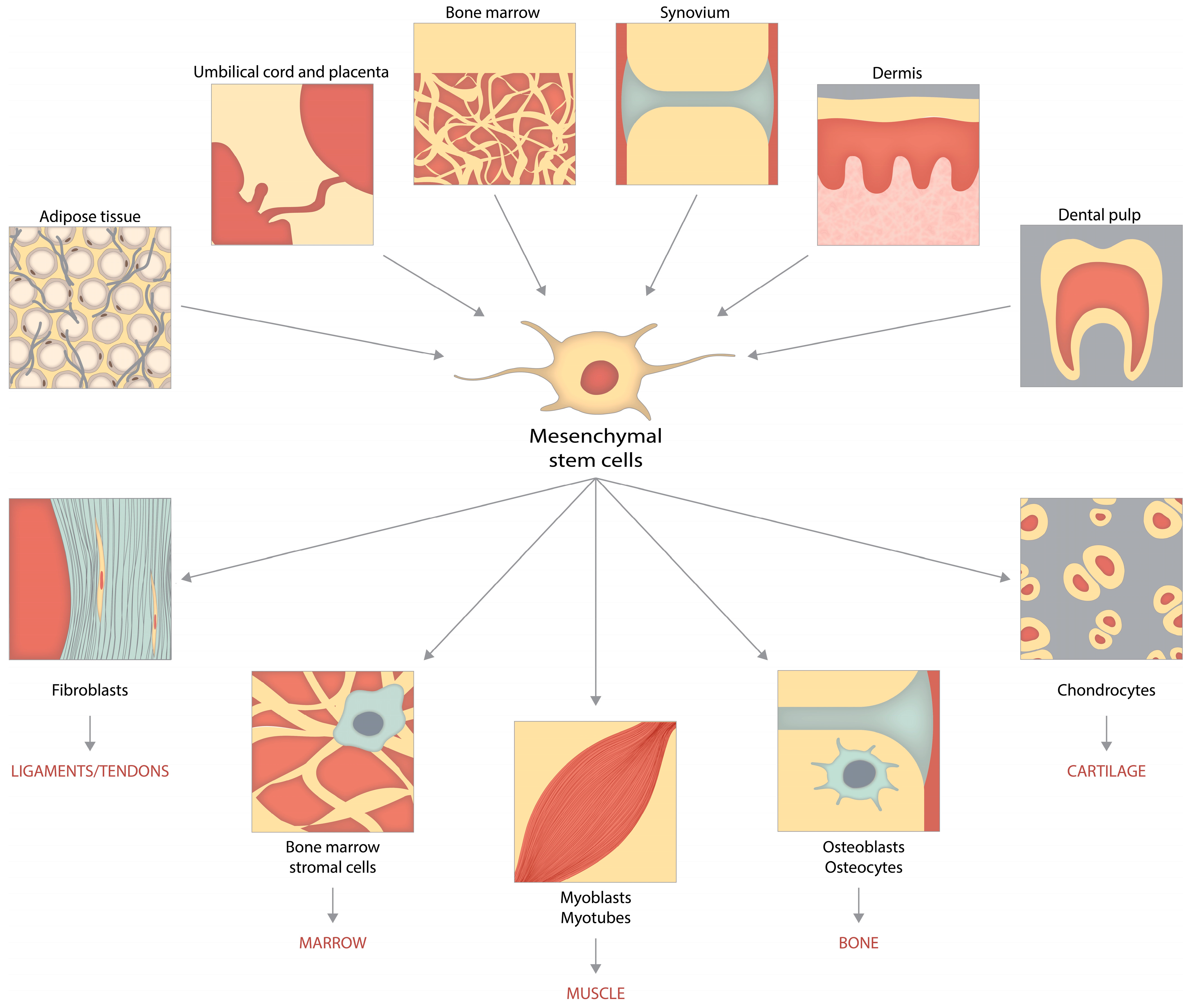
4.2. Sources of MSCs

Different sources of MSCs exhibit distinct properties, and each has its own set of benefits and drawbacks.
In terms of their ability to increase, umbilical cord (UC) and amniotic fluid-derived MSCs surpass fat and bone marrow-derived MSCs (BM-MSCs) [184,185].
MSCs acquired from the UC, amnion, and adipose tissue have a better immunomodulatory ability than MSCs generated from BM-MSCs; nevertheless, placental MSCs have the lowest immunomodulatory capacity [184].
Both adipose tissue-derived MSCs (AD-MSCs) and BM-MSCs have a morphology similar to that of fibroblasts [186]. These cells express CD29, CD44, CD73, CD90, and CD105; however, they do not express CD14, CD31, CD34, CD45, and CD106; in addition, they do not express HLA-DR or c-kit [186,187]. AD-MSCs displayed a more substantial capacity for adipogenic differentiation, while BM-MSCs demonstrated a greater ability for osteogenic development when both types of cells were developed in vitro [186,187].
The ability of BM-MSCs, synovial membrane-derived MSCs (SM-MSCs), and AD-MSCs to differentiate was investigated by Sakaguchi et al. [188,189]. When compared to other types of cells, they discovered that SM-MSCs contained approximately 3000 nucleated cells per mg and were the sort of cell that was most capable of chondrogenesis. In addition, they found that SM-MSCs and infrapatellar fat pad-MSCs (IPFP-MSCs) had a higher proliferative capacity than AD-MSCs and that the pellets created by SM-MSCs and IPFP-MSCs were able to produce more cartilage matrix than the pellets made by AD-MSCs [190].

4.2.1. BM-MSCs for OA Therapy

Because of their accessibility, rapid cell proliferation, long-term differentiation capabilities, and minimal immunological exclusion, BM-MSCs are the most widely employed source of therapeutic MSCs [191]. Numerous studies have established acceptable clinical outcomes in patients with knee OA who underwent either intra-articular injection or the surgical implantation of autologous BM-MSCs [144,192,193].
BM-MSCs have a wide range of properties that are likely to be helpful in treating genetic, mechanical, and age-related deterioration in illnesses such as OA [194].
BM-MSCs have immunomodulatory capabilities due to significantly limiting the proliferation of inflammatory T cells, monocytes, and dendritic cells via direct cell-to-cell contact. They also release anti-inflammatory mediators such as PGE-2, IDO, IL-1Ra, and IL-10 [194,195].
These cells affect the local OA microenvironment by activating chondrogenic progenitor cells and promoting their maturation into mature chondrocytes through the production of BMPs and TGF1 [196]. When differentiation indicators such as BMP-7 and TGF-1 are utilized, it is well documented that BM-MSCs develop in vitro into chondrocytes. Differentiation of BM-MSCs into chondrocytes in vitro has been demonstrated using differentiation cues such as BMP-7 and TGF-1.
A similar method could be used to differentiate BM-MSCs in vivo. By raising the amounts of BMP-7 and TGF-1 in the local joint environment, BM-MSCs can develop into chondrogenic progenitor cells (CPCs) in vivo, mediated by changes in the expression of master regulatory genes such as Sox9, homeobox A (HoxA), HoxD, and Gli3. Chondroblasts, which are unequivocally increased for collagen types II B, IX, and XI, develop from CPCs. The CPCs then mature into mature chondrocytes under the control of balanced collagen X (Col X) expression and release collagen II, which is made up of sulphated GAG (sGAG) building blocks and helps maintain the structural integrity of hyaline cartilage [197]. Collagen X overexpression has been linked to chondrocyte hypertrophy and fibrous cartilage synthesis; hence, the regulated expression of Col X would likely result in hyaline cartilage deposition [198].
While BM-MSCs have many advantages, they also have certain drawbacks. The donor factors, such as disease severity and age, significantly impact the yields and the capacity for differentiation and repair [199]. MSCs only make up 0.001–0.01% of the cells in the BM, which makes things even more complicated [109,198]. Furthermore, the risk of infection during the separation of BM cells cannot be discounted. This procedure can be performed under local anesthesia and guided by ultrasound or fluoroscopic imaging to improve accuracy [200,201].
The first clinical trial of BM-MSCs transplantation for the treatment of articular cartilage abnormalities was conducted nearly two decades ago. The study comprised 24 patients with high tibial osteotomy-related knee OA, with half getting autologous BM-MSCs transplantation and the other half serving as controls. Metachromasia was identified in most of the examined tissue regions after 42 weeks, with partial presence of tissue resembling hyaline cartilage. The cell-transplanted group had substantially better arthroscopy and histology grading scores than the cell-free control group, suggesting that transplantation of BM-MSCs has promise as a therapeutic approach for osteoarthritis [202,203,204].
In 2008, Centeno and colleagues described a patient who received a percutaneous injection of BM-MSCs and reported significant cartilage growth, reduced discomfort, and improved joint mobility. They then presented a case study of 339 patients, revealing that after MSC therapy, only 6.9 percent of patients who needed total knee replacement surgery (69 percent of the patient group) required replacement surgery again. At 11 months, 60 percent of patients reported more than 50% pain relief, whereas 40% reported more than 75% [205]. A cohort of 30 individuals with chronic knee pain, unresponsive to traditional treatment methods, demonstrated significant improvements in several functional measures and cartilage integrity after receiving intra-articular injections of BM-MSCs [206].
Davatchi et al. [207] revealed their findings on the use of autologous BM-MSCs for OA therapy (n = 4) in 2011. They injected approximately 8 to 9 106 cells into the knee cavities of patients. They discovered that it shortened the time it took for pain to emerge while walking, and that patients’ visual analog scale (VAS) scores dropped from 8090 to 4565. They were unable to identify any improvement in X-rays, though. This trial was prolonged to five years following treatment, and researchers discovered that the beneficial effects of BM-MSCs began to fade after six months. Still, they remained significantly better at five years compared to baseline [144].
In 2013, Orozco et al. carried out a clinical study on osteoarthritis (n = 12). During this study, they injected 4107 BM-MSCs into the knee joints of patients with a Kellgren and Lawrence (KL) grade of 2–4. They found that the pain was relieved within three months and continued to improve for at least a year. Additionally, the Lequesne and Western Ontario and McMaster Universities Osteoarthritis Index (WOMAC) ratings improved significantly. In addition, the quantitative MRI data on cartilage quality demonstrated an increase over the previous two years [192].
Additionally, the same research team reported a study in 2016 employing 4107 BM-MSCs to treat KL grade 2–3 OA. The findings of this study were reported in the journal Cell Reports. The daily activities VAS score was around 58.27 at the baseline visit, but it dropped to 19.47 at the one-year follow-up, and it further declined to 14.62, 14.93 at the four-year follow-up, with no severe adverse effects being recorded [208].
In 2014, Vangsness et al. conducted a double-blind, randomized controlled study (RCT) to assess the efficacy and safety of intra-articular injections of allogeneic BM-MSCs in individuals with OA of the knee who had undergone partial medial meniscectomy. In 18 patients who received 50,106 BM-MSCs and 18 patients who received 150,106 BM-MSCs, there were indications of meniscus regeneration and a considerable reduction in knee pain compared to 18 control patients who did not get an injection [209].
In 2015, Vega et al. randomly allocated 30 patients with knee OA to one of two treatment groups: 15 received intra-articular injections of 40,106 allogeneic human BM-MSCs and 15 received intra-articular injections of HA [206]. Patients treated with BM-MSCs reported a significant reduction in knee discomfort, an improvement in knee function, an increase in cartilage quality in post-treatment MRI defects, and no adverse effects.
Gupta et al. investigated the efficacy and safety of intra-articular injections of allogeneic human BM-MSCs in knee OA patients. Sixty OA patients were randomized to receive 25, 50, 75, or 150,106 BM-MSCs or no BM-MSCs. The dosage of 25,106 BM-MSCs was the most effective for alleviating OA knee pain, with no significant side effects [210].
Chahal et al. [211] released their research on using 1, 10, or 50 million BM-MSCs to treat KL grade 3–4 OA in 2019. They found that neither the morphological cartilage scores nor the T2 relaxation levels increased. Using cartilage catabolic markers, they did show putative chondroprotective benefits at 50 million BM-MSCs dosages. In addition, they discovered that the therapy lowered the levels of pro-inflammatory CD14+ CD16+ monocyte/macrophage maker and pro-inflammatory IL-12p40 in synovial fluid.

4.2.2. AD-MSCs for OA Therapy

In 2001, human AD-MSCs were recognized as a potential reservoir of mesenchymal stem cells (MSCs) owing to their ease of access, abundant supply, and immunosuppressive characteristics. AD-MSCs can be obtained in substantial quantities and concentrated to a greater extent (about 500 times more) than BM-MSCs by simple approaches and with the use of local anesthetic. Furthermore, the differentiation potential of AD-MSCs is less influenced by the donor’s age, as shown by previous studies [212,213,214].
AD-MSCs may be isolated from various human body sites [214]; however, AD-MSCs isolated from different areas have different characteristics [215]. AD-MSCs can be isolated from the upper arm, medial thigh, buttocks, trochanteric, superficial deep abdominal depots, and even the IPFP of the knee joint. The stromal vascular fraction (SVF) comprises 2 to 6 million cells and can be retrieved in 1 mL lipoaspirate [192]. The number of AD-MSCs in one gram of AD-MSCs can range from 5000 to 200,000 [193,203]. In other words, we could extract between 0.5 and 20 million AD-MSCs from 100 g of AD-MSCs from a patient.
AD-MSCs can differentiate into adipocytes, osteoblasts, chondrocytes, and endothelial cells due to their mesodermal origin. In addition, it has been demonstrated that they can grow into cells of ectodermal and endodermal origin, such as vascular smooth muscle cells, keratinocytes, hepatocytes, beta-islet cells, neuron-like cells, and glial lineages [216,217,218,219].
The most frequent procedure for isolating AD-MSCs is the enzymatic breakdown of fat tissue [152,220]. Adipose tissue SVF including AD-MSCs and other endothelial and hematopoietic cells is removed using an enzyme (collagenase, dispase, or trypsin) [221]. AD-MSCs are described as plastic-adherent, cultured, multipotent cell populations that have undergone serial passages. In addition, some non-enzymatic digesting methods, such as centrifugation and filtering, are occasionally used. However, it should be noted that the yield range is relatively extensive.
Numerous clinical studies conducted in recent years have shown the efficacy of AD-MSCs in therapeutic applications.
Bui et al. found that non-expanded SVF isolated from adipose tissue and processed in PRP resulted in notable improvements in pain score and cartilage layer thickness in 21 patients with grade II and III OA [222].
Jo et al. investigated the efficacy and safety of intra-articular injections of autologous human AD-MSCs in patients with knee OA in phase I and phase II clinical studies. In phase I, three patients were administered 1107, 5107, and 1108 AD-MSCs, whereas, in phase II, nine patients received a high dose of 1 108 AD-MSCs [223]. Patients who received 1108 AD-MSCs reported decreased OA knee pain, improved knee function, hyaline-like cartilage regeneration in articular cartilage defects, and no severe adverse effects.
Eighteen patients with symptomatic and severe knee OA were administered autologous AD-MSCs at three different doses: low (2106 cells), medium (10,106 cells), and high (50,106 cells). Six-month follow-up revealed no clinically significant adverse effects, and patients treated with low-dose AD-MSCs experienced substantial pain relief and functional enhancement [224].
Fodor’s research showed that the treatment of OA knees with 14.1 million viable, nucleated SVF cells resulted in a statistically significant improvement in WOMAC and VAS ratings, which remained stable after one year [225].
A clinical study evaluated the efficacy of AD-MSCs to that of microfracture alone in treating moderate to severe knee OA in 80 patients aged 18 to 50 years old. Following 24 months, the group that received treatment with AD-MSCs exhibited heightened signal intensity about tissue repair and improved knee injury and osteoarthritis outcome scores (KOOS) concerning pain and symptoms. However, no notable variances were observed in other sub-scores, including daily living activity, sports and recreation, and quality of life. These findings indicate the potential efficacy of AD-MSCs in facilitating tissue repair and alleviating pain [226].
In a phase IIb knee OA clinical study that was active-controlled and double-blinded, investigators successfully used 5107 AD-MSCs to treat the patients. After 12 months, the majority of patients in the group that was given AD-MSCs showed a 70% improvement. In addition, MRI findings at 12 months showed a substantial increase in articular cartilage volume in the AD-MSC group compared to the HA group [227].
Hurley et al. [228] found that 16 separate studies had reported employing AD-MSCs as a therapy for OA using various approaches. AD-MSCs that were isolated from the IPFP and injected into an OA knee showed improved mobility and function and a reduction in pain levels. There were no adverse effects from the treatment. After two years of follow-up, patients reported significantly reduced levels of discomfort and had cartilage regeneration, as demonstrated by MRI [229]. Nepali et al. discovered that orbital AD-MSCs have higher concentrations of CD73, CD90, CD105, and CD146, but lower concentrations of CD31, CD45, and HLA-DR [215].
Kim et al. demonstrated an increase in HLA-ABC and HLA-DR expression in AD-MSCs after IFN treatment, which raises concerns about the usage of allogenic AD-MSCs [230].
A phase Iib randomized, placebo-controlled clinical investigation of AD-MSCs was conducted on 24 patients with knee OA. After six months of follow-up, the AD-MSCs-treated group had a higher WOMAC score, better function, and less pain, demonstrating the therapeutic potential of AD-MSCs [231].
In the NCT01585857 trial, it was found that patients receiving injections of 2106 cells had a higher baseline pain and WOMAC score than those receiving more significant dosages [232].
Importantly, these trials indicated that autologous AD-MSCs transplantation has a low risk of adverse events and does not cause graft rejection or malignancy in recipients, making it a feasible treatment option for OA [210]. To fully comprehend the defining phenotypes and enhance the therapeutic efficiency of these MSCs, additional research is required on donor-matched AD-MSCs from various isolation sites and their associated properties [200,215].

4.2.3. UC-MSCs for OA Therapy

Recent research has shown that umbilical cord mesenchymal stem cells (UC-MSCs) can be helpful source of therapeutic stem cells due to their advantages over BM-MSCs in terms of their capacity for proliferation, functional differentiation, and immunomodulatory effects [127,233].
UC-MSCs have rapid self-renewal and differentiation capabilities, which are beneficial for the modulation of immune responses and repairing damaged tissue. These cells are very easy to obtain and do not cause any discomfort during extraction [234]. UC-MSCs multiply at a pace approximately three to four times faster than that of AD-MSCs. In addition, it is common knowledge that UC-MSCs are capable of secreting several growth factors that contribute to skin renewal. These growth factors include HGF, epithelial growth factor (EGF), collagen type 1, and growth differentiation factor-11 (GDF-11) [230,235,236].
MSCs can be extracted from the UC compartments such as Wharton’s jelly, perivascular tissue, and cord blood [237]. MSCs derived from UC can be obtained painlessly and with fewer ethical considerations.
Prior research has demonstrated that UC-MSCs exhibit undesirable features, such as early morphological changes and a faster loss of amplification capability and diminished attachment efficiency [238,239].
UC-MSCs have been evaluated in several clinical trials for their efficacy in OA treatment.
A 6-month safety and efficacy trial of UC-MSC transplantation was conducted on 36 patients with moderate or severe knee OA. On the Lysholm, WOMAC, and short form-36 (SF-36) measures, the group receiving cell therapy did better than the control group. The therapeutic potential of UC-MSCs in joint improvement was demonstrated by the fact that there was no recurrence of knee soreness throughout the follow-up period [240].
In a separate clinical trial, three patients with knee OA were injected intra-articularly with 5–7 UC-MSCs. During a 3-month follow-up period, biochemical parameters did not alter between pre- and post-treatment in any three patients [241].
Park et al. discovered that a UC-MSCs product was safe and effective for the regeneration of hyaline-like cartilage in knee OA after seven years of follow-up. UCB-MSCs were employed to treat knee OA with excellent clinical results and highly qualified regeneration [242].

4.2.4. ESC-Induced MSCs (ESC-MSCs) for OA Treatment

Exosomes made from human ESC-MSCs exhibited exceptional cartilage regeneration, including complete repair of hyaline cartilage, in rats with osteochondral defects [91].
ESC-MSCs are developed and differentiated indefinitely into any three embryonic germ cell lines: ectoderm, endoderm, and mesoderm [243]. Due to their capacity for continuous self-renewal, ESC-MSCs offer an inexhaustible source of stem cells and chondrocytes for cartilage repair. However, the most significant barrier to employing ESC-MSCs for cartilage matrix regeneration is the ethical complexity and low survival rate of human ESC-MSCs following cell mass disintegration [244]. In addition, ESC-MSCs differentiation into chondrocytes and cartilage regeneration are challenging processes requiring a complex microenvironment, three-dimensional structure, and a specific mechanotransduction signal [245].
Studies found that exosomes produced from ESC-MSCs reduced cartilage breakdown and matrix degradation in a medial meniscus instability model by enhancing Col II and aggrecan while lowering ADMTS5 expression. They also reduced the maximal and total OARSI scores, which resulted in milder OA pathology [246].
Furthermore, it was observed that exosomes and microparticles derived from BM-MSC exhibited a dose-dependent effect on enhancing the expression of anabolic cartilage markers, precisely collagen type II (Col II) and aggrecan, in chondrocytes with osteoarthritis-like characteristics. Additionally, these exosomes and microparticles demonstrated inhibitory effects on catabolic markers, such as MMP-13 and ADAMTS5, as well as inflammatory markers. Moreover, they were found to mitigate articular cartilage damage and subchondral bone degradation, as evidenced by experimental data [247].
When ESCs were coupled with polydimethylsiloxane (PDMS) scaffolds, initial expression of chondrogenic markers Sox9 was boosted, followed by increased production of Col II (a cartilage-specific marker) and decreased expression of octamer-binding transcription factor 4 (Oct4). It did not, however, promote hypertrophic cell differentiation [245].
Exosomes obtained from the 3D culture of umbilical MSCs were more chondroprotective than exosomes obtained from 2D culture systems [248]. This meant that they significantly increased chondrocyte proliferation, migration, and matrix synthesis, as well as improving gross appearance and attenuating cartilage defects in an animal model.

4.2.5. SM-MSCs for OA Treatment

In 2001, De Bari et al. discovered the first SM-MSCs. These cells, much like AD-MSCs, can be separated from some sites, such as the cotyloid fossa or the paralabral synovium, and they display site-specific properties. Compared to other types of MSCs, it is interesting to note that SM-MSCs have a proliferative solid potential, the ability to differentiate into a wide variety of lineages, and low immunogenicity [249,250].
SM-MSCs have a more tremendous chondrogenic potential than MSCs derived from other sources and are predicted to be used more frequently for cartilage repair and joint homeostasis therapies [227,251]. This is due to the increased expression of type II collagen, aggrecan, and Sox9, which gives them a higher chondrogenic potential [227,252].
According to Sakaguchi et al., both SM-MSCs and BM-MSCs have a greater capacity for osteogenic and adipogenic differentiation than other MSCs. Despite this, SM-MSCs generate relatively low-density expansions in vitro compared to BM-MSCs [189,250].
In an experimental model of OA in rats, SM-MSCs were administered by frequent injections. These SM-MSCs exhibited migratory behavior towards the synovium and maintained their undifferentiated state. Furthermore, they demonstrated increased expression levels of chondroprotective proteins, including BMP-2 and an anti-inflammatory gene known as TSG-6 [253].
This demonstrates that SM-MSCs control MSCs characteristics and may also limit the progression of OA via genetic machinery.
In a single retrospective study [254], SM-MSCs have been examined concerning human knee OA. Forty-eight months following surgery, they saw clinical improvement and safe defect filling, as confirmed by second-look arthroscopy and MRI.

4.2.6. IPFP for OA Treatment

The study of IPFP, a form of fatty tissue, has gained significant attention due to its potential to reduce inflammation and slow cartilage degeneration. The IPFP is located inside the knee’s anterior compartment. It is an intra-capsular component that houses around 20 cm3 of AD-MSCs. The collection of these data may occur during either arthroscopic or open knee surgery [255,256].
IPFP-MSCs may serve as a source of autologous stem cells. These MSCs show greater chondrogenic capacity than BM-MSCs and AD-MSCs [257]. IPFP-MSCs are characterized by the presence of cell markers such as CD9, CD10, CD13, CD29, CD44, CD49e, CD59, CD105, CD106, and CD166 [258]. In addition, these cells are capable of adipo-, chondro-, and osteogenic differentiation [258,259].
Toghraie et al. [260] injected IPFP-MSCs intra-articularly directly into the knees of rabbits with experimental OA. The IFP-MSCs that were used had undergone in vitro expansion and cultivation. They were then supplied in a single dosage of 1 million cells suspended in 1 mL of media 12 weeks after the surgical procedure. Rabbits that received IPFP-MSCs had significantly reduced levels of cartilage degradation, osteophyte development, and subchondral sclerosis compared to the control group at a postoperative period of 20 weeks.
IPFP adipocytes treated with PRP and HA induced chondrogenesis and inhibited adipocyte-mediated inflammation [251]. IPFP is also a significant source of perivascular stem cells (PSCs). IPFP contains both of these types of stem cells. Due to their ability to maintain the structural integrity of the telomere, IPFP-PSCs retain their unique and tenacious growth capacities even after several expansions [261,262]. The chondrogenic activity of PSCs extracted from IPFP was much higher than that of PSCs obtained from subcutaneous adipose tissues.
Coculture of chondrocytes and IPFP-MSCs in the presence of chitosan/HA nanoparticles enhanced chondrogenic efficiency, indicating that coculture in the presence of appropriate stimuli may aid in cartilage regeneration in an osteoarthritic knee [263].
Spasovski et al. [264] found that treatment with IPFP-MSCs improves clinical symptoms and reduces pain after three months, with the highest levels of success happening after six months of treatment.

4.2.7. iPSCs for OA Treatment

iPSCs are reprogrammed somatic cells, comparable to ESCs. When patient-specific somatic cells are differentiated into iPSCs, the risk of cross-reactivity and immunogenicity is significantly reduced [265].
The application of iPSCs extends beyond cartilage regeneration to discovering medicines that enhance chondrogenesis or prevent cartilage degradation [266]. These findings indicated that iPSCs have significant potential for cartilage repair in OA. However, the fundamental restrictions in iPSC creation are low efficiency and heterogeneity in transcription factor needs in somatic cells. Furthermore, undifferentiated iPSCs contaminate differentiated MSCs, enhancing tumorigenicity and restricting their use in cell-based regenerative therapy [267,268].
Zhu et al. generated embryonic body development from iPSCs, differentiated into chondrocytes and implanted to restore cartilage in an OA-affected rat [251]. Exosomes produced by MSCs derived from induced pluripotent stem cells (iMSCs) were superior to exosomes of SM-MSCs in repairing cartilage in rats with osteoarthritis [247].
Curcumin, a naturally occurring NF-κB inhibitor, protects MSCs from the harmful effects of pro-inflammatory cytokines, thereby creating a suitable microenvironment for MSCs to undergo chondrogenesis; this strategy may promote in vitro cartilage regeneration. Therefore, curcumin may be considered a possible new therapeutic for the preventative treatment of OA cases with minimal cartilage damage [269].

4.3. Comparisons between Different Forms of MSCs

The etiology of early-onset OA may be attributed to sporadic mutations occurring seldom in monogenetic disorders associated with OA. In contrast, late-onset OA is often characterized by a complex clinical manifestation encompassing prominent genetic differences and other risk factors [35].
Different sources of MSCs have unique characteristics and have their advantages and disadvantages. BM-MSCs were the first MSCs studied; however, due to their significant invasiveness and restricted availability inside living organisms, they have been replaced by MSCs obtained from other sources. BM-derived stromal cells are widely used as the primary clinical source of MSCs. The components above are obtained from a biological matrix consisting of hematopoietic stem cells, endothelial progenitor cells, and associated cytokines and growth factors [270].
Previous studies have used stromal cells obtained from adipose tissue as a viable alternative cell line. The production of stem cells in adipose tissue is comparatively greater. Furthermore, it has been shown that cells originating from adipose tissue have a higher lifespan than cells produced from bone marrow. The research conducted by Muthu et al. examined the disparity between these two cellular groups. The investigation findings indicated that over one year, adipose tissue-derived AD-MSCs showed a higher level of safety and consistent effectiveness in enhancing pain relief, functional improvement, and radiological results [270,271].
The AD-MSCs exhibited inferior cell shape and matrix formation compared to BM-MSCs in the context of in vitro chondrogenesis. However, their ability to undergo adipogenic differentiation was shown to be similar. AD-MSCs have a high degree of genetic and morphological stability, making them particularly suitable for purposes. Furthermore, when subjected to a prolonged incubation period, these MSCs demonstrate superior proliferation capabilities.
Due to their strong in vitro proliferation ability, low immunogenicity, simplicity of isolation and culture, and persistent multidirectional differentiation potential, UC-MSCs are often used. Examining gene expression demonstrated that BM-MSCs exhibit elevated levels of genes associated with osteogenic development compared to UC-MSCs [272].
Previous studies have shown that SM-MSCs have a superior capacity for chondrogenesis compared to MSCs produced from alternative tissue sources. The potential solution may be in the expression of growth differentiation factor 5 (GDF5), a factor often seen in articular cartilage. Nevertheless, there are ongoing issues with the enzymatic digestion process to isolate these cells [270].
iPSCs derived from human sources have distinct characteristics, including an augmented regeneration potential compared to traditional stem cells. Moreover, iPSC-MSCs possess the ability to modify extensively differentiated adult somatic cells via the use of genetic engineering techniques. In theory, it is possible to transform all adult somatic cells into iPSC-MSCs, which have been seen to possess a greater capacity for proliferation compared to traditional MSCs [272].
The IPFP is a noteworthy and auspicious reservoir of MSCs that effectively addresses OA. Research has shown that these MSCs had superior chondrogenic capabilities to those produced from bone marrow or subcutaneous fat [270].
UC, amniotic membrane, and fat sources exhibit greater accessibility and possess a better potential for proliferation, but the proliferative power of BM-MSCs is comparatively insufficient [272].
Regarding MSCs inside the tissue, the UC-MSCs have the greatest concentration, followed by the amniotic fluid and adipose tissue. In relation to the proliferation potential of MSCs, it is seen that UC-MSCs and amniotic-derived MSCs exhibit distinct advantages, followed by AD-MSCs and BM-MSCs. When comparing the immunomodulatory ability of different types of MSCs, it has been shown that umbilical cord, amnion, and AD-MSCs exhibit more excellent immune regulation abilities compared to BM-MSCs. Conversely, placental MSCs have the lowest level of immunomodulatory potential. Compared to cytokine secretion patterns, it has been shown that UC-MSCs exhibit a higher secretion of cell growth factors than BM-MSCs [16].

5. Conclusions

OA is the most prevalent joint disease worldwide, especially among the elderly. However, there is no effective treatment for OA. Environmental, metabolic, and inflammatory factors play an essential role in the development of OA’s pathogenesis. Non-pharmacological and pharmacological approaches to treating OA are covered in depth in this article. The primary treatment goals for OA are to reduce pain and slow or halt the disease’s progression.
Joint replacement is the only treatment alternative to pain medication for severe osteoarthritis. Although joint replacement is an effective treatment for symptomatic end-stage osteoarthritis, the results may be unsatisfactory due to developments in implant technology.
Innovative techniques for treating OA are available thanks to developments in cell-based therapy. It is a prominent topic in regenerative medicine because of its ability to mend cartilage and regulate the immune system. Cellular therapy techniques for OA may benefit from the multipotency, self-renewal, and immunomodulatory characteristics of MSCs, and their easy availability and isolation, according to numerous preclinical and clinical investigations. Bone marrow and AD-MSCs are the most commonly used MSC sources, although many studies investigate their potential for cartilage regeneration when using MSCs. In general, they have exhibited promising outcomes in cartilage repair and the treatment of OA.
A potential treatment option for OA of the knee joint is the application of MSCs directly to the affected area. Studies that are well-designed and have enough follow-up are the ones that show positive results.
Positive outcomes from preclinical and clinical trials suggest that medications produced from MSCs could be used to treat OA. However, this technique presents numerous challenges, necessitating the creation of standardized remedies before their therapeutic recommendation.
The physiological characteristics and cellular activities of MSCs in vitro and in vivo, including immune regulatory processes and the differentiation role of key mediators, are not well understood. Cell culture conditions, cell density, timing and location of MSCs transplantation, and MSCs pretreatment all affect MSCs therapeutic efficacy in both preclinical and clinical studies.
To limit immunological rejection and achieve the appropriate amount of MSCs, the collection, delivery, and preservation of a patient’s stem cells must be thoroughly investigated.
Given that MSCs are multipotent cells with a high capacity for cell proliferation and differentiation and that both autologous and allogeneic MSCs have been used in several studies, the risks associated with tumorigenesis and severe adverse immunoregulatory responses should be further evaluated in preclinical and clinical trials.
To maximize the potential of MSCs in OA therapy while avoiding the adverse consequences, these issues should be elucidated correctly in future studies.

Author Contributions

Conceptualization, R.G. and L.A.M.; methodology, R.G. and L.A.M.; software, A.C. and M.-A.B.; validation, E.R., R.G. and L.A.M.; formal analysis, I.-I.R. and I.R.M.; investigation, I.-I.R. and L.A.M.; resources, A.C.; data curation, L.A.M., R.G. and M.-A.B.; writing—original draft preparation, R.G. and L.A.M.; writing—review and editing, L.A.M. and E.R.; visualization, I.R.M. and A.C.; supervision, E.R. and L.A.M.; project administration, E.R. All authors have read and agreed to the published version of the manuscript.

Funding

This research received no external funding.

Institutional Review Board Statement

Not applicable.

Informed Consent Statement

Not applicable.

Data Availability Statement

Not applicable.

Conflicts of Interest

The authors declare no conflict of interest.

References

  1. Lambova, S.; Müller-Ladner, U. Editorial: Osteoarthritis—Current Insights in Pathogenesis, Diagnosis and Treatment. Curr. Rheumatol. Rev. 2018, 14, 91–97. [Google Scholar] [CrossRef] [PubMed]
  2. Hunter, D.; Bierma-Zeinstra, S. Osteoarthritis. Lancet 2019, 393, 1745–1759. [Google Scholar] [CrossRef] [PubMed]
  3. Abhishek, A.; Doherty, M. Diagnosis and Clinical Presentation of Osteoarthritis. Rheum. Dis. Clin. N. Am. 2013, 39, 45–66. [Google Scholar] [CrossRef] [PubMed]
  4. Jo, C.; Chai, J.; Jeong, E.; Oh, S.; Shin, J.; Shim, H.; Yoon, K. Intra-Articular Injection of Mesenchymal Stem Cells for the Treatment of Osteoarthritis of the Knee: A 2-Year Follow-Up Study. Am. J. Sports Med. 2017, 45, 2774–2783. [Google Scholar] [CrossRef] [PubMed]
  5. Sun, H.; Gong, T.; Jiang, Y.; Zhang, S.; Zhao, Y.; Wu, Q. Global, Regional, and National Prevalence and Disability-Adjusted Life-Years for Infertility in 195 Countries and Territories, 1990–2017: Results from a Global Burden of Disease Study, 2017. Aging 2019, 11, 10952–10991. [Google Scholar] [CrossRef]
  6. Blagojevic, M.; Jinks, C.; Jeffery, A.; Jordan, K. Risk Factors for Onset of Osteoarthritis of the Knee in Older Adults: A Systematic Review and Meta-Analysis. Osteoarthr. Cartil. 2010, 18, 24–33. [Google Scholar] [CrossRef]
  7. Carballo, C.; Nakagawa, Y.; Sekiya, I.; Rodeo, S. Basic Science of Articular Cartilage. Clin. Sports Med. 2017, 36, 413–425. [Google Scholar] [CrossRef]
  8. Glyn-Jones, S.; Palmer, A.; Agricola, R.; Price, A.; Vincent, T.; Weinans, H.; Carr, A. Osteoarthritis. Lancet 2015, 386, 376–387. [Google Scholar] [CrossRef]
  9. Loeser, R.; Goldring, S.; Scanzello, C.; Goldring, M. Osteoarthritis: A Disease of the Joint as an Organ. Arthritis Rheum. 2012, 64, 1697–1707. [Google Scholar] [CrossRef]
  10. Gignac, M.; Irvin, E.; Cullen, K.; Van Eerd, D.; Beaton, D.; Mahood, Q.; McLeod, C.; Backman, C. Men and Women’s Occupational Activities and the Risk of Developing Osteoarthritis of the Knee, Hip, or Hands: A Systematic Review and Recommendations for Future Research. Arthritis Care Res. 2020, 72, 378–396. [Google Scholar] [CrossRef]
  11. Ostrowska, M.; Maśliński, W.; Prochorec-Sobieszek, M.; Nieciecki, M.; Sudoł-Szopińska, I. Cartilage and Bone Damage in Rheumatoid Arthritis. Reumatol./Rheumatol. 2018, 56, 111–120. [Google Scholar] [CrossRef]
  12. Okikiade, A.; Osharode, A.; Oyewole, A.; Ogunesan, D.; Oladejo, D.; Oshobu, I.; Browne, K. Understanding the Role of Inflammation in Secondary Osteoarthritis. Asian J. Med. Health Res. 2022, 20, 60–74. [Google Scholar] [CrossRef]
  13. Lozada, C.J. Osteoarthritis. Available online: https://emedicine.medscape.com/article/330487-overview#a5 (accessed on 9 September 2023).
  14. Chen, D.; Shen, J.; Zhao, W.; Wang, T.; Han, L.; Hamilton, J.; Im, H. Osteoarthritis: Toward a Comprehensive Understanding of Pathological Mechanism. Bone Res. 2017, 5, 16044. [Google Scholar] [CrossRef] [PubMed]
  15. Vina, E.; Kwoh, C. Epidemiology of Osteoarthritis: Literature Update. Curr. Opin. Rheumatol. 2018, 30, 160–167. [Google Scholar] [CrossRef] [PubMed]
  16. Xiang, X.-N.; Zhu, S.-Y.; He, H.-C.; Yu, X.; Xu, Y.; He, C.-Q. Mesenchymal Stromal Cell-Based Therapy for Cartilage Regeneration in Knee Osteoarthritis. Stem Cell Res. Ther. 2022, 13, 14. [Google Scholar] [CrossRef]
  17. Shah, K.; Zhao, A.G.; Sumer, H. New Approaches to Treat Osteoarthritis with Mesenchymal Stem Cells. Stem Cells Int. 2018, 2018, 5373294. [Google Scholar] [CrossRef] [PubMed]
  18. Tiku, M.; Sabaawy, H. Cartilage Regeneration for Treatment of Osteoarthritis: A Paradigm for Nonsurgical Intervention. Ther. Adv. Musculoskelet. Dis. 2015, 7, 76–87. [Google Scholar] [CrossRef] [PubMed]
  19. Felson, D. Osteoarthritis as a Disease of Mechanics. Osteoarthr. Cartil. 2013, 21, 10–15. [Google Scholar] [CrossRef] [PubMed]
  20. Bastiaansen-Jenniskens, Y.; Koevoet, W.; de Bart, A.; van der Linden, J.; Zuurmond, A.; Weinans, H.; Verhaar, J.; van Osch, G.; DeGroot, J. Contribution of Collagen Network Features to Functional Properties of Engineered Cartilage. Osteoarthr. Cartil. 2008, 16, 359–366. [Google Scholar] [CrossRef]
  21. Nelson, A.; Allen, K.; Golightly, Y.; Goode, A.; Jordan, J. A Systematic Review of Recommendations and Guidelines for the Management of Osteoarthritis: The Chronic Osteoarthritis Management Initiative of the U.S. Bone and Joint Initiative. Semin. Arthritis Rheum. 2014, 43, 701–712. [Google Scholar] [CrossRef]
  22. Mehana, E.; Khafaga, A.; El-Blehi, S. The Role of Matrix Metalloproteinases in Osteoarthritis Pathogenesis: An Updated Review. Life Sci. 2019, 234, 116786. [Google Scholar] [CrossRef]
  23. Murphy, G. What Are the Roles of Metalloproteinases in Cartilage and Bone Damage? Ann. Rheum. Dis. 2005, 64, iv44–iv47. [Google Scholar] [CrossRef] [PubMed]
  24. Goldring, M.; Otero, M.; Plumb, D.; Dragomir, C.; Favero, M.; EI Hachem, K.; Hashimoto, K.; Roach, H.; Olivotto, E.; Borzì, R.; et al. Roles of Inflammatory and Anabolic Cytokines in Cartilage Metabolism: Signals and Multiple Effectors Converge Upon MMP-13 Regulation in Osteoarthritis. Eur. Cells Mater. 2011, 21, 202–220. [Google Scholar] [CrossRef] [PubMed]
  25. Sophia Fox, A.; Bedi, A.; Rodeo, S. The Basic Science of Articular Cartilage: Structure, Composition, and Function. Sports Health 2009, 1, 461–468. [Google Scholar] [CrossRef] [PubMed]
  26. Imhof, H.; Sulzbacher, I.; Grampp, S.; Czerny, C.; Youssefzadeh, S.; Kainberger, F. Subchondral Bone and Cartilage Disease. Investig. Radiol. 2000, 35, 581–588. [Google Scholar] [CrossRef] [PubMed]
  27. Kidd, B. Mechanisms of Pain in Osteoarthritis. HSS J. 2012, 8, 26–28. [Google Scholar] [CrossRef]
  28. Wang, H.; Bai, J.; He, B.; Hu, X.; Liu, D. Osteoarthritis and the Risk of Cardiovascular Disease: A Meta-Analysis of Observational Studies. Sci. Rep. 2016, 6, 39672. [Google Scholar] [CrossRef]
  29. Wallace, I.; Worthington, S.; Felson, D.; Jurmain, R.; Wren, K.; Maijanen, H.; Woods, R.; Lieberman, D. Knee Osteoarthritis Has Doubled in Prevalence Since the Mid-20th Century. Proc. Natl. Acad. Sci. USA 2017, 114, 9332–9336. [Google Scholar] [CrossRef]
  30. Veronese, N.; Cereda, E.; Maggi, S.; Luchini, C.; Solmi, M.; Smith, T.; Denkinger, M.; Hurley, M.; Thompson, T.; Manzato, E.; et al. Osteoarthritis and Mortality: A Prospective Cohort Study and Systematic Review with Meta-Analysis. Semin. Arthritis Rheum. 2016, 46, 160–167. [Google Scholar] [CrossRef]
  31. Kim, J.; Kim, H.; Lee, D. Survival of Opening Versus Closing Wedge High Tibial Osteotomy: A Meta-Analysis. Sci. Rep. 2017, 7, 7296. [Google Scholar] [CrossRef]
  32. Wearing, S.; Hennig, E.; Byrne, N.; Steele, J.; Hills, A. Musculoskeletal Disorders Associated with Obesity: A Biomechanical Perspective. Obes. Rev. 2006, 7, 239–250. [Google Scholar] [CrossRef] [PubMed]
  33. Musumeci, G.; Aiello, F.; Szychlinska, M.; Di Rosa, M.; Castrogiovanni, P.; Mobasheri, A. Osteoarthritis in the XXIst Century: Risk Factors and Behaviours That Influence Disease Onset and Progression. Int. J. Mol. Sci. 2015, 16, 6093–6112. [Google Scholar] [CrossRef] [PubMed]
  34. Deshpande, B.; Katz, J.; Solomon, D.; Yelin, E.; Hunter, D.; Messier, S.; Suter, L.; Losina, E. Number of Persons with Symptomatic Knee Osteoarthritis in the US: Impact of Race and Ethnicity, Age, Sex, and Obesity. Arthritis Care Res. 2016, 68, 1743–1750. [Google Scholar] [CrossRef]
  35. Coaccioli, S.; Sarzi-Puttini, P.; Zis, P.; Rinonapoli, G.; Varrassi, G. Osteoarthritis: New Insight on Its Pathophysiology. J. Clin. Med. 2022, 11, 6013. [Google Scholar] [CrossRef] [PubMed]
  36. Reynard, L.; Barter, M. Osteoarthritis Year in Review 2019: Genetics, Genomics and Epigenetics. Osteoarthr. Cartil. 2020, 28, 275–284. [Google Scholar] [CrossRef]
  37. Tachmazidou, I.; Hatzikotoulas, K.; Southam, L.; Esparza-Gordillo, J.; Haberland, V.; Zheng, J.; Johnson, T.; Koprulu, M.; Zengini, E.; Steinberg, J.; et al. Identification of New Therapeutic Targets for Osteoarthritis through Genome-Wide Analyses of UK Biobank Data. Nat. Genet. 2019, 51, 230–236. [Google Scholar] [CrossRef]
  38. Loeser, R. Aging Processes and the Development of Osteoarthritis. Curr. Opin. Rheumatol. 2013, 25, 108–113. [Google Scholar] [CrossRef]
  39. Kelly, T.; Yang, W.; Chen, C.; Reynolds, K.; He, J. Global Burden of Obesity in 2005 and Projections to 2030. Int. J. Obes. 2008, 32, 1431–1437. [Google Scholar] [CrossRef]
  40. Conde, J.; Scotece, M.; Gómez, R.; Lopez, V.; Gómez-Reino, J.; Gualillo, O. Adipokines and Osteoarthritis: Novel Molecules Involved in the Pathogenesis and Progression of Disease. Arthritis 2011, 2011, 203901. [Google Scholar] [CrossRef]
  41. Eckel, R.; Grundy, S.; Zimmet, P. The Metabolic Syndrome. Lancet 2005, 365, 1415–1428. [Google Scholar] [CrossRef]
  42. Steenvoorden, M.; Huizinga, T.; Verzijl, N.; Bank, R.; Ronday, H.; Luning, H.; Lafeber, F.; Toes, R.; DeGroot, J. Activation of Receptor for Advanced Glycation End Products in Osteoarthritis Leads to Increased Stimulation of Chondrocytes and Synoviocytes. Semin. Arthritis Rheum. 2005, 54, 253–263. [Google Scholar] [CrossRef]
  43. Slemenda, C. Quadriceps Weakness and Osteoarthritis of the Knee. Ann. Intern. Med. 1997, 127, 97–104. [Google Scholar] [CrossRef]
  44. Daste, C.; Kirren, Q.; Akoum, J.; Lefèvre-Colau, M.; Rannou, F.; Nguyen, C. Physical Activity for Osteoarthritis: Efficiency and Review of Recommandations. Jt. Bone Spine 2021, 88, 105207. [Google Scholar] [CrossRef] [PubMed]
  45. Kraus, V.; Sprow, K.; Powell, K.; Buchner, D.; Bloodgood, B.; Piercy, K.; George, S.; Kraus, W. Effects of Physical Activity in Knee and Hip Osteoarthritis: A Systematic Umbrella Review. Med. Sci. Sports Exerc. 2019, 51, 1324–1339. [Google Scholar] [CrossRef] [PubMed]
  46. Simopoulos, A. An Increase in the Omega-6/Omega-3 Fatty Acid Ratio Increases the Risk for Obesity. Nutrients 2016, 8, 128. [Google Scholar] [CrossRef]
  47. Dai, Z.; Lu, N.; Niu, J.; Felson, D.; Zhang, Y. Dietary Fiber Intake in Relation to Knee Pain Trajectory. Arthritis Care Res. 2017, 69, 1331–1339. [Google Scholar] [CrossRef] [PubMed]
  48. Roos, E. Joint Injury Causes Knee Osteoarthritis in Young Adults. Curr. Opin. Rheumatol. 2005, 17, 195–200. [Google Scholar] [CrossRef]
  49. Larsson, S.; Englund, M.; Struglics, A.; Lohmander, L. Interleukin-6 and Tumor Necrosis Factor Alpha in Synovial Fluid Are Associated with Progression of Radiographic Knee Osteoarthritis in Subjects with Previous Meniscectomy. Osteoarthr. Cartil. 2015, 23, 1906–1914. [Google Scholar] [CrossRef] [PubMed]
  50. Kapoor, M.; Martel-Pelletier, J.; Lajeunesse, D.; Pelletier, J.; Fahmi, H. Role of Proinflammatory Cytokines in the Pathophysiology of Osteoarthritis. Nat. Rev. Rheumatol. 2010, 7, 33–42. [Google Scholar] [CrossRef]
  51. Sokolove, J.; Lepus, C. Role of Inflammation in the Pathogenesis of Osteoarthritis: Latest Findings and Interpretations. Ther. Adv. Musculoskelet. Dis. 2013, 5, 77–94. [Google Scholar] [CrossRef]
  52. Melchiorri, C.; Meliconi, R.; Frizziero, L.; Silvestri, T.; Pulsatelli, L.; Mazzetti, I.; Borz, R.; Uguccioni, M.; Facchini, A. Enhanced and Coordinated In Vivo Expression of Inflammatory Cytokines and Nitric Oxide Synthase by Chondrocytes from Patients with Osteoarthritis. Arthritis Rheum. 1998, 41, 2165–2174. [Google Scholar] [CrossRef]
  53. Saklatvala, J. Tumour Necrosis Factor A Stimulates Resorption and Inhibits Synthesis of Proteoglycan in Cartilage. Nature 1986, 322, 547–549. [Google Scholar] [CrossRef]
  54. Lefebvre, V.; Peeters-Joris, C.; Vaes, G. Modulation by Interleukin 1 and Tumor Necrosis Factor α of Production of Collagenase, Tissue Inhibitor of Metalloproteinases and Collagen Types in Differentiated and Dedifferentiated Articular Chondrocytes. Biochim. Biophys. Acta Mol. Cell Res. 1990, 1052, 366–378. [Google Scholar] [CrossRef]
  55. Li, H.; Xie, S.; Qi, Y.; Li, H.; Zhang, R.; Lian, Y. TNF-A Increases the Expression of Inflammatory Factors in Synovial Fibroblasts by Inhibiting the PI3K/AKT Pathway in a Rat Model of Monosodium Iodoacetate-Induced Osteoarthritis. Exp. Ther. Med. 2018, 16, 4737–4744. [Google Scholar] [CrossRef] [PubMed]
  56. Alaaeddine, N.; Olee, T.; Hashimoto, S.; Creighton-Achermann, L.; Lotz, M. Production of the Chemokine RANTES by Articular Chondrocytes and Role in Cartilage Degradation. Arthritis Rheum. 2001, 44, 1633–1643. [Google Scholar] [CrossRef] [PubMed]
  57. Homandberg, G.; Hui, F. Association of Proteoglycan Degradation with Catabolic Cytokine and Stromelysin Release from Cartilage Cultured with Fibronectin Fragments. Arch. Biochem. Biophys. 1996, 334, 325–331. [Google Scholar] [CrossRef] [PubMed]
  58. Primorac, D.; Molnar, V.; Rod, E.; Jeleč, Ž.; Čukelj, F.; Matišić, V.; Vrdoljak, T.; Hudetz, D.; Hajsok, H.; Borić, I. Knee Osteoarthritis: A Review of Pathogenesis and State-of-the-Art Non-Operative Therapeutic Considerations. Genes 2020, 11, 854. [Google Scholar] [CrossRef]
  59. Alaaeddine, N.; Di Battista, J.; Pelletier, J.; Kiansa, K.; Cloutier, J.; Martel-Pelletier, J. Inhibition of Tumor Necrosis Factor α-Induced Prostaglandin E2 Production by the Antiinflammatory Cytokines Interleukin-4, Interleukin-10, and Interleukin-13 in Osteoarthritic Synovial Fibroblasts: Distinct Targeting in the Signaling Pathways. Arthritis Rheum. 1999, 42, 710–718. [Google Scholar] [CrossRef] [PubMed]
  60. Porée, B.; Kypriotou, M.; Chadjichristos, C.; Beauchef, G.; Renard, E.; Legendre, F.; Melin, M.; Gueret, S.; Hartmann, D.; Malléin-Gerin, F.; et al. Interleukin-6 (IL-6) and/or Soluble IL-6 Receptor Down-Regulation of Human Type II Collagen Gene Expression in Articular Chondrocytes Requires a Decrease of Sp1·Sp3 Ratio and of the Binding Activity of Both Factors to the COL2A1 Promoter. J. Biol. Chem. 2008, 283, 4850–4865. [Google Scholar] [CrossRef]
  61. Chenoufi, H.; Diamant, M.; Rieneck, K.; Lund, B.; Stein, G.; Lian, J. Increased Mrna Expression and Protein Secretion of Interleukin-6 in Primary Human Osteoblasts Differentiated In Vitro from Rheumatoid and Osteoarthritic Bone. J. Cell Biochem. 2001, 81, 666–678. [Google Scholar] [CrossRef]
  62. Askari, A.; Naghizadeh, M.; Homayounfar, R.; Shahi, A.; Afsarian, M.; Paknahad, A.; Kennedy, D.; Ataollahi, M. Increased Serum Levels of IL-17A and IL-23 Are Associated with Decreased Vitamin D3 and Increased Pain in Osteoarthritis. PLoS ONE 2016, 11, e0164757. [Google Scholar] [CrossRef] [PubMed]
  63. van der Kraan, P.; Blaney Davidson, E.; van den Berg, W. Bone Morphogenetic Proteins and Articular Cartilage. Osteoarthr. Cartil. 2010, 18, 735–741. [Google Scholar] [CrossRef] [PubMed]
  64. Chevalier, X.; Goupille, P.; Beaulieu, A.; Burch, F.; Bensen, W.; Conrozier, T.; Loeuille, D.; Kivitz, A.; Silver, D.; Appleton, B. Intraarticular Injection of Anakinra in Osteoarthritis of the Knee: A Multicenter, Randomized, Double-Blind, Placebo-Controlled Study. Arthritis Rheum. 2009, 61, 344–352. [Google Scholar] [CrossRef] [PubMed]
  65. Melrose, J.; Fuller, E.; Roughley, P.; Smith, M.; Kerr, B.; Hughes, C.; Caterson, B.; Little, C. Fragmentation of Decorin, Biglycan, Lumican and Keratocan Is Elevated in Degenerate Human Meniscus, Knee and Hip Articular Cartilages Compared with Age-Matched Macroscopically Normal and Control Tissues. Arthritis Res. Ther. 2008, 10, R79. [Google Scholar] [CrossRef] [PubMed]
  66. Midwood, K.; Sacre, S.; Piccinini, A.; Inglis, J.; Trebaul, A.; Chan, E.; Drexler, S.; Sofat, N.; Kashiwagi, M.; Orend, G.; et al. Tenascin-C Is an Endogenous Activator of Toll-Like Receptor 4 That Is Essential for Maintaining Inflammation in Arthritic Joint Disease. Nat. Med. 2009, 15, 774–780. [Google Scholar] [CrossRef]
  67. Liu-Bryan, R.; Terkeltaub, R. Chondrocyte Innate Immune Myd88-Dependent Signaling Drives Pro-Catabolic Effects of the Endogenous TLR2/TLR4 Ligands LMW-HA and HMGB1. Arthritis Rheum. 2010, 62, 2004–2012. [Google Scholar] [CrossRef]
  68. Lotz, M.; Loeser, R. Effects of Aging on Articular Cartilage Homeostasis. Bone 2012, 51, 241–248. [Google Scholar] [CrossRef]
  69. Bolduc, J.; Collins, J.; Loeser, R. Reactive Oxygen Species, Aging and Articular Cartilage Homeostasis. Free Radic. Biol. Med. 2019, 132, 73–82. [Google Scholar] [CrossRef]
  70. Loeser, R.; Collins, J.; Diekman, B. Ageing and the Pathogenesis of Osteoarthritis. Nat. Rev. Rheumatol. 2016, 12, 412–420. [Google Scholar] [CrossRef] [PubMed]
  71. Wang, Y.; Zhao, X.; Lotz, M.; Terkeltaub, R.; Liu-Bryan, R. Mitochondrial Biogenesis Is Impaired in Osteoarthritis Chondrocytes But Reversible via Peroxisome Proliferator-Activated Receptor Γ Coactivator 1A. Arthritis Rheum. 2015, 67, 2141–2153. [Google Scholar] [CrossRef]
  72. Hunter, D.; Felson, D. Osteoarthritis. BMJ 2006, 332, 639–642. [Google Scholar] [CrossRef] [PubMed]
  73. Hochberg, M.; Altman, R.; April, K.; Benkhalti, M.; Guyatt, G.; McGowan, J.; Towheed, T.; Welch, V.; Wells, G.; Tugwell, P. American College of Rheumatology 2012 Recommendations for the Use of Nonpharmacologic and Pharmacologic Therapies in Osteoarthritis of the Hand, Hip, and Knee. Arthritis Care Res. 2012, 64, 465–474. [Google Scholar] [CrossRef] [PubMed]
  74. Bhatia, D.; Bejarano, T.; Novo, M. Current Interventions in the Management of Knee Osteoarthritis. J. Pharm. Bioallied. Sci. 2013, 5, 30–38. [Google Scholar] [CrossRef] [PubMed]
  75. Zhang, W.; Nuki, G.; Moskowitz, R.; Abramson, S.; Altman, R.; Arden, N.; Bierma-Zeinstra, S.; Brandt, K.; Croft, P.; Doherty, M.; et al. OARSI Recommendations for the Management of Hip and Knee Osteoarthritis. Osteoarthr. Cartil. 2010, 18, 476–499. [Google Scholar] [CrossRef]
  76. Vincent, H.; Heywood, K.; Connelly, J.; Hurley, R. Obesity and Weight Loss in the Treatment and Prevention of Osteoarthritis. PM&R 2012, 4, S59–S67. [Google Scholar]
  77. Fransen, M.; McConnell, S.; Harmer, A.; Van der Esch, M.; Simic, M.; Bennell, K. Exercise for Osteoarthritis of the Knee: A Cochrane Systematic Review. Br. J. Sports Med. 2015, 49, 1554–1557. [Google Scholar] [CrossRef] [PubMed]
  78. Vincent, K.; Vincent, H. Resistance Exercise for Knee Osteoarthritis. PM&R 2012, 4, S45–S52. [Google Scholar]
  79. Latham, N.; Liu, C. Strength Training in Older Adults: The Benefits for Osteoarthritis. Clin. Geriatr. Med. 2010, 26, 445–459. [Google Scholar] [CrossRef]
  80. Thoumie, P.; Marty, M.; Avouac, B.; Pallez, A.; Vaumousse, A.; Pipet, L.; Monroche, A.; Graveleau, N.; Bonnin, A.; Amor, C.; et al. Effect of Unloading Brace Treatment on Pain and Function in Patients with Symptomatic Knee Osteoarthritis: The ROTOR Randomized Clinical Trial. Sci. Rep. 2018, 8, 10519. [Google Scholar] [CrossRef]
  81. Wilson, B.; Rankin, H.; Barnes, C. Long-Term Results of an Unloader Brace in Patients with Unicompartmental Knee Osteoarthritis. Orthopedics 2011, 34, e334–e337. [Google Scholar] [CrossRef]
  82. Cohen, E.; Lee, Y. A Mechanism-Based Approach to the Management of Osteoarthritis Pain. Curr. Osteoporos. Rep. 2015, 13, 399–406. [Google Scholar] [CrossRef] [PubMed]
  83. Balmaceda, C. Evolving Guidelines in the Use of Topical Nonsteroidal Anti-Inflammatory Drugs in the Treatment of Osteoarthritis. BMC Musculoskelet. Disord. 2014, 15, 27. [Google Scholar] [CrossRef] [PubMed]
  84. Zhang, W.; Robertson, W.; Zhao, J.; Chen, W.; Xu, J. Emerging Trend in the Pharmacotherapy of Osteoarthritis. Front. Endocrinol. 2019, 10, 431. [Google Scholar] [CrossRef] [PubMed]
  85. Craig, D.; Bates, C.; Davidson, J.; Martin, K.; Hayes, P.; Simpson, K. Staggered Overdose Pattern and Delay to Hospital Presentation Are Associated with Adverse Outcomes Following Paracetamol-Induced Hepatotoxicity. Br. J. Clin. Pharmacol. 2012, 73, 285–294. [Google Scholar] [CrossRef] [PubMed]
  86. Machado, G.; Maher, C.; Ferreira, P.; Pinheiro, M.; Lin, C.; Day, R.; McLachlan, A.; Ferreira, M. Efficacy and Safety of Paracetamol for Spinal Pain and Osteoarthritis: Systematic Review and Meta-Analysis of Randomised Placebo Controlled Trials. BMJ 2015, 350, h1225. [Google Scholar] [CrossRef] [PubMed]
  87. Zhang, W.; Moskowitz, R.; Nuki, G.; Abramson, S.; Altman, R.; Arden, N.; Bierma-Zeinstra, S.; Brandt, K.; Croft, P.; Doherty, M.; et al. OARSI Recommendations for the Management of Hip and Knee Osteoarthritis, Part II: OARSI Evidence-Based, Expert Consensus Guidelines. Osteoarthr. Cartil. 2008, 16, 137–162. [Google Scholar] [CrossRef]
  88. Manheimer, E.; Cheng, K.; Linde, K.; Lao, L.; Yoo, J.; Wieland, S.; van der Windt, D.; Berman, B.; Bouter, L. Acupuncture for Peripheral Joint Osteoarthritis. Cochrane Database Syst. Rev. 2010. [Google Scholar] [CrossRef] [PubMed]
  89. Erickson, G.; Gimble, J.; Franklin, D.; Rice, H.; Awad, H.; Guilak, F. Chondrogenic Potential of Adipose Tissue-Derived Stromal Cells In Vitro and In Vivo. Biochem. Biophys. Res. Commun. 2002, 290, 763–769. [Google Scholar] [CrossRef] [PubMed]
  90. Derry, S.; Conaghan, P.; Da Silva, J.; Wiffen, P.; Moore, R. Topical Nsaids for Chronic Musculoskeletal Pain in Adults. Cochrane Database Syst. Rev. 2016, 4, CD007400. [Google Scholar] [CrossRef] [PubMed]
  91. McAlindon, T.; Bannuru, R.; Sullivan, M.; Arden, N.; Berenbaum, F.; Bierma-Zeinstra, S.; Hawker, G.; Henrotin, Y.; Hunter, D.; Kawaguchi, H.; et al. OARSI Guidelines for the Non-Surgical Management of Knee Osteoarthritis. Osteoarthr. Cartil. 2014, 22, 363–388. [Google Scholar] [CrossRef]
  92. Dowell, D.; Haegerich, T.; Chou, R. CDC Guideline for Prescribing Opioids for Chronic Pain—United States, 2016. JAMA 2016, 315, 1624–1625. [Google Scholar] [CrossRef]
  93. Faúndez, J.; Cotoras, P.; Irarrázaval, S. Are Intraarticular Steroids Effective for Knee Osteoarthritis? Medwave 2016, 16, e6599. [Google Scholar] [CrossRef] [PubMed]
  94. Abou-Raya, S.; Abou-Raya, A.; Helmii, M. Duloxetine for the Management of Pain in Older Adults with Knee Osteoarthritis: Randomised Placebo-Controlled Trial. Age Ageing 2012, 41, 646–652. [Google Scholar] [CrossRef] [PubMed]
  95. Mason, L.; Moore, R.; Derry, S.; Edwards, J.; McQuay, H. Systematic Review of Topical Capsaicin for the Treatment of Chronic Pain. BMJ 2004, 328, 991. [Google Scholar] [CrossRef]
  96. Patel, S.; Dhillon, M.; Aggarwal, S.; Marwaha, N.; Jain, A. Treatment with Platelet-Rich Plasma Is More Effective Than Placebo for Knee Osteoarthritis. Am. J. Sports Med. 2013, 41, 356–364. [Google Scholar] [CrossRef] [PubMed]
  97. Li, S.; Ma, F.; Pang, X.; Tang, B.; Lin, L. Synthesis of Chondroitin Sulfate Magnesium for Osteoarthritis Treatment. Carbohydr. Polym. 2019, 212, 387–394. [Google Scholar] [CrossRef] [PubMed]
  98. Zhang, W.; Ouyang, H.; Dass, C.; Xu, J. Current Research on Pharmacologic and Regenerative Therapies for Osteoarthritis. Bone Res. 2016, 4, 15040. [Google Scholar] [CrossRef]
  99. Hunter, D.; Pike, M.; Jonas, B.; Kissin, E.; Krop, J.; McAlindon, T. Phase 1 Safety and Tolerability Study of BMP-7 in Symptomatic Knee Osteoarthritis. BMC Musculoskelet. Disord. 2010, 11, 232. [Google Scholar] [CrossRef]
  100. Nguyen, C.; Boutron, I.; Baron, G.; Coudeyre, E.; Berenbaum, F.; Poiraudeau, S.; Rannou, F. Evolution of Pain at 3 Months by Oral Resveratrol in Knee Osteoarthritis (ARTHROL): Protocol for a Multicentre Randomised Double-Blind Placebo-Controlled Trial. BMJ Open 2017, 7, e017652. [Google Scholar] [CrossRef]
  101. Amirkhizi, F.; Ghoreishy, S.; Hamedi-Shahraki, S.; Asghari, S. Higher Dietary Phytochemical Index Is Associated with Lower Odds of Knee Osteoarthritis. Sci. Rep. 2022, 12, 9059. [Google Scholar] [CrossRef]
  102. Rönn, K.; Reischl, N.; Gautier, E.; Jacobi, M. Current Surgical Treatment of Knee Osteoarthritis. Arthritis 2011, 2011, 454873. [Google Scholar] [CrossRef]
  103. Reid, M.; Eccleston, C.; Pillemer, K. Management of Chronic Pain in Older Adults. BMJ 2015, 350, h532. [Google Scholar] [CrossRef]
  104. Moseley, J.; O’Malley, K.; Petersen, N.; Menke, T.; Brody, B.; Kuykendall, D.; Hollingsworth, J.; Ashton, C.; Wray, N. A Controlled Trial of Arthroscopic Surgery for Osteoarthritis of the Knee. N. Engl. J. Med. 2002, 347, 81–88. [Google Scholar] [CrossRef]
  105. de l’Escalopier, N.; Anract, P.; Biau, D. Surgical Treatments for Osteoarthritis. Ann. Phys. Rehabil. Med. 2016, 59, 227–233. [Google Scholar] [CrossRef] [PubMed]
  106. Gao, L.; Goebel, L.; Orth, P.; Cucchiarini, M.; Madry, H. Subchondral Drilling for Articular Cartilage Repair: A Systematic Review of Translational Research. Dis. Models Mech. 2018, 11, dmm034280. [Google Scholar] [CrossRef]
  107. Brittberg, M.; Lindahl, A.; Nilsson, A.; Ohlsson, C.; Isaksson, O.; Peterson, L. Treatment of Deep Cartilage Defects in the Knee with Autologous Chondrocyte Transplantation. N. Engl. J. Med. 1994, 331, 889–895. [Google Scholar] [CrossRef] [PubMed]
  108. Medvedeva, E.; Grebenik, E.; Gornostaeva, S.; Telpuhov, V.; Lychagin, A.; Timashev, P.; Chagin, A. Repair of Damaged Articular Cartilage: Current Approaches and Future Directions. Int. J. Mol. Sci. 2018, 19, 2366. [Google Scholar] [CrossRef] [PubMed]
  109. Mistry, H.; Connock, M.; Pink, J.; Shyangdan, D.; Clar, C.; Royle, P.; Court, R.; Biant, L.; Metcalfe, A.; Waugh, N. Autologous Chondrocyte Implantation in the Knee: Systematic Review and Economic Evaluation. Health Technol. Assess. 2017, 21, 1–294. [Google Scholar] [CrossRef]
  110. Bijlsma, J.; Berenbaum, F.; Lafeber, F. Osteoarthritis: An Update with Relevance for Clinical Practice. Lancet 2011, 377, 2115–2126. [Google Scholar] [CrossRef]
  111. Pittenger, M.; Mackay, A.; Beck, S.; Jaiswal, R.; Douglas, R.; Mosca, J.; Moorman, M.; Simonetti, D.; Craig, S.; Marshak, D. Multilineage Potential of Adult Human Mesenchymal Stem Cells. Science 1999, 284, 143–147. [Google Scholar] [CrossRef]
  112. Caplan, A. Mesenchymal Stem Cells. J. Orthop. Res. 1991, 9, 641–650. [Google Scholar] [CrossRef]
  113. Lanzillotti, C.; De Mattei, M.; Mazziotta, C.; Taraballi, F.; Rotondo, J.; Tognon, M.; Martini, F. Long Non-Coding Rnas and Micrornas Interplay in Osteogenic Differentiation of Mesenchymal Stem Cells. Front. Cell Dev. Biol. 2021, 9, 646032. [Google Scholar] [CrossRef] [PubMed]
  114. Li, S.; Wu, J. TGF-Β/SMAD Signaling Regulation of Mesenchymal Stem Cells in Adipocyte Commitment. Stem Cell Res. Ther. 2020, 11, 41. [Google Scholar] [CrossRef] [PubMed]
  115. Testa, S.; Riera, C.; Fornetti, E.; Riccio, F.; Fuoco, C.; Bernardini, S.; Baldi, J.; Costantini, M.; Foddai, M.; Cannata, S.; et al. Skeletal Muscle-Derived Human Mesenchymal Stem Cells: Influence of Different Culture Conditions on Proliferative and Myogenic Capabilities. Front. Physiol. 2020, 11, 553198. [Google Scholar] [CrossRef] [PubMed]
  116. Badyra, B.; Sułkowski, M.; Milczarek, O.; Majka, M. Mesenchymal Stem Cells as a Multimodal Treatment for Nervous System Diseases. Stem Cells Transl. Med. 2020, 9, 1174–1189. [Google Scholar] [CrossRef] [PubMed]
  117. Chen, F.; Rousche, K.; Tuan, R. Technology Insight: Adult Stem Cells in Cartilage Regeneration and Tissue Engineering. Nat. Clin. Pract. Rheumatol. 2006, 2, 373–382. [Google Scholar] [CrossRef] [PubMed]
  118. Charbord, P. Bone Marrow Mesenchymal Stem Cells: Historical Overview and Concepts. Hum. Gene Ther. 2010, 21, 1045–1056. [Google Scholar] [CrossRef]
  119. Huang, L.; Zhang, C.; Gu, J.; Wu, W.; Shen, Z.; Zhou, X.; Lu, H. A Randomized, Placebo-Controlled Trial of Human Umbilical Cord Blood Mesenchymal Stem Cell Infusion for Children with Cerebral Palsy. Cell Transplant. 2018, 27, 325–334. [Google Scholar] [CrossRef]
  120. Ding, D.; Chang, Y.; Shyu, W.; Lin, S. Human Umbilical Cord Mesenchymal Stem Cells: A New Era for Stem Cell Therapy. Cell Transplant. 2015, 24, 339–347. [Google Scholar] [CrossRef]
  121. Antoniadou, E.; David, A. Placental Stem Cells. Best. Pract. Res. Clin. Obstet. Gynaecol. 2016, 31, 13–29. [Google Scholar] [CrossRef]
  122. Wang, Y.; Hong, A.; Yen, T.; Hong, H. Isolation of Mesenchymal Stem Cells from Human Alveolar Periosteum and Effects of Vitamin D on Osteogenic Activity of Periosteum-Derived Cells. J. Vis. Exp. 2018. [Google Scholar] [CrossRef]
  123. Sakaguchi, Y.; Sekiya, I.; Yagishita, K.; Ichinose, S.; Shinomiya, K.; Muneta, T. Suspended Cells from Trabecular Bone by Collagenase Digestion Become Virtually Identical to Mesenchymal Stem Cells Obtained from Marrow Aspirates. Blood 2004, 104, 2728–2735. [Google Scholar] [CrossRef]
  124. Minteer, D.; Young, M.; Lin, Y.; Over, P.; Rubin, J.; Gerlach, J.; Marra, K. Analysis of Type II Diabetes Mellitus Adipose-Derived Stem Cells for Tissue Engineering Applications. J. Tissue Eng. 2015, 6, 204173141557921. [Google Scholar] [CrossRef] [PubMed]
  125. Amemiya, M.; Tsuji, K.; Sekiya, I.; Muneta, T.; Koga, H. Characteristics and Usefulness of Synovial Fluid-Derived Stem Cells Compared with Synovium-Derived Stem Cells. Osteoarthr. Cartil. 2018, 26, S23–S24. [Google Scholar] [CrossRef]
  126. Hollands, P.; Aboyeji, D.; Orcharton, M. Dental Pulp Stem Cells in Regenerative Medicine. Br. Dent. J. 2018, 224, 747–750. [Google Scholar] [CrossRef]
  127. Saghahazrati, S.; Ayatollahi, S.; Kobarfard, F.; Minaei, B. The Protective Effects of Cultured Mesenchymal Stem Cells onto the Surface of Electrospun Poly-L-Lactide Acid Scaffolds Coated with Matricaria chamomilla L. Oil in Streptozotocin-Induced Diabetic Rabbits. Iran. Red Crescent Med. J. 2019, 21, e85247. [Google Scholar] [CrossRef]
  128. Dominici, M.; Le Blanc, K.; Mueller, I.; Slaper-Cortenbach, I.; Marini, F.; Krause, D.; Deans, R.; Keating, A.; Prockop, D.; Horwitz, E. Minimal Criteria for Defining Multipotent Mesenchymal Stromal Cells. The International Society for Cellular Therapy Position Statement. Cytotherapy 2006, 8, 315–317. [Google Scholar] [CrossRef]
  129. Johnstone, B.; Hering, T.; Caplan, A.; Goldberg, V.; Yoo, J. In Vitro chondrogenesis of Bone Marrow-Derived Mesenchymal Progenitor Cells. Exp. Cell Res. 1998, 238, 265–272. [Google Scholar] [CrossRef]
  130. Schmitt, B.; Ringe, J.; Häupl, T.; Notter, M.; Manz, R.; Burmester, G.; Sittinger, M.; Kaps, C. BMP2 Initiates Chondrogenic Lineage Development of Adult Human Mesenchymal Stem Cells in High-Density Culture. Differentiation 2003, 71, 567–577. [Google Scholar] [CrossRef]
  131. Indrawattana, N.; Chen, G.; Tadokoro, M.; Shann, L.; Ohgushi, H.; Tateishi, T.; Tanaka, J.; Bunyaratvej, A. Growth Factor Combination for Chondrogenic Induction from Human Mesenchymal Stem Cell. Biochem. Biophys. Res. Commun. 2004, 320, 914–919. [Google Scholar] [CrossRef]
  132. Iturriaga, L.; Hernáez-Moya, R.; Erezuma, I.; Dolatshahi-Pirouz, A.; Orive, G. Advances in Stem Cell Therapy for Cartilage Regeneration in Osteoarthritis. Expert Opin. Biol. Ther. 2018, 18, 883–896. [Google Scholar] [CrossRef]
  133. Singer, N.; Caplan, A. Mesenchymal Stem Cells: Mechanisms of Inflammation. Annu. Rev. Pathol. Mech. Dis. 2011, 6, 457–478. [Google Scholar] [CrossRef] [PubMed]
  134. van der Kraan, P. Stem Cell Therapy in Osteoarthritis: A Step Too Far? BioDrugs 2013, 27, 175–180. [Google Scholar] [CrossRef] [PubMed]
  135. Luque-Campos, N.; Contreras-López, R.; Jose Paredes-Martínez, M.; Torres, M.; Bahraoui, S.; Wei, M.; Espinoza, F.; Djouad, F.; Elizondo-Vega, R.; Luz-Crawford, P. Mesenchymal Stem Cells Improve Rheumatoid Arthritis Progression by Controlling Memory T Cell Response. Front. Immunol. 2019, 10, 798. [Google Scholar] [CrossRef]
  136. Fan, L.; Hu, C.; Chen, J.; Cen, P.; Wang, J.; Li, L. Interaction between Mesenchymal Stem Cells and B-Cells. Int. J. Mol. Sci. 2016, 17, 650. [Google Scholar] [CrossRef]
  137. Regmi, S.; Pathak, S.; Kim, J.; Yong, C.; Jeong, J. Mesenchymal Stem Cell Therapy for the Treatment of Inflammatory Diseases: Challenges, Opportunities, and Future Perspectives. Eur. J. Cell Biol. 2019, 98, 151041. [Google Scholar] [CrossRef]
  138. Jiang, W.; Xu, J. Immune Modulation by Mesenchymal Stem Cells. Cell Prolif. 2019, 53, e12712. [Google Scholar] [CrossRef] [PubMed]
  139. Aggarwal, S.; Pittenger, M. Human Mesenchymal Stem Cells Modulate Allogeneic Immune Cell Responses. Blood 2005, 105, 1815–1822. [Google Scholar] [CrossRef]
  140. Beyth, S.; Borovsky, Z.; Mevorach, D.; Liebergall, M.; Gazit, Z.; Aslan, H.; Galun, E.; Rachmilewitz, J. Human Mesenchymal Stem Cells Alter Antigen-Presenting Cell Maturation and Induce T-Cell Unresponsiveness. Blood 2005, 105, 2214–2219. [Google Scholar] [CrossRef]
  141. Murphy, J.; Fink, D.; Hunziker, E.; Barry, F. Stem Cell Therapy in a Caprine Model of Osteoarthritis. Arthritis Rheum. 2003, 48, 3464–3474. [Google Scholar] [CrossRef]
  142. Wu, L.; Leijten, J.; Georgi, N.; Post, J.; van Blitterswijk, C.; Karperien, M. Trophic Effects of Mesenchymal Stem Cells Increase Chondrocyte Proliferation and Matrix Formation. Tissue Eng. Part A 2011, 17, 1425–1436. [Google Scholar] [CrossRef] [PubMed]
  143. Pak, J. Regeneration of Human Bones in Hip Osteonecrosis and Human Cartilage in Knee Osteoarthritis with Autologous Adipose-Tissue-Derived Stem Cells: A Case Series. J. Med. Case Rep. 2011, 5, 296. [Google Scholar] [CrossRef] [PubMed]
  144. Davatchi, F.; Sadeghi Abdollahi, B.; Mohyeddin, M.; Nikbin, B. Mesenchymal Stem Cell Therapy for Knee Osteoarthritis: 5 Years Follow-Up of Three Patients. Int. J. Rheum. Dis. 2015, 19, 219–225. [Google Scholar] [CrossRef]
  145. Wolfstadt, J.; Cole, B.; Ogilvie-Harris, D.; Viswanathan, S.; Chahal, J. Current Concepts. Sports Health 2014, 7, 38–44. [Google Scholar] [CrossRef]
  146. Filardo, G.; Madry, H.; Jelic, M.; Roffi, A.; Cucchiarini, M.; Kon, E. Mesenchymal Stem Cells for the Treatment of Cartilage Lesions: From Preclinical Findings to Clinical Application in Orthopaedics. Knee Surg. Sports Traumatol. Arthrosc. 2013, 21, 1717–1729. [Google Scholar] [CrossRef]
  147. De Windt, T.; Saris, D.; Slaper-Cortenbach, I.; van Rijen, M.; Gawlitta, D.; Creemers, L.; de Weger, R.; Dhert, W.; Vonk, L. Direct Cell–Cell Contact with Chondrocytes Is a Key Mechanism in Multipotent Mesenchymal Stromal Cell-Mediated Chondrogenesis. Tissue Eng. Part A 2015, 21, 2536–2547. [Google Scholar] [CrossRef]
  148. Mancuso, P.; Raman, S.; Glynn, A.; Barry, F.; Murphy, J. Mesenchymal Stem Cell Therapy for Osteoarthritis: The Critical Role of the Cell Secretome. Front. Bioeng. Biotechnol. 2019, 7, 9. [Google Scholar] [CrossRef]
  149. Ren, G.; Zhang, L.; Zhao, X.; Xu, G.; Zhang, Y.; Roberts, A.; Zhao, R.; Shi, Y. Mesenchymal Stem Cell-Mediated Immunosuppression Occurs via Concerted Action of Chemokines and Nitric Oxide. Cell Stem Cell 2008, 2, 141–150. [Google Scholar] [CrossRef] [PubMed]
  150. Baksh, D.; Song, L.; Tuan, R. Adult Mesenchymal Stem Cells: Characterization, Differentiation, and Application in Cell and Gene Therapy. J. Cell Mol. Med. 2004, 8, 301–316. [Google Scholar] [CrossRef] [PubMed]
  151. Bunnell, B.; Flaat, M.; Gagliardi, C.; Patel, B.; Ripoll, C. Adipose-Derived Stem Cells: Isolation, Expansion and Differentiation. Methods 2008, 45, 115–120. [Google Scholar] [CrossRef] [PubMed]
  152. Bourin, P.; Bunnell, B.; Casteilla, L.; Dominici, M.; Katz, A.; March, K.; Redl, H.; Rubin, J.; Yoshimura, K.; Gimble, J. Stromal Cells from the Adipose Tissue-Derived Stromal Vascular Fraction and Culture Expanded Adipose Tissue-Derived Stromal/Stem Cells: A Joint Statement of the International Federation for Adipose Therapeutics and Science (IFATS) and the International Society for Cellular Therapy (ISCT). Cytotherapy 2013, 15, 641–648. [Google Scholar] [PubMed]
  153. Yu, G.; Wu, X.; Dietrich, M.; Polk, P.; Scott, L.; Ptitsyn, A.; Gimble, J. Yield and Characterization of Subcutaneous Human Adipose-Derived Stem Cells by Flow Cytometric and Adipogenic Mrna Analyzes. Cytotherapy 2010, 12, 538–546. [Google Scholar] [CrossRef] [PubMed]
  154. Viswanathan, S.; Shi, Y.; Galipeau, J.; Krampera, M.; Leblanc, K.; Martin, I.; Nolta, J.; Phinney, D.; Sensebe, L. Mesenchymal Stem Versus Stromal Cells: International Society for Cell & Gene Therapy (ISCT®) Mesenchymal Stromal Cell Committee Position Statement on Nomenclature. Cytotherapy 2019, 21, 1019–1024. [Google Scholar] [PubMed]
  155. Saeed, H.; Ahsan, M.; Saleem, Z.; Iqtedar, M.; Islam, M.; Danish, Z.; Khan, A. Mesenchymal Stem Cells (MSCs) as Skeletal Therapeutics—An Update. J. Biomed. Sci. 2016, 23, 41. [Google Scholar] [CrossRef] [PubMed]
  156. Rajabzadeh, N.; Fathi, E.; Farahzadi, R. Stem Cell-Based Regenerative Medicine. Stem Cell Investig. 2019, 6, 19. [Google Scholar] [CrossRef] [PubMed]
  157. Farajkhoda, T. An Overview on Ethical Considerations in Stem Cell Research in Iran and Ethical Recommendations. Int. J. Reprod. Biomed. 2017, 15, 67–74. [Google Scholar] [CrossRef] [PubMed]
  158. Lo, B.; Parham, L. Ethical Issues in Stem Cell Research. Endocr. Rev. 2009, 30, 204–213. [Google Scholar] [CrossRef]
  159. Shi, Y.; Inoue, H.; Wu, J.; Yamanaka, S. Induced Pluripotent Stem Cell Technology: A Decade of Progress. Nat. Rev. Drug Discov. 2016, 16, 115–130. [Google Scholar] [CrossRef]
  160. Zhang, M.; Wang, L.; An, K.; Cai, J.; Li, G.; Yang, C.; Liu, H.; Du, F.; Han, X.; Zhang, Z.; et al. Lower Genomic Stability of Induced Pluripotent Stem Cells Reflects Increased Non-Homologous End Joining. Cancer Commun. 2018, 38, 49. [Google Scholar] [CrossRef]
  161. Serakinci, N.; Fahrioglu, U.; Christensen, R. Mesenchymal Stem Cells, Cancer Challenges and New Directions. Eur. J. Cancer 2014, 50, 1522–1530. [Google Scholar] [CrossRef]
  162. Centeno, C.; Markle, J.; Dodson, E.; Stemper, I.; Williams, C.; Hyzy, M.; Ichim, T.; Freeman, M. Treatment of Lumbar Degenerative Disc Disease-Associated Radicular Pain with Culture-Expanded Autologous Mesenchymal Stem Cells: A Pilot Study on Safety and Efficacy. J. Transl. Med. 2017, 15, 197. [Google Scholar] [CrossRef]
  163. Riecke, J.; Johns, K.; Cai, C.; Vahidy, F.; Parsha, K.; Furr-Stimming, E.; Schiess, M.; Savitz, S. A Meta-Analysis of Mesenchymal Stem Cells in Animal Models of Parkinson’s Disease. Stem Cells Dev. 2015, 24, 2082–2090. [Google Scholar] [CrossRef] [PubMed]
  164. Ng, A.; Alexander, W. Haematopoietic Stem Cells: Past, Present and Future. Cell Death Discov. 2017, 3, 17002. [Google Scholar] [CrossRef] [PubMed]
  165. Yamamoto, R.; Wilkinson, A.; Nakauchi, H. Changing Concepts in Hematopoietic Stem Cells. Science 2018, 362, 895–896. [Google Scholar] [CrossRef] [PubMed]
  166. Morgan, R.; Gray, D.; Lomova, A.; Kohn, D. Hematopoietic Stem Cell Gene Therapy: Progress and Lessons Learned. Cell Stem Cell 2017, 21, 574–590. [Google Scholar] [CrossRef]
  167. Bianco, P.; Robey, P.; Simmons, P. Mesenchymal Stem Cells: Revisiting History, Concepts, and Assays. Cell Stem Cell 2008, 2, 313–319. [Google Scholar] [CrossRef]
  168. Lindroos, B.; Suuronen, R.; Miettinen, S. The Potential of Adipose Stem Cells in Regenerative Medicine. Stem Cell Rev. Rep. 2010, 7, 269–291. [Google Scholar] [CrossRef]
  169. Meirelles, L.; Chagastelles, P.; Nardi, N. Mesenchymal Stem Cells Reside in Virtually All Post-Natal Organs and Tissues. J. Cell Sci. 2006, 119, 2204–2213. [Google Scholar] [CrossRef]
  170. Krampera, M.; Marconi, S.; Pasini, A.; Galiè, M.; Rigotti, G.; Mosna, F.; Tinelli, M.; Lovato, L.; Anghileri, E.; Andreini, A.; et al. Induction of Neural-Like Differentiation in Human Mesenchymal Stem Cells Derived from Bone Marrow, Fat, Spleen and Thymus. Bone 2007, 40, 382–390. [Google Scholar] [CrossRef]
  171. Steffenhagen, C.; Dechant, F.; Oberbauer, E.; Furtner, T.; Weidner, N.; Küry, P.; Aigner, L.; Rivera, F. Mesenchymal Stem Cells Prime Proliferating Adult Neural Progenitors toward an Oligodendrocyte Fate. Stem Cells Dev. 2012, 21, 1838–1851. [Google Scholar] [CrossRef]
  172. Jafarian, A.; Taghikhani, M.; Abroun, S.; Pourpak, Z.; Allahverdi, A.; Soleimani, M. Generation of High-Yield Insulin Producing Cells from Human Bone Marrow Mesenchymal Stem Cells. Mol. Biol. Rep. 2014, 41, 4783–4794. [Google Scholar] [CrossRef] [PubMed]
  173. Gabr, M.; Zakaria, M.; Refaie, A.; Abdel-Rahman, E.; Reda, A.; Ali, S.; Khater, S.; Ashamallah, S.; Ismail, A.; Ismail, H.; et al. From Human Mesenchymal Stem Cells to Insulin-Producing Cells: Comparison between Bone Marrow- and Adipose Tissue-Derived Cells. BioMed Res. Int. 2017, 2017, 3854232. [Google Scholar] [CrossRef]
  174. Kennea, N.; Waddington, S.; Chan, J.; O’Donoghue, K.; Yeung, D.; Taylor, D.; Al-Allaf, F.; Pirianov, G.; Themis, M.; Edwards, A.; et al. Differentiation of Human Fetal Mesenchymal Stem Cells into Cells with an Oligodendrocyte Phenotype. Cell Cycle 2009, 8, 1069–1079. [Google Scholar] [CrossRef] [PubMed]
  175. Loo, S.; Wong, N. Advantages and Challenges of Stem Cell Therapy for Osteoarthritis (Review). Biomed. Rep. 2021, 15, 67. [Google Scholar] [CrossRef]
  176. Keating, A. Mesenchymal Stromal Cells: New Directions. Cell Stem Cell 2012, 10, 709–716. [Google Scholar] [CrossRef] [PubMed]
  177. Prockop, D.; Youn Oh, J. Mesenchymal Stem/Stromal Cells (MSCs): Role as Guardians of Inflammation. Mol. Ther. 2012, 20, 14–20. [Google Scholar] [CrossRef] [PubMed]
  178. Su, W.; Zhou, M.; Zheng, Y.; Fan, Y.; Wang, L.; Han, Z.; Kong, D.; Zhao, R.; Wu, J.; Xiang, R.; et al. Bioluminescence Reporter Gene Imaging Characterize Human Embryonic Stem Cell-Derived Teratoma Formation. J. Cell Biochem. 2011, 112, 840–848. [Google Scholar] [CrossRef]
  179. Glynn, S.; Busch, M.; Dodd, R.; Katz, L.; Stramer, S.; Klein, H.; Simmons, G.; Kleinman, S.; Shurin, S. Emerging Infectious Agents and the Nation’s Blood Supply: Responding to Potential Threats in the 21St Century. Transfusion 2012, 53, 438–454. [Google Scholar] [CrossRef]
  180. Theo, J. Regenerative Therapy in Osteoarthritis of the Knee. J. Musculoskelet. Disord. Treat. 2016, 2, 13. [Google Scholar] [CrossRef]
  181. Breitbach, M.; Bostani, T.; Roell, W.; Xia, Y.; Dewald, O.; Nygren, J.; Fries, J.; Tiemann, K.; Bohlen, H.; Hescheler, J.; et al. Potential Risks of Bone Marrow Cell Transplantation into Infarcted Hearts. Blood 2007, 110, 1362–1369. [Google Scholar] [CrossRef]
  182. Akay, I.; Oxmann, D.; Helfenstein, A.; Mentlein, R.; Schünke, M.; Hassenpflug, J.; Kurz, B. Tumor Risk by Tissue Engineering: Cartilaginous Differentiation of Mesenchymal Stem Cells Reduces Tumor Growth. Osteoarthr. Cartil. 2010, 18, 389–396. [Google Scholar] [CrossRef]
  183. Rai, V.; Dilisio, M.; Dietz, N.; Agrawal, D. Recent Strategies in Cartilage Repair: A Systemic Review of the Scaffold Development and Tissue Engineering. J. Biomed. Mater. Res. Part A 2017, 105, 2343–2354. [Google Scholar] [CrossRef] [PubMed]
  184. Somoza, R.; Welter, J.; Correa, D.; Caplan, A. Chondrogenic Differentiation of Mesenchymal Stem Cells: Challenges and Unfulfilled Expectations. Tissue Eng. Part B Rev. 2014, 20, 596–608. [Google Scholar] [CrossRef]
  185. Oryan, A.; Kamali, A.; Moshiri, A.; Baghaban Eslaminejad, M. Role of Mesenchymal Stem Cells in Bone Regenerative Medicine: What Is the Evidence? Cells Tissues Organs. 2017, 204, 59–83. [Google Scholar] [CrossRef]
  186. Xu, L.; Liu, Y.; Sun, Y.; Wang, B.; Xiong, Y.; Lin, W.; Wei, Q.; Wang, H.; He, W.; Wang, B.; et al. Tissue Source Determines the Differentiation Potentials of Mesenchymal Stem Cells: A Comparative Study of Human Mesenchymal Stem Cells from Bone Marrow and Adipose Tissue. Stem Cell Res. Ther. 2017, 8, 275. [Google Scholar] [CrossRef] [PubMed]
  187. Heo, J.; Choi, Y.; Kim, H.; Kim, H. Comparison of Molecular Profiles of Human Mesenchymal Stem Cells Derived from Bone Marrow, Umbilical Cord Blood, Placenta and Adipose Tissue. Int. J. Mol. Med. 2015, 37, 115–125. [Google Scholar] [CrossRef]
  188. Tsuji, W. Adipose-Derived Stem Cells: Implications in Tissue Regeneration. World J. Stem Cells 2014, 6, 312–321. [Google Scholar] [CrossRef]
  189. Sakaguchi, Y.; Sekiya, I.; Yagishita, K.; Muneta, T. Comparison of Human Stem Cells Derived from Various Mesenchymal Tissues: Superiority of Synovium as a Cell Source. Arthritis Rheum. 2005, 52, 2521–2529. [Google Scholar] [CrossRef] [PubMed]
  190. Mochizuki, T.; Muneta, T.; Sakaguchi, Y.; Nimura, A.; Yokoyama, A.; Koga, H.; Sekiya, I. Higher Chondrogenic Potential of Fibrous Synovium– and Adipose Synovium–Derived Cells Compared with Subcutaneous Fat–Derived Cells: Distinguishing Properties of Mesenchymal Stem Cells in Humans. Arthritis Rheum. 2006, 54, 843–853. [Google Scholar] [CrossRef]
  191. Volarevic, V.; Gazdic, M.; Simovic Markovic, B.; Jovicic, N.; Djonov, V.; Arsenijevic, N. Mesenchymal Stem Cell-Derived Factors: Immuno-Modulatory Effects and Therapeutic Potential. BioFactors 2017, 43, 633–644. [Google Scholar] [CrossRef] [PubMed]
  192. Orozco, L.; Munar, A.; Soler, R.; Alberca, M.; Soler, F.; Huguet, M.; Sentís, J.; Sánchez, A.; García-Sancho, J. Treatment of Knee Osteoarthritis with Autologous Mesenchymal Stem Cells. Transplantation 2013, 95, 1535–1541. [Google Scholar] [CrossRef]
  193. Wong, K.; Lee, K.; Tai, B.; Law, P.; Lee, E.; Hui, J. Injectable Cultured Bone Marrow–Derived Mesenchymal Stem Cells in Varus Knees with Cartilage Defects Undergoing High Tibial Osteotomy: A Prospective, Randomized Controlled Clinical Trial with 2 Years’ Follow-Up. Arthroscopy 2013, 29, 2020–2028. [Google Scholar] [CrossRef]
  194. Liu, Q.; Zheng, H.; Chen, X.; Peng, Y.; Huang, W.; Li, X.; Li, G.; Xia, W.; Sun, Q.; Xiang, A. Human Mesenchymal Stromal Cells Enhance the Immunomodulatory Function of CD8+CD28 Regulatory T Cells. Cell. Mol. Immunol. 2014, 12, 708–718. [Google Scholar] [CrossRef]
  195. Nauta, A.; Fibbe, W. Immunomodulatory Properties of Mesenchymal Stromal Cells. Blood 2007, 110, 3499–3506. [Google Scholar] [CrossRef] [PubMed]
  196. Freitag, J.; Bates, D.; Boyd, R.; Shah, K.; Barnard, A.; Huguenin, L.; Tenen, A. Mesenchymal Stem Cell Therapy in the Treatment of Osteoarthritis: Reparative Pathways, Safety and Efficacy—A Review. BMC Musculoskelet. Disord. 2016, 17, 230. [Google Scholar] [CrossRef]
  197. Gupta, P.; Das, A.; Chullikana, A.; Majumdar, A. Mesenchymal Stem Cells for Cartilage Repair in Osteoarthritis. Stem Cell Res. Ther. 2012, 3, 25. [Google Scholar] [CrossRef] [PubMed]
  198. Wang, M.; Shen, J.; Jin, H.; Im, H.; Sandy, J.; Chen, D. Recent Progress in Understanding Molecular Mechanisms of Cartilage Degeneration during Osteoarthritis. Ann. N. Y. Acad. Sci. 2011, 1240, 61–69. [Google Scholar] [CrossRef]
  199. Fan, X.; Zhang, Y.; Li, X.; Fu, Q. Mechanisms Underlying the Protective Effects of Mesenchymal Stem Cell-Based Therapy. Cell Mol. Life Sci. 2020, 77, 2771–2794. [Google Scholar] [CrossRef] [PubMed]
  200. Mohamed-Ahmed, S.; Fristad, I.; Lie, S.; Suliman, S.; Mustafa, K.; Vindenes, H.; Idris, S. Adipose-Derived and Bone Marrow Mesenchymal Stem Cells: A Donor-Matched Comparison. Stem Cell Res. Ther. 2018, 9, 25. [Google Scholar] [CrossRef] [PubMed]
  201. Madry, H.; Gao, L.; Eichler, H.; Orth, P.; Cucchiarini, M. Bone Marrow Aspirate Concentrate-Enhanced Marrow Stimulation of Chondral Defects. Stem Cells Int. 2017, 2017, 1609685. [Google Scholar] [CrossRef]
  202. Haynesworth, S.; Goshima, J.; Goldberg, V.; Caplan, A. Characterization of Cells with Osteogenic Potential from Human Marrow. Bone 1992, 13, 81–88. [Google Scholar] [CrossRef] [PubMed]
  203. Wakitani, S.; Imoto, K.; Yamamoto, T.; Saito, M.; Murata, N.; Yoneda, M. Human Autologous Culture Expanded Bone Marrow Mesenchymal Cell Transplantation for Repair of Cartilage Defects in Osteoarthritic Knees. Osteoarthr. Cartil. 2002, 10, 199–206. [Google Scholar] [CrossRef] [PubMed]
  204. Busse, D. Increased Knee Cartilage Volume Indegenerative Joint Disease Usingpercutaneously Implanted, Autologousmesenchymal Stem Cells. Pain Physician 2008, 11, 343–353. [Google Scholar] [CrossRef]
  205. Centeno, C.; Schultz, J.; Cheever, M.; Robinson, B.; Freeman, M.; Marasco, W. Safety and Complications Reporting on the Re-Implantation of Culture-Expanded Mesenchymal Stem Cells Using Autologous Platelet Lysate Technique. Curr. Stem Cell Res. Ther. 2010, 5, 81–93. [Google Scholar] [CrossRef] [PubMed]
  206. Vega, A.; Martín-Ferrero, M.; Del Canto, F.; Alberca, M.; García, V.; Munar, A.; Orozco, L.; Soler, R.; Fuertes, J.; Huguet, M.; et al. Treatment of Knee Osteoarthritis with Allogeneic Bone Marrow Mesenchymal Stem Cells. Transplantation 2015, 99, 1681–1690. [Google Scholar] [CrossRef]
  207. Davatchi, F.; Abdollahi, B.; Mohyeddin, M.; Shahram, F.; Nikbin, B. Mesenchymal Stem Cell Therapy for Knee Osteoarthritis. Preliminary Report of Four Patients. Int. J. Rheum. Dis. 2011, 14, 211–215. [Google Scholar] [CrossRef]
  208. Soler, R.; Orozco, L.; Munar, A.; Huguet, M.; López, R.; Vives, J.; Coll, R.; Codinach, M.; Garcia-Lopez, J. Final Results of a Phase I–II Trial Using Ex Vivo Expanded Autologous Mesenchymal Stromal Cells for the Treatment of Osteoarthritis of the Knee Confirming Safety and Suggesting Cartilage Regeneration. Knee 2016, 23, 647–654. [Google Scholar] [CrossRef]
  209. Vangsness, C.; Farr, J.; Boyd, J.; Dellaero, D.; Mills, C.; LeRoux-Williams, M. Adult Human Mesenchymal Stem Cells Delivered via Intra-Articular Injection to the Knee Following Partial Medial Meniscectomy. J. Bone Jt. Surg. 2014, 96, 90–98. [Google Scholar] [CrossRef]
  210. Gupta, P.; Chullikana, A.; Rengasamy, M.; Shetty, N.; Pandey, V.; Agarwal, V.; Wagh, S.; Vellotare, P.; Damodaran, D.; Viswanathan, P.; et al. Efficacy and Safety of Adult Human Bone Marrow-Derived, Cultured, Pooled, Allogeneic Mesenchymal Stromal Cells (Stempeucel®): Preclinical and Clinical Trial in Osteoarthritis of the Knee Joint. Arthritis Res. Ther. 2016, 18, 301. [Google Scholar] [CrossRef]
  211. Chahal, J.; Gómez-Aristizábal, A.; Shestopaloff, K.; Bhatt, S.; Chaboureau, A.; Fazio, A.; Chisholm, J.; Weston, A.; Chiovitti, J.; Keating, A.; et al. Bone Marrow Mesenchymal Stromal Cell Treatment in Patients with Osteoarthritis Results in Overall Improvement in Pain and Symptoms and Reduces Synovial Inflammation. Stem Cells Transl. Med. 2019, 8, 746–757. [Google Scholar] [CrossRef]
  212. Zuk, P.; Zhu, M.; Mizuno, H.; Huang, J.; Futrell, J.; Katz, A.; Benhaim, P.; Lorenz, H.; Hedrick, M. Multilineage Cells from Human Adipose Tissue: Implications for Cell-Based Therapies. Tissue Eng. 2001, 7, 211–228. [Google Scholar] [CrossRef] [PubMed]
  213. Chen, H.; Lee, M.; Chen, C.; Chuang, S.; Chang, L.; Ho, M.; Hung, S.; Fu, Y.; Wang, Y.; Wang, H.; et al. Proliferation and Differentiation Potential of Human Adipose-Derived Mesenchymal Stem Cells Isolated from Elderly Patients with Osteoporotic Fractures. J. Cell Mol. Med. 2012, 16, 582–592. [Google Scholar] [CrossRef]
  214. Jin, Y.; Lee, J. Mesenchymal Stem Cell Therapy for Bone Regeneration. Clin. Orthop. Surg. 2018, 10, 271–278. [Google Scholar] [CrossRef]
  215. Nepali, S.; Park, M.; Lew, H.; Kim, O. Comparative Analysis of Human Adipose-Derived Mesenchymal Stem Cells from Orbital and Abdominal Fat. Stem Cells Int. 2018, 2018, 3932615. [Google Scholar] [CrossRef] [PubMed]
  216. Caplan, A.; Dennis, J. Mesenchymal Stem Cells as Trophic Mediators. J. Cell Biochem. 2006, 98, 1076–1084. [Google Scholar] [CrossRef] [PubMed]
  217. Hernigou, P.; Mathieu, G.; Poignard, A.; Manicom, O.; Beaujean, F.; Rouard, H. Percutaneous Autologous Bone-Marrow Grafting for Nonunions. J. Bone Jt. Surg. 2006, 88, 322–327. [Google Scholar] [CrossRef]
  218. Centeno, C.; Pitts, J.; Al-Sayegh, H.; Freeman, M. Efficacy of Autologous Bone Marrow Concentrate for Knee Osteoarthritis with and without Adipose Graft. BioMed Res. Int. 2014, 2014, 370621. [Google Scholar] [CrossRef] [PubMed]
  219. Shapiro, S.; Kazmerchak, S.; Heckman, M.; Zubair, A.; O’Connor, M. A Prospective, Single-Blind, Placebo-Controlled Trial of Bone Marrow Aspirate Concentrate for Knee Osteoarthritis. Am. J. Sports Med. 2016, 45, 82–90. [Google Scholar] [CrossRef] [PubMed]
  220. Gentile, P.; Calabrese, C.; De Angelis, B.; Pizzicannella, J.; Kothari, A.; Garcovich, S. Impact of the Different Preparation Methods to Obtain Human Adipose-Derived Stromal Vascular Fraction Cells (AD-SVFs) and Human Adipose-Derived Mesenchymal Stem Cells (AD-MSCs): Enzymatic Digestion Versus Mechanical Centrifugation. Int. J. Mol. Sci. 2019, 20, 5471. [Google Scholar] [CrossRef]
  221. Patrikoski, M.; Juntunen, M.; Boucher, S.; Campbell, A.; Vemuri, M.; Mannerström, B.; Miettinen, S. Development of Fully Defined Xeno-Free Culture System for the Preparation and Propagation of Cell Therapy-Compliant Human Adipose Stem Cells. Stem Cell Res. Ther. 2013, 4, 27. [Google Scholar] [CrossRef]
  222. Van Pham, P.; Bui, K.; Duong, T.; Nguyen, N.; Nguyen, T.; Le, V.; Mai, V.; Phan, N.; Le, D.; Ngoc, N. Symptomatic Knee Osteoarthritis Treatment Using Autologous Adipose Derived Stem Cells and Platelet-Rich Plasma: A Clinical Study. Biomed. Res. Ther. 2014, 1, 2. [Google Scholar] [CrossRef]
  223. Jo, C.; Lee, Y.; Shin, W.; Kim, H.; Chai, J.; Jeong, E.; Kim, J.; Shim, H.; Shin, J.; Shin, I.; et al. Intra-Articular Injection of Mesenchymal Stem Cells for the Treatment of Osteoarthritis of the Knee: A Proof-of-Concept Clinical Trial. Stem Cells 2014, 32, 1254–1266. [Google Scholar] [CrossRef]
  224. Pers, Y.; Rackwitz, L.; Ferreira, R.; Pullig, O.; Delfour, C.; Barry, F.; Sensebe, L.; Casteilla, L.; Fleury, S.; Bourin, P.; et al. Adipose Mesenchymal Stromal Cell-Based Therapy for Severe Osteoarthritis of the Knee: A Phase I Dose-Escalation Trial. Stem Cells Transl. Med. 2016, 5, 847–856. [Google Scholar] [CrossRef]
  225. Fodor, P.; Paulseth, S. Adipose Derived Stromal Cell (ADSC) Injections for Pain Management of Osteoarthritis in the Human Knee Joint. Aesthetic Surg. J. 2015, 36, 229–236. [Google Scholar] [CrossRef]
  226. Koh, Y.; Kwon, O.; Kim, Y.; Choi, Y.; Tak, D. Adipose-Derived Mesenchymal Stem Cells with Microfracture Versus Microfracture Alone: 2-Year Follow-Up of a Prospective Randomized Trial. Arthroscopy 2016, 32, 97–109. [Google Scholar] [CrossRef] [PubMed]
  227. Kubosch, E.; Lang, G.; Furst, D.; Kubosch, D.; Izadpanah, K.; Rolauffs, B.; Sudkamp, N.; Schmal, H. The Potential for Synovium-Derived Stem Cells in Cartilage Repair. Curr. Stem Cell Res. Ther. 2018, 13, 174–184. [Google Scholar] [CrossRef] [PubMed]
  228. Hurley, E.; Yasui, Y.; Gianakos, A.; Seow, D.; Shimozono, Y.; Kerkhoffs, G.; Kennedy, J. Limited Evidence for Adipose-Derived Stem Cell Therapy on the Treatment of Osteoarthritis. Knee Surg. Sports Traumatol. Arthrosc. 2018, 26, 3499–3507. [Google Scholar] [CrossRef]
  229. Koh, Y.; Jo, S.; Kwon, O.; Suh, D.; Lee, S.; Park, S.; Choi, Y. Mesenchymal Stem Cell Injections Improve Symptoms of Knee Osteoarthritis. Arthroscopy 2013, 29, 748–755. [Google Scholar] [CrossRef]
  230. Kim, J.; Jo, C.; Kim, H.; Hwang, Y. Comparison of Immunological Characteristics of Mesenchymal Stem Cells from the Periodontal Ligament, Umbilical Cord, and Adipose Tissue. Stem Cells Int. 2018, 2018, 8429042. [Google Scholar] [CrossRef]
  231. Lee, W.; Kim, H.; Kim, K.; Kim, G.; Jin, W. Intra-Articular Injection of Autologous Adipose Tissue-Derived Mesenchymal Stem Cells for the Treatment of Knee Osteoarthritis: A Phase IIb, Randomized, Placebo-Controlled Clinical Trial. Stem Cells Transl. Med. 2019, 8, 504–511. [Google Scholar] [CrossRef]
  232. Yokota, N.; Hattori, M.; Ohtsuru, T.; Otsuji, M.; Lyman, S.; Shimomura, K.; Nakamura, N. Comparative Clinical Outcomes After Intra-Articular Injection with Adipose-Derived Cultured Stem Cells or Noncultured Stromal Vascular Fraction for the Treatment of Knee Osteoarthritis. Am. J. Sports Med. 2019, 47, 2577–2583. [Google Scholar] [CrossRef]
  233. Ilic, D.; Miere, C.; Lazic, E. Umbilical Cord Blood Stem Cells: Clinical Trials in Non-Hematological Disorders. Br. Med. Bull. 2012, 102, 43–57. [Google Scholar] [CrossRef]
  234. Nagamura-Inoue, T. Umbilical Cord-Derived Mesenchymal Stem Cells: Their Advantages and Potential Clinical Utility. World J. Stem Cells 2014, 6, 195–202. [Google Scholar] [CrossRef]
  235. Chen, J.; Mou, X.; Du, X.; Xiang, C. Comparative Analysis of Biological Characteristics of Adult Mesenchymal Stem Cells with Different Tissue Origins. Asian Pac. J. Trop. Med. 2015, 8, 739–746. [Google Scholar] [CrossRef] [PubMed]
  236. Kim, Y.; Seo, D.; Lee, S.; Lee, S.; An, G.; Ahn, H.; Kwon, D.; Seo, K.; Kang, K. Conditioned Media from Human Umbilical Cord Blood-Derived Mesenchymal Stem Cells Stimulate Rejuvenation Function in Human Skin. Biochem. Biophys. Rep. 2018, 16, 96–102. [Google Scholar] [CrossRef] [PubMed]
  237. Klontzas, M.; Kenanidis, E.; Heliotis, M.; Tsiridis, E.; Mantalaris, A. Bone and Cartilage Regeneration with the Use of Umbilical Cord Mesenchymal Stem Cells. Expert Opin. Biol. Ther. 2015, 15, 1541–1552. [Google Scholar] [CrossRef] [PubMed]
  238. Stanko, P.; Kaiserova, K.; Altanerova, V.; Altaner, C. Comparison of Human Mesenchymal Stem Cells Derived from Dental Pulp, Bone Marrow, Adipose Tissue, and Umbilical Cord Tissue by Gene Expression. Biomed. Pap. 2014, 158, 373–377. [Google Scholar] [CrossRef]
  239. Schmelzer, E.; McKeel, D.; Gerlach, J. Characterization of Human Mesenchymal Stem Cells from Different Tissues and Their Membrane Encasement for Prospective Transplantation Therapies. BioMed Res. Int. 2019, 2019, 6376271. [Google Scholar] [CrossRef]
  240. Kim, Y.; Kim, Y.; Koh, Y. Intra-Articular Injection of Human Synovium-Derived Mesenchymal Stem Cells in Beagles with Surgery-Induced Osteoarthritis. Knee 2021, 28, 159–168. [Google Scholar] [CrossRef]
  241. Liu, C.; Yang, Y.; He, G. Efficacy and Safety of Umbilical Cord-Mesenchymal Stem Cells Transplantation for Treating Osteoarthritis. Osteoarthr. Cartil. 2017, 25, S389. [Google Scholar] [CrossRef]
  242. Park, Y.; Ha, C.; Lee, C.; Yoon, Y.; Park, Y. Cartilage Regeneration in Osteoarthritic Patients by a Composite of Allogeneic Umbilical Cord Blood-Derived Mesenchymal Stem Cells and Hyaluronate Hydrogel: Results from a Clinical Trial for Safety and Proof-of-Concept with 7 Years of Extended Follow-Up. Stem Cells Transl. Med. 2016, 6, 613–621. [Google Scholar] [CrossRef]
  243. Yu, J.; Vodyanik, M.; He, P.; Slukvin, I.; Thomson, J. Human Embryonic Stem Cells Reprogram Myeloid Precursors Following Cell–Cell Fusion. Stem Cells 2006, 24, 168–176. [Google Scholar] [CrossRef] [PubMed]
  244. Watanabe, K.; Ueno, M.; Kamiya, D.; Nishiyama, A.; Matsumura, M.; Wataya, T.; Takahashi, J.; Nishikawa, S.; Nishikawa, S.; Muguruma, K.; et al. A ROCK Inhibitor Permits Survival of Dissociated Human Embryonic Stem Cells. Nat. Biotechnol. 2007, 25, 681–686. [Google Scholar] [CrossRef] [PubMed]
  245. McKee, C.; Hong, Y.; Yao, D.; Chaudhry, G. Compression Induced Chondrogenic Differentiation of Embryonic Stem Cells in Three-Dimensional Polydimethylsiloxane Scaffolds. Tissue Eng. Part A 2017, 23, 426–435. [Google Scholar] [CrossRef]
  246. Zhu, Y.; Wu, X.; Liang, Y.; Gu, H.; Song, K.; Zou, X.; Zhou, G. Repair of Cartilage Defects in Osteoarthritis Rats with Induced Pluripotent Stem Cell Derived Chondrocytes. BMC Biotechnol. 2016, 16, 78. [Google Scholar] [CrossRef]
  247. Zhu, Y.; Wang, Y.; Zhao, B.; Niu, X.; Hu, B.; Li, Q.; Zhang, J.; Ding, J.; Chen, Y.; Wang, Y. Comparison of Exosomes Secreted by Induced Pluripotent Stem Cell-Derived Mesenchymal Stem Cells and Synovial Membrane-Derived Mesenchymal Stem Cells for the Treatment of Osteoarthritis. Stem Cell Res. Ther. 2017, 8, 64. [Google Scholar] [CrossRef]
  248. Yamashita, A.; Morioka, M.; Yahara, Y.; Okada, M.; Kobayashi, T.; Kuriyama, S.; Matsuda, S.; Tsumaki, N. Generation of Scaffoldless Hyaline Cartilaginous Tissue from Human iPSCs. Stem Cell Rep. 2015, 4, 404–418. [Google Scholar] [CrossRef] [PubMed]
  249. Murata, Y.; Uchida, S.; Utsunomiya, H.; Hatakeyama, A.; Nakashima, H.; Chang, A.; Sekiya, I.; Sakai, A. Synovial Mesenchymal Stem Cells Derived from the Cotyloid Fossa Synovium Have Higher Self-Renewal and Differentiation Potential Than Those from the Paralabral Synovium in the Hip Joint. Am. J. Sports Med. 2018, 46, 2942–2953. [Google Scholar] [CrossRef]
  250. Li, N.; Gao, J.; Mi, L.; Zhang, G.; Zhang, L.; Zhang, N.; Huo, R.; Hu, J.; Xu, K. Synovial Membrane Mesenchymal Stem Cells: Past Life, Current Situation, and Application in Bone and Joint Diseases. Stem Cell Res. Ther. 2020, 11, 381. [Google Scholar] [CrossRef]
  251. Ogata, Y.; Mabuchi, Y.; Yoshida, M.; Suto, E.; Suzuki, N.; Muneta, T.; Sekiya, I.; Akazawa, C. Purified Human Synovium Mesenchymal Stem Cells as a Good Resource for Cartilage Regeneration. PLoS ONE 2015, 10, e0129096. [Google Scholar] [CrossRef]
  252. Sasaki, A.; Mizuno, M.; Ozeki, N.; Katano, H.; Otabe, K.; Tsuji, K.; Koga, H.; Mochizuki, M.; Sekiya, I. Canine Mesenchymal Stem Cells from Synovium Have a Higher Chondrogenic Potential Than Those from Infrapatellar Fat Pad, Adipose Tissue, and Bone Marrow. PLoS ONE 2018, 13, e0202922. [Google Scholar] [CrossRef]
  253. Ozeki, N.; Muneta, T.; Koga, H.; Nakagawa, Y.; Mizuno, M.; Tsuji, K.; Mabuchi, Y.; Akazawa, C.; Kobayashi, E.; Matsumoto, K.; et al. Not Single but Periodic Injections of Synovial Mesenchymal Stem Cells Maintain Viable Cells in Knees and Inhibit Osteoarthritis Progression in Rats. Osteoarthr. Cartil. 2016, 24, 1061–1070. [Google Scholar] [CrossRef] [PubMed]
  254. Ramakrishnan, S.; Nicolau, D.V.; Langford, B.; Mahdi, M.; Jeffers, H.; Mwasuku, C.; Krassowska, K.; Fox, R.; Binnian, I.; Glover, V.; et al. Inhaled budesonide in the treatment of early COVID-19 (STOIC): A phase 2, open-label, randomised controlled trial. Lancet Respir. Med. 2021, 9, 763–772, Erratum in Lancet Respir. Med. 2021, 9, e55. [Google Scholar] [CrossRef] [PubMed]
  255. Koh, Y.; Choi, Y. Infrapatellar Fat Pad-Derived Mesenchymal Stem Cell Therapy for Knee Osteoarthritis. Knee 2012, 19, 902–907. [Google Scholar] [CrossRef]
  256. Chuckpaiwong, B.; Charles, H.; Kraus, V.; Guilak, F.; Nunley, J. Age-Associated Increases in the Size of the Infrapatellar Fat Pad in Knee Osteoarthritis as Measured by 3T MRI. J. Orthop. Res. 2010, 28, 1149–1154. [Google Scholar] [CrossRef]
  257. Koga, H.; Muneta, T.; Nagase, T.; Nimura, A.; Ju, Y.; Mochizuki, T.; Sekiya, I. Comparison of Mesenchymal Tissues-Derived Stem Cells for In Vivo Chondrogenesis: Suitable Conditions for Cell Therapy of Cartilage Defects in Rabbit. Cell Tissue Res. 2008, 333, 207–215. [Google Scholar] [CrossRef] [PubMed]
  258. Wickham, M.; Erickson, G.; Gimble, J.; Vail, T.; Guilak, F. Multipotent Stromal Cells Derived from the Infrapatellar Fat Pad of the Knee. Clin. Orthop. Relat. Res. 2003, 412, 196–212. [Google Scholar] [CrossRef]
  259. Ye, K.; Felimban, R.; Traianedes, K.; Moulton, S.; Wallace, G.; Chung, J.; Quigley, A.; Choong, P.; Myers, D. Chondrogenesis of Infrapatellar Fat Pad Derived Adipose Stem Cells in 3D Printed Chitosan Scaffold. PLoS ONE 2014, 9, e99410. [Google Scholar] [CrossRef]
  260. Toghraie, F.; Chenari, N.; Gholipour, M.; Faghih, Z.; Torabinejad, S.; Dehghani, S.; Ghaderi, A. Treatment of Osteoarthritis with Infrapatellar Fat Pad Derived Mesenchymal Stem Cells in Rabbit. Knee 2011, 18, 71–75. [Google Scholar] [CrossRef]
  261. Chen, W.; Lin, C.; Huang, C.; Hsu, W.; Lee, C.; Ou, K.; Dubey, N.; Deng, W. Functional Recovery in Osteoarthritic Chondrocytes through Hyaluronic Acid and Platelet-Rich Plasma–Inhibited Infrapatellar Fat Pad Adipocytes. Am. J. Sports Med. 2016, 44, 2696–2705. [Google Scholar] [CrossRef]
  262. Neri, S.; Guidotti, S.; Lilli, N.; Cattini, L.; Mariani, E. Infrapatellar Fat Pad-Derived Mesenchymal Stromal Cells from Osteoarthritis Patients: In Vitro Genetic Stability and Replicative Senescence. J. Orthop. Res. 2016, 35, 1029–1037. [Google Scholar] [CrossRef]
  263. Huang, S.; Song, X.; Li, T.; Xiao, J.; Chen, Y.; Gong, X.; Zeng, W.; Yang, L.; Chen, C. Pellet Coculture of Osteoarthritic Chondrocytes and Infrapatellar Fat Pad-Derived Mesenchymal Stem Cells with Chitosan/Hyaluronic Acid Nanoparticles Promotes Chondrogenic Differentiation. Stem Cell Res. Ther. 2017, 8, 264. [Google Scholar] [CrossRef]
  264. Spasovski, D.; Spasovski, V.; Baščarević, Z.; Stojiljković, M.; Vreća, M.; Anđelković, M.; Pavlović, S. Intra-Articular Injection of Autologous Adipose-Derived Mesenchymal Stem Cells in the Treatment of Knee Osteoarthritis. J. Gene Med. 2018, 20, e3002. [Google Scholar] [CrossRef]
  265. Saito, T.; Yano, F.; Mori, D.; Ohba, S.; Hojo, H.; Otsu, M.; Eto, K.; Nakauchi, H.; Tanaka, S.; Chung, U.; et al. Generation of Col2a1-EGFP iPS Cells for Monitoring Chondrogenic Differentiation. PLoS ONE 2013, 8, e74137. [Google Scholar] [CrossRef]
  266. Guzzo, R.; O’Sullivan, M. Human Pluripotent Stem Cells: Advances in Chondrogenic Differentiation and Articular Cartilage Regeneration. Mol. Biol. Rep. 2016, 2, 113–122. [Google Scholar] [CrossRef]
  267. Kuroda, T.; Yasuda, S.; Kusakawa, S.; Hirata, N.; Kanda, Y.; Suzuki, K.; Takahashi, M.; Nishikawa, S.; Kawamata, S.; Sato, Y. Highly Sensitive In Vitro Methods for Detection of Residual Undifferentiated Cells in Retinal Pigment Epithelial Cells Derived from Human iPS Cells. PLoS ONE 2012, 7, e37342. [Google Scholar] [CrossRef]
  268. Rezuş, E.; Burlui, A.; Cardoneanu, A.; Macovei, L.A.; Tamba, B.I.; Rezuş, C. from Pathogenesis to Therapy in Knee Osteoarthritis: Bench-to-Bedside. Int. J. Mol. Sci. 2021, 22, 2697. [Google Scholar] [CrossRef] [PubMed]
  269. Buhrmann, C.; Mobasheri, A.; Matis, U.; Shakibaei, M. Curcumin Mediated Suppression of Nuclear Factor-Κb Promotes Chondrogenic Differentiation of Mesenchymal Stem Cells in a High-Density Co-Culture Microenvironment. Arthritis Rheum. 2010, 12, R127. [Google Scholar] [CrossRef] [PubMed]
  270. Giorgino, R.; Albano, D.; Fusco, S.; Peretti, G.M.; Mangiavini, L.; Messina, C. Knee Osteoarthritis: Epidemiology, Pathogenesis, and Mesenchymal Stem Cells: What Else Is New? An Update. Int. J. Mol. Sci. 2023, 24, 6405. [Google Scholar] [CrossRef]
  271. Muthu, S.; Patil, S.C.; Jeyaraman, N.; Jeyaraman, M.; Gangadaran, P.; Rajendran, R.L.; Oh, E.J.; Khanna, M.; Chung, H.Y.; Ahn, B.-C. Comparative Effectiveness of Adipose-Derived Mesenchymal Stromal Cells in the Management of Knee Osteoarthritis: A Meta-Analysis. World J. Orthop. 2023, 14, 23–41. [Google Scholar] [CrossRef]
  272. Lv, Z.; Cai, X.; Bian, Y.; Wei, Z.; Zhu, W.; Zhao, X.; Weng, X. Advances in Mesenchymal Stem Cell Therapy for Osteoarthritis: From Preclinical and Clinical Perspectives. Bioengineering 2023, 10, 195. [Google Scholar] [CrossRef] [PubMed]
Figure 1. MSCs may result from a variety of tissues (bone marrow, adipose tissue, umbilical cord, synovium, dental pulp, dermis) and are able to transform into structures’ cells involved in OA development and management.
Applsci 13 10617 g001
Figure 2. Recognized mechanisms by which MSCs can influence cartilage repair in OA.
Applsci 13 10617 g002
Figure 3. International Society for Cellular Therapy (ISCT) 2006 criteria for MSCs.
Applsci 13 10617 g003
Table 1. Treatment options and their effect in OA.
TreatmentAdministrationTherapeutic Effect in OA
Physical measureslifestyle change, weight loss, adapted exercise, braces
Pharmacological options
   Acetaminophensystemic↓ pain
   NSAIDssystemic, topical↓ pain, ↓ inflammation
   Opioidssystemic↓ pain
   Glucocorticoidsintra-articular injection↓ pain, ↓ inflammation
   Duloxetinesystemic↓ pain
Capsaicintopical↓ pain
PRPintra-articular injection↓ pain, ↓ inflammation, ↑ tissue regeneration, ↑ function
Hyaluronic acidintra-articular injection↓ pain, ↑ function
Glucosamine and chondroitin sulfatesystemic↑ function
MSCsintra-articular injection↑ bone and cartilage regeneration, ↓ pain, ↑ function
Surgical intervention
   Joint debridementarthrotomy, arthroscopy↓ pain, ↑ function
   Marrow stimulation methodssubchondral drilling↑ cartilage repair
   Osteotomy ↓ pain, ↓ degenerative process
   Joint lavagearthroscopy↓ pain, ↓ inflammation
   Arthrodesis ↓ pain
   Autologous chondrocyte implantation ↓ pain, ↑ cartilage repair
   Joint replacement ↓ pain, ↑ function
OA = osteoarthritis, NSAIDs = non-steroidal anti-inflammatory drugs, PRP = platelet-rich plasma, MSCs = mesenchymal stem cells, ↓ = decrease; ↑ = increase.
Disclaimer/Publisher’s Note: The statements, opinions and data contained in all publications are solely those of the individual author(s) and contributor(s) and not of MDPI and/or the editor(s). MDPI and/or the editor(s) disclaim responsibility for any injury to people or property resulting from any ideas, methods, instructions or products referred to in the content.

