Article

A Biomimetic Polynucleotides–Hyaluronic Acid Hydrogel Promotes the Growth of 3D Spheroid Cultures of Gingival Fibroblasts

Histology and Embryology Lab, Department of Medicine and Surgery, University of Parma, Via Volturno 39, 43126 Parma, Italy
*
Author to whom correspondence should be addressed.
Appl. Sci. 2023, 13(2), 743; https://doi.org/10.3390/app13020743
Submission received: 31 October 2022 / Revised: 3 December 2022 / Accepted: 29 December 2022 / Published: 5 January 2023
(This article belongs to the Special Issue Application of Biomaterials for Tissue Engineering)

Abstract

:

Featured Application

A polynucleotide-hyaluronic acid compound represents a promising tool to improve soft tissue regeneration. More advanced 3D culture models are required to better characterize its effects on target cells.

Abstract

(1) Background: Three-dimensional cultures are useful tools to evaluate regenerative approaches in vitro, as they may mimic the spatial arrangement of cells more closely to natural tissues than routine 2D culture methods. (2) Methods: We investigated the effects of a polynucleotide, hyaluronic acid (PN, HA) compound on 3D spheroid cultures of primary gingival fibroblasts, by measuring their morphology over time, cell viability with Calcein-AM, a fluorescent marker, and cell growth potential by re-plating spheroids in attachment-permissive regular culture plates under routine conditions and following them up for 15 days. (3) Results: PN + HA induced an increase in spheroid size and perimeter and a decrease in spheroid circularity, as cells tended to grow and form small peripheral stacks around the spheroid. Levels of cell viability were also higher in this group. After re-plating, only the spheroids previously stimulated with PN + HA dissolved completely during the second week of culture and colonized the plate, thus indicating the retention of a higher level of viability by the cells forming the whole spheroid with this stimulus. (4) Conclusions: Taken together, our data support the idea that the combination of PN and HA has synergic effects on primary fibroblasts and promotes their viability, the growth of 3D cellular constructs, and the retention of a remarkable proliferative potential over the course of the experimental period, making it a promising compound for further investigations.

1. Introduction

Tissue regeneration is the ambitious goal of a rich research field that aims at recreating lost tissues using a wide array of approaches. Tissue regeneration and engineering have therefore prompted the generation of an abundant literature that covers many, if not all, of their possible clinical applications, including the regeneration of periodontal tissues, i.e., the tissues that support teeth, including bone, periodontal ligament and gingiva [1]. Periodontal surgery has greatly benefited from the introduction of regenerative treatments, which help regain the important support tissues lost in common pathologic processes, such as periodontitis. Although some regenerative techniques are already available in periodontics and routinely used, limitations in their predictability are still present and fuel novel lines of investigation on better scaffold materials and improved biological cues [2]. One such possible bioactive compound is polynucleotides, a DNA derivative that has shown promising results in numerous in vitro cell models, e.g., gingival fibroblasts [3] and in clinical trials [4,5,6,7].
An extensive number of studies have been published providing a solid rationale for polynucleotides as “bioreactivating primers” by promoting the physiological restoration of tissues [8]. Polynucleotides have been shown to act on cells via different mechanisms including the supply of nucleotides to support DNA duplication, and thus cell viability, cell growth, wound healing, and synthesis of matrix components [8]. The complexity of tissue regeneration, however, far exceeds the simple need to promote cell survival and growth. The notion that tissues are elements formed by the ordered arrangement of cells and matrix into complex 3D constructs is central to these efforts [9]. This implies that cells should also be provided with specific signals that may act as instructions to control the geometric assembly of cells, such as matrix components. Consistent with this idea, a compound of polynucleotides and hyaluronic acid, a common molecule in extracellular matrices, has been proposed to facilitate overall tissue regeneration [10,11,12,13] as well as to specifically improve periodontal wound healing [14]. The rationale of this compound is therefore to provide a stimulus to support viability while mimicking the extracellular matrix and thus providing provisional support during tissue formation. Such an endeavor, however, requires appropriate tools for testing.
It has long been acknowledged that 2D cultures, in spite of their simplicity, do not represent a reliable model of tissue physiology and are not an ideal environment to investigate tissue engineering [15]. Cells in tissues do not grow, in the majority of cases, as two-dimensional sheets, but develop in 3D matrices, where the interactions between cells, the diffusion of soluble mediators, and the access to nutrients radically differ from the conditions present in a standard culture plate [16]. This also entails that cell reactions to stimuli in a tissue differ from standard 2D cultures, hence the need to set up in vitro models that are closer to in vivo conditions [17]. Among the plethora of 3D culture protocols that are screened and optimized in the literature, spheroids play an important role, as they are a potent culture model that yet proves quite simple to be managed and can be easily standardized [18].
We set off to use the spheroid model to investigate the regenerative potential of a polynucleotide-hyaluronic acid compound on gingival fibroblasts, because, unlike other scaffold-based models (e.g., Matrigel [19]), spheroids do not require a scaffold and are therefore suitable to investigate different stimuli that already include a scaffold molecule-hyaluronic acid, in this case.
The purpose of the present work was, therefore, building on the available evidence on the effects of polynucleotides (PN), alone or in combination with hyaluronic acid (HA) on 2D cultures of gingival fibroblasts, to further investigate their biostimulatory potential in a 3D model using spheroids. In particular, we hypothesized that Hyaluronic acid could improve the effects of Polynucleotides, possibly by providing a provisional extracellular matrix ersatz to facilitate the 3D arrangement of cells in spheroids.

2. Materials and Methods

2.1. Materials

Polynucleotides (PN, Mastelli s.r.l., Sanremo, Italy) comprise DNA of different lengths obtained from salmon trout gonads through a process maximizing both yield and purity [20]. The compounds we tested in this study are Class III medical devices from the commerce: PN (2 mg/mL), HA (20 mg/mL), and PN + HA (10 mg/mL PN and 10 mg/mL HA fixed combination with mannitol).

2.2. Cell Culture

Our cell models are gingival HGF (Human primary fibroblasts; Normal, Human, Adult from a commercial vendor - ATCC, LGC Standards S.R.L., Milan, Italy). Cells were cultured in complete Dulbecco modified MEM (DMEM, LifeTechnologies, Carlsbad, CA, USA) in addition with 10% Fetal Bovine Serum (FBS, LifeTechnologies, Carlsbad, CA, USA), 4 mM L-glutamine (Merck KGaA, Darmstadt, Germany), 100 IU/mL penicillin and 100 μg/mL streptomycin (PenStrep, Merck KGaA, Darmstadt, Germany), in a humidified atmosphere at 37 °C and 5% CO2. We used a Nikon TMS inverted optical microscope (Nikon, Tokyo, Japan) in phase contrast to follow-up the cell culture. We further used a Nikon Digital Sight DS-2Mv acquisition system (Nikon, Tokyo, Japan) and NIS Elements F software (Nikon, Tokyo, Japan) for image analysis.
Upon confluence, we used trypsin–EDTA (Merck KGaA, Darmstadt, Germany) to detach cells, we counted and re-plated them in a new vessel. Four experimental groups were tested: Control, Polynucleotides alone (PN), Hyaluronic acid alone (HA), and a combination of the two (PN + HA).
To investigate their effects on HGF, we plated and stimulated fibroblasts with either culture medium (Control) or 100 µg/mL PN or 100 µg/mL HA or 100 µg/mL PN + HA 24 h after seeding.

2.3. Morphometric Analysis

To conduct morphometric analyses, cells were plated at a density of 6 × 10³ cells/well in 96-well plates with a conical and cell-repelling surface, specifically designed for spheroid cultures (BIOFLOAT™ 96-well U-bottom Plates, faCellitate, Mannheim, Germany). Cells were stimulated 24 h after seeding and then monitored for a week with an inverted optical microscope (Nikon, Tokyo, Japan) to observe any changes in spheroids’ shape and size. The 3D culture images acquired over time were used to carry out a morphometric analysis. Specifically, we used ImageJ (U.S National Institutes of Health, Bethesda, MD, USA) to calculate the area, the perimeter, and the circularity coefficient and investigate the integrity of spheroids for each experimental condition.

2.4. Cell Viability

LIVE/DEAD assays were performed using Calcein-AM (cAM) and Propidium Iodide (PI) to label viable and dead cells, respectively. Briefly, spheroids were cultured for one week and were then transferred into glass-bottomed support, where they were stained for 2 h with cAM (4µg/mL) and PI (2 µg/mL). Images were then collected with the STELLARIS 5 Confocal System (Leica Microsystems, Wetzlar, Germany), equipped with White Light Laser (WLL) and Diode UV Laser, in combination with HyDS spectral detectors and equipped with a DMi8 inverted microscope. cAM was excited with WLL at 494 nm and emission spectral recorded in the range from 499–544 nm and PI with WLL at 549 nm and emission spectral from 577–720 nm. Images of confocal serial planes (Z STACK) were acquired using an HC PL APO 10×/0.30, processed through the LAS X software, and shown as a “Maximum Intensity” projection. For each sample, three spheroids were assayed.

2.5. Cell Behavior

To assess the effect of compounds on cell viability and/or migration, after a week of culture the spheroids were re-plated on a standard 96-well culture plate. Cultures were maintained under routine conditions for 15 days and images were collected every other day by an inverted optical microscope equipped with a camera.

2.6. Statistical Analysis

We used Prism X (GraphPad, La Jolla, CA, USA) to analyze the data. We have expressed the data as means ± standard deviation (mean ± SD) of repeated experiments. We investigated differences between the experimental group using an ANOVA test and a, as commonly accepted, we considered p value < 0.05 was considered a statistically significant difference.

3. Results

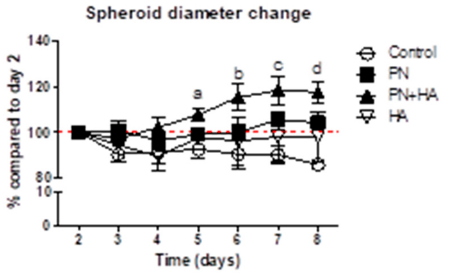
3.1. PN + HA Promotes the Dimensional Growth of Spheroids

Gingival fibroblasts were cultured for eight days on cell-repelling surfaces in complete DMEM to form spheroids and then observe possible changes in their morphology. We assessed that by measuring the longest diameter of the spheroid (Figure 1). The size of control spheroids remained stable for the whole duration of the experiment, and no significant changes were observed. If anything, the diameter of control spheroids tended to progressively decrease, to about 85% of the original size. Similarly, the addition of 100 ug/mL PN or HA did not significantly affect the size of spheroids over time. Noticeably, the initial size of the spheroids in the presence of PN or HA was smaller than control samples (Figure S1). The addition of the PN + HA compound, however, increased the size of the spheroids, which, by day 5, were significantly bigger than the control ones and ended up being about 20% larger than their original size (Figure 1).

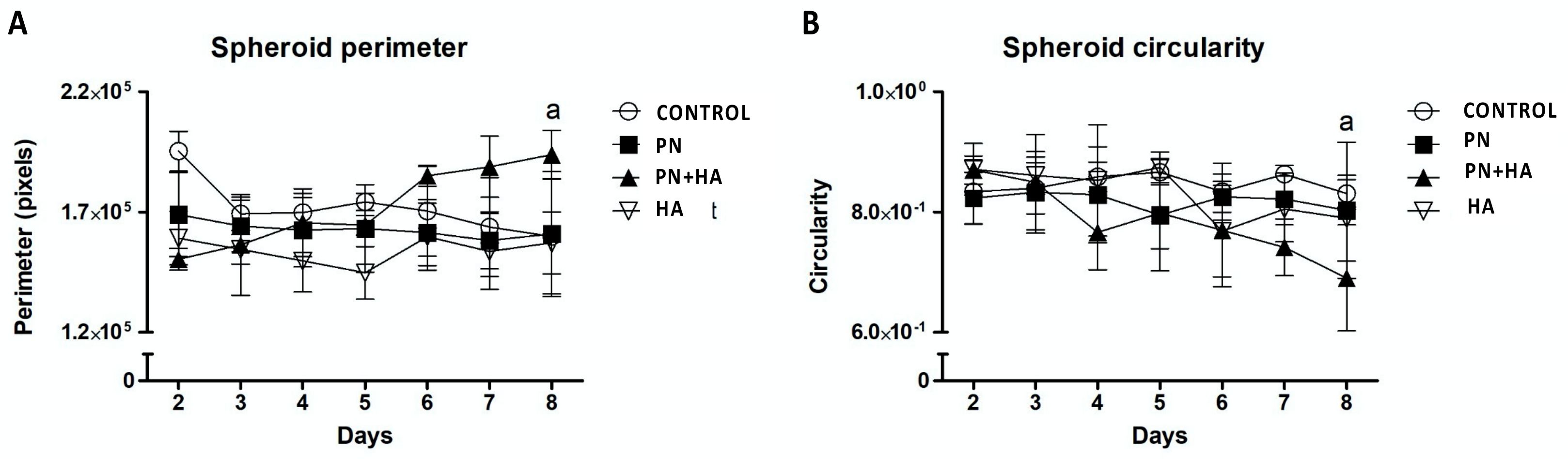
3.2. PN + HA Induces Morphological Changes in Spheroids

To better characterize the morphological changes of spheroids under different conditions, we acquired microphotographs of the spheroids and analyzed them using the freely available software ImageJ. Consistently with the measurement of their diameter, the perimeter of control spheroids remained overall stable from day 3 to day 8 of culture, though a significant drop was observed between days 2 and 3. No effect on cell perimeter was instead observed for cells stimulated with PN or HA alone. In contrast, the perimeter of spheroids stimulated with PN + HA gradually and significantly increased (Figure 2A). We also assessed spheroid circularity, a shape parameter that ranges from 0 to 1, with 1 meaning a perfect circle and 0 a line. As expected, spheroids generally fared quite high (around 0.8) on this scale, as they tend to be circular structures. Somewhat in agreement with the preceding observations, no changes in shape were detected during the eight-day observation period for Control, PN or HA samples. We, however, observed a significant and progressive decrease in circularity in PN+HA samples, indicating that the cell clusters became less circular, because of the presence of localized growth spurs, which were visible as small cell heaps protruding out of spheroids along their perimeter (Figure 2B).

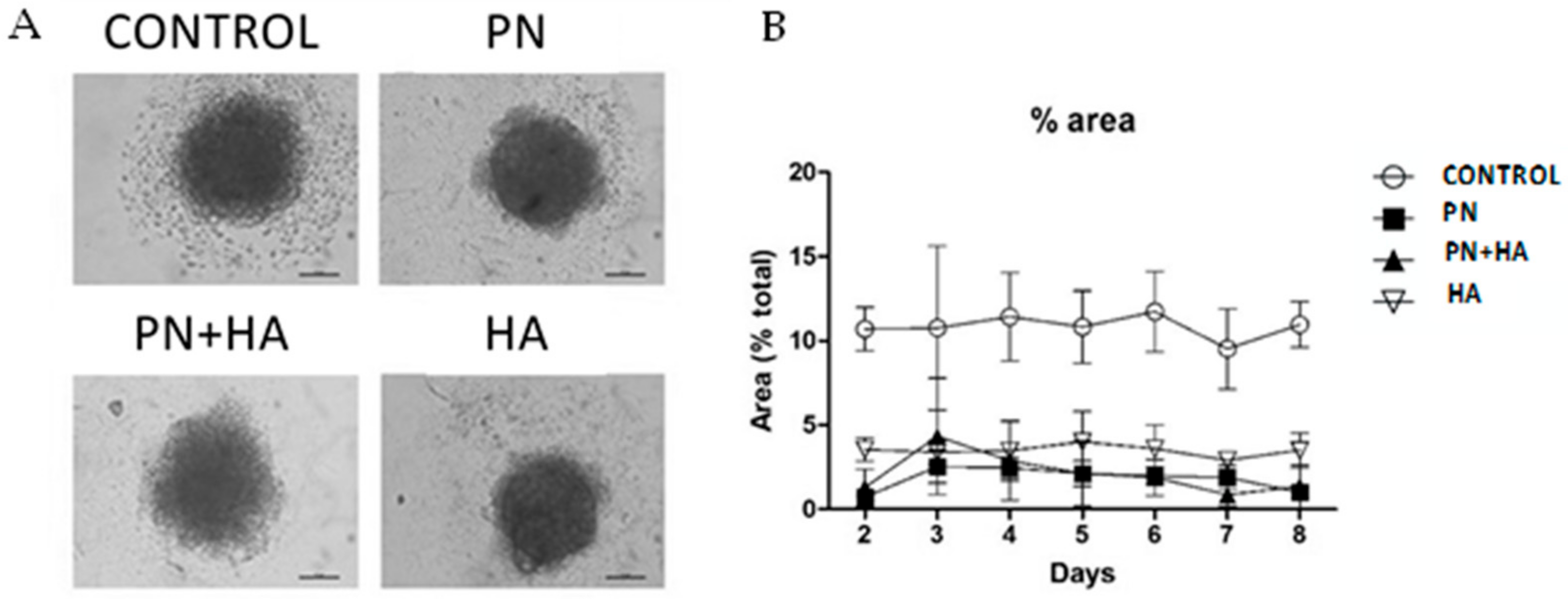
3.3. PN Preserves the Integrity of Cell Spheroids

A well-visible feature of control spheroids in culture was that a thick cloud of particles appeared around the spheroid soon after seeding (Figure 3A) and remained throughout the experiment. These particles can be interpreted as cell debris, as they did not appear to be compatible with whole cells (based on shape and size), nor with living organisms (based on metabolic assays, data not shown). The amount of this debris remained quite constant during the whole experiment. Such particles were not observed around all spheroids, but they were more abundant in control samples (Figure 3A). Treated samples did not present nearly as many particles, although samples with HA tended to have slightly more particles than PN-treated spheroids. We quantitated the particle-covered area around the spheroids using ImageJ and plotted it (Figure 3B).

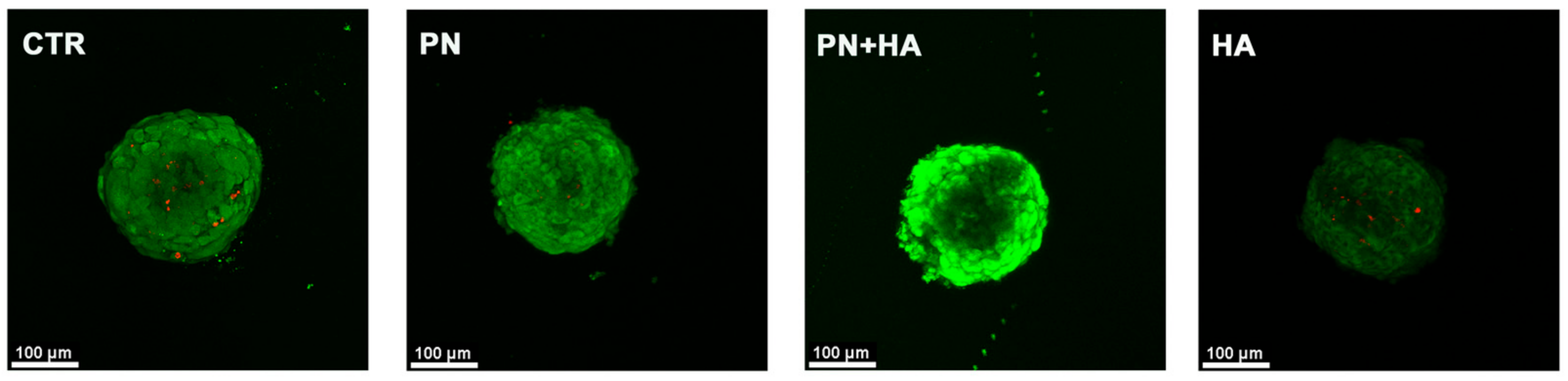
3.4. PN Increases Cell Viability within Spheroids

We then moved on to investigate cell viability within spheroids, as it can be challenging for cells to remain viable in these constructs, due to uneven access to nutrients in the different parts of the spheroid because of its morphology. To do that, we stained spheroids with calcein-AM, a dye that becomes fluorescent only after being internalized and cleaved by viable cells, or Propidium Iodide, a compound that, on the contrary, can pass the cell membrane and enter cells only if they are dead, and observed the samples using confocal microscopy (Figure 4). Cells in spheroids appeared viable, without detectable dead cells, and outer cells tended to be more strongly labeled than those insides. A stronger fluorescent signal of calcein in the peripheral area of spheroids has been previously described in the literature and appears to be associated with higher esterase activity of the cells in the outer layer of the cluster [21]. Fluorescence, however, appeared more intense in PN + HA-treated samples, as compared to HA-treated spheroids, and even when compared to PN alone (Figure 4).

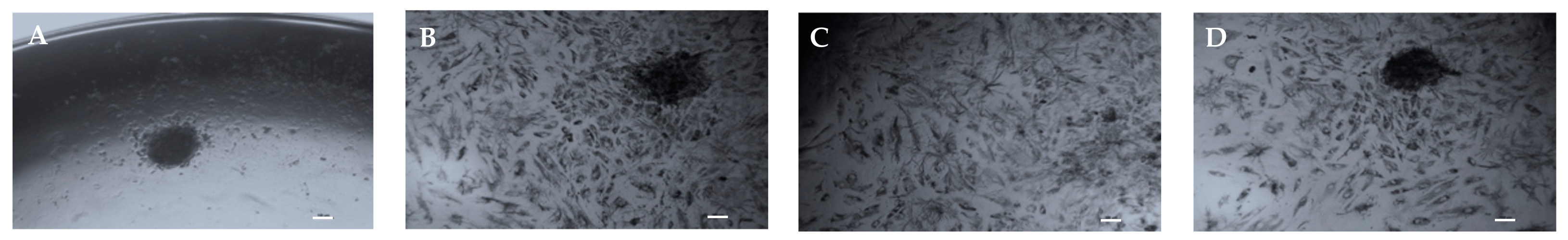
3.5. PN Increases Cell Proliferation and Migration from Spheroids

Spheroids are commonly created using special plates that do not allow for cell attachment. Once they get re-plated to regular culture substrate, however, cells within spheroids can attach to culture plastic and spheroids may disassemble. We investigated whether our stimuli would promote different behavior in spheroids, once they were replated on common culture plates under normal culture conditions, i.e., in the absence of further stimuli (Figure 5), and followed up for 15 days. Noticeably, control spheroids, even after being re-plated, maintained their shape for the whole 15 days of culture, though a few cells appeared scattered around spheroids (Figure S2). Treated spheroids, on the contrary, colonized the culture plate quite evenly, and by day 15 most of the plate surface appeared covered in viable, dividing cells, although with some remarkable differences. As cells proliferated and migrated on the plate, the spheroids themselves appeared to disassemble in the PN + HA treated samples, where they lost their integrity already after one week of culture (T6), and during the course of the second week of culture they disappeared completely: by day 15 the plate was covered by a dense layer of cells and no spheroid was observable (Figure 5). A noticeable decrease in spheroid density was also observed in PN samples, whereas the spheroids maintained their integrity in HA-alone samples.

4. Discussion

It has been previously shown that the combination of PN and HA exhibits emerging properties that clinically support tissue healing and protection [14,22,23,24,25]. These have been investigated using in vitro models that have shown that a combination of PN and HA promotes cell viability and matrix synthesis in human gingival fibroblasts in regular 2D cultures [26]. However, we concede that standard cultures can only offer very partial insights into the biological properties of such compounds [27] and we were thus prompted to pursue a more thorough investigation of these molecules, resorting to spheroids, a more complex and more challenging in vitro condition. To obtain spheroids, we cultured fibroblasts on special round-bottomed multiwell-plates, whose surfaces had been specifically treated to inhibit cell attachment. Cells, thus unable to adhere to the substrate, attached only to one another and formed spheroids very quickly.
Our data suggest that in the absence of stimuli only the cells that were originally seeded formed spheroids, and this culture condition did not promote cell growth, as the size of the spheroid remained unchanged over the course of the experiment (Figure 1). The size of control spheroids tended to slightly decrease over time, and this may be easily interpreted as a consequence of cell death. This hypothesis is supported also by the presence of abundant cell debris around control spheroids. Taken together, it can be therefore assumed that gingival fibroblasts seeded into the plates in the absence of external stimuli promptly formed spheroids but were unable to further grow. Although most cells apparently survived, a sizable fraction of the initially plated cells died and detached from spheroids.
The spheroid condition is possibly quite challenging for primary fibroblasts, as these did not appear to proliferate even in the presence of PN or HA alone, from what can be inferred from spheroid size measurements. In contrast, the addition of a combination of PN and HA supported an increase in spheroid size, and thus, possibly, an increase in cell number, although alternative explanations cannot be ruled out. It is possible that the increase in spheroid size is caused by the deposition of abundant extracellular matrix, which would be consistent with our previous data [3,26], or a combination of both phenomena. Our present data on spheroid circularity, however, suggest that gingival fibroblasts may have grown within PN + HA-treated spheroids, or they have migrated and rearranged. The decrease in spheroid circularity index in PN + HA samples from day 5 on (Figure 2B) corresponds to the observed apparition of little cell stacks at the periphery of the spheroids (Figure S1), which indicate that the cells in the outer layers of the cluster did have the tendency to grow or move out of the spheroid. It has been shown that spheroids are characterized by strong inequalities in the distribution of their nutrients and oxygen conditions, and, as a consequence, of cell activity, with peripheral cells more viable than those in the center, as confirmed by our confocal data [21]. This may have been brought about by a possible scaffolding activity of hyaluronic acid [28], which may have provided cells with a provisional matrix on which they could reposition themselves, in combination with the action of PN, which is known to promote cell viability in several cell models [11]. Interestingly, however, neither HA nor PN alone exerted such an effect on fibroblasts, suggesting that their combination may have synergic, non-linear effects that should be further investigated. Similarly, our confocal microscopy observations (Figure 4) attest to the increased cell viability in the presence of both PN and HA, which would explain why cells are capable of growing around spheroids under those conditions.
Furthermore, the re-plating experiment (Figure 5 and Figure S2) offered some important insights into the biology of cells in spheroid models under PN stimuli. One of the most striking observations is that control spheroids preserved their integrity over a 15-day culture period. This may be interpreted as an indication of a lack of proliferative potential in these spheroids, which were not able to give rise to a new cell progeny and colonize the culture plate. HA-treated samples were instead capable to do that, although spheroids were maintained. This suggests that only a small fraction of the cells that made up the spheroids participated in the plate colonization process. Cell sprouts appeared quite late in culture, possibly because only a few cell clones were activated and were this capable of spreading on the plate once they divided long enough and could migrate, possibly using the available HA as an aid to enhance their motility. Most of the cells in HA-treated spheroids, however, remained where they were, and the spheroids maintained their size and density, as observed by staining intensity. In the presence of PN, on the contrary, and even more so in the presence of a combination of PN + HA, the spheroids lost some of their density or were even disassembled completely (Figure 5). This can possibly mean that the majority of (or all) the cells forming the spheroids were reactivated and participated in plate colonization. As such, these data can be interpreted as proof that cells in PN, HA-treated spheroids maintained a higher degree of viability and were available for further adaptation and migration. Based on the available data it is not possible to identify how the combination of these stimuli may act this way, possibly by providing both a scaffold for spheroids and cells to attach and a pro-survival stimulus but the synergy of these compounds appears capable of maintaining cell viability across the whole spheroid, unlike the other tested treatments. It can be hypothesized that a combination of PN + HA provides both an extracellular matrix-like support to cells and a viability stimulus that, by working in synergy, have an overall positive trophic effect on cultured cells, maintaining their regeneration potential for a longer time.

5. Conclusions

Further studies are needed to better characterize the biological activity of PN in combination with HA, underpinning our observations, and more specifically the individual role of hyaluronic acid and polynucleotides in the physiology of 3D cells clusters, to optimize such compounds and their clinical effectiveness in tissue regeneration.

Supplementary Materials

The following supporting information can be downloaded at: https://www.mdpi.com/article/10.3390/app13020743/s1, Figure S1: Spheroid dimensions under different culture conditions; Figure S2: Spheroid replating.

Author Contributions

Conceptualization, C.G., S.G. and S.B.; methodology, S.B. and M.T.C.; software, C.G. and M.T.C.; investigation, M.T.C., M.L.V. and P.G.; resources, S.G.; data curation, C.G. writing—original draft preparation, C.G.; writing—review and editing, S.G., S.B. and M.T.C.; supervision, S.B. and P.G.; project administration, S.G. funding acquisition, S.G. All authors have read and agreed to the published version of the manuscript.

Funding

This research received no external funding.

Data Availability Statement

The data presented in this study are available on request from the corresponding author.

Conflicts of Interest

The authors declare no conflict of interest.

References

  1. Paternoster, J.L.; Vranckx, J.J. State of the Art of Clinical Applications of Tissue Engineering in 2021. Tissue Eng. Part B Rev. 2022, 28, 592–612. [Google Scholar] [CrossRef] [PubMed]
  2. Liu, Y.; Guo, L.; Li, X.; Liu, S.; Du, J.; Xu, J.; Hu, J.; Liu, Y. Challenges and Tissue Engineering Strategies of Periodontal Guided Tissue Regeneration. Tissue Eng. Part C Methods 2022, 28, 405–419. [Google Scholar] [CrossRef] [PubMed]
  3. Colangelo, M.T.; Govoni, P.; Belletti, S.; Squadrito, F.; Guizzardi, S.; Galli, C. Polynucleotide Biogel Enhances Tissue Repair, Matrix Deposition and Organization. J. Biol. Regul. Homeost. Agents 2021, 35, 355–362. [Google Scholar] [CrossRef] [PubMed]
  4. Cavallini, M.; Bartoletti, E.; Maioli, L.; Massirone, A.; Pia Palmieri, I.; Papagni, M.; Priori, M.; Trocchi, G. Consensus Report on the Use of PN-HPTTM (Polynucleotides Highly Purified Technology) in Aesthetic Medicine. J. Cosmet. Dermatol. 2021, 20, 922–928. [Google Scholar] [CrossRef] [PubMed]
  5. Cavallini, M.; de Luca, C.; Prussia, G.; Raichi, M. PN-HPT® (Polynucleotides Highly Purified Technology) in Facial Middle Third Rejuvenation. Exploring the Potential. J. Cosmet. Dermatol. 2022, 21, 615–624. [Google Scholar] [CrossRef]
  6. Gennero, L.; Denysenko, T.; Calisti, G.F.; Vercelli, A.; Vercelli, C.M.; Amedeo, S.; Mioletti, S.; Parino, E.; Montanaro, M.; Melcarne, A.; et al. Protective Effects of Polydeoxyribonucleotides on Cartilage Degradation in Experimental Cultures. Cell Biochem. Funct. 2013, 31, 214–227. [Google Scholar] [CrossRef]
  7. Giarratana, L.S.; Marelli, B.M.; Crapanzano, C.; de Martinis, S.E.; Gala, L.; Ferraro, M.; Marelli, N.; Albisetti, W. A Randomized Double-Blind Clinical Trial on the Treatment of Knee Osteoarthritis: The Efficacy of Polynucleotides Compared to Standard Hyaluronian Viscosupplementation. Knee 2014, 21, 661–668. [Google Scholar] [CrossRef]
  8. Colangelo, M.T.; Galli, C.; Guizzardi, S. The Effects of Polydeoxyribonucleotide on Wound Healing and Tissue Regeneration: A Systematic Review of the Literature. Regen. Med. 2020, 15, 1801–1821. [Google Scholar] [CrossRef]
  9. Scognamiglio, C.; Soloperto, A.; Ruocco, G.; Cidonio, G. Bioprinting Stem Cells: Building Physiological Tissues One Cell at a Time. Am. J. Physiol.-Cell Physiol. 2020, 319, C465–C480. [Google Scholar] [CrossRef]
  10. Pia Palmieri, I.; Raichi, M. Biorevitalization of Postmenopausal Labia Majora, the Polynucleotide/Hyaluronic Acid Option. Obstet. Gynecol. Rep. 2019, 3, 1–5. [Google Scholar] [CrossRef]
  11. Guizzardi, S.; Uggeri, J.; Belletti, S.; Cattarini, G. Hyaluronate Increases Polynucleotides Effect on Human Cultured Fibroblasts. J. Cosmet. Dermatol. Sci. Appl. 2013, 3, 124–128. [Google Scholar] [CrossRef] [Green Version]
  12. Dallari, D.; Sabbioni, G.; del Piccolo, N.; Carubbi, C.; Veronesi, F.; Torricelli, P.; Fini, M. Efficacy of Intra-Articular Polynucleotides Associated With Hyaluronic Acid Versus Hyaluronic Acid Alone in the Treatment of Knee Osteoarthritis. Clin. J. Sport Med. 2020, 30, 1–7. [Google Scholar] [CrossRef] [PubMed]
  13. Zhang, L.; Lei, N.; Chang, R.; Yang, C.; Li, Q.; Zuo, N.; Gu, Y. Efficacy of Intra-Articular Polynucleotides Associated with Hyaluronic Acid vs. Hyaluronic Acid Alone in the Treatment of Knee Osteoarthritis: A Systematic Review and Meta-Analysis of Randomized Clinical Trial. Medicine 2020, 99, e20689. [Google Scholar] [CrossRef] [PubMed]
  14. Pilloni, A.; Rojas, M.A.; Dh, C.T.; Carere, M.; de Filippis, A.; Marsala Dh, R.L.; Marini, L. Clinical Effects of the Adjunctive Use of a Polynucleotides and Hyaluronic Acid Based Gel in the Subgingival Re-Instrumentation of Residual Periodontal Pockets: A Randomized, Split-Mouth Clinical Trial. J. Periodontol. 2022, 1–10. [Google Scholar] [CrossRef]
  15. Duval, K.; Grover, H.; Han, L.-H.; Mou, Y.; Pegoraro, A.F.; Fredberg, J.; Chen, Z. Modeling Physiological Events in 2D vs. 3D Cell Culture. Physiology 2017, 32, 266–277. [Google Scholar] [CrossRef] [Green Version]
  16. Schindler, M.; Nur-E-Kamal, A.; Ahmed, I.; Kamal, J.; Liu, H.-Y.; Amor, N.; Ponery, A.S.; Crockett, D.P.; Grafe, T.H.; Chung, H.Y.; et al. Living in Three Dimensions: 3D Nanostructured Environments for Cell Culture and Regenerative Medicine. Cell Biochem. Biophys. 2006, 45, 215–227. [Google Scholar] [CrossRef]
  17. Wolff, A.; Frank, M.; Staehlke, S.; Peters, K. A Comparative Study on the Adipogenic Differentiation of Mesenchymal Stem/Stromal Cells in 2D and 3D Culture. Cells 2022, 11, 1313. [Google Scholar] [CrossRef]
  18. Jubelin, C.; Muñoz-Garcia, J.; Griscom, L.; Cochonneau, D.; Ollivier, E.; Heymann, M.-F.; Vallette, F.M.; Oliver, L.; Heymann, D. Three-Dimensional in Vitro Culture Models in Oncology Research. Cell Biosci. 2022, 12, 155. [Google Scholar] [CrossRef]
  19. Kozlowski, M.T.; Crook, C.J.; Ku, H.T. Towards Organoid Culture without Matrigel. Commun. Biol. 2021, 4, 1387. [Google Scholar] [CrossRef]
  20. Squadrito, F.; Bitto, A.; Irrera, N.; Pizzino, G.; Pallio, G.; Minutoli, L.; Altavilla, D. Pharmacological Activity and Clinical Use of PDRN. Front. Pharmacol. 2017, 8, 224. [Google Scholar] [CrossRef]
  21. Bigelow, C.E.; Mitra, S.; Knuechel, R.; Foster, T.H. ALA- and ALA-Hexylester-Induced Protoporphyrin IX Fluorescence and Distribution in Multicell Tumour Spheroids. Br. J. Cancer 2001, 85, 727–734. [Google Scholar] [CrossRef] [Green Version]
  22. Stagni, C.; Rocchi, M.; Mazzotta, A.; del Piccolo, N.; Rani, N.; Govoni, M.; Vivarelli, L.; Veronesi, F.; Fini, M.; Dallari, D. Randomised, Double-Blind Comparison of a Fixed Co-Formulation of Intra-Articular Polynucleotides and Hyaluronic Acid versus Hyaluronic Acid Alone in the Treatment of Knee Osteoarthritis: Two-Year Follow-Up. BMC Musculoskelet. Disord. 2021, 22, 773. [Google Scholar] [CrossRef]
  23. de Caridi, G.; Massara, M.; Acri, I.; Zavettieri, S.; Grande, R.; Butrico, L.; de Franciscis, S.; Serra, R. Trophic Effects of Polynucleotides and Hyaluronic Acid in the Healing of Venous Ulcers of the Lower Limbs: A Clinical Study. Int. Wound J. 2016, 13, 754–758. [Google Scholar] [CrossRef]
  24. Segreto, F.; Carotti, S.; Marangi, G.F.; Francesconi, M.; Scaramuzzino, L.; Gratteri, M.; Caldaria, E.; Morini, S.; Persichetti, P. The Use of Acellular Porcine Dermis, Hyaluronic Acid and Polynucleotides in the Treatment of Cutaneous Ulcers: Single Blind Randomised Clinical Trial. Int. Wound J. 2020, 17, 1702–1708. [Google Scholar] [CrossRef]
  25. Maioli, L. Polynucleotides Highly Purified Technology and Nucleotides for the Acceleration and Regulation of Normal Wound Healing. Aesthetic Med. 2020, 6, 48–52. [Google Scholar]
  26. Colangelo, M.T.; Belletti, S.; Govoni, P.; Guizzardi, S.; Galli, C. A Biomimetic Polynucleotides–Hyaluronic Acid Hydrogel Promotes Wound Healing in a Primary Gingival Fibroblast Model. Appl. Sci. 2021, 11, 4405. [Google Scholar] [CrossRef]
  27. Al Shboul, S.; DeLuca, V.J.; Al Dweiri, Y.; Saleh, T. Can 3D Bioprinting Solve the Mystery of Senescence in Cancer Therapy? Ageing Res. Rev. 2022, 81, 101732. [Google Scholar] [CrossRef]
  28. AlHowaish, N.A.; AlSudani, D.I.; AlMuraikhi, N.A. Evaluation of a Hyaluronic Acid Hydrogel (Restylane Lyft) as a Scaffold for Dental Pulp Regeneration in a Regenerative Endodontic Organotype Model. Odontology 2022, 110, 726–734. [Google Scholar] [CrossRef]
Figure 1. Spheroids were cultured on special cell-repellant culture plates and followed up for eight days. We measured spheroid diameter in vehicle-treated samples (Control) and compared it to Polynucleotides alone (PN), Hyaluronic acid alone (HA), or Polynucleotides in combination with Hyaluronic acid (PN + HA) groups, expressed as a percentage of the initial diameter. a = p < 0.05 PN + HA vs. all groups; b = p < 0.01 PN + HA vs. all groups; c, d = p < 0.001 PN + HA vs. all groups.
Applsci 13 00743 g001
Figure 2. Changes in spheroid morphology as determined by ImageJ analysis. (A) Perimeter and (B) circularity of spheroids stimulated with vehicle (Control), Polynucleotides (PN) with or without Hyaluronic acid (PN + HA and HA respectively). a = p < 0.05 PN + HA vs. all groups.
Applsci 13 00743 g002
Figure 3. A thick cloud of particles appeared around the spheroid soon after seeding. (A) Microphotographs of spheroids after eight days of culture under different conditions. Magnification 10× (B) Quantification of debris area in the different cultural groups over time. The debris area in control groups is significantly higher than all other groups at all time points.
Applsci 13 00743 g003
Figure 4. Microphotographs of spheroids under different culture conditions after eight days of culture at Confocal Microscope after labeling with calcein-AM (green) or propidium iodide (red). Magnification = 10×.
Applsci 13 00743 g004
Figure 5. Microphotographs of spheroids stimulated with (A) vehicle, (B) Polynucleotides, (C) Polynucleotides and Hyaluronic acid, (D) Hyaluronic acid, after re-plating into regular culture plates and cultured for 15 days. Spheroids that had been previously cultured in PN + HA dissolved completely after replating and their cells colonized the new plate. Spheroids previously cultured under different conditions were mostly retained, although some of their cells did spread out and grew on the new plates. Magnification 4×. Bar = 50 μm.
Applsci 13 00743 g005
Disclaimer/Publisher’s Note: The statements, opinions and data contained in all publications are solely those of the individual author(s) and contributor(s) and not of MDPI and/or the editor(s). MDPI and/or the editor(s) disclaim responsibility for any injury to people or property resulting from any ideas, methods, instructions or products referred to in the content.

