Article

Influence of Silver Nanoparticles on Color Stability of Room-Temperature-Vulcanizing Maxillofacial Silicone Subjected to Accelerated Artificial Aging

1
Department of Prosthodontics, College of Dentistry, University of Sulaimani, Sulaimani 46001, Iraq
2
Department of Periodontics, College of Dentistry, University of Sulaimani, Sulaimani 46001, Iraq
3
Department of Physics, Nanotechnology Research Laboratory, College of Science, University of Sulaimani, Sulaimani 46001, Iraq
*
Author to whom correspondence should be addressed.
Appl. Sci. 2023, 13(20), 11201; https://doi.org/10.3390/app132011201
Submission received: 17 September 2023 / Revised: 7 October 2023 / Accepted: 10 October 2023 / Published: 12 October 2023
(This article belongs to the Section Applied Dentistry and Oral Sciences)

Abstract

:
In this in vitro study, we assessed the color stability of an A-2186 room-temperature-vulcanizing (RTV) silicone elastomer by incorporating silver nanoparticles during accelerated artificial aging. Using five intrinsic silicone pigment types, including no pigment (colorless), red, blue, mocha, and a combination of the three, we created 160 disk-shaped specimens. These were evenly distributed across 20 experimental groups, each containing 8 samples (n = 8). The specimens underwent aging for 250 and 500 h in an artificial aging chamber. A colorimeter was used to measure the values of L*a*b* according to the Commission Internationale de L’Éclairage (CIE) standards. The 50:50% perceptibility threshold (∆E* = 1.1) and acceptability threshold (∆E* = 3.0) were used in the interpretation of the recorded color differences. At the 0.05 level of significance, the one-way analysis of variance (ANOVA) and Tukey post hoc test were used in the statistical analysis. Fourier-transform infrared spectroscopy with attenuated total reflectance (FTIR-ATR) showed that 0.2% AgNPs after 500 h of aging protected the silicone elastomer matrix and all characteristic bonds of the silicone elastomer. In contrast, silicone without AgNPs showed the distortion of all bonds after 500 h. Chromatic alterations (∆E* > 0) were observed in all specimen groups, surpassing the perceptible threshold (1.1 units), except for mocha, with 0.2% AgNPs after 250 h of aging, which remained below the perceptible threshold (∆E* = 0.97). All groups demonstrated ∆E* values below the acceptable threshold, except for the red color, which exhibited a highly significant color change (p = 0.000). This study determined that all specimens, including colorless silicone, underwent color changes (∆E* > 0), with red displaying a notably significant chromatic alteration. Additionally, AgNPs demonstrated substantial protection of the silicone and reduced the color change across all groups and colors, with enhanced efficacy corresponding to higher AgNP concentrations (0.2% AgNPs > 0.15% AgNPs > 0.1% AgNPs).

1. Introduction

Maxillofacial prosthetics is a branch of prosthodontics that focuses on treating and managing maxillofacial abnormalities [1]. Extraoral maxillofacial prostheses are frequently utilized to restore facial components that have been lost due to congenital defects, trauma, or surgical removal. These prostheses serve to restore both functional and aesthetic aspects, thereby greatly enhancing the overall quality of life for patients [1,2,3,4,5,6,7]. Silicone, also known as polydimethylsiloxane, is frequently utilized in the fabrication of the extraoral sections of the maxillofacial prosthesis. For more than half a century, it has been utilized in the fabrication of facial prosthetics for patients because of its longevity, resistance, biocompatibility, and ease of manipulation [4,8,9,10,11,12,13,14,15]. In spite of this, there are specific worries regarding the durability of silicone prostheses, in addition to the maintenance that is required for them [16,17,18,19]. The color stability of prostheses made with silicone elastomer is only guaranteed for a period of six to twelve months [20,21]. The discoloration of the silicone elastomers is the primary factor that leads to the need for the replacement of facial prostheses [7,19,22,23,24]. The color change of the facial prosthesis is brought on by a combination of factors, including exposure to ultraviolet (UV) radiation, air pollution, pigments that are naturally present in the material, and the use of cleaning solvents [7,25,26,27,28]. Even though ultraviolet light only makes up a very small percentage of the total radiation that is emitted by the sun, it has a significant influence on the facial prostheses’ ability to maintain their color. All wavelengths that fall between 100 and 400 nanometers are included in the definition of ultraviolet radiation. To simulate the conditions of the natural environment, a large number of researchers have constructed artificial weathering or aging chambers. These chambers are helpful for determining the overall deterioration of materials and have been widely used [25,27,29].
Polymers, such as silicone materials, have the ability to absorb the energy of UV photons, which can lead to the breakdown of polymeric chains and the production of new radicals that can cause additional damage to polymer networks. Polymers also have the ability to produce new radicals that can cause additional damage to polymer networks. As a result of this process, the surface might develop cracks, it might lose its shine; it might turn a different color; or it might fade. In addition, there is a possibility that the material’s molecular weight will decrease, as well as its elasticity [30].
In the process of defining color notations, the CIE-Lab system is frequently applied. This system was developed by the Commission Internationale de L’Éclairage (CIE). ∆E* is the notation that is used in this system to refer to the overall color difference caused by all changes in color coordinates. It has been stated that the perceptibility threshold for silicone maxillofacial prosthetics with light skin tone is ∆E* of 1.1, while the acceptability threshold is ∆E* of 3.0 [31,32].
Nano-oxide particles have been incorporated into various polymers so that they can provide protection against ultraviolet (UV) rays and also function as a strengthening agent. The strengthening mechanism of the particles can be traced back to an increasing chemical reactivity of the particles themselves. The increased chemical reactivity exhibited by these particles confers upon them the capacity to engage in reactions with polymer chains, thereby imparting stabilization to the chains. There is a hypothesis suggesting that the minuscule dimensions of nano-oxides play a role in enhancing their ability to absorb ultraviolet (UV) light and facilitate the scattering of UV photons. As a consequence, this leads to a reduction in the overall absorption of UV energy by the polymer network. This is supported by the fact that nano-oxides have both of these properties [33,34].
Silver nanoparticles, also known as AgNPs, are one of the most exciting and significant nanomaterials that are used in biomedical applications today. The fields of nanoscience and nanotechnology, and nanomedicine in particular, have found AgNPs to be of critical importance in recent years. AgNPs are finding growing use in a variety of fields due to the exceptional physical and chemical properties that they possess. Some of these fields include medicine, food, health care, consumer goods, and industry, to name just a few. Optical, electrical, and thermal properties, in addition to high electrical conductivity and biological characteristics, are all incorporated into this category [35,36,37,38].
In spite of the widespread application of maxillofacial silicone elastomer, there has been no research carried out to investigate the effect that AgNPs have on the color stability of this material. In light of this, the purpose of this study was to investigate the effect that AgNPs have on the ability of an A-2186 maxillofacial silicone elastomer to maintain its color. It was hypothesized that adding AgNPs to A-2186 maxillofacial silicone would not preserve the color stability of the silicone after it was artificially aged. It was also hypothesized that the aging procedure would not cause the color of the A-2186 silicone to change.

2. Materials and Methods

2.1. Materials

The materials used in this study were obtained from their respective manufacturers and are listed below in Table 1.

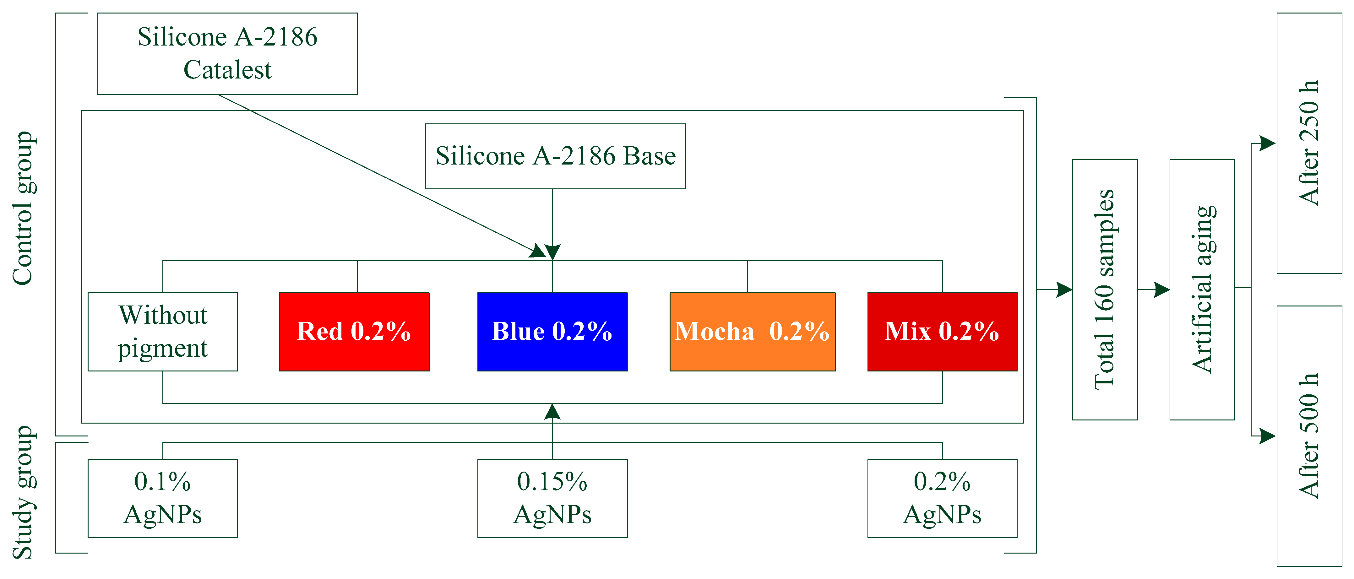
2.2. Experimental Design and Sample Preparation

In total, 160 disk-shaped specimens, each with a 2 mm thickness and a 20 mm diameter, were manufactured and evenly spread among 20 experimental groups, with each group containing 8 samples (n = 8) [39]. The control specimens were fabricated without adding AgNPs (0% AgNPs). In comparison, the study specimens were fabricated by mixing varying concentrations of silver nanoparticles (0.1%, 0.15%, and 0.2% by weight) with the silicone and each pigment (i.e., no pigment, red, blue, mocha, and RBM). Table 2 shows the distribution of the specimens.
Clear acrylic sheets were laser-cut to create the molds. For each mold, two clear acrylic plates with the exact exterior dimensions of the mold and a thickness of 6 mm to bear clamping force were cut to sandwich the mold between them. According to the manufacturer’s instructions, A-2186 silicone elastomer can be obtained as a base (part A) and a catalyst (part B), and they can be combined at a ratio of 10:1 by weight. The weight of the color was equivalent to 0.2% of the total weight of the silicone [40,41,42,43,44].
For the preparation of the A-2186 silicone specimens without color and AgNPs, parts A and B of the silicone were initially measured in a digital electronic weight balance (Nimbus® Analytical, Adam Equipment, Oxford, CT, USA) and then mixed per the manufacturer’s instructions in a vacuum mixer (AX-2000C; Aixin Medical Equipment Co., Ltd., Tianjin, China) for 5 min at a speed of 360 rpm and a vacuum of −0.09 MPa. The silicone specimens with each color were then prepared by weighing and combining the color with part A of the A-2186 silicone and then vacuum-mixing for 10 min. However, for the first 2 min, only mixing was performed without vacuuming to avoid color suctioning. For the production of the study group specimens, the color and AgNPs were weighed and then added to the A-2186 silicone part A. They were then mixed for 10 min in a vacuum mixer. To prevent the suction of the color and AgNPs, the vacuum was turned off for the first 2 min, just as it had been for the previous silicone specimen preparation with the only color content. The mixing bowl was then left aside to cool down to room temperature since the mixer’s rotation creates heat, which decreases the material’s working time. Then, part B was added to the vacuum mixer and mixed for an additional 5 min. The mixture was then loaded into the molds using a metallic spatula and placed in a vacuum chamber for 2 min to remove any air bubbles that may have formed during the loading process. After that, the molds were put in a pressure pot (Pentola A pressione typodont; leone s.p.a., Sesto Fiorentino, Italy) at 0.2 MPa for 2 min to smooth the mixture’s surface and rupture any superficial air bubbles [45]. Following that, the molds were sealed and secured using G-clamps, and the manufacturer-recommended 24 h setting period was followed. The specimens were removed from the molds, cleaned with water and liquid detergent, and dried with tissue paper. The specimens were then cut using scissors. Before testing, samples that had visible defects were discarded. All specimens were housed in a lightproof black box to prevent any possible color changes. Color measurements were performed using a digital colorimeter (WR10QC colorimeter, FRU, Guangzhou, China). An aging chamber (Weather-Ometer device QUV, made by Atlas Electric Devices Company, Chicago, IL, USA) was used to artificially age the samples in line with ASTM G15445 cycle 1 [46]. After 250 and 500 h, another reading was recorded, and ∆E* was calculated using the formula below. The procedural framework employed in our study is elucidated in Figure 1.
∆E* = [(∆L)2 + (∆a)2 + (∆b)2]1/2
where L = lightness, a = green-red coordinate, and b = blue-yellow coordinate.

2.3. Characterization and Statistical Analysis

The well-known characterization method known as Fourier-transform infrared spectroscopy-attenuated total reflectance (FTIR-ATR) (Tensor 27, Bruker Optics, Ettlingen, Germany) was used to investigate the impact of aging on the silicone elastomer, the AgNPs on protecting the silicone, and artificial aging on the silicone bonds. Multiple one-way ANOVAs were performed using statistical software (IBM SPSS Statistics, v24; IBM Corp., Armonk, NY, USA) to see whether there were differences in color among the groups; post hoc Tukey tests were used for multiple comparisons when the one-way ANOVA was significant (p = 0.05).

3. Results

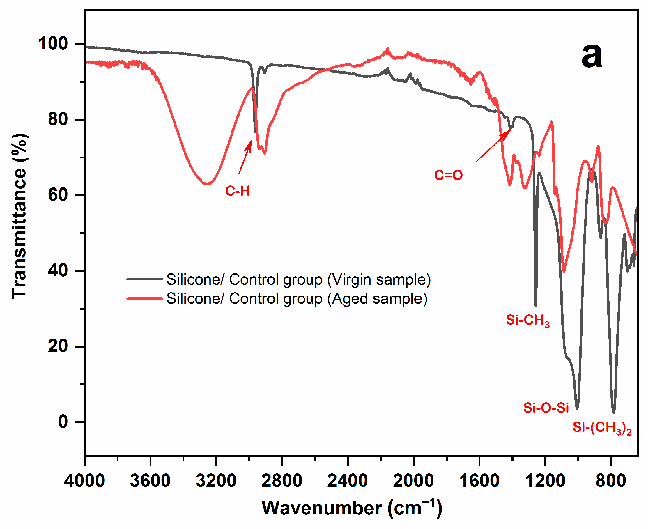
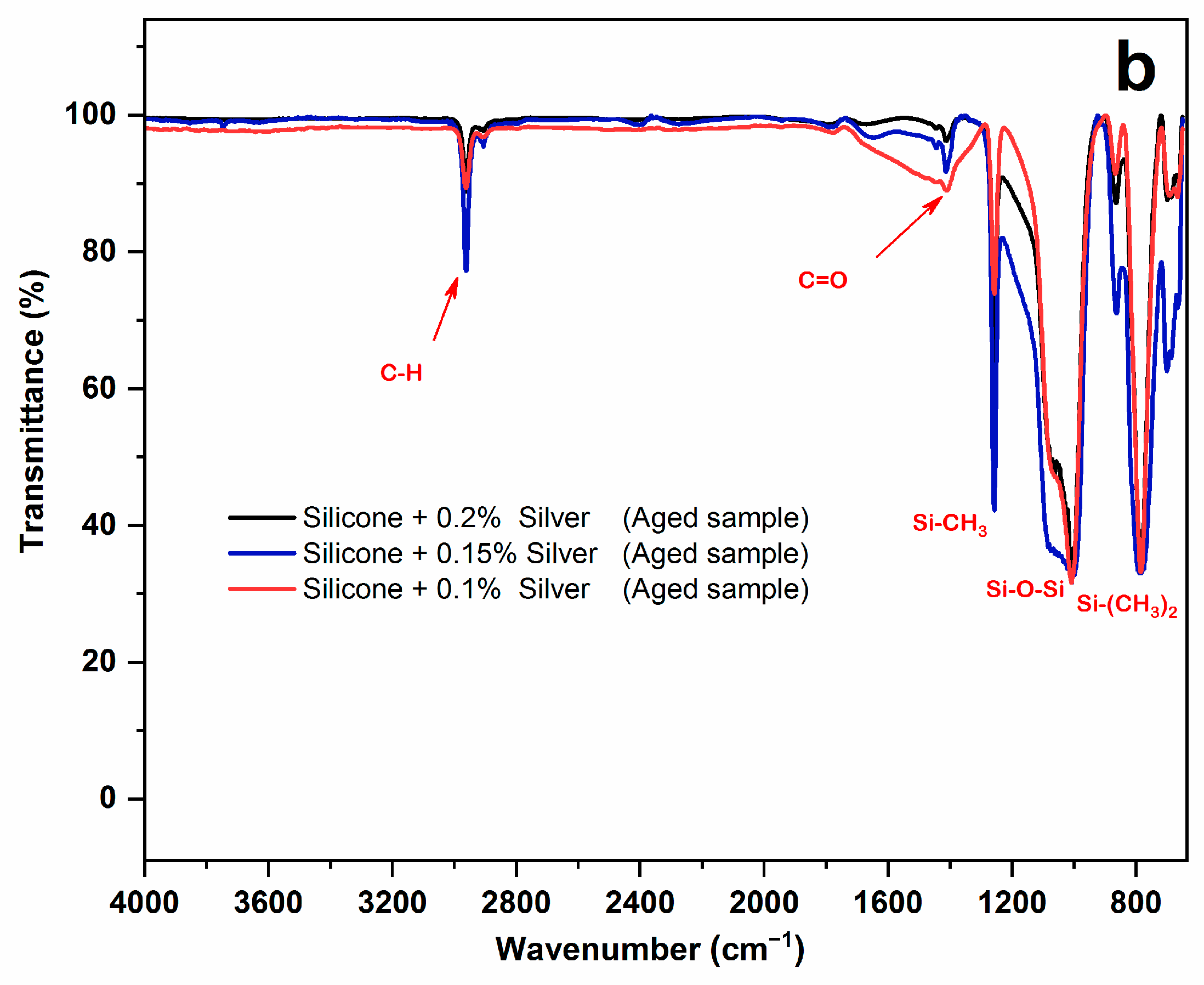
3.1. FTIR-ATR Result

Figure 2a,b shows the FTIR-ATR results for the silicone elastomer before and after 500 h of artificial aging without AgNPs and with 0.2% AgNPs, respectively. Prior to artificial aging, as can be seen from Figure 2a, all of the characteristic peaks of silicone were exhibited, including the Si-O-Si (1018.14 cm−1), Si-(CH3)2 (800.46 cm−1), C-H (3000 cm−1), and Si-CH3 (1255 cm−1) bonds. Nonetheless, after 500 h of artificial aging, the peaks had deteriorated.
Figure 2b shows the FTIR-ATR results of silicone elastomer with 0.1, 0.15, and 0.2% AgNPs after 500 h of artificial aging. When 0.2% AgNPs were added to the silicone elastomer, the silicone exhibited all of its characteristic peaks, indicating that the AgNPs did not affect the silicone’s matrix and bonds. Even after 500 h of artificial aging, it was also found that all of the silicone peaks remained intact and undeterred. This demonstrates the protective role of AgNPs within the silicone, protecting the silicone elastomer matrix and silicone peaks.

3.2. Color Change Results

The means and standard deviations of ∆E* in the control and AgNPs groups for all of the colors, including colorless, red, blue, mocha, and their mixtures, subjected to 250 h of artificial aging, are presented in Table 3. However, Table 4 displays the same findings for 500 h of artificial aging. An ANOVA table of the color alterations (∆E*) following the artificial aging is shown in Table 5. All evaluated specimens from the control and AgNP groups experienced a chromatic alteration (∆E* > 0). The ∆E* values for all groups were found to be above the perceptible threshold (1.1 units), except for mocha with 0.2% AgNPs after 250 h of aging, where the ∆E* value was below the perceptible threshold (∆E* = 0.97). However, ∆E* was determined to be below the acceptable clinical threshold of 3.0 units, demonstrating acceptable aging-dependent color changes, except for red in all groups and blue in the control group after 500 h of aging.
It is crucial to note that there were relevant variations in the ∆E* values among the groups. For the colorless specimens, only the control group that underwent 500 h of aging showed a significant change (∆E* = 2.41 (0.22)) according to the perceptible threshold (p = 0.027). All other groups were over the perceptible threshold, but the differences were barely perceptible and were still below the acceptable threshold. After 250 and 500 h, all groups with and without AgNPs showed a highly significant change in the perceptible and acceptable threshold for red (p = 0.000). For the color blue, only the control group, after 500 h of aging, displayed a significant change (∆E* = 3.15 (0.44)) relative to the perceptible threshold (p = 0.000). The blue color with 0.2% AgNPs after 500 h of aging showed a significant decrease in color change (∆E* = 1.49 (0.28)) compared to the acceptable threshold (p = 0.002). According to the perceptible threshold (p = 0.019) for mocha, the control group aged for 500 h exhibited a significant change (∆E* = 2.44 (0.24)). However, it is essential to mention that the least color change for mocha was found for the 0.2% AgNPs subjected to 250 h of aging, which was found to be lower than the perceptible threshold (∆E* = 0 0.97 (0.23)). Based on the perceptible threshold for the mixed color (∆E* = 1.1), the control group underwent substantial changes after 250 and 500 h of aging (p = 0.048 and p = 0.001, respectively). Also, in terms of the perceptible threshold (p = 0.04), after 500 h, the mixed color containing 0.1 percent AgNPs (∆E* = 2.70 (0.26%)) altered dramatically. However, all of the groups were below the acceptable threshold (∆E* = 3.0).

4. Discussion

The findings derived from this investigation provide evidence for the rejection of the null hypothesis posited, as the silver nanoparticles (AgNPs) demonstrated a crucial function in safeguarding the silicone material. The color of the A-2186 maxillofacial silicone was notably altered as a result of artificial aging, particularly leading to a substantial change in color (∆E > 3.0) within the red spectrum. In addition, it is worth noting that all groups demonstrated a variation in color, including colorless, red, blue, mocha, and mixture. Despite the fact that certain color alterations in the control group exceeded the perceptibility threshold of 1.1 by a significant margin, only the red color exhibited a level of significance surpassing the acceptability threshold of 3.0 [47]. This implies that the color change in the silicone specimens in the present study is not clinically significant, except for the red color. The perceptibility threshold is defined as the color difference that the human eye notices. In contrast, the acceptability threshold is considered the color difference that is acceptable in terms of aesthetics [48]. Changing the material’s color in a clinical setting is permissible if the change is below the acceptable threshold and above the detectable threshold. This indicates that the change in a material’s color can be noticed clinically while being aesthetically appealing.
Consistent with the anticipated outcomes, the color of the additive-free silicone samples that were artificially aged exhibited a noticeable alteration. Silicone has the ability to undergo UV radiation absorption, resulting in the fragmentation of polymer chains and the generation of free radicals [30]. After 500 h of artificial aging, the unpigmented group exhibited a considerable color change in relation to the perceptibility threshold. Hatamleh et al. [49] reported that the unpigmented group underwent a significant color change in their study, attributing it to the continual chemical polymerization. Our findings follow prior studies [25,50] that found colorless silicone to be susceptible to chromatic alteration, even after the addition of pigments and nano-oxides.
The red color showed a significant color change in terms of both perceptibility and acceptability (p < 0.000) [48,51,52]. Due to the findings of Kiat-Amnuay et al., red was selected as the intrinsic color for this study. In their study, the red pigment was shown to have the most significant detrimental influence on the color stability of silicone elastomers [29]. Beatty et al. [2] studied the effect of UV light exposure on the color of dry-pigmented maxillofacial elastomers. They discovered that after 400 h of exposure, red cosmetic dry earth pigment exhibited considerable color changes.
The efficacy of artificial aging can be leveraged to assess the color stability of maxillofacial materials in a prompt, efficient, and proficient manner. There has been a suggestion that the stability of color in silicone, whether artificially aged or not, may be compromised when nanoparticles (NPs) are present [42,50,53].
Ultraviolet (UV) light, which falls within a narrow segment of the electromagnetic spectrum that is not visible to the human eye, is known to primarily induce detrimental effects on colorants and polymers. The excitation of electrons within the nanoparticles of a given medium occurs as a result of their induced vibrational motion upon exposure to ultraviolet radiation. Due to the comparatively smaller sizes of nanoparticles in relation to ultraviolet light wavelengths, a portion of the incident ultraviolet light is scattered while another portion is concurrently absorbed [54]. In accordance with these fundamental principles, UV protection arises as a result of the absorption and scattering of nanoparticles. Due to their nanoparticle nature and ability to scatter and absorb UV radiation, AgNPs have the potential to offer UV protection that is comparable to existing methods. The present study employed a vacuum mixer to achieve uniform dispersion of silver nanoparticles and silicone pigments within the silicone elastomer matrix. This process led to a significant decrease in the extent of color alteration observed in the silicone material. A higher degree of ultraviolet (UV) shielding can be attained with the reduction in the size of nano-oxide particles. Hence, it can be inferred that the utilization of silver nanoparticles has the potential to maintain the color integrity of various materials.
There were particular concerns regarding this in vitro study that also need to be considered. The study employed a low concentration of silver nanoparticles (AgNPs) and only one type of silicone elastomer. Rather than relying on outdoor aging to assess color stability, this study used artificial aging over only 500 h. To determine how AgNPs impact the stability of various colored elastomers used in the production of maxillofacial prosthetic silicone in clinical practice requires more investigation. It is necessary to investigate the impact of varying concentrations of AgNPs on the color stability of A-2186 and other maxillofacial silicones. The impact of AgNPs on the color stability of maxillofacial silicone during exposure to natural outdoor weathering must be evaluated. Additionally, the range and duration of artificial aging essential for color change must be assessed.

5. Conclusions

An A-2186 RTV silicone elastomer with silver nanoparticles was tested for color stability under accelerated aging in this extensive in vitro study. All specimens changed color after 250 and 500 h, which was an important finding. Polymerization caused the colorless silicone to change color. After 250 and 500 h of artificial aging, significant changes in red were observed, exceeding the established thresholds (p < 0.000). As the concentration increased (0.2% > 0.15% > 0.1%), the silver nanoparticles protected the silicone and reduced the color change.
In particular, mocha showed the least variation, demonstrating its remarkable chromatic resilience. These results improve our understanding of silicone elastomer color stability. We gained more insight into the silver nanoparticles’ concentration-dependent effects and the different behaviors of the colorless and mocha specimens. Silver nanoparticles’ transformative potential as a powerful protective measure in material science holds promise for diverse applications.

Author Contributions

Methodology, K.M.; formal analysis, K.M., F.Z. and B.A.; investigation, K.M. and B.A.; data curation, K.M. and B.A.; writing—original draft, F.Z. and B.A.; writing—review and editing, F.Z. and B.A.; visualization, K.M. and B.A.; supervision, F.Z. and B.A.; project administration, F.Z. All authors have read and agreed to the published version of the manuscript.

Funding

This research received no external funding.

Data Availability Statement

All the required data related to the current study are embedded in this manuscript.

Acknowledgments

The authors are grateful to the Nanotechnology Research Laboratory, Department of Physics, University of Sulaimani, for laboratory support.

Conflicts of Interest

The authors declare that they have no known competing financial interest or personal relationships that could have appeared to influence the work reported in this paper.

References

  1. Roberts, A.C. Facial Reconstruction by Prosthetic Means. Br. J. Oral Surg. 1966, 4, 157–182. [Google Scholar] [CrossRef] [PubMed]
  2. Beatty, M.W.; Mahanna, G.K.; Dick, K.; Jia, W. Color Changes in Dry-Pigmented Maxillofacial Elastomer Resulting from Ultraviolet Light Exposure. J. Prosthet. Dent. 1995, 74, 493–498. [Google Scholar] [CrossRef]
  3. Kiat-amnuay, S.; Johnston, D.A.; Powers, J.M.; Jacob, R.F. Color Stability of Dry Earth Pigmented Maxillofacial Silicone A-2186 Subjected to Microwave Energy Exposure. J. Prosthodont. Implant. Esthet. Reconstr. Dent. 2005, 14, 91–96. [Google Scholar] [CrossRef] [PubMed]
  4. Goiato, M.C.; Pesqueira, A.A.; dos Santos, D.M.; Zavanelli, A.C.; do Prado Ribeiro, P. Color Stability Comparison of Silicone Facial Prostheses Following Disinfection. J. Prosthodont. Implant. Esthet. Reconstr. Dent. 2009, 18, 242–244. [Google Scholar] [CrossRef] [PubMed]
  5. Hecker, D.M. Maxillofacial Rehabilitation of a Large Facial Defect Resulting from an Arteriovenous Malformation Utilizing a Two-Piece Prosthesis. J. Prosthet. Dent. 2003, 89, 109–113. [Google Scholar] [CrossRef] [PubMed]
  6. Goiato, M.C.; Pesqueira, A.A.; Ramos da Silva, C.; Gennari Filho, H.; Micheline Dos Santos, D. Patient Satisfaction with Maxillofacial Prosthesis. Literature Review. J. Plast. Reconstr. Aesthet. Surg. 2009, 62, 175–180. [Google Scholar] [CrossRef] [PubMed]
  7. Mancuso, D.N.; Goiato, M.C.; dos Santos, D.M. Color Stability after Accelerated Aging of Two Silicones, Pigmented or Not, for Use in Facial Prostheses. Braz. Oral Res. 2009, 23, 144–148. [Google Scholar] [CrossRef]
  8. Barnhart, G.W. A New Material and Technic in the Art of Somato-Prosthesis. J. Dent. Res. 1960, 39, 836–844. [Google Scholar] [CrossRef] [PubMed]
  9. Chu, C.C.; Fischer, T.E. Evaluation of Sunlight Stability of Polyurethane Elastomers for Maxillofacial Use. I. J. Biomed. Mater. Res. 1978, 12, 347–359. [Google Scholar] [CrossRef] [PubMed]
  10. Sanchez, R.A.; Moore, D.J.; Cruz, D.L.; Chappell, R. Comparison of the Physical Properties of Two Types of Polydimethyl Siloxane for Fabrication of Facial Prostheses. J. Prosthet. Dent. 1992, 67, 679–682. [Google Scholar] [CrossRef] [PubMed]
  11. Polyzois, G.L. Evaluation of a New Silicone Elastomer for Maxillofacial Prostheses. J. Prosthodont. 1995, 4, 38–41. [Google Scholar] [CrossRef] [PubMed]
  12. Andres, C.J.; Haug, S.P.; Munoz, C.A.; Bernal, G. Effects of Environmental Factors on Maxillofacial Elastomers: Part I-Literature Review. J. Prosthet. Dent. 1992, 68, 327–330. [Google Scholar] [CrossRef] [PubMed]
  13. Aziz, T.; Waters, M.; Jagger, R. Analysis of the Properties of Silicone Rubber Maxillofacial Prosthetic Materials. J. Dent. 2003, 31, 67–74. [Google Scholar] [CrossRef] [PubMed]
  14. Polyzois, G.L. Color Stability of Facial Silicone Prosthetic Polymers after Outdoor Weathering. J. Prosthet. Dent. 1999, 82, 447–450. [Google Scholar] [CrossRef] [PubMed]
  15. Hatamleh, M.M.; Polyzois, G.L.; Nuseir, A.; Hatamleh, K.; Alnazzawi, A. Mechanical Properties and Simulated Aging of Silicone Maxillofacial Elastomers: Advancements in the Past 45 Years. J. Prosthodont. 2016, 25, 418–426. [Google Scholar] [CrossRef] [PubMed]
  16. Chang, T.-L.; Garrett, N.; Roumanas, E.; Beumer, J., 3rd. Treatment Satisfaction with Facial Prostheses. J. Prosthet. Dent. 2005, 94, 275–280. [Google Scholar] [CrossRef] [PubMed]
  17. Ariani, N.; Visser, A.; van Oort, R.P.; Kusdhany, L.; Rahardjo, T.B.W.; Krom, B.P.; van der Mei, H.C.; Vissink, A. Current State of Craniofacial Prosthetic Rehabilitation. Int. J. Prosthodont. 2013, 26, 57–67. [Google Scholar] [CrossRef] [PubMed]
  18. Nemli, S.K.; Aydin, C.; Yilmaz, H.; Bal, B.T.; Arici, Y.K. Quality of Life of Patients with Implant-Retained Maxillofacial Prostheses: A Prospective and Retrospective Study. J. Prosthet. Dent. 2013, 109, 44–52. [Google Scholar] [CrossRef] [PubMed]
  19. Brandão, T.B.; Vechiato Filho, A.J.; de Souza Batista, V.E.; Ribeiro, A.C.P.; Nary Filho, H.; Chilvarquer, I.; Nunn, M.E.; Santos-Silva, A.R.; Barão, V.A.R.; Wee, A.G. Assessment of Treatment Outcomes for Facial Prostheses in Patients with Craniofacial Defects: A Pilot Retrospective Study. J. Prosthet. Dent. 2017, 118, 235–241. [Google Scholar] [CrossRef] [PubMed]
  20. Lemon, J.C.; Chambers, M.S.; Jacobsen, M.L.; Powers, J.M. Color Stability of Facial Prostheses. J. Prosthet. Dent. 1995, 74, 613–618. [Google Scholar] [CrossRef] [PubMed]
  21. Ishigami, T.; Tanaka, Y.; Kishimoto, Y.; Okadac, M. A Facial Prosthesis Made of Porcelain Fused to Metal: A Clinical Report. J. Prosthet. Dent. 1997, 77, 564–567. [Google Scholar] [CrossRef] [PubMed]
  22. Al-Harbi, F.A.; Ayad, N.M.; Saber, M.A.; ArRejaie, A.S.; Morgano, S.M. Mechanical Behavior and Color Change of Facial Prosthetic Elastomers after Outdoor Weathering in a Hot and Humid Climate. J. Prosthet. Dent. 2015, 113, 146–151. [Google Scholar] [CrossRef] [PubMed]
  23. Eleni, P.N.; Krokida, M.K.; Polyzois, G.L.; Gettleman, L. Effect of Different Disinfecting Procedures on the Hardness and Color Stability of Two Maxillofacial Elastomers over Time. J. Appl. Oral Sci. 2013, 21, 278–283. [Google Scholar] [CrossRef] [PubMed]
  24. Hatamleh, M.M.; Watts, D.C. Porosity and Color of Maxillofacial Silicone Elastomer. J. Prosthodont. Implant. Esthet. Reconstr. Dent. 2011, 20, 60–66. [Google Scholar] [CrossRef]
  25. Gary, J.J.; Huget, E.F.; Powell, L.D. Accelerated Color Change in a Maxillofacial Elastomer with and without Pigmentation. J. Prosthet. Dent. 2001, 85, 614–620. [Google Scholar] [CrossRef]
  26. Guiotti, A.M.; Goiato, M.C. Dimensional Changing and Maintenance of Details Evaluations of a Silicone for Use in Maxillofacial Prosthesis. J. Dent. Res. 2003, 82, 250. [Google Scholar]
  27. Kiat-Amnuay, S.; Mekayarajjananonth, T.; Powers, J.M.; Chambers, M.S.; Lemon, J.C. Interactions of Pigments and Opacifiers on Color Stability of MDX4-4210/Type A Maxillofacial Elastomers Subjected to Artificial Aging. J. Prosthet. Dent. 2006, 95, 249–257. [Google Scholar] [CrossRef] [PubMed]
  28. Fernandes, A.Ú.R.; Goiato, M.C.; Batista, M.A.J.; Santos, D.M. dos Color Alteration of the Paint Used for Iris Painting in Ocular Prostheses. Braz. Oral Res. 2009, 23, 386–392. [Google Scholar] [CrossRef]
  29. Kiat-amnuay, S.; Lemon, J.C.; Powers, J.M. Effect of Opacifiers on Color Stability of Pigmented Maxillofacial Silicone A-2186 Subjected to Artificial Aging. J. Prosthodont. 2002, 11, 109–116. [Google Scholar] [PubMed]
  30. Bangera, B.S.; Guttal, S.S. Evaluation of Varying Concentrations of Nano-Oxides as Ultraviolet Protective Agents When Incorporated in Maxillofacial Silicones: An in Vitro Study. J. Prosthet. Dent. 2014, 112, 1567–1572. [Google Scholar] [CrossRef]
  31. Paravina, R.D.; Majkic, G.; del Mar Perez, M.; Kiat-amnuay, S. Color Difference Thresholds of Maxillofacial Skin Replications. J. Prosthodont. Implant. Esthet. Reconstr. Dent. 2009, 18, 618–625. [Google Scholar] [CrossRef] [PubMed]
  32. Paravina, R.D.; Powers, J.M. Esthetic Color Training in Dentistry; Elsevier Mosby: St. Louis, MO, USA, 2004; ISBN 9780323028387. [Google Scholar]
  33. Han, Y.; Kiat-amnuay, S.; Powers, J.M.; Zhao, Y. Effect of Nano-Oxide Concentration on the Mechanical Properties of a Maxillofacial Silicone Elastomer. J. Prosthet. Dent. 2008, 100, 465–473. [Google Scholar] [CrossRef] [PubMed]
  34. Akash, R.N.; Guttal, S.S. Effect of Incorporation of Nano-Oxides on Color Stability of Maxillofacial Silicone Elastomer Subjected to Outdoor Weathering. J. Prosthodont. Off. J. Am. Coll. Prosthodont. 2015, 24, 569–575. [Google Scholar] [CrossRef] [PubMed]
  35. Mukherjee, P.; Ahmad, A.; Mandal, D.; Senapati, S.; Sainkar, S.R.; Khan, M.I.; Parishcha, R.; Ajaykumar, P.V.; Alam, M.; Kumar, R. Fungus-Mediated Synthesis of Silver Nanoparticles and Their Immobilization in the Mycelial Matrix: A Novel Biological Approach to Nanoparticle Synthesis. Nano Lett. 2001, 1, 515–519. [Google Scholar] [CrossRef]
  36. Li, W.-R.; Xie, X.-B.; Shi, Q.-S.; Zeng, H.-Y.; Ou-Yang, Y.-S.; Chen, Y.-B. Antibacterial Activity and Mechanism of Silver Nanoparticles on Escherichia coli. Appl. Microbiol. Biotechnol. 2010, 85, 1115–1122. [Google Scholar] [CrossRef]
  37. Gurunathan, S.; Park, J.H.; Han, J.W.; Kim, J.-H. Comparative Assessment of the Apoptotic Potential of Silver Nanoparticles Synthesized by Bacillus Tequilensis and Calocybe Indica in MDA-MB-231 Human Breast Cancer Cells: Targeting P53 for Anticancer Therapy. Int. J. Nanomed. 2015, 10, 4203. [Google Scholar] [CrossRef] [PubMed]
  38. Zhang, X.-F.; Liu, Z.-G.; Shen, W.; Gurunathan, S. Silver Nanoparticles: Synthesis, Characterization, Properties, Applications, and Therapeutic Approaches. Int. J. Mol. Sci. 2016, 17, 1534. [Google Scholar] [CrossRef] [PubMed]
  39. Sonnahalli, N.K.; Chowdhary, R. Effect of Nanoparticles on Color Stability and Mechanical and Biological Properties of Maxillofacial Silicone Elastomer: A Systematic Review. J. Indian Prosthodont. Soc. 2020, 20, 244–254. [Google Scholar] [CrossRef] [PubMed]
  40. Filié Haddad, M.; Coelho Goiato, M.; Micheline Dos Santos, D.; Moreno, A.; Filipe D’almeida, N.; Alves Pesqueira, A. Color Stability of Maxillofacial Silicone with Nanoparticle Pigment and Opacifier Submitted to Disinfection and Artificial Aging. J. Biomed. Opt. 2011, 16, 95004. [Google Scholar] [CrossRef] [PubMed]
  41. Goiato, M.C.; Haddad, M.F.; Pesqueira, A.A.; Moreno, A.; Dos Santos, D.M.; Bannwart, L.C. Effect of Chemical Disinfection and Accelerated Aging on Color Stability of Maxillofacial Silicone with Opacifiers. J. Prosthodont. Implant. Esthet. Reconstr. Dent. 2011, 20, 566–569. [Google Scholar] [CrossRef]
  42. Han, Y.; Zhao, Y.; Xie, C.; Powers, J.M.; Kiat-amnuay, S. Color Stability of Pigmented Maxillofacial Silicone Elastomer: Effects of Nano-Oxides as Opacifiers. J. Dent. 2010, 38 (Suppl. S2), e100–e105. [Google Scholar] [CrossRef] [PubMed]
  43. Guiotti, A.M.; Goiato, M.C.; Dos Santos, D.M.; Vechiato-Filho, A.J.; Cunha, B.G.; Paulini, M.B.; Moreno, A.; de Almeida, M.T.G. Comparison of Conventional and Plant-Extract Disinfectant Solutions on the Hardness and Color Stability of a Maxillofacial Elastomer after Artificial Aging. J. Prosthet. Dent. 2016, 115, 501–508. [Google Scholar] [CrossRef]
  44. Pesqueira, A.A.; Goiato, M.C.; dos Santos, D.M.; Haddad, M.F.; do Prado Ribeiro, P.; Coelho Sinhoreti, M.A.; Sundefeld, M.L.M.M. Effect of Disinfection and Accelerated Aging on Color Stability of Colorless and Pigmented Facial Silicone. J. Prosthodont. Implant. Esthet. Reconstr. Dent. 2011, 20, 305–309. [Google Scholar] [CrossRef]
  45. Abdalqadir, M.; Faraj, S.; Azhdar, B. An Evaluation of a Technique to Improve the Mechanical Properties of Maxillofacial Silicone Elastomers with Zinc Oxide Nanoparticles. J. Prosthet. Dent. 2021, 128, 531–538. [Google Scholar] [CrossRef] [PubMed]
  46. ASTM G-154-06; Standard Practice for Operating Fluorescent Light Apparatus for UV Exposure of Nonmetallic Materials. ASTM Internation: West Conshohocken, PA, USA, 2016; pp. 646–654.
  47. Kulkarni, R.S.; Nagda, S.J. Colour Stability of Maxillofacial Silicone Elastomers: A Review of the Literature. Eur. J. Prosthodont. Restor. Dent. 2014, 22, 108–115. [Google Scholar] [PubMed]
  48. Khashayar, G.; Bain, P.A.; Salari, S.; Dozic, A.; Kleverlaan, C.J.; Feilzer, A.J. Perceptibility and Acceptability Thresholds for Colour Differences in Dentistry. J. Dent. 2014, 42, 637–644. [Google Scholar] [CrossRef] [PubMed]
  49. Hatamleh, M.M.; Watts, D.C. Effect of Extraoral Aging Conditions on Color Stability of Maxillofacial Silicone Elastomer. J. Prosthodont. Implant. Esthet. Reconstr. Dent. 2010, 19, 536–543. [Google Scholar] [CrossRef] [PubMed]
  50. dos Santos, D.M.; Goiato, M.C.; Sinhoreti, M.A.C.; Fernandes, A.U.R.; Ribeiro, P.d.P.; Dekon, S.F.d.C. Color Stability of Polymers for Facial Prosthesis. J. Craniofac. Surg. 2010, 21, 54–58. [Google Scholar] [CrossRef] [PubMed]
  51. Paravina, R.; Pérez Gómez, M.d.M.; Ghinea, R.I. Acceptability and Perceptibility Thresholds in Dentistry: A Comprehensive Review of Clinical and Research Applications. J. Esthet. Restor. Dent. 2019, 31, 103–112. [Google Scholar] [CrossRef]
  52. Gómez-Polo, C.; María, M.; Gomez-Polo, M.; Montero, J. Colour Thresholds of the Gingival Chromatic Space. J. Dent. 2020, 103, 103502. [Google Scholar] [CrossRef]
  53. Nada, H.E.; Ahmad, M.A.; Moustafa, N.A. Evaluation of intrinsic color stability of facial silicone elastomer reinforced with different nanoparticles. Alex. Dent. J. 2016, 41, 50–54. [Google Scholar] [CrossRef]
  54. Li, R.; Yabe, S.; Yamashita, M.; Momose, S.; Yoshida, S.; Yin, S.; Sato, T. UV-Shielding Properties of Zinc Oxide-Doped Ceria Fine Powders Derived via Soft Solution Chemical Routes. Mater. Chem. Phys. 2002, 75, 39–44. [Google Scholar] [CrossRef]
Figure 1. Methodological flow diagram.
Applsci 13 11201 g001
Figure 2. (a) FTIR-ATR results of silicone elastomer without AgNPs before and after 500 h of artificial aging (black line = silicone before aging, red line = silicone after aging). (b) FTIR-ATR results of silicone elastomer with 0.1%, 0.15%, and 0.2% AgNPs after 500 h of artificial aging.
Applsci 13 11201 g002aApplsci 13 11201 g002b
Table 1. Materials used in this study.
MaterialsLot No.Manufacturer
A-2186 maxillofacial silicon elastomer (part A)T 72533Factor II Inc., Lakeside, AZ, USA
A-2186 maxillofacial silicon elastomer (part B)T 72533Factor II Inc., Lakeside, AZ, USA
Silver nanoparticles
99.99% purity, 30–50 nm, SSA: ~16–20 m2/g, density: 10.5 g/cm3
spherical, black
CAS: 7440-22-4Us Research Nanomaterials, Inc., Houston, TX, USA
Dry pigment (brilliant red)
P212
B19DTechnovent Ltd., Bridgend, UK
Dry pigment (blue)
P216
B19ATechnovent Ltd., Bridgend, UK
Dry pigment (mocha)
P213
B19DTechnovent Ltd., Bridgend, UK
Table 2. Sample distribution of this study.
Control GroupsA-2186
Silicone
(Without Color)
A-2186
+
(Red 0.2%)
A-2186
+
(Blue 0.2%)
A-2186
+
(Mocha 0.2%)
A-2186
+
(Mix 0.2%)
Study groupsA-2186 +
(0.1% Ag NPs)
A-2186
+ (0.2% Red) +
(0.1% Ag NPs)
A-2186
+ (0.2% Blue) +
(0.1% Ag NPs)
A-2186
+ (0.2% Mocha) +
(0.1% Ag NPs)
A-2186
+ (0.2% Mix) +
(0.1% Ag NPs)
A-2186 +
(0.15% Ag NPs)
A-2186
+ (0.2% Red) +
(0.15% Ag NPs)
A-2186
+ (0.2% Blue) +
(0.15% Ag NPs)
A-2186
+ (0.2% Mocha) +
(0.15% Ag NPs)
A-2186
+ (0.2% Mix) +
(0.15% Ag NPs)
A-2186 +
(0.2% Ag NPs)
A-2186
+ (0.2% Red) +
(0.2% Ag NPs)
A-2186
+ (0.2% Blue) +
(0.2% Ag NPs)
A-2186
+ (0.2% Mocha) +
(0.2% Ag NPs)
A-2186
+ (0.2% Mix) +
(0.2% Ag NPs)
Table 3. Means and standard deviations of ∆E* for control and AgNP groups after 250 h of aging.
PigmentsControlAgNPs (%)
0.1 AgNPs0.15 AgNPs0.2 AgNPs
Colorless
(Silicone only)
1.98 (0.33)1.77 (0.47)1.67 (0.58)1.47 (0.28)
Red26.71 (2.24)14.03 (0.40)13.11 (0.35)11.65 (0.70)
Blue2.03 (0.40)1.71 (0.32)1.58 (0.39)1.12 (0.33)
Mocha1.41 (0.31)1.22 (0.29)1.17 (0.31)0.97 (0.23)
Mix
(Red + Blue + Mocha)
2.36 (0.40)2.14 (0.36)2.05 (0.19)1.92 (0.24)
Bolded numbers: ∆E* > 1.1 (50:50% perceptibility threshold); bolded underlined numbers: ∆E* > 3.0 (50:50% acceptability threshold).
Table 4. Means and standard deviations of ∆E* for control and AgNPs groups after 500 h of aging.
PigmentsControlAgNPs (%)
0.1 AgNPs0.15 AgNPs0.2 AgNPs
Colorless
(Silicone only)
2.41 (0.22)2.13 (0.60)2.03 (0.51)1.85 (0.72)
Red28.32 (0.83)15.47 (0.76)14.65 (1.32)12.88 (1.58)
Blue3.15 (0.44)2.14 (0.55)1.93 (0.53)1.49 (0.28)
Mocha2.44 (0.24)2.02 (0.41)1.74 (0.46)1.44 (0.49)
Mix
(Red + Blue + Mocha)
2.70 (0.26)2.37 (0.58)2.16 (0.24)1.92 (0.22)
Bolded numbers: DE* > 1.1 (50:50% perceptibility threshold); bolded underlined numbers: DE* > 3.0 (50:50% acceptability threshold).
Table 5. ANOVA results for color changes (∆E*) after artificial aging.
Sum of SquaresdfMean SquareFSig.
Between Groups14418.48440360.462894.4060.000
Within Groups115.6662870.403
Total14534.150327
Disclaimer/Publisher’s Note: The statements, opinions and data contained in all publications are solely those of the individual author(s) and contributor(s) and not of MDPI and/or the editor(s). MDPI and/or the editor(s) disclaim responsibility for any injury to people or property resulting from any ideas, methods, instructions or products referred to in the content.

