Article

Retromalleolar Groove Deepening in Recurrent Peroneal Tendon dislocation: Short- and Medium-Term Functional Outcomes

1
Department of Orthopedics and Trauma Surgery, University of Verona, Surgical Center “P. Confortini”, P.le A. Stefani, 1, 37126 Verona, Italy
2
Orthopaedics and Traumatology Unit, “Maggiore della Carità” Hospital, Department of Health Sciences, University of Piemonte Orientale, 28100 Novara, Italy
3
IRCCS Istituto Ortopedico Rizzoli, 1st Orthopaedics and Traumatology Clinic, University of Bologna, 40136 Bologna, Italy
*
Author to whom correspondence should be addressed.
Appl. Sci. 2023, 13(20), 11468; https://doi.org/10.3390/app132011468
Submission received: 20 August 2023 / Revised: 16 October 2023 / Accepted: 17 October 2023 / Published: 19 October 2023
(This article belongs to the Section Applied Biosciences and Bioengineering)

Abstract

:
Recurrent peroneal tendon dislocation is a rare and often underrecognized condition. It may result from acute injuries, more commonly seen in athletes, or from untreated ankle sprains leading to chronic ankle instability. The aim of this study is to retrospectively evaluate short- and medium-term functional outcomes of the surgical technique involving deepening of the retromalleolar groove for the management of recurrent peroneal tendon dislocation. Nine patients, one of whom had bilateral dislocation, were enrolled in this study. The study group consisted of six males and three females, with a mean age of 31.2 years. CT scans were used to measure the depth of the neo-grooves, while the EFAS score (European Foot and Ankle Society Score) and AOFAS score (American Orthopedic Foot and Ankle Score) were used to assess functional results. The medium follow-up period was 4.8 years. CT scan data revealed a mean increase of 4.69 mm in the depth of the neo-retromalleolar groove. AOFAS scores improved from a mean preoperative value of 74.4 to 86.9 after surgery, and EFAS scores increased from a mean preoperative score of 19.7 to 31. Statistical analysis of clinical scores yielded significant results with a p-value < 0.005. All patients returned to their previous sports activities within an average time of 7.7 months, reporting no pain or limitations. There were no recurrences observed. The retromalleolar groove deepening technique demonstrated excellent results in the treatment of recurrent peroneal tendon dislocation, evidenced by significant improvements in clinical scores, functional recovery, successful return to sports, and high patient satisfaction.

1. Introduction

Peroneal tendon dislocation is a rare condition, often initially misdiagnosed as a lateral ankle sprain during initial medical evaluation. Tendon tears are frequently associated with this condition. Acute injuries are more prevalent among athletes engaged in sports such as soccer and skiing, while chronic injuries are linked to a history of repetitive ankle sprains resulting in functional instability [1,2]. Nonetheless, subluxation or dislocation may also arise as secondary effects of neurological disorders and calcaneal fractures [3].
Risk factors for subluxation or full dislocation include retromalleolar groove dysplasia, characterized by a convex or flat fibular groove, superior peroneal retinaculum incompetence, posterior lateral fibular spurring, low-lying brevis and quartus anomalies, and a high-arch foot type [1,4,5,6].
Prior research has examined the configuration of the retromalleolar groove and its significant role in maintaining tendon stability. It has been observed that the shape of this groove can vary among individuals. Edwards conducted a study on cadavers and categorized the shape of the groove as follows: concave in 82% of cases, flat in 11%, and convex in 7%. The average depth of the groove ranged from 2 to 4 mm, while the typical width was around 9 mm. Additionally, there is a fibrocartilaginous ridge present that extends the surface area laterally [4,7,8]. Different authors have demonstrated how the low-lying brevis and quartus anomalies have the capacity to congest the retromalleolar groove, resulting in the relaxation of the SPR (short peroneal retinaculum). This relaxation is correlated with the occurrence of longitudinal splitting, tenosynovitis, and tendon dislocation [5].
The mechanism of dislocation typically involves a forceful contraction of the peroneal muscles during sudden dorsiflexion and inversion of the foot, resulting in the disruption of the superior peroneal retinaculum (SPR) [9]. The primary structure preventing the subluxation of peroneal tendons within the fibular groove is the superior band of the SPR. In 1987, Oden introduced a fourth grade to the classification system for peroneal instability, which was originally proposed by Eckert and Davies in 1976 [10].
From a clinical perspective, patients often describe a sensation of popping or snapping in the ankle region and a feeling of instability, especially when walking on uneven surfaces or during rotational movements of the ankle [11]. Some patients can replicate this instability during a clinical examination by actively circumducting the foot, starting in plantar flexion and proceeding into active eversion, which may not necessarily cause pain [12,13].
Once more, a physician can intentionally induce tendon subluxation or dislocation during a clinical examination by applying resistance while the patient performs plantar flexion combined with eversion or by forcibly dorsiflexing the foot. As the patient walks, the peroneal tendons may be observed dislocating anteriorly or laterally along the distal fibula.
The role of imaging in diagnosis and management is a subject of debate, with clinical evidence being the primary basis. Conventional radiography is typically used in acute cases. The presence of the ‘fleck’ sign on radiographs is a clear indicator of SPR avulsion and may suggest tendon subluxation or dislocation. Ultrasound imaging provides the advantage of real-time, dynamic visualization of peroneal tendon dislocation [14]. For a comprehensive assessment of peroneal tendon integrity, SPR condition, and tenosynovitis, MRI is considered the preferred imaging modality [15].
Computed tomography (CT) is useful for evaluating osseous anatomy, such as the morphology of the retromalleolar groove, hypertrophied peroneal tubercle, or lateral wall impingement, especially following calcaneal fractures. Saupe et al. adapted a CT assessment of the retromalleolar groove to MRI and identified four distinct types [16].
Treatment approaches include both nonoperative and surgical methods, contingent upon the acuteness of the injury and associated findings.
Nonoperative intervention remains a subject of debate, with the literature suggesting a success rate of less than 50% [17,18]. This approach involves physiotherapy, the use of non-steroidal anti-inflammatory drugs, and immobilization in a short cast for a duration of 6 weeks to facilitate the healing of the superior peroneal retinaculum (SPR). Neglecting to address peroneal tendon disorders can result in enduring lateral ankle discomfort and significant functional challenges. Repeated dislocations necessitate surgical intervention [19,20].
Numerous authors have detailed various techniques for surgical treatment, such as groove deepening, tendon rerouting, bone block procedures, and tendon graft reconstruction. Groove deepening can be achieved through direct or indirect methods. The direct method involves elevating the cartilaginous floor of the retromalleolar groove, creating sufficient concavity for tendon movement. On the other hand, the indirect method uses a 3.5 mm drill to deepen the groove without disturbing the floor, helping to stabilize the peroneal tendons.
Rerouting the peroneal tendons under the calcaneofibular ligament has been attempted, but it comes with high complication rates like sural nerve issues and ankle instability, making it unpopular. Alternatively, distal fibula rotation or translation following a sagittal osteotomy can act as a physical barrier against peroneal dislocation, but it is associated with complications like bone displacement and tendon problems. Some surgeons have explored using the Achilles, peroneus brevis, and plantaris tendons to reinforce the SPR through a transosseous fibular tunnel, with varying degrees of success [21,22].
The aim of this study was to retrospectively evaluate the short- and medium-term functional outcomes of the retromalleolar groove deepening technique, as described by Shawen and Anderson [23], in the management of recurrent peroneal tendon dislocation.

2. Materials and Methods

The surgical database of our institution (Ormaweb, Dedalus Spa 2020 O4C 5.1.0) facilitated the search and retrieval of patients who had undergone surgery for recurrent peroneal tendon dislocation.
The inclusion criteria were patients afflicted with recurrent peroneal tendon dislocation who were treated with the deepening of the retromalleolar groove (DRG) and had intraoperative confirmation of dysplasia. Exclusion criteria encompassed patients with acute injuries, those managed conservatively, and individuals treated using alternative surgical techniques.

2.1. Demographic

In total, 9 patients (10 ankles) were included in the study, consisting of 3 women and 6 men (refer to Table 1). They underwent retromalleolar groove deepening using the Shawen and Anderson technique [23]. The mean age at the time of surgery was 31.2 years, with an age range of 14 to 51 years. Among these patients, seven (70%) reported a history of sports-related traumatic injuries, such as soccer, dance, climbing, and skiing, while the remaining patients had experienced domestic ankle sprains. Left-sided injuries predominated, accounting for 8 out of 10 ankles. The average duration of nonsurgical treatment before surgery was 11.7 ± 8.6 months, with a range from 7 to 16.5 months.

2.2. Clinical Evaluation

Since all patients were dealing with a chronic stage characterized by recurrent peroneal tendon dislocation, they exhibited clinical symptoms, including the tendons being positioned laterally or anteriorly to the fibula, tenderness over the dislocated tendons, and a certain degree of weakness in active eversion. Additionally, all patients tested positive during a provocative test used to assess the dynamic stability of the tendons. This test involves resisting active eversion with a dorsiflexed ankle while the patient lies prone with the knee flexed at 90°. During this examination, the examiner evaluates for signs of subluxation, enabling the observation of dynamic tendon instability [24].

2.3. Radiographic Measures

Conventional weight-bearing foot and ankle radiographic views were conducted to evaluate hindfoot realignment. Additionally, CT scans were utilized for 8 patients, while 1 patient underwent an MRI to measure the mean depth of the neofibular grooves. To standardize the measurement, a reference point was established in axial and sagittal sections, located 1 cm proximal to the distal tip of the lateral malleolus, as defined by Level 2 in Matcuk’s paper [25].
To establish this reference point, we initially identified a coronal scan passing through the center of the fibula. Subsequently, we pinpointed the level situated 1 cm proximal to the tip of the lateral malleolus. Utilizing the measurement tools within the Synapse PACS software (version 7.1, Copyright © 2015–2022 FUJIFILM Medical Systems USA, Inc., Lexington, MA, USA), we measured the width and maximum depth of the groove between a cross-sectional section passing through this level and a lower one, positioned 0.5 cm away from the malleolar apex. It was consistently observed that within this range, the section with the maximum depth of the groove remained consistently located.

2.4. Clinical Scores

Each patient underwent a clinical examination and completed two questionnaires: the American Orthopaedic Foot and Ankle Society Ankle–Hindfoot Evaluation Scale (AOFAS-AHES) [26] and the European Foot and Ankle Society score (EFAS) [27]. These assessments were carried out to evaluate the functional outcomes following surgical treatment in comparison to their preoperative condition. Additionally, we assessed the time it took for patients to return to activity (RTA) and their activity levels after surgery.
It is important to note that all patients provided informed consent for the utilization of their medical records and personal data upon admission. Ethical committee approval was not required to produce this retrospective article. The study was conducted in accordance with relevant guidelines, regulations, and the Declaration of Helsinki, as revised in 2013.

2.5. Retromalleolar Groove Deepening Technique

The patient is lying in the lateral decubitus position. A curvilinear skin incision is made along the posterior aspect of the distal fibula. After meticulous subcutaneous preparation, the SPR and peroneal tendon sheaths are explored. The SPR is incised, leaving a cuff on the fibrocartilage ridge for eventual repair and closure, and the peroneal tip is exposed. Then, sequentially reaming with a 3.5 mm drill is performed on the fibula, moving from distal to proximal and parallel to the retromalleolar groove, reaching into the subchondral bone and impacting the posterior cortex. This preserves the floor of the groove, which is tamped down into the drilled tunnel, thereby deepening it. The goal is to deepen the retromalleolar groove and provide a new, more stable bed for the peroneal tendons. (Figure 1). Any tear of the peroneal tendons is addressed by debridement and tubulization. A direct repair of the SPR is performed, and it is reattached into the fibula after shaving down the insertion site until fresh bleeding bone is visible.
In the case of hindfoot varus, the authors add the Dwyer procedure with a lateral wedge removal of the calcaneus.

2.6. Post-Operative Care and Rehabilitation

Patients were discharged the day after surgery with thromboprophylaxis therapy, using two crutches and an Aircast for six weeks, with no weight bearing for the first three weeks, followed by full weight bearing. At the end of this period, they began gradual stretching, strengthening, and proprioceptive exercises for two months. Cycling and swimming started six weeks after surgery. Patients were allowed to return to their sport four months after surgery.

2.7. Statistics

Data from the AOFAS-AHES score and EFAS score before and after the surgical treatment were compared using the Student t-test. Statistical analysis was performed using SPSS software (version 24.0; IBM, Addison, TX, USA). A significance level of p < 0.005 was considered.

3. Results

Surgical repair and deepening of the retromalleolar groove were performed on nine patients (10 ankles), including three women and six men, with a median follow-up period of 4.2 years (minimum 6 months–maximum 6 years). Intraoperative confirmation of dysplasia with a flat retromalleolar groove was observed in all patients. Associated tendon tears were identified in six out of the nine patients (four tears of the peroneus longus and two tears in two peroneus brevis), which were treated with debridement and tubulization. Three patients presented an overcrowded groove due to a peroneus quartus muscle (one patient) and hypertrophic peroneus longus (two patients), which were surgically debulked or excised to decompress the fibular groove.

3.1. Clinical Results

The AOFAS-AHES score and EFAS score indicated favorable outcomes in terms of pain reduction, ankle function recovery, and patient satisfaction (Table 2 and Table 3).
Out of the nine patients, eight successfully resumed regular physical activity within 7 months (±1.4; minimum 5–maximum 9 months) at the same level as before surgery and expressed satisfaction with the surgical results. Within our group, two cases of mild ankle stiffness and three patients with minor paresthesia on the lateral aspect of the foot, without functional implications, experienced full recovery within 6 months. Only one patient exhibited poor scores and limited improvement; this individual had a collagen disease and encountered two ankle dislocations with rehabilitation difficulties.

3.2. Radiologic Results

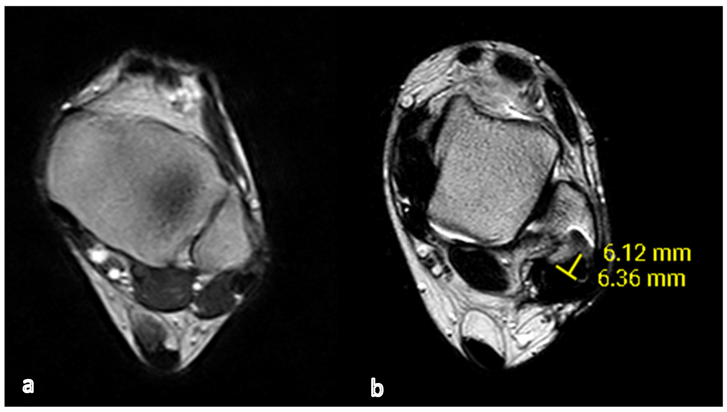
Measurements obtained from CT scans (Figure 2, Figure 3 and Figure 4) or MRI (Figure 5) scans revealed significantly deeper neo-fibular grooves. The mean depth of the neo-grooves was 4.50 mm (±2.02), and the mean width was 8.30 mm (±1.58).
The relationship between the depth of neo-grooves and clinical outcomes was found to be statistically insignificant. The AOFAS score increased from a mean preoperative value of 74.4 to 86.9 after surgery, while the EFAS score improved from a mean preoperative score of 19.7 to 31. Statistical analysis of the clinical scores showed a statistically significant difference with a p-value of <0.005. No instances of recurrence were observed.

4. Discussion

This study showed the effectiveness and validity of the retromalleolar groove deepening in recurrent peroneal tendon dislocation in patients with a flat distal fibula. All patients achieved excellent clinical outcomes, and no recurrence occurred. The mean time to return to sports is quite short, and in 7 months, all patients could perform physical activities again. Only one patient affected by collagen disease incurred several difficulties during rehabilitation, with instability and chronic pain.
The groove deepening combined with the repair of SPR is a valuable technique in the treatment of recurrent peroneal tendon dislocation.
Undiagnosed peroneal tendon disorders can potentially lead to long-lasting complications, such as recurrent dislocations, peroneal tendon damage or degeneration, chronic lateral ankle discomfort, and tendon inflammation. Therefore, it is crucial to identify the pathology and assess the necessity for appropriate treatment.
Surgical intervention is necessary for chronic peroneal tendon dislocation, whether in athletes or non-athletes. According to the ESSKA-AFAS international consensus statement on peroneal tendon pathologies, surgical stabilization is recommended as the primary treatment, with a particular focus on deepening the retromalleolar groove in athletes [28].
Numerous operative techniques have been described. Currently, there have been no prospective randomized studies conducted to determine the superiority of one approach over another [19,29].
All these procedures focus on stabilizing the peroneal tendons within the retrofibular groove, whether achieved through the correction of anatomical malformations or the reinforcement of the SPR.
A systematic review from 2016 indicated that patients who underwent both superior peroneal retinaculum (SPR) repair and deepening of the retromalleolar groove had a higher likelihood of returning to sports compared to those who only underwent SPR repair [30].
But, only a few years later, in 2020, Seung-Hwan Park et al. proposed that in cases of traumatic peroneal tendon dislocation, reattaching the peroneal retinaculum without deepening the groove may be adequate. This is because the primary concern in traumatic cases is the detachment of the superior retinaculum, which serves as the primary restraint for the peroneal tendon [31]. Surely, this technique is less invasive in comparison with others [32]; however, in cases of patients with a congenital convex fibular groove, this straightforward repair is unlikely to provide stability to the subluxated tendon. Raikin et al. indicated the effectiveness of this technique in their series of 14 surgically treated patients with peroneal dislocation. During the surgical procedure, thirteen patients were found to have a convex peroneal groove, and all fourteen had an intact peroneal retinaculum [33].
Research has demonstrated that the morphology of the retromalleolar groove is a contributing factor in peroneal tendon pathology, with flat and convex grooves showing the strongest association [34].
The surgical technique used in this study preserves the natural fibrocartilage within the malleolar groove, facilitating smoother peroneal tendon gliding and lowering the risk of tendon shearing [35]. However, some authors have expressed concerns about this technique, citing worries about the increased force needed to compress the posterior cortex of the fibula, which could potentially harm the fibrocartilage or even result in a fibula fracture [36]. This complication was not observed in our study.
According to the authors, the deepening groove technique should be considered when intraoperative confirmation reveals groove dysplasia.
A recent meta-analysis [37] compared four surgical techniques in patients with chronic peroneal instability: SPR repair or replacement, groove-deepening procedures (often combined with additional SPR operations), rerouting procedures, and bony procedures. Surgical treatment yields excellent clinical and functional outcomes in patients with chronic peroneal instability, while less favorable results were observed with rerouting and bony procedures.
A ‘sliding bone-block procedure’ can be employed, as described by Kelly (1920) and DuVries (1965). In this technique, a section of bone is excised from the lateral side of the fibula and repositioned more posteriorly to deepen the malleolar groove (MG). Potential issues are associated with bone block procedures: excessive post-operative wear and tear on the peroneal tendons can occur, especially if the procedure is carried out too distally, where compression and shear forces are more significant [38].
The surgical technique of rerouting the peroneal tendons behind the calcaneofibular ligament does not directly address the restoration of the superior peroneal retinaculum (SPR) anatomy. Instead, it relies on the calcaneofibular ligament as a natural alternative restraint. It involves dividing the calcaneofibular ligament, repositioning the tendons behind it, and then suturing the calcaneofibular ligament back together. Martens et al. employed this technique and achieved excellent results in 11 patients during a 30-month follow-up with no recurrence, but 2 patients experienced sural nerve injuries [39].
Several authors have also outlined procedures for strengthening or enhancing weakened superior peroneal retinaculum (SPR) through soft tissue transfer, such as for the Achilles tendon, peroneus brevis tendon, plantaris tendon, and peroneus quartus tendon [40].
In summary, our study investigated the medium to long-term clinical outcomes of the surgical deepening of the retro-malleolar peroneal groove for recurrent peroneal tendon dislocation. Employing EFAS and AOFAS scores, most patients achieved excellent results, indicating significant functional improvement and enhanced quality of life post-surgery. Notably, no cases of recurrence were observed, underscoring the high efficacy of the technique in preventing symptom relapse. Furthermore, a noteworthy observation was the return to sports activities within approximately 7 months post-intervention, highlighting both the success of the procedure and its capacity for rapid physical activity resumption. However, it is important to acknowledge the limitation of our study, which lies in its small sample size. The results, therefore, should be interpreted with caution, and larger-scale investigations are warranted for more comprehensive validation.
In conclusion, our results demonstrate that surgical deepening of the retro-malleolar peroneal groove is a highly effective and safe option for patients with recurrent peroneal tendon dislocation. This procedure consistently yields excellent outcomes, preventing recurrence and enabling swift resumption of daily and sporting activities.

Author Contributions

B.M. and E.M.S. contributed to the study conceptualization and design. Material preparation and data collection and analysis were performed by L.A.; statistics and software were performed by T.M.; the first draft of the manuscript was written by L.A.; writing—review and editing: E.M.S., M.L. and A.M. All authors have read and agreed to the published version of the manuscript.

Funding

This research received no external funding.

Institutional Review Board Statement

All subjects gave their informed consent for inclusion before they participated in the study. The study was conducted in accordance with the Declaration of Helsinki.

Informed Consent Statement

Informed consent was obtained from all subjects involved in the study.

Data Availability Statement

The datasets used and analyzed during the current study are available from the corresponding author (Ludovica Auregli MD) on reasonable request. In order to ensure the confidentiality of data and to avoid data loss or manipulation, precautionary measures have been taken: the data are restricted to authorized members only. Authorized members are Magnan MD Prof, Samaila MD Prof, Auregli MD, Maluta MD, Leigheb MD Prof, and Mazzotti MD. All information about the patients enrolled in the study is stored on a secure server, and database access is protected through a password that periodically changes. Only authorized members have the password. Paper material about the clinical evaluation is kept in a cabinet in the office of Professor Samaila.

Conflicts of Interest

The authors declare no conflict of interest.

References

  1. Dombek, M.F.; Lamm, B.M.; Saltrick, K.; Mendicino, R.W.; Catanzariti, A.R. Peroneal tendon tears: A retrospective review. J. Foot Ankle Surg. 2003, 42, 250–258. [Google Scholar] [CrossRef] [PubMed]
  2. Espinosa, N.; Maurer, M.A. Peroneal tendon dislocation. Eur. J. Trauma. Emerg. Surg. 2015, 41, 631–637. [Google Scholar] [CrossRef]
  3. Kwaadu, K.Y.; Fleming, J.J.; Florek, D. Superior peroneal retinacular injuries in calcaneal fractures. J. Foot Ankle Surg. 2015, 54, 458–463. [Google Scholar] [CrossRef] [PubMed]
  4. Mabit, C.; Salanne, P.; Boncoeur-Martel, M.P.; Fiorenza, F.; Valleix, D.; Descottes, B.; Caix, M. La gouttière rétromalléolaire latérale: Étude radio-anatomique [The lateral retromalleolar groove: A radio-anatomic study]. Bull. Assoc. Anat. 1996, 80, 17–21. [Google Scholar]
  5. Wang, X.T.; Rosenberg, Z.S.; Mechlin, M.B.; Schweitzer, M.E. Normal variants and diseases of the peroneal tendons and superior peroneal retinaculum: MR imaging features. Radiographics 2005, 25, 587–602. [Google Scholar] [CrossRef]
  6. Klammer, G.; Benninger, E.; Espinosa, N. The varus ankle and instability. Foot Ankle Clin. 2012, 17, 57–82. [Google Scholar] [CrossRef]
  7. Edwards, M. The relations of the peroneal tendons to the fibula, calcaneus, and cuboideum. Am. J. Anat. 1927, 42, 213–252. [Google Scholar] [CrossRef]
  8. Athavale, S.A.; Swathi Vangara, S.V. Anatomy of the superior peroneal tunnel. J. Bone Joint Surg. 2011, 93, 564–571. [Google Scholar] [CrossRef]
  9. Lugo-Pico, J.G.; Kaiser, J.T.; Sanchez, R.A.; Aiyer, A.A. Peroneal Tendinosis and Subluxation. Clin. Sports Med. 2020, 39, 845–858. [Google Scholar] [CrossRef]
  10. Oden, R.R. Tendon injuries about the ankle resulting from skiing. Clin. Orthop. Relat. Res. 1987, 216, 63–69. [Google Scholar] [CrossRef]
  11. Philbin, T.M.; Landis, G.S.; Smith, B. Peroneal tendon injuries. J. Am. Acad. Orthop. Surg. 2009, 17, 306–317. [Google Scholar] [CrossRef] [PubMed]
  12. Adamson, C.; Cymet, T. Ankle sprains: Evaluation, treatment, rehabilitation. Md. Med. J. 1997, 46, 530–537. [Google Scholar] [PubMed]
  13. Hutchinson, B.L.; Gustafson, L.S. Chronic peroneal tendon subluxation. New surgical technique and retrospective analysis. J. Am. Podiatr. Med. Assoc. 1994, 84, 511–517. [Google Scholar] [PubMed]
  14. Neustadter, J.; Raikin, S.M.; Nazarian, L.N. Dynamic sonographic evaluation of peroneal tendon subluxation. AJR Am. J. Roentgenol. 2004, 183, 985–988. [Google Scholar] [CrossRef]
  15. Rosenberg, Z.S.; Bencardino, J.; Astion, D.; Schweitzer, M.E.; Rokito, A.; Sheskier, S. MRI features of chronic injuries of the superior peroneal retinaculum. AJR Am. J. Roentgenol. 2003, 181, 1551–1557. [Google Scholar] [CrossRef]
  16. Saupe, N.; Mengiardi, B.; Pfirrmann, C.W.; Vienne, P.; Seifert, B.; Zanetti, M. Anatomic variants associated with peroneal tendon disorders: MR imaging findings in volunteers with asymptomatic ankles. Radiology 2007, 242, 509–517. [Google Scholar] [CrossRef]
  17. Stover, C.N.; Bryan, D.R. Traumatic dislocation of the peroneal tendons. Am. J. Surg. 1962, 103, 180–186. [Google Scholar] [CrossRef]
  18. Escalas, F.; Figueras, J.M.; Merino, J.A. Dislocation of the peroneal tendons. Long-term results of surgical treatment. J. Bone Joint Surg. Am. 1980, 62, 451–453. [Google Scholar] [CrossRef]
  19. Ferran, N.A.; Oliva, F.; Maffulli, N. Recurrent subluxation of the peroneal tendons. Sports Med. 2006, 36, 839–846. [Google Scholar] [CrossRef]
  20. Selmani, E.; Gjata, V.; Gjika, E. Current concepts review: Peroneal tendon disorders. Foot Ankle Int. 2006, 27, 221–228. [Google Scholar] [CrossRef]
  21. Davda, K.; Malhotra, K.; O’Donnell, P.; Singh, D.; Cullen, N. Peroneal tendon disorders. EFORT Open Rev. 2017, 2, 281–292. [Google Scholar] [CrossRef] [PubMed]
  22. Coughlin, M.J.; Schon, L.C. Disorders of tendons. In Mann’s Surgery of the Foot and Ankle, 9th ed.; Coughlin, M.J., Mann, R.A., Saltzman, C.L., Eds.; Mosby Elsevier: Philadelphia, PA, USA, 2013. [Google Scholar]
  23. Shawen, S.B.; Anderson, R.B. Indirect Groove Deepening in the Management of Chronic Peroneal Tendon Dislocation. Tech. Foot Ankle Surg. 2004, 3, 118–125. [Google Scholar] [CrossRef]
  24. Safran, M.R.; O’Malley, D., Jr.; Fu, F.H. Peroneal tendon subluxation in athletes: New exam technique, case reports, and review. Med. Sci. Sports Exerc. 1999, 31 (Suppl. S7), S487–S492. [Google Scholar] [CrossRef] [PubMed]
  25. Matcuk, G.R. Fibular groove morphology and measurements on MRI: Correlation with fibularis tendon abnormalities. Surg. Radiol. Anat. 2019, 41, 75–85. [Google Scholar] [CrossRef]
  26. Leigheb, M.; Janicka, P.; Andorno, S.; Marcuzzi, A.; Magnani, C.; Grassi, F. Italian translation, cultural adaptation and validation of the “American Orthopaedic Foot and Ankle Society’s (AOFAS) ankle-hindfoot scale”. Acta Biomed. 2016, 87, 38–45. [Google Scholar]
  27. Richter, M.; Agren, P.H.; Besse, J.L.; Cöster, M.; Kofoed, H.; Maffulli, N.; Rosenbaum, D.; Steultjens, M.; Alvarez, F.; Boszczyk, A.; et al. EFAS Score—Multilingual development and validation of a patient-reported outcome measure (PROM) by the score committee of the European Foot and Ankle Society (EFAS). Foot Ankle Surg. 2018, 24, 185–204. [Google Scholar] [CrossRef]
  28. Van Dijk, P.A.; Miller, D.; Calder, J.; DiGiovanni, C.W.; Kennedy, J.G.; Kerkhoffs, G.M.; Kynsburtg, A.; Havercamp, D.; Guillo, S.; Oliva, X.M.; et al. The ESSKA-AFAS international consensus statement on peroneal tendon pathologies. Knee Surg. Sports Traumatol. Arthrosc. 2018, 26, 3096–3107. [Google Scholar] [CrossRef]
  29. Heckman, D.S.; Reddy, S.; Pedowitz, D.; Wapner, K.L.; Parekh, S.G. Operative treatment for peroneal tendon disorders. J. Bone Joint Surg. Am. 2008, 90, 404–418. [Google Scholar] [CrossRef]
  30. Van Dijk, P.A.; Gianakos, A.L.; Kerkhoffs, G.M.; Kennedy, J.G. Return to sports and clinical outcomes in patients treated for peroneal tendon dislocation: A systematic review. Knee Surg. Sports Traumatol. Arthrosc. 2016, 24, 1155–1164. [Google Scholar] [CrossRef]
  31. Park, S.H.; Choi, Y.R.; Lee, J.; Seo, J.; Lee, H.S. Treatment of Recurrent Peroneal Tendon Dislocation by Peroneal Retinaculum Reattachment Without Fibular Groove Deepening. J. Foot Ankle Surg. 2021, 60, 994–997. [Google Scholar] [CrossRef]
  32. Adachi, N.; Fukuhara, K.; Tanaka, H.; Nakasa, T.; Ochi, M. Superior retinaculoplasty for recurrent dislocation of peroneal tendons. Foot Ankle Int. 2006, 27, 1074–1078. [Google Scholar] [CrossRef]
  33. Raikin, S.M.; Elias, I.; Nazarian, L.N. Intrasheath subluxation of the peroneal tendons. J. Bone Joint Surg. Am. 2008, 90, 992–999. [Google Scholar] [CrossRef] [PubMed]
  34. Mabit, C.; Salanne, P.H.; Blanchard, F. The retromalleolar groove of the fibula: A radio-anatomical study. Foot Ankle Surg. 1999, 5, 179–186. [Google Scholar] [CrossRef]
  35. Hwang, I.M.; Del Sol, S.R.; Jenkins, S.M.; Bryant, S.A.; Gardner, B.B.; McGahan, P.; Chen, J. Open Peroneal Tendon Stabilization With Fibular Groove Deepening. Arthrosc. Tech. 2022, 11, e347–e352. [Google Scholar] [CrossRef]
  36. Van Dijk, P.A.D.; Vopat, B.G.; Guss, D.; Younger, A.; DiGiovanni, C.W. Retromalleolar Groove Deepening in Recurrent Peroneal Tendon Dislocation: Technique Tip. Orthop. J. Sports Med. 2017, 5, 2325967117706673. [Google Scholar] [CrossRef] [PubMed]
  37. Lootsma, J.; Wuite, S.; Hoekstra, H.; Matricali, G.A. Surgical treatment options for chronic instability of the peroneal tendons: A systematic review and proportional meta-analysis. Arch. Orthop. Trauma Surg. 2023, 143, 1903–1913. [Google Scholar] [CrossRef] [PubMed]
  38. Kumai, T.; Benjamin, M. The histological structure of the malleolar groove of the fibula in man: Its direct bearing on the displacement of peroneal tendons and their surgical repair. J. Anat. 2003, 203, 257–262. [Google Scholar] [CrossRef] [PubMed]
  39. Martens, M.A.; Noyez, J.F.; Mulier, J.C. Recurrent dislocation of the peroneal tendons. Results of rerouting the tendons under the calcaneofibular ligament. Am. J. Sport. Med. 1986, 14, 148–150. [Google Scholar] [CrossRef]
  40. Thomas, J.L.; Sheridan, L.; Graviet, S. A modification of the Ellis Jones procedure for chronic peroneal subluxation. J. Foot Surg. 1992, 31, 454–458. [Google Scholar]
Figure 1. Intra-operative images of a 38-year-old male with peroneal tendon dislocation and SPR rupture: (a) retrograde drilling of the fibula to ream out the fibular core; (b) the posterior cortex rim is impacted to deepen the retromalleolar groove; and (c) repositioning the peroneal tendon within the groove.
Applsci 13 11468 g001
Figure 2. CT scan of a 28-year-old male. (a) Preoperative view that demonstrates a flat groove; (b) post-operative view with the DRG and a concave retromalleolar groove.
Applsci 13 11468 g002
Figure 3. CT scan of a 44-year-old male. (a) Flat malleolus in the preoperative view; (b) CT scan after deepening of the retromalleolar groove.
Applsci 13 11468 g003
Figure 4. CT scan of a 47-year-old male. (a) Preoperative view. (b) Post-operative CT scan that shows the deeper neo-fibular groove.
Applsci 13 11468 g004
Figure 5. MRI of a 39-year-old male. (a) Preoperative MRI; (b) post-operative view with measurement of the neo-fibular groove.
Applsci 13 11468 g005
Table 1. Characteristics of the study population.
Patients’ DataMean (Range)
Sex
Male6
Female3
Age, years31.2 (14–51)
Side of injury
Left8
Right2
Activity during injury
Soccer3
Dance1
Climbing ski2
Other (domestic accident)3
Time from injury to surgery
(months)
11.7 ± 8.6 (7–16.5)
Table 2. Clinical score results.
ScoreMean ± SD (N = 10)p-Value
Follow-up (years) 4.2 years
(min 6 months–max 6 years)
AOFAS score <0.005
pre-op74.4 (sd 17.17)
post-op86.9 (sd 21.23)
EFAS score <0.005
pre-op19.7 (sd 7.50)
post-op31 (sd 14.82)
Table 3. Preoperative and post-operative evaluation of AOFAS and EFAS scores.
Case N.AOFAS
Pre-op
AOFAS
Post-op
EFAS
Pre-op
EFAS
Post-op
182 pt87 pt16 pt31 pt
248 pt38 pt17 pt3 pt
374 pt96 pt25 pt38 pt
485 pt100 pt27 pt40 pt
592 pt100 pt26 pt40 pt
675 pt100 pt20 pt40 pt
781 pt100 pt12 pt40 pt
848 pt88 pt12 pt34 pt
961 pt60 pt10 pt4 pt
1098 pt100 pt32 pt40 pt
Mean74.4 pt86.9 pt19.7 pt31 pt
StDev17.1721.237.5014.82
Disclaimer/Publisher’s Note: The statements, opinions and data contained in all publications are solely those of the individual author(s) and contributor(s) and not of MDPI and/or the editor(s). MDPI and/or the editor(s) disclaim responsibility for any injury to people or property resulting from any ideas, methods, instructions or products referred to in the content.