Article

Optimization of the Antifungal Property in a Composite of Polyurethane and Silver Nanoparticles against the Trichophyton rubrum Fungus

by
Armando Mares Castro
1,*,
Anayansi Estrada Monje
2,
Alejandra Imelda Saldívar Campos
3 and
Anayansi Zaragoza Estrada
4
1
Industrial Engineering División, Tecnológico Nacional de México/ITS de Purísima del Rincón, Blvd. Del Valle 2301, Guardarrayas, Purísima del Rincón 36413, Mexico
2
Industrial Processes and Energy Division, Centro de Innovación Aplicada en Tecnologías Competitivas, Omega 201 Col. Industrial Delta, León 37545, Mexico
3
Engineering Area, Universidad Iberoamericana León, Blvd. Jorge Vértiz Campero 1640 Col. Cañada de Alfaro, León 37238, Mexico
4
Engineeering and Materials Chemistry Division, Centro de Investigación en Materiales Avanzados, Av. Miguel de Cervantes 120, Complejo Industrial Chihuahua, Chihuahua 31136, Mexico
*
Author to whom correspondence should be addressed.
Appl. Sci. 2023, 13(21), 12028; https://doi.org/10.3390/app132112028
Submission received: 8 September 2023 / Revised: 16 October 2023 / Accepted: 24 October 2023 / Published: 4 November 2023
(This article belongs to the Section Materials Science and Engineering)

Abstract

:
This research aims to analyze and optimize the antifungal property of a composite material made of polyurethane (PUR) and silver nanoparticles (AgNPs) against the fungus Trichophyton rubrum to find the optimal parameters that allow the highest inhibition of the fungal growth to be used in healthcare aimed at the population vulnerable to fungal infections, such as people suffering from diabetes mellitus, thus creating an opportunity for the generation of antifungal products for healthcare applications. This study analyzes the effect of three variables on the antifungal properties of the composite material: nanoparticle size, concentration, and the application of an ultrasonic treatment as a method to aid the dispersion of the AgNPs into PUR matrix. The proposed methodology includes tests in accordance with a standar to evaluate the growth inhibition of the fungi on a culture medium. A categorical logistic regression model was adjusted from 23 factorial design with five replicates, which was optimized by the use of multi-objective genetic algorithms. The experimental factors showed a significant effect on the growth inhibition of the fungus, and the optimal levels were determined.

Graphical Abstract

1. Introduction

Nanotechnology is an emerging area of scientific research with a wide range of applications in microbiology and biotechnology [1]. Silver nanoparticles (AgNPs) are aggregates of a relatively small number of atoms, ranging from small sizes consisting of only a few, to larger nanoparticles containing more than 100,000 atoms [2]. Clusters are a type of nanoparticle on the scale range of nanometers containing a small number of atoms [3]; their properties differ from those of both molecules and atoms and are very dependent on size [4]. This characteristic is one of the objectives of the present study; thus, two sizes of AgNPs are examined.
AgNPs display unique physical and chemical properties, such as adsorption capacity, fast diffusion rate, and changing surface characteristics [5]. Their application in nanobiotechnology, biosensors, cell structure imaging, and targeted drug delivery are well known [1]. Biological resources, including bacteria, plants, algae, fungi, and biomolecules, have been proved to be excellent candidates for AgNPs synthesis [6]. To determine numerous properties, namely size, geometry, shape, crystallinity, and surface area, different techniques, including transmission electron microscopy (TEM), scanning electron microscopy (SEM), and UV-vis spectroscopy, can be applied [4].
A property of interest of AgNPs reported in the literature is the biocide capacity against different types of microorganisms, which will be analyzed thoroughly in the state-of-the-art section. In this research, it is of particular interest the analysis of the growth inhibition capacity of a composite PUR and AgNPs material on the fungus Trichophyton rubrum, propriety that could present potential applications on products able to inhibit the proliferation of Trichophyton fungi, responsible for diseases including tinea pedis and tinea cruris [7].
PUR is considered one of the most versatile polymeric materials due to the wide range of materials with different properties that can be obtained from it, such as rigid and flexible foams, coatings, and thermoplastic materials [8]. These materials are chemically complex and usually obtained via chemical reactions between diisocyanates and polyols, which can be polyester or polyether-type [9]. Derived from the versatility of the properties that can be obtained, there have been developed, for example, fire-resistant PUR nanocomposites [10]. In recent years, due to their excellent chemical stability, biocompatibility, and low cytotoxicity, some PURs have been widely used for biomedical applications [11]. However, the cytotoxicity of its raw materials can limit their use in certain applications, for example, the toxicity of 4,4-methylene diphenyl diisocyanate (MDI) in the formulation [12]; few studies have evaluated the effect of the PUR chemical structure on the cytotoxicity. Dominik Grzeda et al., indicate that the isocyanate index highly influences the chemical structure of polyurethane foams, also causing changes in cytotoxicity [13]. This parameter must be carefully considered to ensure biocompatibility [13,14]. PUR nanocomposites have also been used to remove inks and dyes [15]. The current medical PUR research has been focused on a new approach: metals, such as silver, and copper oxide nanoparticles as a new type of biocide agent [16,17]. Thus, the proposed methodology for this research focuses on the use of polyester PUR and silver nanoparticles of different sizes to obtain a composite material with antifungal properties.
This paper is based on the analysis of three control variables of interest: the size and concentration of AgNPs, and the use of an ultrasound method during the mixing of AgNPs and PUR to evaluate the effect of these factors on the growth inhibition of the fungus Trichophyton rubrum on culture medium in the presence of the composite material. Through a 23-factorial design with five replicates, 40 experimental runs were evaluated in accordance with ASTM-G21-15-4 [18]. The fungus Trichophyton rubrum presents a filamentous shape and subtle color, making its quantification via image processing impossible. Hence, it was evaluated qualitatively using observation categories by the standard and subsequently adjusted to an ordinal logistic model. The optimization of the model was made using genetic algorithms due to the presence of logarithmic elements. This technique calculated the optimal parameters of the PUR and AgNPs composite material to obtain the highest growth inhibition of the fungus Trichophyton rubrum.
The relevance of this study lies in the possible applications of a PUR and AgNPs composite material to obtain devices (such as insoles) that can be used by people who are continuously exposed to fungi from the Trichophyton family, which are highly contagious, can cause allergic reactions and eczema, require long treatments, and are especially dangerous for people with sensitive skin. The research on AgNPs is relatively new, and this study contributes to the application of the nanoparticles in polymeric materials, highlighting the potential benefits for humankind.

1.1. Properties of AgNPs against Microorganisms and Fungi

Linima VK et al. [19] report highly promising antimicrobial, antiviral, and anticancer properties from using AgNPs, including antibacterial and antifungal activity against Salmonella typhi, Staphylococcus aureus, and the fungus Aspergillus flavus. According to laboratory testing, the bactericidal activity against Staphylococcus aureus and Escherichia coli presented by the AgNPs could be related to a high concentration of the nanoparticles; however, very high concentrations could cause toxicity [20,21]. Other microorganisms, such as Pseudomonas stutzeri, have shown resistance and accumulation against AgNPs [22].
AgNPs have been demonstrated to be an effective antimicrobial agent against multiple pathogenic microorganisms, such as the fungus Aspergillus flavus, with positive outcomes being reported for biological synthesis in the laboratory [23]. AgNPs obtained via biosynthesis using the fungus Aspergillus melleus have shown antibacterial and cytotoxic activity against S. aureus and E. coli, which creates a solid background for antifungal pharmaceutical and cosmetic applications based on biosynthesized AgNPs [24]. An example of a cosmetic antifungal application is fluconazole combined with AgNPs, which is effective against fungi such as Candida albicans, Phoma glomerata, and Fusarium semitectum [25].
AgNPs have shown properties that inhibit the growth of different types of fungi, including Aspergillus fumigates, A. niger, A. flavus, Trichophyton rubrum, Candida albicans, and Penicillium spp. According to the current hypothesis, AgNPs alter the permeability of the cell membrane, therefore causing cell death by producing reactive oxygen species (ROS) and free radicals, which cause denaturalization and damage to the nucleic acids, proton pump, and cell wall, as well as peroxidation of lipids [26].
Reported studies using AgNPs against fungi from the Trichophyton family have shown efficient antifungal and antimycotic capacities. Robles–Martínez et al. presented an article about an Allium sativum extract and AgNPs with a reported growth inhibition of 94% against T. rubrum [27]. The AgNPs presented a concentration-dependent antifungal activity against fungal infections caused by T. rubrum [28]. Assis Da Silva et al. [29] reported antifungal activity against the fungus T. mentagrophytes in concentrations from 20 to 100 µgL−1. Other studies combining photodynamic therapy with AgNPs [30] and AgNPs-decorated zinc oxide [31] were also demonstrated to be highly effective at inhibiting the growth of fungi from the Trichophyton family.

1.2. The Shape of Nanoparticles and Their Applications on Polyurethanes

The morphology of AgNPs has been reported as a relevant factor for evaluating antimicrobial activity, and the most common shapes include plates, rods, and nanoparticles [32]. The effects of these morphologies have been tested on S. aureus and E. coli [33]. Pat et al. [34] studied the antibacterial activity of spherical, rod-shaped, and truncated triangle AgNPs against E. coli; their results showed that nanotriangles had the highest biocide activity, followed by nanospheres and rod-shaped nanoparticles. As noted, the evaluation of morphology is highly important for nanometric systems. The cellular toxicity is also related to the nanoparticle size, which is crucial to consider while designing AgNPs for biomedical applications [35].
In materials research, the application of AgNPs in polymers and subsequent visualization via SEM techniques has allowed us to corroborate the efficiency of their antibacterial and antifungal activity. Studies on thermoplastic polyurethane (TPU) with incorporated AgNPs have successfully evaluated the antiviral activity of the composite material against the spring viraemia of carp (SVC) virus and SARS-CoV-2 [36]; similarly, powdered coatings made of PUR with AgNPs as a biocide agent have been developed, demonstrating efficiency against the SARS-CoV-2 virus [37].
The concept of active PUR with AgNPs and copper nanoparticles (CuNPs) has been reported in the literature. The preparation of the composite material is described as the mixture of liquid polyether (the monomer for polyurethane synthesis) with the silver and copper components, followed by the PUR synthesis; this allows us to consider these materials as medical supplies with biocidal activity, given that the samples did not exhibit cytotoxicity against the tested cells in tissue culture (rat hypodermis) and biodegradation products were not observed in the culture medium [38]. The cationic surfactants of PUR have been evaluated, specifically, their effect on the stability of AgNPs and surface and biological activity, showing biocide activity against bacteria and fungi [39].
Other applications of AgNPs have been developed for footwear manufacturing, mainly leather and synthetic elements, aiming to reduce the growth of skin disease-causing fungi [40,41]. Given the increasing number of products that incorporate AgNPs, it is crucial to analyze the potential risks of their use on humans; data of interest can be found in Winhoven SWP et al. [40]

1.3. References for Laboratory Testing

This study was made under the ASTM-G21-15-4 standard [18] to evaluate the antifungal activity of PUR/AgNPs composite materials using a mixture of the fungi Aspergillus brasiliensis, Penicillium funiculosum, Chaetomium globosum, Trichoderma virens, and Aureobadisium pullulans; subsequently, the mechanical, optical, and electrical properties were tested following the corresponding ASTM standards.
It is essential to point out that the tests were conducted in triplicate and classified categorically in three levels due to the growth of the fungus being slow, and its filamentous shape made it difficult to evaluate via image processing. Considering the qualitative nature of the results, an ordinal logistic regression model was adjusted, which is adequate for categoric-type results. The statistical analysis allowed us to evaluate the effects of each experimental variable of interest. The model was later optimized using genetic algorithms since it demonstrates higher precision when handling logarithmic elements and the curved functions.
The contributions of this research take place in a context where nanotechnology is starting to peak in biomedical applications. The potential applications for generating articles from the PUR/AgNPs composite materials benefiting the population susceptible to contracting fungal infections caused by T. rubrum present themselves as elements with a high added value considering their benefits and improved quality of life. An additional element is the proposal of a methodology to evaluate the antifungal properties of composite materials against fungi from the Trichophyton family, which is not currently available for general use.

2. Materials and Methods

2.1. Materials

Two different sizes of AgNPs dispersed in ethanol were evaluated, with an average diameter of 15 and 45 nm, and were obtained from the local provider NABICRON (Irapuato, Mexico). The supplier reports a concentration of 10,000 ppm of AgNPs, obtained via green synthesis, presenting a molecular weight of 107.87, a specific gravity of 0.9915 (25 °C), a boiling point of 100 °C, and a pH of 3–4.
The polyol-isocyanate system utilized is polyester type, soft, presenting a fine cell structure. The polyol was acquired from SIMON QUIMICA (León, Mexico) and is a blend of mainly polyester-type polyols, while the isocyanate is brand ACON 314 provided by SIMON QUIMICA is an aromatic diisocyanate prepolymer. The ratio of the mixture is 100 polyol/78 isocyanate at a temperature of 40+/−3 °C and a demolding time of 3–5 min. The system’s physical properties are the following: free density 100–150 g/L, packing density 280–300 g/L, and Shore A hardness 5–10. The provider of the polyol-isocyanate system dispersed the AgNPs/ethanol solution in the polyol per the following instructions:
  • 5 g of the 15 nm AgNPs/ethanol dispersion for a concentration of 12.66 mg of AgNPs per 1 kg of polyol. (Small AgNPs size, low concentration)
  • 5 g of the 45 nm AgNPs/ethanol dispersion for a concentration of 12.66 mg of AgNPs per 1 kg of polyol. (Large AgNPs size, low concentration)
  • 15 g of the 15 nm AgNPs/ethanol dispersion for a concentration of 38 mg of AgNPs per 1 kg of polyol. (Small AgNPs size, high concentration)
  • 15 g of the 45 nm AgNPs/ethanol dispersion for a concentration of 38 mg of AgNPs per 1 kg of polyol. (Large AgNPs size, high concentration)
Among the chemical and laboratory supplies used is lactophenol blue for fungal staining, zinc sulfate, sodium dioctyl sulfosuccinate, Sabourand dextrose agar, Petri dishes, inoculation loop, dissection forceps, digital shaker-mixer, Hielscher UP200Ht ultrasonic homogenizer with sonotrodes, laboratory incubators, and a microscope. The methodology was executed in specialized laboratories, following the parameters and procedures by the ASTM-G21-15-4 [18] standard. The research methodology is shown in Figure 1.

2.2. Definition of Experimental Design

The experimental array used for the research is a factorial design 23 with five replicates. This experimental design allows the study of the effect of three different factors with two levels each, and it consists of eight different treatments. Using this design, seven effects can be studied: the three main, double, and triple interaction. The objective of the study focuses on analyzing the effects of the following: the AgNPs size ( X 1 ), either low-small 15 nm- or high-large 45 nm-; the concentration of nanoparticles ( X 2 ), with a low concentration of 5 g of AgNPs/ethanol dispersion and a high-concentration of 10 g of AgNPs/ethanol dispersion; and applying the ultrasonic treatment on the polyol for the AgNPs dispersion ( X 3 ), with low −0 s- and high −10 s levels. The experimental matrix is shown in Table 1.
The values Y i j correspond to a categoric quantitative evaluation, which is related to the level of the fungal growth inhibition in the culture medium surrounding the PUR and AgNPs composite material. Each treatment and its replicates were carried out in triplicate, and the average of the three observed growths for each treatment was used. Due to the filamentous morphology of the studied fungus, it was impossible to quantify it using image processing tools; therefore, it was decided to use a categoric-type of response, evaluating through categories based on the growth observed using the microscope.

2.3. Sample Preparation

For each experimental sample, 44.69 g of previously heated to 40 °C polyol (containing the AgNPs/ethanol dispersion) and 34.86 g of isocyanate were weighed, 0.45 g of catalyst was added to complete the PUR formulation, considering that these elements should not be mixed until the PUR foaming is desired. In Figure 2, the process for the weighing of each constitutive element of PUR is illustrated.

2.4. Ultrasonic Treatment Application

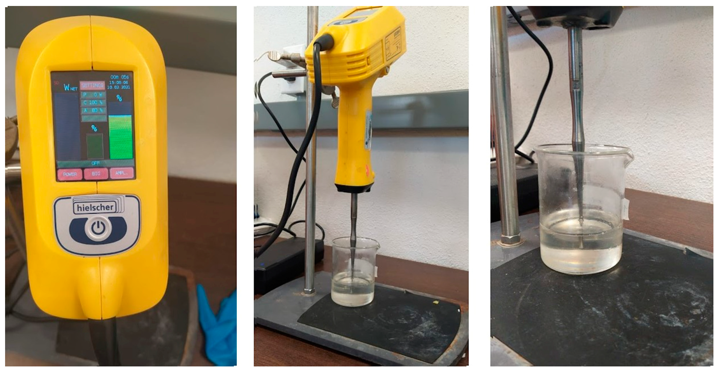
The third factor of interest in the treatment is applying an ultrasonic treatment and analyzing its effect on fungal growth inhibition. The AgNPs were dispersed using an ultrasonic processor (Model UP200Ht, Hielscher, Teltow, Germany). Its maximum power input and frequency are 400 W and 20 kHz, respectively. A 7 mm microprobe (S26d7, Sonotrode) was immersed in the dispersion of AgNPs/Ethanol; the sonication amplitude (tip movement) was 10–100%. The ultrasound was applied to the corresponding treatments for 10 s. The device was placed on a retort stand while applying the treatment on the polyol and AgNP mixture samples; the process is shown in Figure 3.

2.5. Obtention of PUR/AgNPs Composite Materials

The previously heated and weighted polyol and isocyanate are poured into a container, and the catalyst is immediately added. The mixture is placed on the paddles of the mechanical mixer for 10 s until the color changes to pale gray. Afterward, the sample is removed from the mixer and left to foam, leaving it undisturbed for at least one hour for a better result. The process is illustrated in Figure 4.

2.6. Characterization of the PUR/AgNPs Composite Materials

The AgNPs dispersion was characterized by using an emission scanning electron microscope, FE-SEM, JSM-7401 F, Jeol Ltd., Akishima, Japan.

2.7. Culture Medium Preparation

Using the streak plate inoculation method, a sample of a stock culture was inoculated. The mother culture was composed of pure T. rubrum fungi (consult American Type Culture Collection, ATCC, Manassas, VA, USA), and it was in optimal refrigerated storage conditions at −20 °C. This process was conducted in triplicate on potato dextrose agar (PDA) and Sabouraud dextrose agar. The cultures were incubated at 26 °C for 10 days. In triplicate, samples of 5 mm in diameter from the mother culture were inoculated in the center of the culture medium. The cultures were incubated at 26 °C with a relative humidity of at least 85% for 10 days.
Samples with a surface area of 50 × 50 mm were cut from the material following ASTM-G21-15-4 [29], with an approximate thickness of 3 mm, ensuring that the surfaces of the pieces were flat and even. The samples received a treatment using 96% ethanol for 30 min, followed by a 24-h drying period and a second treatment with UV light for 2 h.

2.8. Activation and Inoculation of the Fungi on the Culture Medium

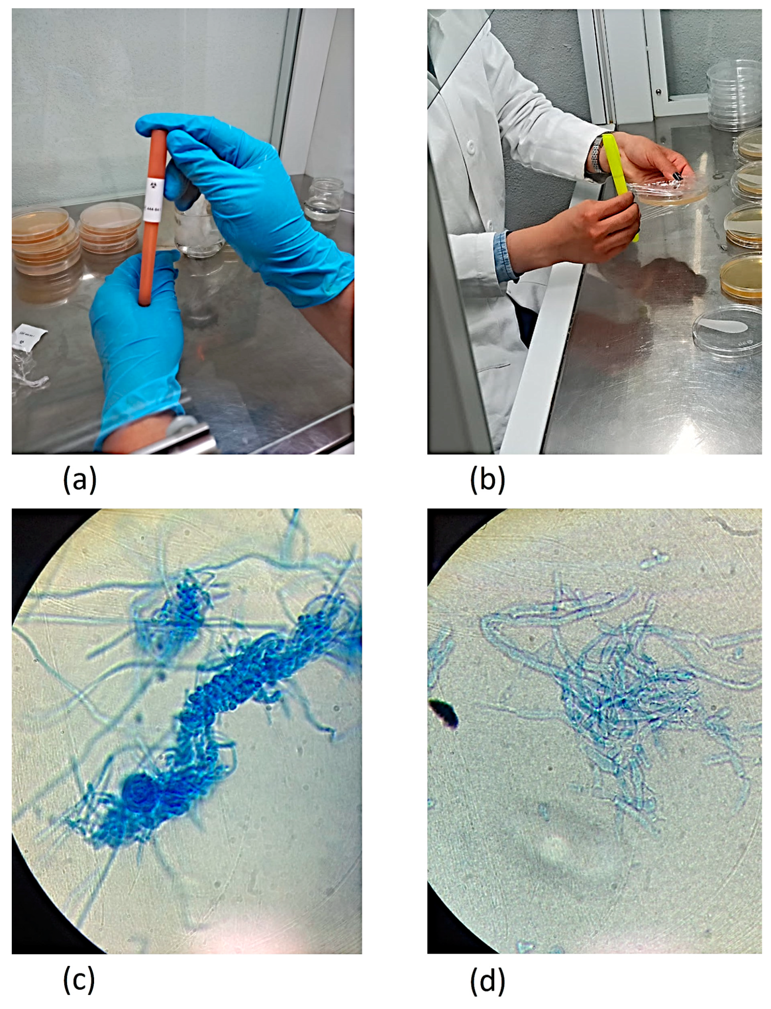
To visualize the fungal structure of T. rubrum at a microscopic level, a differential staining was carried out with lactophenol cotton blue using the imprinting technique, which consists of placing an impression of the surface of the fungus on a solid structure using adhesive tape. Then, 20 µL of the dye were added and the sample was observed under the microscope at 40×.
Each culture medium had 10 mL of sterilized distilled water added. The suspension was homogenized by scraping the surface of the culture medium where fungal colonies were visible, also liberating the spores. The samples were filtrated using gauze and placed in glass flasks. The suspended cells were visually identified for the inoculation essays. This process was undertaken through the Neubauer chamber, dyeing the spores by adding 20 µL of malachite green dye to 1 mL of the previously obtained cell suspension. A sample was placed in the chamber and observed at 40×.
Preparation for the mass tests: Following the disinfection treatments for the PUR pieces and the preparation of the spore suspension, essays were carried out using nutritive salts culture medium added with 50 mg/mL of kanamycin. The PUR pieces were placed in the center of the culture medium, ensuring they did not touch any other agar surface. The 50 µL aliquots of the spore suspension were inoculated, one in the center of the PUR piece, one on the right, and one on the left. The aliquots at the sides of the piece were directly on the culture medium, equidistant, and not in direct contact with the PUR piece nor with the walls of the Petri dish. Each sample of the PUR/AgNPs composite material was conducted in triplicate, with a control for the viability of the cells. The essays were incubated at 26 °C, with a relative humidity of at least 85% for 15 days at least, registering the fungal growth daily. The fungus preparation processes and its visualization with lactophenol cotton blue are illustrated in Figure 5.

2.9. Fungal Incubation

In accordance with the standard, the Petri dishes were placed inside an incubator at 28–30 °C and a relative humidity of at least 85%. The duration of the standard test is 28 days of incubation. The test can be terminated in less than 28 days for samples that exhibit a growth index of two or more.

2.10. Fungal Growth Evaluation

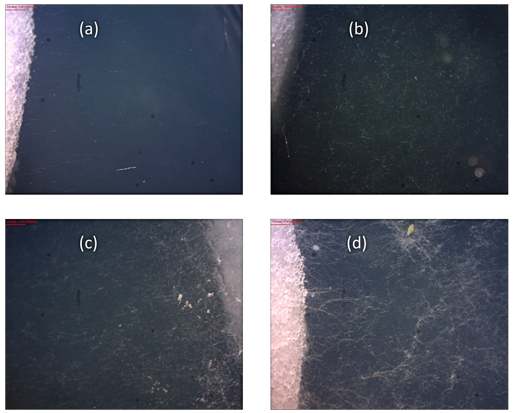
The images obtained under the microscope of the T. rubrum fungus show a filamentous shape that is difficult to quantify using image processing techniques. The ASTM-G21-15-4 [18] standard states the observation of visible effects after taking the samples out of the incubator. The suggested classification by the standard is indicated in Table 2.
Figure 6 illustrates examples of fungal growth according to the classification 1, 2, 3, and 4 shown in Table 2. Based on this classification, each treatment was assessed, and a number, according to Table 2, was assigned. The obtained classification number represents the average value of the triplicate samples. The experimental results are shown in Table 3.

2.11. Ordinal Logistic Regression Model

As is shown, the experimental design presents the case of a categoric-type variable, which is a particular case that can be approached using generalized linear model techniques [42]. The generalized linear models use a common algorithm for estimating the parameters using maximum likelihood, using weighted least squares with an adjusted dependent variable, and do not require preliminary guesses at the parameter values. When a categoric-type response is obtained, the best option among the generalized linear models are ordinal logistic regression models, which are appliable to both nominal and ordinal responses with more than two categories, in which a natural order can exist among the response categories [43,44]
The default link function for these models is logit, which accumulates the cumulative log odds. The cumulative log odds of the coefficient of a response belonging to a category with a value less than or equal to the category j ,   P y c j . The ordinal models are usually based on the assumption that the effects of the predictor variables are the same for every category on the logarithmic scale, i.e., the model has different intercepts but equal slopes. This model is called parallel regression on the proportional odds model. This is the default for ordinal responses, the proportional odds model is:
ln P y     c 1 P y   >   c 1 = ln π 1 π 2 + + π k = α 1 + β 1 X 1 + β 2 X 2 + + β p X p , ln P y     c 2 P y   >   c 2 = ln π 1 + π 2 π 3 + + π k = α 1 + β 1 X 1   + β 2 X 2 + + β p X p , ln P y     c k 1 P y   >   c k 1 = ln π 1 + π 2 + + π k 1 π k = α k 1 + β 1 X 1 + β 2 X 2 + + β p X p ,
where π j = P y = j is the probability that an output is in the j category, k is the number of categories, and p is the number of predictor variables. Using the nominal response model as a base and assuming that the coefficient for the last category is cero, the probability of being in each category is:
P y     k = π 1 + π 2 + + π k = exp α j + l = 1 p β jl x l   1 + exp α j + l = 1 p β jl x l   ,   j = 1 , , j 1
Derived from the density functions of the individual probabilities, the expression is maximized to generate the optimal values of β j l . The log-likelihood cannot be used by itself as an adjustment measure, considering it depends on the sample size; however, it can be used for comparing two models. For ordinal logistic regression, n independent multinominal vectors exist, each with j categories. These observations are denoted by y 1 , , y n , where y i = y i 1 , , y ij and Σ k y i j = m i is fixed for each i. From the ith observation the contribution of the log-likelihood is:
L π k ; y i = Σ k y ij log π k
The total log-likelihood is the sum of the contributions of each one n observations:
L π ; y = Σ k L π k , y i

2.12. Optimization by Genetic Algorithms

Genetic algorithms (GAs) are among the most used global search optimization techniques or metaheuristics. GAs are adaptative computational techniques widely used for optimization issues. Their main advantage over traditional numeric optimization techniques lies in a higher probability of finding the global optimum in the presence of highly non-linear, discontinuous, non-differentiable, or stochastic functions.
The GAs can be used to solve both constrained and unconstrained optimization problems through a technique that imitates the natural selection that happens in biological evolution. The algorithm changes repeatedly a population of individual solutions in an iterative process that involves selection, reproduction, mutation, crossover, and migration techniques. The fitness function is the function to be optimized, equivalent to the objective function in a traditional optimization scheme.
Global optimization can be used when having a set of objectives where a tradeoff that simultaneously satisfies all ends is required. GAs present a variable called multi-objective genetic algorithm (MOGAs), which allows one to solve multiple objective problems. Given that the ordinal logistic regression model generates a set of models to the probabilities on different categories, the aim would be to optimize (minimize) simultaneously the probabilities that there is growth in each category using the selection of the optimal levels on the experimental variables.
The objective function of the MOGAs optimization problem for the analyzed case is presented in Equation (5). The objective is to obtain the level combination that minimizes the probability of fungal growth; the optimization problem can be expressed as:
Min   F Y 1 X , F Y 2 X , F Y 3 X s.t.   1     X i     1 X i R
where F(Y1(X)), F(Y2(X)), and F(Y3(X)) are the fitness functions to be simultaneously minimized using MOGAs and correspond to cumulative odds models on each category as indicated in Equation (2); the parameters used for the optimization on MATLAB R2021b are shown on Table 4.

3. Results

3.1. Analysis of the Main Effects and Interactions

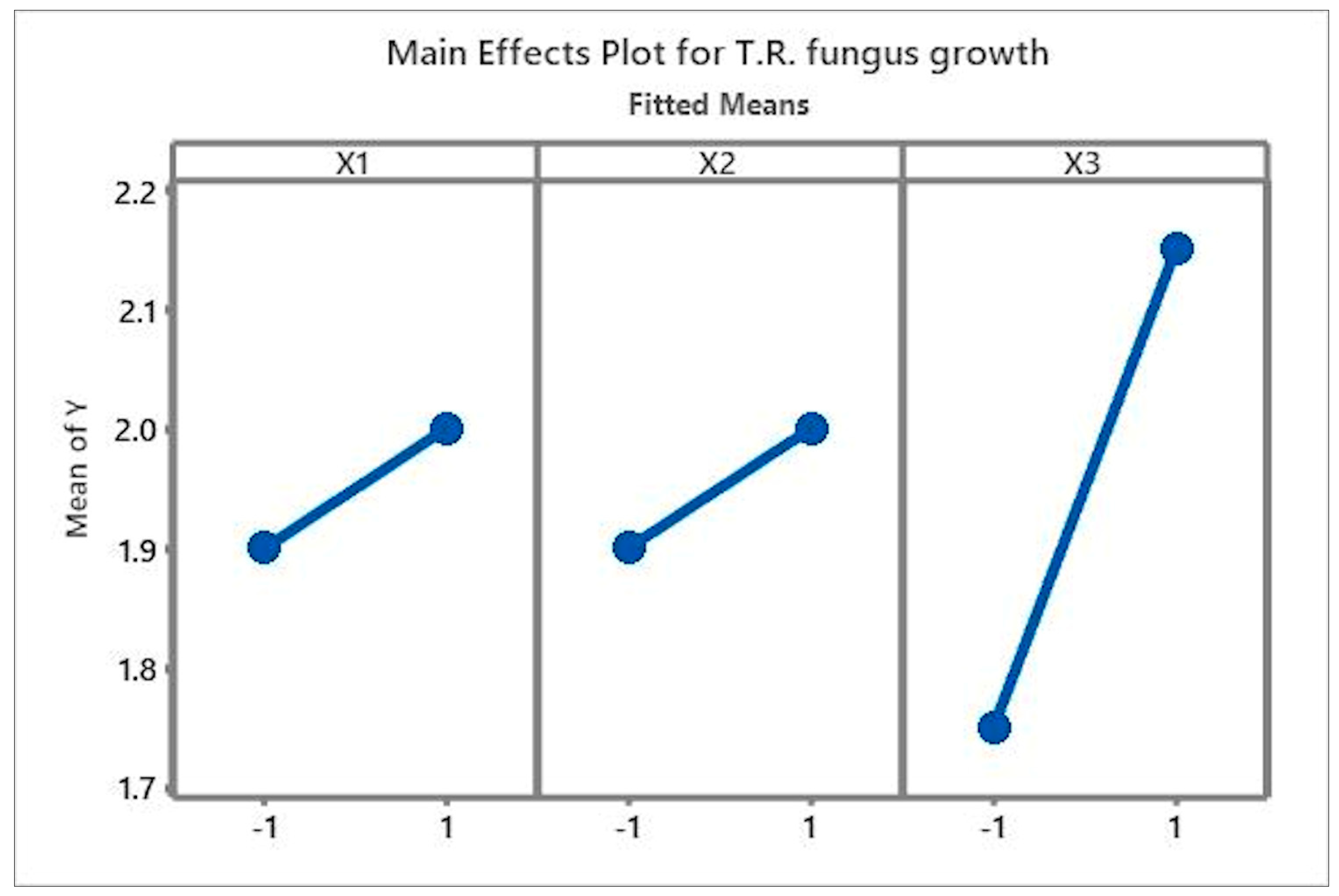
A preliminary analysis of the effects on the factorial design is shown in Figure 7. There is a low effect of the three factors, X1 Size of the AgNPs, X2 Concentration of AgNPs, and X3 Ultrasonic treatment on the samples, on the fungal growth based on a qualitative evaluation. The factors present a similar effect on the fungal growth, obtaining the highest inhibition on their low values. This analysis indicates a more noticeable change in the variable, indicating that the ultrasonic treatment shows a significant effect over the fungal growth inhibition.
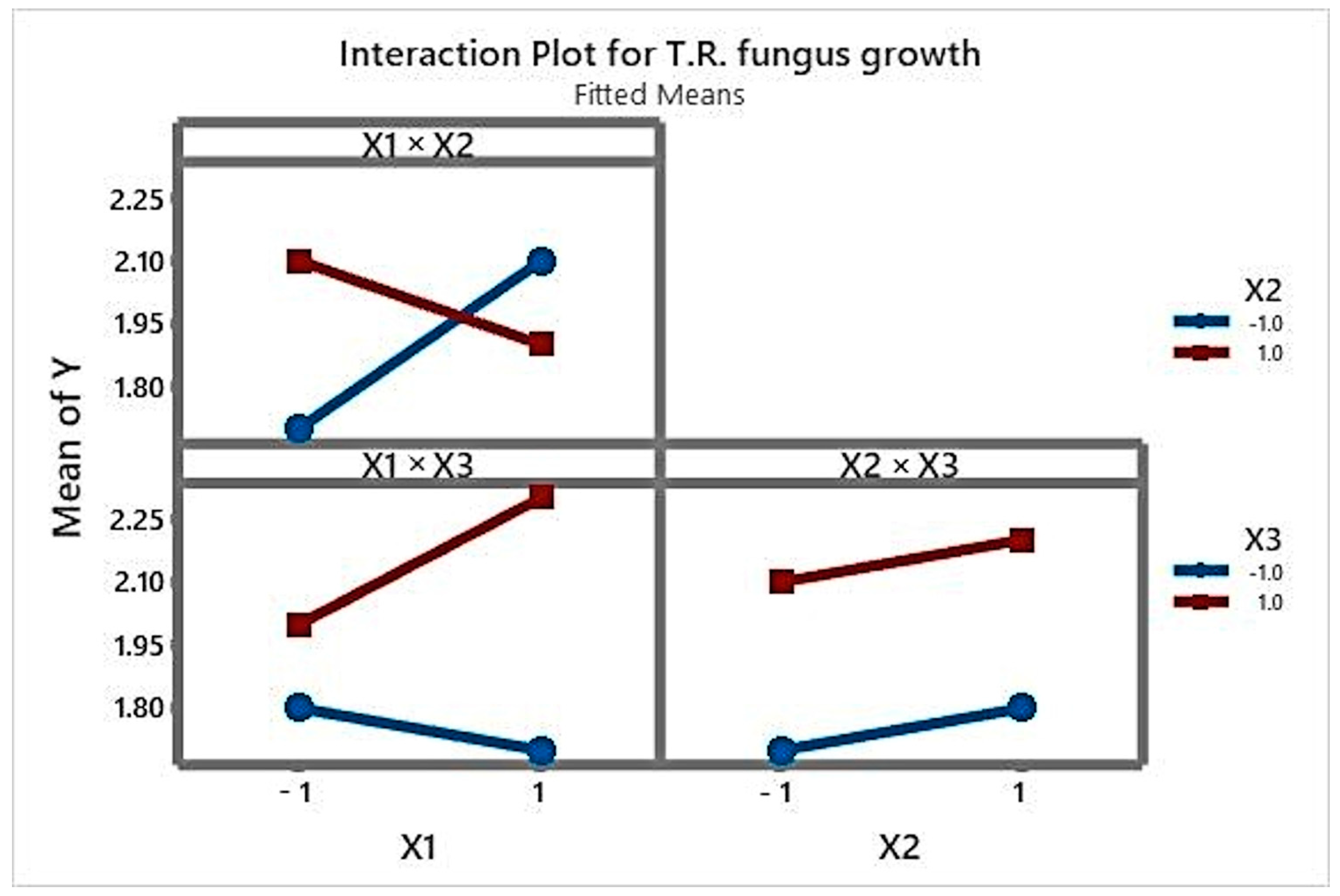
The graphic of the interactions, Figure 8, shows the effect of the interaction between the nanoparticle size and concentration, considering that inverted slopes can be observed on their high and low levels; therefore, both slopes change at the same time. Between the nanoparticle size and the ultrasonic treatment, the effect of the interaction can be observed as well, and it is opposite to the previous case. The nanoparticle size does not show interaction with the ultrasonic treatment due to their slopes being equal.
This means that a strong interaction exists between the size of the nanoparticle and the concentration used. The effect is much more significant in the case of the concentration and the nanoparticle size than on the concentration and ultrasonic treatment, i.e., if the high concentration of nanoparticles is used with a low ultrasonic treatment time, less fungal growth is observed.

3.2. Adjusting the Ordinal Logistic Regression Model

The logit link function was used to adjust the ordinal logistic regression model, presenting convergence on the 8th step of the log-likelihood with a 43.5042 value. The test to verify that all slopes are zero indicates a p-value of 0.394, which indicates that the correlation between the response variable and the predictors is not statistically significant. The value for Pearson’s goodness of fit test is 0.587, and the p-value for the goodness of fit test using standard deviation is 0.418, both values higher than 0.05. There is no evidence indicating the model is inadequate. In Table 5, the statistics for the ordinal logistic regression model are shown.
The p-values for the intercepts are less than 0.05, which indicates that they are statistically significant. At the same time, the p-values for the principal effects and their interactions are more extensive than 0.05, indicating that they are not statistically significant. The conditional probability models are:
Y 1 X = ln π 1 π 2 + π 3   + π 4 = 0.8444     0.1155 X 1     0.1039 X 2     0.5033 X 3 + 0.3284 X 2     0.1599 X 1 X 3     0.0594 X 2 X 3     0.518 X 1 X 2 X 3 Y 2 X = ln π 1 + π 2 π 3 + π 4 = 1.4194     0.1155 X 1     0.1039 X 2     0.5033 X 3 + 0.3284 X 2     0.1599 X 1 X 3     0.0594 X 2 X 3     0.518 X 1 X 2 X 3 Y 3 X = ln π 1 + π 2 + π 3 π 4 = 1.4194     0.1155 X 1     0.1039 X 2     0.5033 X 3 + 0.3284 X 2     0.1599 X 1 X 3     0.594 X 2 X 3     0.518 X 1 X 2 X 3

3.3. MOGAs Optimization

The regression models obtained in Equation (6) are substituted into Equation (2) to define the three objectives to minimize according to the multi-objective optimization problem described in Equation (5). Defined below are the models for each objective to be minimized using the multi-objective genetic algorithm.
F Y 1 X = exp Y 1 X / 1     exp Y 1 X F Y 2 X = exp Y 2 X / 1     exp Y 2 X F Y 3 X = exp Y 3 X / 1     exp Y 3 X
The first model is a function of the category in which less growth exists; therefore, it can be taken as a priority when selecting the best solution of the Pareto Front obtained by the multi-objective genetic algorithm shown in Table 6.
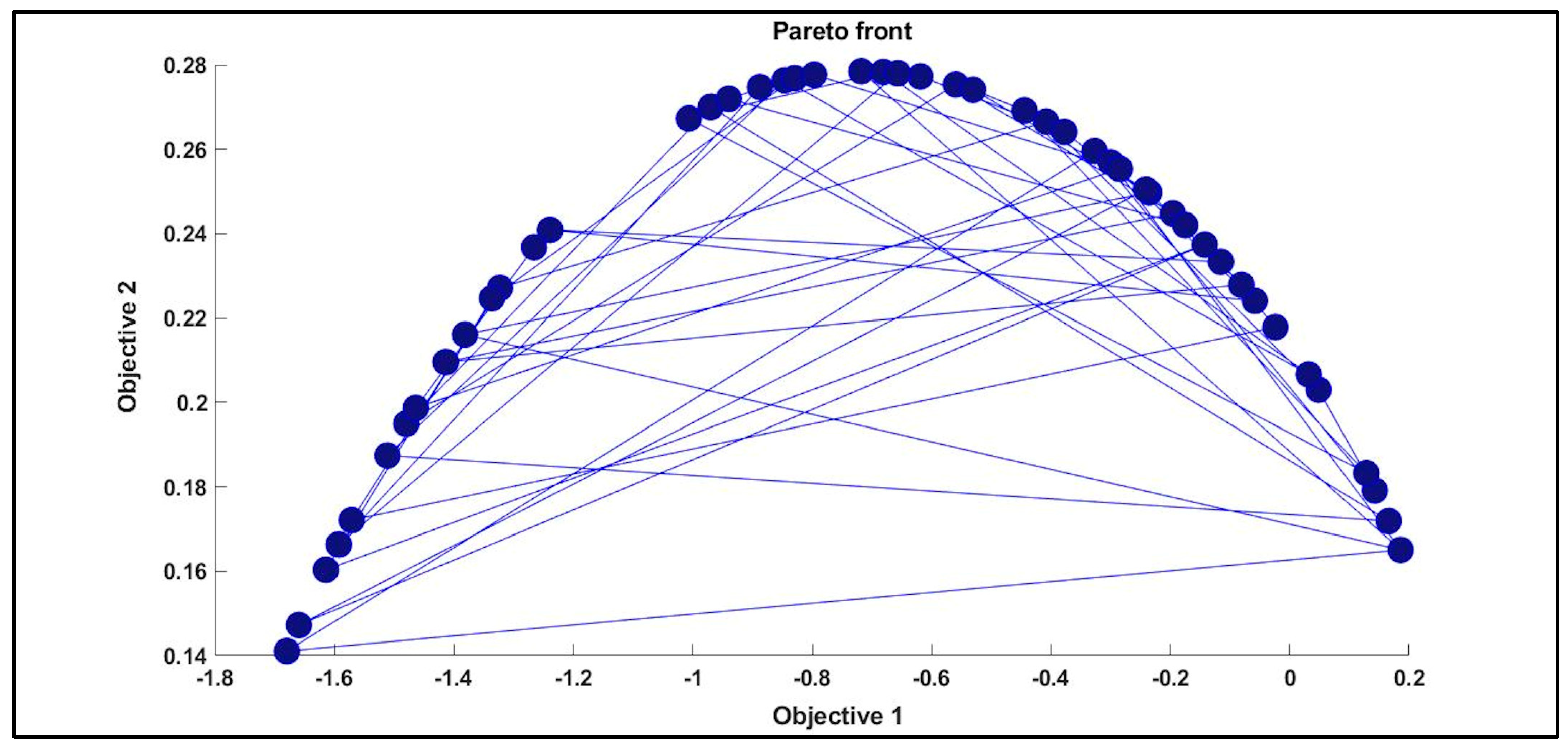
The set of solutions of the Pareto Front (Figure 9) meets the requirements of the optimization problem. The solution closest to zero was selected for the first objective, related to the lesser fungal growth probability. In Figure 9, the set of solutions between the first and second objectives is shown, where the point corresponding to the solution closest to zero for the first objective can be determined, and it corresponds to the solution in Table 6.
The optimization result indicates that the optimal fungal growth inhibition can be obtained using large size nanoparticles with a high concentration, using an ultrasonic treatment for 2 and 2.5 s. The three experimental factors showed an effect on fungal growth inhibition; therefore, they are factors to consider while designing products based on composite materials from PUR and AgNPs.
The ordinal logistic regression model is more accurate for categoric-type responses and can slightly modify the levels of the graphics for the main observed effects (see Figure 7).

3.4. SEM Analysis of the Samples

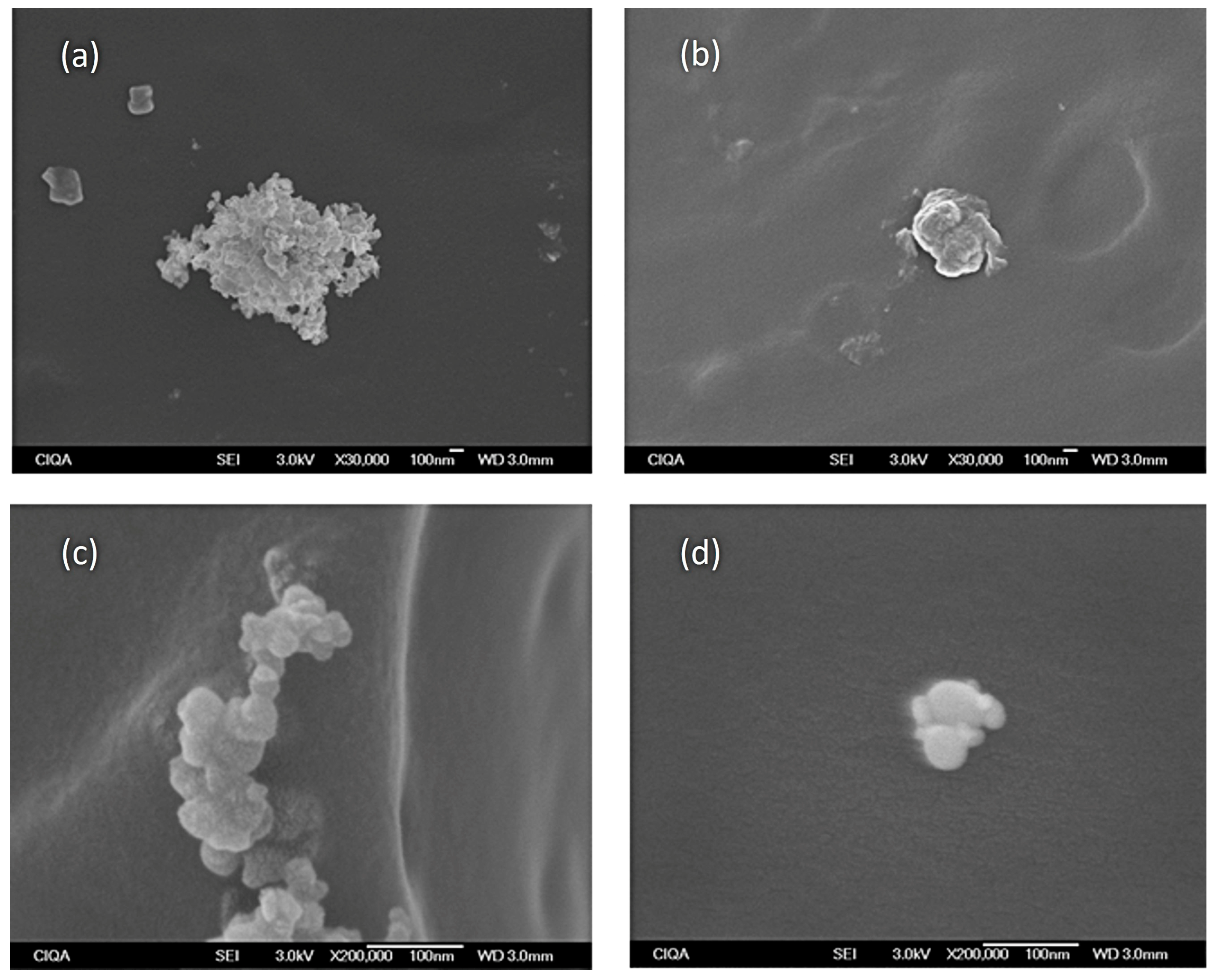
The SEM micrographs showed a satisfactory dispersion of the nanoparticles for both sizes, although in low quantity and small sizes, usually forming agglomerates. In Figure 10, the micrographs taken of the composite materials with 15 and 45 nm AgNPs, with and without ultrasonic treatment, can be observed.
In micrograph from Figure 10a, an agglomerate of AgNPs formed by smaller nanoparticles (15 nm) can be observed when the ultrasound treatment has not been applied to the system. When the ultrasound treatment is applied while obtaining the composite material, it can be observed that the agglomerates considerably diminish in size, as can be appreciated in micrograph (Figure 10b). The nanoparticles, especially metallic ones like silver, strongly tend to agglomerate. The formation of agglomerates causes a reduction of the total particle surface and, therefore, could reduce the biocide properties.
For the 45 nm AgNPs samples, agglomerates are also observed in the micrographs; however, the agglomerates are smaller in size (see Figure 10a,c); thus, a larger surface area and higher biocide activity could be expected in comparison to the composite materials containing 15 nm AgNPs.
By applying the ultrasound treatment on the composite materials containing 45 nm AgNPs, the agglomerates reduce in size, becoming even smaller than the agglomerates found in 15 nm AgNPs containing composite materials after the ultrasound treatment. These results are consistent with the optimization analysis of the studied parameters, in which it was established that to obtain the lesser T. rubrum fungal growth, 45 nm nanoparticles, high concentrations, and ultrasonic treatment should be used.
These results are consistent with those reported in the literature, which indicate that the formation of agglomerates tends to reduce the nanotoxicity [44]. As for the effect of the concentration, it is logical that composite materials show a higher fungal growth inhibition when the presence of nanoparticles in the medium is higher.

4. Conclusions

The applications for AgNPs are fascinating and could bring numerous benefits to humankind. The research on the biocide efficiency of AgNPs continuously reports success cases that suggest highly encouraging scenarios for their day-to-day use of diverse products. Said products can serve as preventive devices against the damage caused by the numerous pathogenic microorganisms coming into contact with the skin. The fungus Trichophyton rubrum and other fungi from the Trichophyton family are of interest due to being of medical concern, considering the known damage they cause to the skin and nails of infected patients and the high cost and length of the treatments able to eradicate it, such as the prolonged use of topic lotions and pills that can damage the organism through consumption.
In this study, essential effects of the AgNP size, the concentration, and the use of an ultrasonic sound for dispersion were identified in inhibiting the growth of T. rubrum. The optimization analysis showed that to present an appropriate antifungal property, the size of the AgNPs should be larger to avoid promoting the formation of agglomerates, and a proper dispersion system such as ultrasound should be used. Knowing these aspects could significantly impact the design of products for human consumption, which could benefit people with vulnerable skin, such as diabetes mellitus patients and people suffering from other conditions.
This type of fungi cannot be quantified due to their shape; thus, their analysis presents a significant challenge. In this study, a methodology for the qualitative evaluation of growth was applied, a model using ordinal logistic regression, and MOGAs for optimization were applied on an approach that can be generalized for similar cases. Future research on this line contemplates using a more complex experimental design, analyzing the effect of inhibiting additional factors and implementing more options for quantifying the growth to obtain more precise results.

Author Contributions

Conceptualization, A.M.C. and A.E.M.; methodology, A.M.C., A.E.M. and A.I.S.C.; software, A.M.C. and A.E.M.; validation, A.M.C., A.E.M. and A.Z.E.; formal analysis, A.M.C., A.E.M., A.I.S.C. and A.Z.E.; investigation, A.M.C., A.E.M. and A.Z.E.; resources, A.M.C., A.E.M. and A.I.S.C.; data curation, A.M.C. and A.Z.E.; writing—original draft preparation, A.M.C. and A.E.M.; writing—review and editing, A.M.C., A.E.M. and A.I.S.C.; translation to English, A.Z.E.; visualization, A.M.C. and A.E.M.; supervision, A.M.C., A.E.M. and A.Z.E.; project administration, A.M.C.; funding acquisition, A.M.C. and A.I.S.C. All authors have read and agreed to the published version of the manuscript.

Funding

This research was funded by PRODEP grant number 511-6/2029-8623 in “Convocatoria Fortalecimiento CAs”, Idea Guanajuato in Convocatoria Mentefactura Tecnológica Submodalidad Ciencia Productiva grant number S016.C14.QC3550.

Institutional Review Board Statement

Not applicable.

Informed Consent Statement

Not applicable.

Data Availability Statement

The data that support the findings of this study are available on request from the corresponding author (A.M.).

Acknowledgments

We thank Centro de Innovación Aplicada en Tecnologías Competitivas and Instituto Tecnológico Superior de Purísima del Rincón for the facilities granted for the use of equipment and laboratories.

Conflicts of Interest

The authors declare no conflict of interest.

References

  1. Mobeen, H.; Safdar, M.; Fatima, A.; Afzal, S.; Zaman, H.; Mehdi, Z. Emerging Applications of Nanotechnology in Context to Immunology: A Comprehensive Review. Front. Bioeng. Biotechnol. 2022, 10, 1024871. [Google Scholar] [CrossRef]
  2. Kim, H.-A.; Lee, B.-T.; Na, S.-Y.; Kim, K.-W.; Ranville, J.F.; Kim, S.-O.; Jo, E.; Eom, I.-C. Characterization of Silver Nanoparticle Aggregates Using Single Particle-Inductively Coupled Plasma-Mass Spectrometry (SpICP-MS). Chemosphere 2017, 171, 468–475. [Google Scholar] [CrossRef] [PubMed]
  3. Joudeh, N.; Linke, D. Nanoparticle Classification, Physicochemical Properties, Characterization, and Applications: A Comprehensive Review for Biologists. J. Nanobiotechnology 2022, 20, 262. [Google Scholar] [CrossRef] [PubMed]
  4. Sahoo, S.; Hormozi-Nezhad, M.R. Gold and Silver Nanoparticles: Synthesis and Applications; Elsevier: Amsterdam, The Netherlands, 2023; ISBN 9780323994545. [Google Scholar]
  5. Zhang, X.-F.; Liu, Z.-G.; Shen, W.; Gurunathan, S. Silver Nanoparticles: Synthesis, Characterization, Properties, Applications, and Therapeutic Approaches. Int. J. Mol. Sci. 2016, 17, 1534. [Google Scholar] [CrossRef] [PubMed]
  6. Dhaka, A.; Chand Mali, S.; Sharma, S.; Trivedi, R. A Review on Biological Synthesis of Silver Nanoparticles and Their Potential Applications. Results Chem. 2023, 6, 101108. [Google Scholar] [CrossRef]
  7. Chanyachailert, P.; Leeyaphan, C.; Bunyaratavej, S. Cutaneous Fungal Infections Caused by Dermatophytes and Non-Dermatophytes: An Updated Comprehensive Review of Epidemiology, Clinical Presentations, and Diagnostic Testing. J. Fungi 2023, 9, 669. [Google Scholar] [CrossRef] [PubMed]
  8. Ahirwar, D.; Telang, A.; Purohit, R.; Namdev, A. A Short Review on Polyurethane Polymer Composite. Mater. Today Proc. 2022, 62, 3804–3810. [Google Scholar] [CrossRef]
  9. Witkiewicz, W.; Zieliński, A. Properties of the Polyurethane (PU) Light Foams. Adv. Mater. Sci. 2006, 6, 35–51. [Google Scholar]
  10. Eugenia, M.M.; Adolfo, T.L.; Rodolfo, E. Characterization of Polyurethane Nanocomposites for Flame Retardant Applications. J. Chem. Chem. Eng. 2018, 12, 60–73. [Google Scholar] [CrossRef]
  11. Cui, M.; Chai, Z.; Lu, Y.; Zhu, J.; Chen, J. Developments of Polyurethane in Biomedical Applications: A Review. Resour. Chem. Mater. 2023, 2, 262–276. [Google Scholar] [CrossRef]
  12. Wendels, S.; Avérous, L. Biobased Polyurethanes for Biomedical Applications. Bioact. Mater. 2021, 6, 1083–1106. [Google Scholar] [CrossRef] [PubMed]
  13. Grzęda, D.; Węgrzyk, G.; Nowak, A.; Idaszek, J.; Szczepkowski, L.; Ryszkowska, J. Cytotoxic Properties of Polyurethane Foams for Biomedical Applications as a Function of Isocyanate Index. Polymers 2023, 15, 2754. [Google Scholar] [CrossRef] [PubMed]
  14. Okrasa, M.; Leszczyńska, M.; Sałasińska, K.; Szczepkowski, L.; Kozikowski, P.; Majchrzycka, K.; Ryszkowska, J. Viscoelastic Polyurethane Foams for Use in Seals of Respiratory Protective Devices. Materials 2021, 14, 1600. [Google Scholar] [CrossRef] [PubMed]
  15. Sana, S.S.; Haldhar, R.; Parameswaranpillai, J.; Chavali, M.; Kim, S.-C. Silver Nanoparticles-Based Composite for Dye Removal: A Comprehensive Review. Clean. Mater. 2022, 6, 100161. [Google Scholar] [CrossRef]
  16. Morena, A.G.; Stefanov, I.; Ivanova, K.; Pérez-Rafael, S.; Sánchez-Soto, M.; Tzanov, T. Antibacterial Polyurethane Foams with Incorporated Lignin-Capped Silver Nanoparticles for Chronic Wound Treatment. Ind. Eng. Chem. Res. 2020, 59, 4504–4514. [Google Scholar] [CrossRef]
  17. Xu, C.; Akakuru, O.U.; Ma, X.; Zheng, J.; Zheng, J.; Wu, A. Nanoparticle-Based Wound Dressing: Recent Progress in the Detection and Therapy of Bacterial Infections. Bioconjug. Chem. 2020, 31, 1708–1723. [Google Scholar] [CrossRef]
  18. ASTM D ASTM-G21-15-4; Standard Practice for Determining Resistance of Synthetic Polymeric Materials to Fungi. ASTM International: Conshohocken, PA, USA, 2015. Available online: https://www.astm.org/g0021-15r21e01.html (accessed on 5 August 2023).
  19. Linima, V.K.; Ragunathan, R.; Johney, J. Biogenic Synthesis of RICINUS COMMUNIS Mediated Iron and Silver Nanoparticles and Its Antibacterial and Antifungal Activity. Heliyon 2023, 9, e15743. [Google Scholar] [CrossRef]
  20. Gil-Korilis, A.; Cojocaru, M.; Berzosa, M.; Gamazo, C.; Andrade, N.J.; Ciuffi, K.J. Comparison of Antibacterial Activity and Cytotoxicity of Silver Nanoparticles and Silver-Loaded Montmorillonite and Saponite. Appl. Clay Sci. 2023, 240, 106968. [Google Scholar] [CrossRef]
  21. Zhao, G.; Stevens, J.S.E. Multiple Parameters for the Comprehensive Evaluation of the Susceptibility of Escherichia Coli to the Silver Ion. Biometals 1998, 11, 27–32. [Google Scholar] [CrossRef]
  22. Slawson, R.M.; Trevors, J.T.; Lee, H. Silver Accumulation and Resistance in Pseudomonas Stutzeri. Arch. Microbiol. 1992, 158, 398–404. [Google Scholar] [CrossRef]
  23. Vigneshwaran, N.; Ashtaputre, N.M.; Varadarajan, P.V.; Nachane, R.P.; Paralikar, K.M.; Balasubramanya, R.H. Biological Synthesis of Silver Nanoparticles Using the Fungus Aspergillus Flavus. Mater. Lett. 2007, 61, 1413–1418. [Google Scholar] [CrossRef]
  24. Skanda, S.; Bharadwaj, P.S.J.; Datta Darshan, V.M.; Sivaramakrishnan, V.; Vijayakumar, B.S. Proficient Mycogenic Synthesis of Silver Nanoparticles by Soil Derived Fungus Aspergillus Melleus SSS-10 with Cytotoxic and Antibacterial Potency. J. Microbiol. Methods 2022, 199, 106517. [Google Scholar] [CrossRef]
  25. Gajbhiye, M.; Kesharwani, J.; Ingle, A.; Gade, A.; Rai, M. Fungus-Mediated Synthesis of Silver Nanoparticles and Their Activity against Pathogenic Fungi in Combination with Fluconazole. Nanomedicine 2009, 5, 382–386. [Google Scholar] [CrossRef]
  26. Mansoor, S.; Zahoor, I.; Baba, T.R.; Padder, S.A.; Bhat, Z.A.; Koul, A.M.; Jiang, L. Fabrication of Silver Nanoparticles against Fungal Pathogens. Front. Nanotechnol. 2021, 3, 679358. [Google Scholar] [CrossRef]
  27. Robles-Martínez, M.; González, J.F.C.; Pérez-Vázquez, F.J.; Montejano-Carrizales, J.M.; Pérez, E.; Patiño-Herrera, R. Antimycotic Activity Potentiation of Allium Sativum Extract and Silver Nanoparticles against Trichophyton Rubrum. Chem. Biodivers. 2019, 16, e1800525. [Google Scholar] [CrossRef] [PubMed]
  28. Mohsen, L.Y.; Fadhil Alsaffar, M.; Ahmed Lilo, R.; Khalil Al-Shamari, A. Silver Nanoparticles That Synthesis by Using Trichophyton Rubrum and Evaluate Antifungal Activity. Arch. Razi Inst. 2022, 77, 2145–2149. [Google Scholar]
  29. da Silva, C.A.; Ribeiro, B.M.; Trotta, C.D.V.; Perina, F.C.; Martins, R.; Abessa, D.M.d.S.; Barbieri, E.; Simões, M.F.; Ottoni, C.A. Effects of Mycogenic Silver Nanoparticles on Organisms of Different Trophic Levels. Chemosphere 2022, 308, 136540. [Google Scholar] [CrossRef]
  30. Wijesiri, N.; Yu, Z.; Tang, H.; Zhang, P. Antifungal Photodynamic Inactivation against Dermatophyte Trichophyton Rubrum Using Nanoparticle-Based Hybrid Photosensitizers. Photodiagnosis Photodyn. Ther. 2018, 23, 202–208. [Google Scholar] [CrossRef]
  31. Patiño-Herrera, R.; Catarino-Centeno, R.; Robles-Martínez, M.; Zarate, M.G.M.; Flores-Arriaga, J.C.; Pérez, E. Antimycotic Activity of Zinc Oxide Decorated with Silver Nanoparticles against Trichophyton Mentagrophytes. Powder Technol. 2018, 327, 381–391. [Google Scholar] [CrossRef]
  32. Helmlinger, J.; Sengstock, C.; Groß-Heitfeld, C.; Mayer, C.; Schildhauer, T.A.; Köller, M.; Epple, M. Silver Nanoparticles with Different Size and Shape: Equal Cytotoxicity, but Different Antibacterial Effects. RSC Adv. 2016, 6, 18490–18501. [Google Scholar] [CrossRef]
  33. Sadeghi, B.; Garmaroudi, F.S.; Hashemi, M.; Nezhad, H.R.; Nasrollahi, A.; Ardalan, S.; Ardalan, S. Comparison of the Anti-Bacterial Activity on the Nanosilver Shapes: Nanoparticles, Nanorods and Nanoplates. Adv. Powder Technol. 2012, 23, 22–26. [Google Scholar] [CrossRef]
  34. Pal, S.; Tak, Y.K.; Song, J.M. Does the Antibacterial Activity of Silver Nanoparticles Depend on the Shape of the Nanoparticle? A Study of the Gram-Negative Bacterium Escherichia coli. Appl. Environ. Microbiol. 2007, 73, 1712–1720. [Google Scholar] [CrossRef] [PubMed]
  35. Kim, T.-H.; Kim, M.; Park, H.-S.; Shin, U.S.; Gong, M.-S.; Kim, H.-W. Size-Dependent Cellular Toxicity of Silver Nanoparticles. J. Biomed. Mater. Res. A 2012, 100A, 1033–1043. [Google Scholar] [CrossRef]
  36. Díaz-Puertas, R.; Rodríguez-Cañas, E.; Bello-Perez, M.; Fernández-Oliver, M.; Mallavia, R.; Falco, A. Viricidal Activity of Thermoplastic Polyurethane Materials with Silver Nanoparticles. Nanomaterials 2023, 13, 1467. [Google Scholar] [CrossRef] [PubMed]
  37. Bechtold, M.; Valério, A.; de Souza, A.A.U.; de Oliveira, D.; Franco, C.V.; Serafim, R.; Souza, S.M.A.G.U. Synthesis and Application of Silver Nanoparticles as Biocidal Agent in Polyurethane Coating. J. Coatings Technol. Res. 2020, 17, 613–620. [Google Scholar] [CrossRef]
  38. Savelyev, Y.; Gonchar, A.; Movchan, B.; Gornostay, A.; Vozianov, S.; Rudenko, A.; Rozhnova, R.; Travinskaya, T. Antibacterial Polyurethane Materials with Silver and Copper Nanoparticles. Mater. Today Proc. 2017, 4, 87–94. [Google Scholar] [CrossRef]
  39. El-Rahman, N.R.A.; Bekhit, M.; Fekry, M. Fabrication and Evaluation of Polyurethane Cationic Surfactants, and Their Potential on Silver Nanoparticles Stability, Surface Activity, and Biological Activity. Egypt. J. Pet. 2022, 31, 23–31. [Google Scholar] [CrossRef]
  40. Wijnhoven, S.W.P.; Peijnenburg, W.J.G.M.; Herberts, C.A.; Hagens, W.I.; Oomen, A.G.; Heugens, E.H.W.; Roszek, B.; Bisschops, J.; Gosens, I.; Van De Meent, D.; et al. Nano-Silver—A Review of Available Data and Knowledge Gaps in Human and Environmental Risk Assessment. Nanotoxicology 2009, 3, 109–138. [Google Scholar] [CrossRef]
  41. Monje, A.E.; Reséndiz, J.R.H. Synthesis of Urethane Base Composite Materials with Metallic Nanoparticles. MRS Proc. 2013, 1547, 141–147. [Google Scholar] [CrossRef]
  42. McCullagh, P.; Nelder, J.A. Generalized Linear Models; Chapman and Hall: London, UK, 1983; pp. 1–19. [Google Scholar]
  43. Dobson Annette, J. Normal Linear Models. In An Intruction to Generalized Linear models; Chatfield, C., Zidek, J., Eds.; Chapman and Hall: Vancouver, BC, Canada, 2001; pp. 179–196. [Google Scholar]
  44. Bae, E.; Lee, B.-C.; Kim, Y.; Choi, K.; Yi, J. Effect of Agglomeration of Silver Nanoparticle on Nanotoxicity Depression. Korean J. Chem. Eng. 2013, 30, 364–368. [Google Scholar] [CrossRef]
Figure 1. Proposed methodology.
Applsci 13 12028 g001
Figure 2. Sample preparation: (a) pre-heating polyol and isocyanate on a laboratory heating plate, (b) polyol weighing, (c) isocyanate weighing, and (d) catalyst weighing in a syringe.
Applsci 13 12028 g002
Figure 3. Homogenization process of the samples using ultrasound. Note: Hielscher is the brand of ultrasound equipment.
Applsci 13 12028 g003
Figure 4. (a) Mixture of the PUR components, (b) start of the foaming, and (c) completed foaming.
Applsci 13 12028 g004
Figure 5. (a) Preparation of the culture medium and fungal activation, (b) inoculation of the fungus on the culture medium, and (c,d) visualization of the fungus T. rubrum using lactophenol cotton blue dye.
Applsci 13 12028 g005
Figure 6. Examples of the observed fungal growth are (a) traces of growth below 10% (1), (b) light growth from 10 to 30% (2), (c) medium growth from 30 to 60% (3), and (d) heavy growth from 60% to complete coverage (4).
Applsci 13 12028 g006
Figure 7. Graph showing the main effects on the average fungal growth on variables X1 size of the AgNPs, X2 concentration of AgNPs, and X3 ultrasonic treatment on polyol. The blue dots represent the average effects of each of the control factors on the average fungal growth of the fungus when changing from low and high levels, respectively.
Applsci 13 12028 g007
Figure 8. Graph of the interactions for the average fungal growth among the variables X1 size of the AgNPs, X2 concentration of AgNPs, and X3 ultrasonic treatment on polyol.
Applsci 13 12028 g008
Figure 9. Pareto Front for the set of solutions between objective 1 and objective 2 for the multi-objective optimization problem. The blue points represent the feasible solutions for the optimization problem and are intersections between the vectors (the blue lines) generated in the process according to the optimization problem.
Applsci 13 12028 g009
Figure 10. SEM micrographs of the samples with 15 nm AgNPs, (a) without ultrasound, (b) with ultrasound, and samples with 45 nm AgNPs, (c) without ultrasound, and (d) with ultrasound.
Applsci 13 12028 g010
Table 1. Experimental matrix.
Treatment X 1 X 2 X 3 Y 1 Y 2 Y 3 Y 4 Y 5
115 nm5 g0 s Y 11 Y 12 Y 13 Y 14 Y 15
245 nm5 g0 s Y 21 Y 22 Y 23 Y 24 Y 25
315 nm10 g0 s Y 31 Y 32 Y 33 Y 34 Y 35
445 nm10 g0 s Y 41 Y 42 Y 43 Y 44 Y 45
515 nm5 g 10 s Y 51 Y 52 Y 53 Y 54 Y 55
645 nm5 g10 s Y 61 Y 62 Y 63 Y 64 Y 65
715 nm10 g10 s Y 71 Y 72 Y 73 Y 74 Y 75
845 nm10 g10 s Y 81 Y 82 Y 83 Y 84 Y 85
Table 2. Rating according to the observed growth of the fungus on specimens. Adapted with oermission from [18] Copyrighted by ASTM International, 2013.
Observed Growth on SpecimensRating
None0
Traces of growth (less than 10%)1
Light growth (10 to 30%)2
Medium growth (30 to 60%)3
Heavy growth (60% to complete coverage)4
Table 3. Experimental results.
Treatment X 1 X 2 X 3 Y 1 Y 2 Y 3 Y 4 Y 5
115 nm5 g0 s21112
245 nm5 g0 s22231
315 nm10 g0 s42221
445 nm10 g0 s11122
515 nm5 g10 s22132
645 nm5 g10 s42113
715 nm10 g10 s11323
845 nm10 g10 s32322
The reported values in the table represent the average of the triplicate samples for each test.
Table 4. Parameters for the genetic algorithm.
ParameterValue
Number of variables3
Population size50
Limits[−1, −1, −1]; [1, 1, 1]
Selection functionUniform stochastic
Initial value[−10, 10]
Elite counting0.05 × initial population
Crossover fraction0.8
Mutation function Dependent on restrictions
Migration directionForward
Generations100 × Number of variables
Stagnant generations50
Tolerance of the function1 × 10−6
Table 5. Coefficients for the ordinal logistic regression model.
PredictorCoefSE CoefZp
α1−0.8444830.368023−2.290.022
α21.419370.4038283.510.000
α33.206500.7480004.290.000
X1−0.1154660.304130−0.380.704
X2−0.1038870.304001−0.340.733
X3−0.5033270.310272−1.620.105
X1 ×X20.3284450.3056801.070.283
X1 × X3−0.1599360.304776−0.520.600
X2 × X3−0.05941710.303632−0.200.845
X1 × X2 × X3−0.5176290.310828−1.670.096
Table 6. Optimal solution selected from the Pareto Front.
Pareto Front Solution X 1 X 2 X 3 F Y 1 X F Y 2 X F ( Y 3 X )
280.983570.98818−0.660400.031830.206630.06601
Disclaimer/Publisher’s Note: The statements, opinions and data contained in all publications are solely those of the individual author(s) and contributor(s) and not of MDPI and/or the editor(s). MDPI and/or the editor(s) disclaim responsibility for any injury to people or property resulting from any ideas, methods, instructions or products referred to in the content.