Article

Ex Vivo Analysis of Ability of Osseodensification to Improve Dental Implant Primary Stability Using Xenograft Bone Walls

by
Henrique Ferreira
1,
Carlos Fernando Mourão
2,*,
Rafael Coutinho Mello-Machado
3,
Kayvon Javid
4,
Rodrigo dos Santos Pereira
1 and
Plinio Mendes Senna
5
1
Department of Dental Research and Innovation, University of Grande Rio—UNIGRANRIO, Duque de Caxias 25071-202, RJ, Brazil
2
Department of Periodontology, Tufts University School of Dental Medicine, Boston, MA 02111, USA
3
Department of Implant Dentistry, Universidade Iguaçu, Nova Iguaçu 26260-045, RJ, Brazil
4
Department of Oral Surgery, Fluminense Federal University, Niterói 24020-140, RJ, Brazil
5
Department of Prosthodontics, State University of Rio de Janeiro, Rio de Janeiro 24033-900, RJ, Brazil
*
Author to whom correspondence should be addressed.
Appl. Sci. 2023, 13(22), 12337; https://doi.org/10.3390/app132212337
Submission received: 18 October 2023 / Revised: 8 November 2023 / Accepted: 12 November 2023 / Published: 15 November 2023
(This article belongs to the Special Issue Novel Biomaterial and Implant in Dentistry)

Abstract

:
Osseodensification is a technique that involves compressing bone using specialized drilling instruments to increase bone–implant contact. The present study aimed to evaluate the structure of a xenograft bone (XB) wall created within an implantation site and how it affects the initial stability of dental implants. Six segments of pig ribs, representing low-density bone, were used in the experiment. Four different drilling conditions were created for each section using a tapered bur system associated with bovine xenograft bone: clockwise (cutting mode—CW) or counterclockwise (densification mode—CCW). The bone samples were then placed individually in microtomography equipment to define a volume of interest (VOI) 50% larger than the osteotomy. Mathematical calculations of bone volume, trabecular thickness and separation, and total porosity were performed. An implant with a diameter of 4.0 mm and a length of 11.5 mm was then inserted into each osteotomy. The final insertion torque (IT) and resonance frequency analysis/implant stability quotient (ISQ) values were recorded. The groups were compared using ANOVA and Tukey’s post hoc test. The results show that the use of xenograft bone produced densification at the apex region, with higher bone volume and trabecular thickness, and reduced trabecular separation compared with the CW group (p < 0.05). The CW + XB group demonstrated a similar porosity to the CCW group and similar values of IT and ISQ (p > 0.05). Compared with the other groups, CCW + XB exhibited the lowest percentual porosity and the highest values of IT and ISQ (p < 0.05). We concluded that the use of a xenograft bone wall before implant placement can improve the primary stability of dental implants.

1. Introduction

Bone tissue, a specialized connective tissue, exhibits a substantial capacity for regeneration and remodeling, which is crucial for maintaining structural and functional integrity [1,2]. However, localized bone osseointegration, the structural and functional connection between living bone and the surface of a load-bearing implant, is fundamental to the long-term success of dental implants [3,4]. Adequate primary stability, defined as the mechanical stability of an implant at the time of placement, is widely recognized as an essential prerequisite for osseointegration to occur [1,5]. Primary stability is influenced by the quality and quantity of native bone, the surgical technique, and implant features. Low primary stability in poor-quality bone increases the risk of micromotion and fibrous encapsulation, compromising the establishment of secondary stability and ultimately leading to implant failure [1,6,7].
Secondary stability originates from biological fixation as newly formed woven bone transitions to mature lamellar bone. Implant micromotion, excessive loading, poor bone quality, and surgical trauma can all negatively impact secondary stability [4,7,8]. Low secondary stability is associated with reduced long-term survival [9,10]. Therefore, enhancing early stabilization is key to avoiding micromotion, facilitating effective osseointegration, and enabling earlier loading protocols [5,11,12]. Numerous surgical techniques have been developed to improve primary stability in low-density bone, thereby setting the stage for optimal secondary stability and osseointegration [13,14,15,16,17].
Two quantitative methods exist for clinically evaluating implant stability: insertion torque (IT) and resonance frequency analysis, also known as implant stability quotient (ISQ) [7,8]. Insertion torque provides a direct measure of the rotational friction between the implant and bone as the implant is placed. Higher IT values indicate greater primary stability, with prior research demonstrating IT values above 30 Ncm are considered ideal [17]. ISQ utilizes vibration analysis to characterize the resonance frequency of the implant-bone complex, providing an indirect measure of stiffness and micromobility. While IT provides a one-time measure of primary stability at placement, ISQ can be utilized at multiple time points to monitor the progression of secondary stability via bone regeneration and remodeling at the implant interface [18].
Osseodensification (OD) is a technique that involves the use of specialized drilling instruments to compress bone, thereby increasing bone–implant contact and friction [13,19,20]. However, its effectiveness is limited by the density of the patient’s natural bone. Studies have shown that OD is more effective than traditional osteotomy preparation in low-density-bone models [21,22,23,24,25]. It is possible that the benefits of OD could be enhanced by utilizing particulate bone graft walls at the surgical site, although further investigations are necessary. Therefore, this study compared the effectiveness of OD-prepared implants with and without xenograft bone walls in enhancing primary implant stability in low-density bone.

2. Materials and Methods

2.1. Sample Preparation

In this ex vivo study, pig ribs were acquired from a local retail meat market, specifically from pigs within the age range of 10–12 months and weight range of 80–100 kg. After removing the periosteum, each rib was meticulously cleaned using a solution of hydrogen peroxide and saline to remove excess fat and tissues. Following cleaning, 60 mm segments with a consistent cortical thickness of 1.0 ± 0.5 mm were isolated. This simulation of low-density bone was referenced from previous studies [26,27]. The samples were then preserved at −20 °C and later thawed at room temperature for an hour before experimentation.

2.2. Power Analysis and Sample Size Determination

The researchers conducted a power analysis using data from Bhargava et al. [28] to determine the minimum number of maxillary sinuses needed in each group. Based on a one-tailed test with a significance level of 5% and a statistical power of 80%, a mean difference of 9.1 and a standard deviation of 1.81 were used to determine that a minimum of 5 implant sites were required. The calculus was performed on the website “http://estatistica.bauru.usp.br/calculoamostral/calculos.php”, accessed on 30 July 2023.

2.3. Randomization and Allocation of Implants

The sealed envelope method was adopted to ensure impartial allocation of implants to the bone segments. Sequentially numbered, opaque, sealed envelopes contained the designation of each experimental group. An independent researcher who was not involved in the implant procedure prepared these envelopes. The position of the implant in each bone section was determined by drawing an envelope, ensuring a fully randomized allocation process. A minimum inter-implant distance of 5 mm, determined using an electronic digital caliper (model 500-196-30, Mitutoyo Corporation, Kawasaki, Japan), was maintained throughout the experiment to mitigate interaction effects between adjacent implants. Further details are illustrated in Table 1.

2.4. Drilling Procedure and Implant Installation

All osteotomies were undertaken by an experienced surgeon, utilizing a BLM 600 plus surgical electric micromotor (K Driller, São Paulo, Brazil). The drilling process was standardized to ensure reproducibility; multi-fluted tapered burs with diameters of 2.0 mm (pilot), 2.3 mm, and 3.0 mm (Densah Bur; Versah®, Jackson, MI, USA) were used at a controlled speed of 1200 rpm, applying a consistent force which was monitored using a force gauge to prevent bone overheating. Continuous saline irrigation (at a rate of 30 mL/min) was implemented to facilitate cooling and removal of bone debris. The drilling directions were alternated between clockwise and counterclockwise up to a depth of 11.5 mm.
For implant installation, a specific implant model 4.0 mm in diameter and 11.5 mm in length was utilized (Medens, SP, Brazil). The implant featured a sandblasted, large-grit, acid-etched surface to enhance osseointegration. Subsequent to implant bed preparation, xenograft bone (XB) material, comprising bovine-derived granules ranging between 0.5 and 1.0 mm (Cerabone, Straumann, Bern, Switzerland), was utilized to fill half of the osteotomies.

2.5. Microtomographic Analysis and VOI Determination

Each bone segment underwent micro-CT scanning using a Skyscan 1174 (Bruker, Kontich, Belgium) under specific parameters: 50 kV voltage, 800 μA current, 0.5 mm aluminum filter, and 5200 ms exposure time, with a voxel size of 28 μm and an X-ray energy of 50 kVp. The detailed scanning process spanned approximately 120 min, and image reconstruction was then conducted utilizing NRecon® software (version 2.0) with the specified settings.
The volume of interest (VOI) was demarcated as a cylindrical zone with a diameter of 4.5 mm (50% larger than the osteotomy) and a height of 13 mm, encompassing an approximate volume of 200 mm3. This dimensioning ensured a comprehensive analysis encompassing the immediate regions surrounding the osteotomy site, as illustrated in Figure 1.

2.6. Image Analysis

Using the CT Analyser v.1.14.4.1 (CTAn, Bruker, Kontich, Belgium), transaxial sections of each osteotomy were analyzed to derive quantitative data pertaining to bone volume (BV), trabecular thickness (Tb.Th), trabecular separation (Tb.Sp), and total porosity (Po(tot)). The gray threshold was defined between 46 and 255 for the binarization process. A pixel size of 57.5 µm was considered for the “3D analysis” tool in the software to facilitate detailed analysis.

2.7. Implant Stability Measurements

Implant stability measurements, specifically IT and ISQ, were conducted using specialized equipment. The IT was measured using a digital torquemeter (TQ-8800; Lutron Company, MA, USA) at the final position of the implant (Figure 2). Concurrently, the ISQ was determined using the Osstell ISQ system (W&H group, Gothenburg, Sweden), where transducers were mounted on each implant and secured manually, by capturing two perpendicular measurements and recording the mean value.

2.8. Statistical Analysis

Data normality was assessed using the Shapiro–Wilk test. Subsequently, ANOVA and Tukey’s post hoc test were employed for group comparisons at a 5% significance level, utilizing the SPSS software (version 20, IBM, IL, USA) for comprehensive statistical analysis.

3. Results

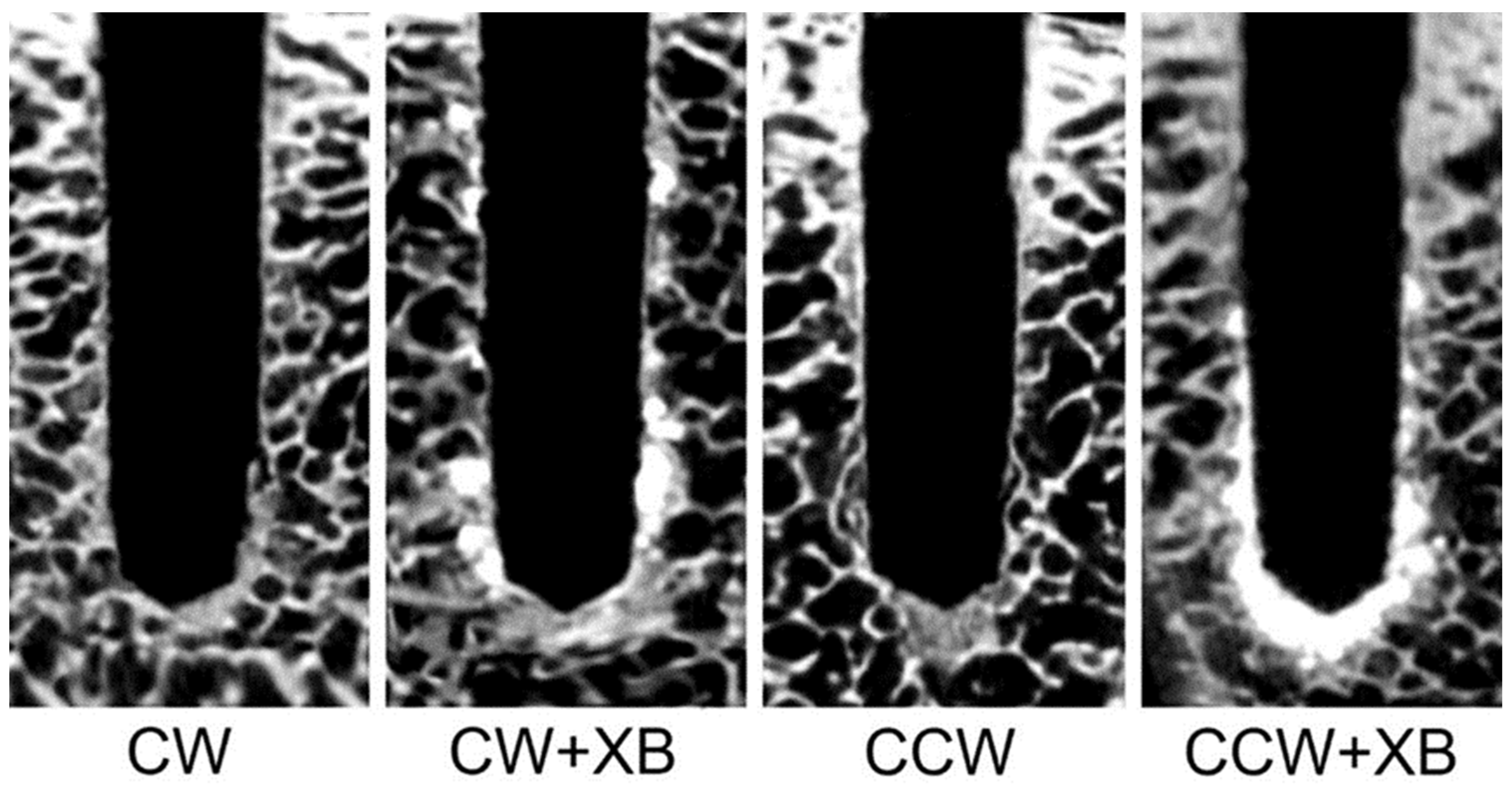
3.1. Analysis of Bone Density and Microstructure Post-Osteotomy

In the microtomographic analysis, discernible variations in bone density and microstructure were found, depending on the osteotomy techniques used (Figure 3). Notably, osteotomies augmented with xenograft bone showcased pronounced densification at both the body and apex regions of the implant sites, irrespective of the drilling direction employed. This densification phenomenon was mirrored by an escalation in bone volume and trabecular thickness, coupled with a marked diminution in trabecular separation within the VOI—a comprehensive representation of which is tabulated in Table 2.
Upon analyzing the porosity data, it was observed that the group subjected to clockwise drilling coupled with XB (CW + XB) exhibited porosity levels analogous to those of the counterclockwise group (CCW). However, the CCW approach complemented with XB integration manifested the most notable decline in percentage porosity within the VOI, showcasing a statistically significant improvement (p < 0.05).

3.2. Evaluation of Implant Stability

The assessment of implant stability, as delineated through IT and ISQ values, indicated a pronounced enhancement in the stability parameters in the groups supplemented with XB (Figure 4 and Figure 5). In particular, the xenograft bone bolstered the IT and ISQ metrics significantly compared with the CW group alone, a trend that was statistically significant (p < 0.05).
Unusually, the evaluations indicated marginal differences between the CW + XB and CCW groups, establishing no significant divergence (p > 0.05). Conversely, the CCW + XB cohort emerged as the group presenting the zenith in both IT and ISQ values, a statistically significant observation (p < 0.05).
The different osteotomy approaches produced different density aspects (Figure 3). The use of XB produced stronger densification at the body and apex of the implantation site regardless of drilling direction, with improvement in bone volume and trabecular thickness and a reduction in trabecular separation inside the VOI (Table 2). The CW + XB group produced the same level of porosity as CCW. CCW + XB reduced the lowest percentual porosity inside the VOI (p < 0.05).

4. Discussion

In the present study, notable improvements were observed in bone volume, trabecular thickness, and implant stability parameters, which were significantly amplified when employing xenograft bone walls in concert with counterclockwise osseodensification [29,30,31,32,33]. This approach not only substantiates the growing body of evidence that advocates for this combination in enhancing early implant stabilization but also highlights the benefits of utilizing low-quality bone to fortify the trabecular spaces. Leveraging the principles of osseodensification, this strategy integrates bone substitutes that have preferably undergone a sintering process during the same surgical phase, potentially fostering a scaffold that ensures greater particle stability at the time of implant installation, thereby granting more advantageous primary stability [20,21,24,29].
The results of this study align with the findings presented in the systematic review and meta-analysis conducted by Inchingolo et al. In their comprehensive evaluation of various studies, they highlighted the advantages of counterclockwise osseodensification, particularly in terms of improved bone-to-implant contact and insertion torque compared with clockwise osseodensification [19,30,31,32]. However, the bone area fraction occupied remained relatively consistent between the two techniques. The micro-CT results of this study support the mechanical benefits of osseodensification, with localized densification that closely aligns with the biomechanical improvements noted in the meta-analysis. These findings suggest a potential shift in strategies for optimizing implant site preparations [33,34,35].
On the other hand, the technique used in the present research was characterized by the compaction of xenograft bone during the preparation of the implant bed to create a graft layer along the surface of the osteotomy site to improve the primary stability. This can lead to preservation and condensation of the implant bed, which is specifically important for low-density bones [1,21,29,36]. Once the osteotomy reaches its final diameter, the implantation bed is filled with xenograft bone particles. Using osseodensification drills, the particles are condensed against the bone walls to achieve a primary stability of over 30 Ncm [12,24,37]. This is done to ensure that any density deficiency in the host tissue is overcome, resulting in optimal stability [37,38].
Furthermore, it remains imperative to highlight that utilizing these substitutes at varying surgical junctures might not yield identical clinical advantages, potentially signaling a decline in the cooperative efficacy between osseodensification and the application of bone substitutes at distinct surgical intervals. Following a bone graft procedure, the bone substitute materials, such as xenografts and alloplastic biomaterials, assume a pivotal role as a scaffold fostering new bone formation. However, the bone that eventually materializes at the surgical site post-healing retains these biomaterials, consequently diminishing the concentration of collagen present, a fundamental component facilitating bone movement during the osseodensification process [39,40,41,42]. Given the prominent presence of bone substitute materials coupled with a diminished collagen concentration, the newly formed bone may not adhere to the established standards of the osseodensification technique, potentially culminating in a heightened susceptibility to bone fracture during the drilling phase [22,31,32]. In light of these findings, this study advocates for the concurrent employment of osseodensification techniques and biomaterials during site preparation immediately preceding the insertion of the dental implant. This strategy, as evidenced by the study outcomes, harbors the potential to enhance primary stability.
Recent research suggests that the osseodensification method can enhance the primary stability of dental implants, with a survival rate of 98.1% after one year [14,30,43]. In the present study, the use of a bovine xenograft bone with counterclockwise drilling increased the bone volume and trabecular thickness, leading to heightened primary stability. This might be a factor that could maintain and/or improve the survival rates for dental implants, especially in cases that need a bone graft around the implant during the surgical procedure [15]. In addition, using a particulate bone graft, such as a xenograft, in the bone wall may be beneficial for immediate loading protocols [28,44,45].
The present ex vivo rib bone model study presents promising evidence for the benefits of using xenograft bone walls in low-density-bone environments. The use of biomaterial particles improved implant stability. However, it is crucial to acknowledge some limitations. In an in vivo setting, factors such as bone remodeling and vascularization over an extended timeframe may impact the integration of xenografts and long-term implant stability [46,47]. Comparing different implant surface topographies and graft materials like allografts would help optimize techniques [10,34,44]. Most importantly, long-term experimental studies and human clinical trials evaluating feasibility, cost-effectiveness, and real-world efficacy are essential before widespread clinical adoption. While encouraging, these ex vivo results represent an early step, and further research is imperative to fully understand the potential of these interventions.

5. Conclusions

This study assessed whether using xenograft bone walls could improve implant stability in low-density-bone environments during osteotomy preparation. Utilizing IT and ISQ, the investigation revealed that xenograft integration significantly augments implant primary stability, marking a pivotal advance in early implant stabilization protocols for compromised bone scenarios.
The analysis demonstrated notable improvements in bone volume and trabecular thickness and a decline in trabecular separation when integrating xenografts with OD processes. The findings suggest a promising future for xenograft bone walls in fostering enhanced bone–implant interactions, thereby expanding the possibilities of early loading protocols and potentially increasing the pool of candidates suitable for implant therapies, especially broadening the prospects for successful implant procedures in low-density-bone sites.

Author Contributions

Conceptualization, H.F. and P.M.S.; methodology, H.F., R.d.S.P. and P.M.S., formal analysis, C.F.M., R.C.M.-M. and P.M.S.; investigation, H.F. and P.M.S.; writing—original draft preparation, H.F., C.F.M., R.C.M.-M., K.J. and P.M.S.; writing—review and editing, H.F., C.F.M., R.C.M.-M., K.J., R.d.S.P. and P.M.S.; supervision, C.F.M., R.C.M.-M., R.d.S.P. and P.M.S. All authors have read and agreed to the published version of the manuscript.

Funding

This research received no external funding.

Institutional Review Board Statement

Not applicable.

Informed Consent Statement

Not applicable.

Data Availability Statement

Data supporting the findings of this study are not publicly available due to privacy concerns. Further details about the data and conditions for access can be obtained from the corresponding author upon reasonable request.

Conflicts of Interest

The authors declare no conflict of interest.

References

  1. Trisi, P.; Berardi, D.; Paolantonio, M.; Spoto, G.; D’Addona, A.; Perfetti, G. Primary stability, insertion torque, and bone density of conical implants with internal hexagon: Is there a relationship? J. Craniofac. Surg. 2013, 24, 841–844. [Google Scholar] [CrossRef] [PubMed]
  2. Punnoose, K.; Kumar, G.A.; Mahesh, B.; Govindarajulu, R.; Amalorpavam, V.; Ebinu, A.; Babu, J.S.; Swarnalatha, C.; Nayyar, A.S. Osseodensification implant site preparation technique and subsequent implant stability: A pilot study. J. Orthod. Sci. 2022, 11, 1–7. [Google Scholar]
  3. Balshi, T.J.; Wolfinger, G.J.; Balshi, S.F.; Nevins, M.; Kim, D.M. Thirty-Two-Year Success of Dental Implants in Periodontally Compromised Dentition. Int. J. Periodontics Restor. Dent. 2018, 38, 827–831. [Google Scholar] [CrossRef]
  4. Albrektsson, T.; Wennerberg, A. On osseointegration in relation to implant surfaces. Clin. Implant. Dent. Relat. Res. 2019, 21 (Suppl. 1), 4–7. [Google Scholar] [CrossRef] [PubMed]
  5. Coelho, P.G.; Suzuki, M.; Guimaraes, M.V.; Marin, C.; Granato, R.; Gil, J.N.; Miller, R.J. Early bone healing around different implant bulk designs and surgical techniques: A study in dogs. Clin. Implant. Dent. Relat. Res. 2010, 12, 202–208. [Google Scholar] [CrossRef]
  6. Barberá-Millán, J.; Larrazábal-Morón, C.; Enciso-Ripoll, J.J.; Pérez-Pevida, E.; Chávarri-Prado, D.; Gómez-Adrián, M.D. Evaluation of the primary stability in dental implants placed in low density bone with a new drilling technique, Osseodensification: An in vitro study. Med. Oral Patol. Oral Cir. Bucal 2021, 26, e361–e367. [Google Scholar] [CrossRef]
  7. Barone, A.; Alfonsi, F.; Derchi, G.; Tonelli, P.; Toti, P.; Marchionni, S.; Covani, U. The effect of insertion torque on the clinical outcome of single implants: A randomized clinical trial. Clin. Implat. Dent. Relat. Res. 2016, 18, 588–600. [Google Scholar] [CrossRef]
  8. Bavetta, G.; Bavetta, G.; Randazzo, V.; Cavataio, A.; Paderni, C.; Grassia, V.; Dipalma, G.; Gargiulo Isacco, C.; Scarano, A.; De Vito, D.; et al. A Retrospective Study on Insertion Torque and Implant Stability Quotient (ISQ) as Stability Parameters for Immediate Loading of Implants in Fresh Extraction Sockets. BioMed Res. Int. 2019, 2019, 9720419. [Google Scholar] [CrossRef]
  9. Beutel, B.G.; Danna, N.R.; Granato, R.; Bonfante, E.A.; Marin, C.; Tovar, N.; Suzuki, M.; Coelho, P.G. Implant design and its effects on osseointegration over time within cortical and trabecular bone. J. Biomed. Mater. Res. B Appl. Biomater. 2016, 104, 1091–1097. [Google Scholar] [CrossRef]
  10. Buser, D.; Schenk, R.; Steinemann, S.; Fiorellini, J.; Fox, C.; Stich, H. Influence of surface characteristics on bone integration of titanium implants. A histomorphometric study in miniature pigs. J. Biomed. Mater. Res. 1991, 25, 889–902. [Google Scholar] [CrossRef]
  11. Brizuela-Velasco, A.; Álvarez-Arenal, Á.; Gil-Mur, F.J.; Herrero-Climent, M.; Chávarri-Prado, D.; Chento-Valiente, Y.; Dieguez-Pereira, M. Relationship between Insertion Torque and Resonance Frequency Measurements, Performed by Resonance Frequency Analysis, in Micromobility of Dental Implants: An In Vitro Study. Implant. Dent. 2015, 24, 607–611. [Google Scholar] [CrossRef] [PubMed]
  12. Marin, C.; Granato, R.; Suzuki, M.; Gil, J.N.; Janal, M.N.; Coelho, P.G. Histomorphologic and histomorphometric evaluation of various endosseous implant healing chamber configurations at early implantation times: A study in dogs. Clin. Oral Implant. Res. 2010, 21, 577–583. [Google Scholar] [CrossRef] [PubMed]
  13. Huwais, S.; Meyer, E.G. A Novel Osseous Densification Approach in Implant Osteotomy Preparation to Increase Biomechanical Primary Stability, Bone Mineral Density, and Bone-to-Implant Contact. Int. J. Oral Maxillofac. Implant. 2017, 32, 26–36. [Google Scholar] [CrossRef] [PubMed]
  14. Huwais, S.; Mazor, Z.; Ioannou, A.L.; Gluckman, H.; Neiva, R. A Multicenter Retrospective Clinical Study with Up-to-5-Year Follow-up Utilizing a Method that Enhances Bone Density and Allows for Transcrestal Sinus Augmentation through Compaction Grafting. Int. J. Oral Maxillofac. Implant. 2018, 33, 1305–1311. [Google Scholar] [CrossRef]
  15. Goiato, M.C.; dos Santos, D.M.; Santiago, J.M., Jr.; Moreno, A.; Pellizzer, E.P. Longevity of dental implants in type IV bone: A systematic review. Int. J. Oral Maxillofac. Surg. 2014, 43, 1108–1116. [Google Scholar] [CrossRef] [PubMed]
  16. Misch, C.E. Density of bone: Effect on treatment plans, surgical approach, healing, and progressive boen loading. Int. J. Oral Implantol. 1990, 6, 23–31. [Google Scholar]
  17. Bilhan, H.; Geckili, O.; Mumcu, E.; Bozdag, E.; Sünbüloğlu, E.; Kutay, O. Influence of surgical technique, implant shape and diameter on the primary stability in cancellous bone. J. Oral Rehabil. 2010, 37, 900–907. [Google Scholar] [CrossRef]
  18. Turkyilmaz, I.; Aksoy, U.; McGlumphy, E.A. Two alternative surgical techniques for enhancing primary implant stability in the posterior maxilla: A clinical study including bone density, insertion torque, and resonance frequency analysis data. Clin. Implat. Dent. Relat. Res. 2008, 10, 231–237. [Google Scholar] [CrossRef]
  19. Jimbo, R.; Giro, G.; Marin, C.; Granato, R.; Suzuki, M.; Tovar, N.; Lilin, T.; Janal, M.; Coelho, P.G. Simplified drilling technique does not decrease dental implant osseointegration: A preliminary report. J. Periodontol. 2013, 84, 1599–1605. [Google Scholar] [CrossRef]
  20. Mello-Machado, R.C.; de Almeida Barros Mourão, C.F.; Javid, K.; Ferreira, H.T.; Montemezzi, P.; Calasans-Maia, M.D.; Senna, P.M.S. Clinical Assessment of Dental Implants Placed in Low-Quality Bone Sites Prepared for the Healing Chamber with Osseodensification Concept: A Double-Blind, Randomized Clinical Trial. Appl. Sci. 2021, 11, 640. [Google Scholar] [CrossRef]
  21. Machado, R.C.M.; da Gama, C.S.; Batista, S.H.; Rizzo, D.; Valiense, H.; Moreira, R.F. Tomographic and clinical findings, pre-, trans-, and post-operative, of osseodensification in immediate loading. Int. J. Growth Factors Stem Cells Dent. 2018, 1, 101–105. [Google Scholar] [CrossRef]
  22. Lopez, C.D.; Alifarag, A.M.; Torroni, A.; Tovar, N.; Diaz-Siso, J.R.; Witek, L.; Rodriguez, E.D.; Coelho, P.G. Osseodensification for enhancement of spinal surgical hardware fixation. J. Mech. Behav. Biomed. Mater. 2017, 69, 275–281. [Google Scholar] [CrossRef]
  23. Padhye, N.M.; Padhye, A.M.; Bhatavadekar, N.B. Osseodensification—A systematic review and qualitative analysis of published literature. J. Oral Biol. Craniofac. Res. 2020, 10, 375–380. [Google Scholar] [CrossRef]
  24. Mello-Machado, R.C.; Sartoretto, S.C.; Granjeiro, J.M.; Calasans-Maia, J.A.; de Uzeda, M.; Mourão, C.; Ghiraldini, B.; Bezerra, F.J.B.; Senna, P.M.; Calasans-Maia, M.D. Osseodensification enables bone healing chambers with improved low-density bone site primary stability: An in vivo study. Sci. Rep. 2021, 11, 15436. [Google Scholar] [CrossRef]
  25. Pikos, M.A.; Miron, R.J. Osseodensification: An Overview of Scientific Rationale and Biological Background. Compend. Contin. Educ. Dent. 2019, 40, 217–222; quiz 223. [Google Scholar]
  26. Mikic, M.; Vlahovic, Z.; Stevanović, M.; Arsic, Z.; Mladenovic, R. The Importance of Correlation between CBCT Analysis of Bone Density and Primary Stability when Choosing the Design of Dental Implants-Ex Vivo Study. Tomography 2022, 8, 1293–1306. [Google Scholar] [CrossRef]
  27. Vidyasagar, L.; Salms, G.; Apse, P.; Teibe, U.J.S. The influence of site preparation (countersinking) on initial dental implant stability, an in vitro study using resonance frequency analysis. Stomatologija 2004, 6, 14–16. [Google Scholar]
  28. Bhargava, N.; Perrotti, V.; Caponio, V.C.A.; Matsubara, V.H.; Patalwala, D.; Quaranta, A. Comparison of heat production and bone architecture changes in the implant site preparation with compressive osteotomes, osseodensification technique, piezoelectric devices, and standard drills: An ex vivo study on porcine ribs. Odontology 2023, 111, 142–153. [Google Scholar] [CrossRef]
  29. Lahens, B.; Neiva, R.; Tovar, N.; Alifarag, A.M.; Jimbo, R.; Bonfante, E.A.; Bowers, M.M.; Cuppini, M.; Freitas, H.; Witek, L. Biomechanical and histologic basis of osseodensification drilling for endosteal implant placement in low density bone. An experimental study in sheep. J. Mech. Behav. Biomed. Mater. 2016, 63, 56–65. [Google Scholar] [CrossRef]
  30. Trisi, P.; Berardini, M.; Falco, A.; Vulpiani, M.P. New osseodensification implant site preparation method to increase bone density in low-density bone: In vivo evaluation in sheep. Implant. Dent. 2016, 25, 24. [Google Scholar] [CrossRef]
  31. Inchingolo, A.D.; Inchingolo, A.M.; Bordea, I.R.; Xhajanka, E.; Romeo, D.M.; Romeo, M.; Zappone, C.M.F.; Malcangi, G.; Scarano, A.; Lorusso, F.; et al. The Effectiveness of Osseodensification Drilling Protocol for Implant Site Osteotomy: A Systematic Review of the Literature and Meta-Analysis. Materials 2021, 14, 1147. [Google Scholar] [CrossRef]
  32. Neiva, R.; Tanello, B.; Duarte, W.; Coelho, P.; Witek, L.; Silva, F.J. Effects of osseodensification on Astra TX and EV implant systems. Clin. Oral Implant. Res. 2018, 29, 444. [Google Scholar] [CrossRef]
  33. Oliveira, P.; Bergamo, E.T.P.; Neiva, R.; Bonfante, E.A.; Witek, L.; Tovar, N.; Coelho, P.G. Osseodensification outperforms conventional implant subtractive instrumentation: A study in sheep. Mater. Sci. Eng. C Mater. Biol. Appl. 2018, 90, 300–307. [Google Scholar] [CrossRef]
  34. Bätz, J.; Syrigos, S.; Vorbeck, M.; Prüch, E.; Campbell, G.; Morlock, M. The influence of broach design on bone friction and osseodensification in total hip arthroplasty. Clin. Biomech. 2020, 73, 234–240. [Google Scholar] [CrossRef]
  35. Gaikwad, A.M.; Joshi, A.A.; Nadgere, J.B. Biomechanical and histomorphometric analysis of endosteal implants placed by using the osseodensification technique in animal models: A systematic review and meta-analysis. J. Prosthet. Dent. 2022, 127, 61–70. [Google Scholar] [CrossRef]
  36. Alifarag, A.M.; Lopez, C.D.; Neiva, R.F.; Tovar, N.; Witek, L.; Coelho, P.G. Atemporal osseointegration: Early biomechanical stability through osseodensification. J. Orthop. Res. 2018, 36, 2516–2523. [Google Scholar] [CrossRef]
  37. Karl, M.; Grobecker-Karl, T. Effect of bone quality, implant design, and surgical technique on primary implant stability. Quintessence Int. 2018, 22, 189–198. [Google Scholar]
  38. Lahens, B.; Lopez, C.D.; Neiva, R.F.; Bowers, M.M.; Jimbo, R.; Bonfante, E.A.; Morcos, J.; Witek, L.; Tovar, N.; Coelho, P.G. The effect of osseodensification drilling for endosteal implants with different surface treatments: A study in sheep. J. Biomed. Mater. Res. B Appl. Biomater. 2019, 107, 615–623. [Google Scholar] [CrossRef]
  39. de Carvalho Formiga, M.; Grzech-Leśniak, K.; Moraschini, V.; Shibli, J.A.; Neiva, R. Effects of Osseodensification on Immediate Implant Placement: Retrospective Analysis of 211 Implants. Materials 2022, 15, 3539. [Google Scholar] [CrossRef]
  40. de Carvalho Formiga, M.; da Silva, H.D.P.; Ghiraldini, B.; Siroma, R.S.; Ardelean, L.C.; Piattelli, A.; Shibli, J.A. Effects of Osseodensification on Primary Stability of Cylindrical and Conical Implants-An Ex Vivo Study. J. Clin. Med. 2023, 12, 3736. [Google Scholar] [CrossRef]
  41. Das, N. The New Bone Drilling Concept-Osseodensification (Hydrodynamic Bone Preparation). EC Dent. Sci. 2019, 18, 2345–2355. [Google Scholar]
  42. Bergamo, E.T.P.; Zahoui, A.; Barrera, R.B.; Huwais, S.; Coelho, P.G.; Karateew, E.D.; Bonfante, E.A. Osseodensification effect on implants primary and secondary stability: Multicenter controlled clinical trial. Clin. Implant. Dent. Relat. Res. 2021, 23, 317–328. [Google Scholar] [CrossRef]
  43. Delgado-Ruiz, R.; Gold, J.; Somohano Marquez, T.; Romanos, G. Under-Drilling versus Hybrid Osseodensification Technique: Differences in Implant Primary Stability and Bone Density of the Implant Bed Walls. Materials 2020, 13, 390. [Google Scholar] [CrossRef]
  44. Devlin, H.; Horner, K.; Ledgerton, D. A comparison of maxillary and mandibular bone mineral densities. J. Prosthet. Dent. 1998, 79, 323–327. [Google Scholar] [CrossRef]
  45. do Carmo Filho, L.C.; Faot, F.; de Matos Madruga, M.; Marcello-Machado, R.M.; Bordin, D.; Cury, A.A.D.B. Effect of implant macrogeometry on peri-implant healing outcomes: A randomized clinical trial. Clin. Oral Investig. 2019, 23, 567–575. [Google Scholar] [CrossRef]
  46. Ren, Y.; Senarathna, J.; Grayson, W.L.; Pathak, A.P. State-of-the-art techniques for imaging the vascular microenvironment in craniofacial bone tissue engineering applications. Am. J. Physiol. Cell Physiol. 2022, 323, C1524–C1538. [Google Scholar] [CrossRef]
  47. Javed, F.; Ahmed, H.B.; Crespi, R.; Romanos, G.E. Role of primary stability for successful osseointegration of dental implants: Factors of influence and evaluation. Interv. Med. Appl. Sci. 2013, 5, 162–167. [Google Scholar] [CrossRef]
Figure 1. A cylindrical mask with a diameter of 4.5 mm (50% larger than the osteotomy diameter) was used to determine the volume of interest for mathematical analysis. The mask was colored red.
Applsci 13 12337 g001
Figure 2. Study flow diagram.
Applsci 13 12337 g002
Figure 3. Micro-CT reconstructions illustrating representative middle sections of each osteotomy group. This delineates the variation in bone density and microstructure characteristics resultant of osteotomies conducted in clockwise (CW) and counterclockwise (CCW) drilling directions, with or without xenograft bone (XB) granule integration.
Applsci 13 12337 g003
Figure 4. Histogram representation delineating the variation in insertion torque (IT) values across the different osteotomy approaches.
Applsci 13 12337 g004
Figure 5. Histogram representation illustrating the disparities in implant stability quotient (ISQ) values as dictated by the osteotomy techniques utilized.
Applsci 13 12337 g005
Table 1. Descriptive overview of experimental groups utilized in this study.
GroupDrill DirectionXenograft Bone
CWClockwiseNo
CW + XBClockwiseYes
CCWCounterclockwiseNo
CCW + XBCounterclockwiseYes
Table 2. Comparative analysis of microstructural attributes.
ParametersCWCW + XBCCWCCW + XB
BV (%)37.0 ± 1.1 A55.2 ± 1.9 B50.6 ± 2.3 C56.8 ± 2.1 B
Tb.Th (mm)0.41 ± 0.02 A0.76 ± 0.02 B0.61 ± 0.03 C0.78 ± 0.03 B
Tb.Sp (mm)2.40 ± 0.16 A2.11 ± 0.12 B2.14 ± 0.12 B2.01 ± 0.11 B
Po(tot) (%)64.0 ± 3.1 A48.3 ± 1.9 B49.9 ± 2.9 B43.5 ± 2.4 C
Characteristic quantitative analyses of microstructural properties (mean ± SD) pertaining to bone volume (BV), trabecular thickness (Tb.Th), trabecular separation (Tb.Sp), and overall porosity (Po(tot)), as influenced by osteotomy techniques (clockwise (CW) and counterclockwise (CCW)) coupled with the presence or absence of xenograft bone (XB) granules. Different superscripts denote significant differences between the groups as determined via ANOVA and corroborated with Tukey’s post hoc test (p < 0.05).
Disclaimer/Publisher’s Note: The statements, opinions and data contained in all publications are solely those of the individual author(s) and contributor(s) and not of MDPI and/or the editor(s). MDPI and/or the editor(s) disclaim responsibility for any injury to people or property resulting from any ideas, methods, instructions or products referred to in the content.

