Article

Interleukin-6 Expression of Osteogenic Cell Lines Grown on Laser-Treated and Hydroxyapatite-Coated Titanium Discs

by
Ana Flávia Piquera Santos
1,2,
Lara Cristina Cunha Cervantes
1,2,3,
Roberta Okamoto
2,
Antonio Carlos Guastaldi
4,
Thallita Pereira Queiroz
5,
Layla Panahipour
1,
Reinhard Gruber
1,6,* and
Francisley Ávila Souza
2,*
1
Department of Oral Biology, Medical University of Vienna, 1090 Vienna, Austria
2
Department of Diagnosis and Surgery, School of Dentistry, São Paulo State University (UNESP), Aracatuba 16015-050, Brazil
3
Dentistry School, Brazil University, Fernandópolis 15600-000, Brazil
4
Department of Analytical, Physicochemical, and Inorganic Chemistry, Institute of Chemistry, São Paulo State University (UNESP), Araraquara 14800-060, Brazil
5
Department of Health Science, University of Araraquara—UNIARA, Araraquara 14801-340, Brazil
6
Department of Periodontology, University of Bern, 3012 Bern, Switzerland
*
Authors to whom correspondence should be addressed.
Appl. Sci. 2023, 13(23), 12646; https://doi.org/10.3390/app132312646
Submission received: 28 September 2023 / Revised: 6 November 2023 / Accepted: 9 November 2023 / Published: 24 November 2023
(This article belongs to the Special Issue Laser and Optical Technology in Dentistry)

Abstract

:
The laser treatment and hydroxyapatite coating of dental implants are supposed to enhance osseointegration, but prior to preclinical testing, any negative impact on cell viability should be ruled out. This study aimed to evaluate the response of murine osteogenic cell lineage MC3T3-E1 and the bone marrow-derived stromal cells ST2 to surface modifications of machined titanium discs, e.g., laser treatment without and with hydroxyapatite coating, as well as sandblasting followed by acid etching. Scanning electron microscopy and the contact angle measurements revealed that laser treatment caused a honeycomb surface and higher wettability compared to a machined or sandblasting acid-etched surface. Hydroxyapatite coating, however, not only reduced the viability of MC3T3-E1 and ST2 cells but also provoked the expression and release of interleukin-6. These findings suggest that the laser treatment of titanium supports its hydrophilicity, but adding hydroxyapatite can reduce cell viability and induce the concomitant release of inflammatory cytokines.

1. Introduction

The osseointegration of dental implants is fundamental to the clinical success of this reconstructive therapy aiming to replace tooth loss [1,2]. The process of osseointegration basically follows the conserved sequence of bone regeneration, but it is the topography and roughness of the dental implant that dictates how fast bone forms on the surface. The surface topography and roughness of dental implants affect the in vitro attachment, proliferation, and differentiation of osteogenic cells [3]. Furthermore, in vivo research suggests that surface treatment can support the osseointegration of dental implants [4,5,6]. Methods to improve the surface characteristics of dental implants have extensively been explored, with sandblasting followed by acid etching being commonly used in clinical routine [7]. However, changes in nanotopography have a multifaceted impact, not only modifying surface roughness but also influencing the physical, chemical, and biological properties [1,8]. Surface roughness plays a fundamental role in determining cell behavior, where an optimal range of roughness facilitates cell growth and proliferation. At the same time, cellular deformability plays a substantial role in shaping its response to surface roughness [9]. Using the cell lines HeLa and MDA MB 231, a moderate surface energy and intermediate roughness ratio were favorable for cell adhesion, growth, and proliferation [9]. Furthermore, advancing the osteophilic properties of dental implant surfaces further is helpful in complex clinical scenarios, including immediate post-tooth extraction implants and cases involving patients with weakened bone or impaired wound healing abilities [10,11,12,13,14]. Here, laser and coating with hydroxyapatite nanoparticles seems to be a promising approach to modify and ideally improve the osteoconductive capacity of dental implants.
The laser treatment of implant surfaces is a promising approach to support early osseointegration [5]. Surface modifications include the creation of a nanostructured surface with enhanced roughness [15]. Laser treatment can further improve the resistance to corrosion and wear, eliminate contaminants, reduce infection risk, and improve the biocompatibility of the implant [16]. Laser can thicken the oxide layer on the titanium surface [17]. As a consequence, the laser treatment of implants enhances their removal torque, indicating a stronger bond between implant and bone [18,19]. Hydroxyapatite (HA) coating is another strategy used for enhancing the osseointegration of dental implants [4]. Nanoparticles composed of calcium phosphate exhibit physical properties and porosity resembling human bone [20,21]. Thus, HA is used as a bone substitute [21] and for coating dental implants [22]. One novel approach is to combine the laser treatment and HA coating of dental implants.
Combining laser treatment with HA coating is a promising strategy to enhance the osteophilic properties of dental implants [23]. These implants show increased bone-to-implant contact and faster osseointegration, especially during the first two months after implant placement [4]. Implants treated with laser followed by phosphate deposition showed increased surface roughness, improved surface chemistry, and enhanced biocompatibility for bone cells [24,25]. The use of laser for surface treatment with subsequent phosphate deposition can support the performance of dental implants [1,26]. Cells exposed to rough biomaterials firmly attach and interact with the material, in addition to their high proliferation rates [24]. However, HA nanoparticles can induce inflammation by triggering the activation of monocytes and neutrophils [27]. There is thus a demand for a bioassay to simulate the impact of laser treatment and the HA coating of implant surfaces on the cellular response.
In vitro, HA particles can stimulate the release of cytokines, including interleukin-6 (IL6) [28]. HA has the potential to be a contributing factor to implant-associated inflammation [27,29]. On the other hand, HA-coated and machined titanium discs showed a similar inflammatory response [30]. HA also increased the osteogenic activity [25] and osteogenic differentiation of bone marrow cells [31]. Other studies concluded that the size and shape of HA particles had no impact on the proliferation [32] and differentiation of MC3T3-E1 osteogenic cells [33]. Thus, the in vitro studies are heterogenous with respect to how laser treatment and HA coating affect the in vitro cell response, particularly with respect to their viability and cytokine production. Therefore, although the combination of laser treatment and HA deposition supports the osseointegration of dental implants in vivo [29], it is crucial to assess the inflammatory response of mesenchymal cells exposed to surfaces treated with laser and HA deposition.
The objective of this study was to evaluate the in vitro response of the osteogenic cell line MC3T3-E1 and the bone marrow-derived stromal cells ST2 to the laser treatment and HA deposition of titanium discs.

2. Materials and Methods

2.1. Surfaces

Titanium discs with dimensions of 8 mm in diameter and 3 mm in thickness were generously provided by Titanium Fix (AS Technology, São José dos Campos, São Paulo, Brazil) with two types of surfaces: a machined surface (MS) and sandblasted and acid-etched surface (SBAS). MS discs were also sent to the Institute of Chemistry at São Paulo State University (Araraquara, UNESP). Surface modifications were performed using a laser beam (LS) and a laser beam followed by the deposition of the HA biomimetic method without heat treatment (LHS).

2.2. Laser Beam

The MS discs were firmly affixed in a rotary lathe under the pulsed Yb: 20 W laser equipment (Pulsed Ytterbium Fiber Laser, OmniMark System 20F, Ominitek Tecnologia Ltd., São Paulo, Brazil). The laser worked with a nominal power input of 140 mJ, pulse frequency of 20 kHz, wavelength 1064 nm, and cut-off length 10 µm. The laser beam was directed across the entire surface of the discs for 90 min, and this procedure was carried out at room temperature.

2.3. Laser Beam Followed by Deposition of Hydroxyapatite Biomimetic Method (LHS)

Following laser irradiation, the discs were subjected to a 24 h immersion in a 5 M NaOH solution within an oven set at 60 °C. This step aimed to activate the surface and create a sodium titanate layer. Afterward, the discs were placed in the oven for 3h to allow the surface to dry. Subsequently, they were immersed in a simulated body fluid (SBF) [34], designed to replicate the ionic composition and pH levels of blood plasma. The discs remained submerged in this solution for 4 days at a temperature of 37 °C and a pH of 7.25 to facilitate the formation of a hydroxyapatite coating. The SBF solution was changed every 24 h to ensure a consistent ion concentration.

2.4. Topographic Characterization of Surfaces

The surface properties of the discs were evaluated with the scanning electron microscope EVO LS15, equipped with an EDX microanalysis detector (Zeiss, Oberkochen, Germany). To determine the contact angle (θ), discs were assessed at room temperature with a relative humidity of 75% using the OCA 15Plus Video-Based Optical Contact Angle Measuring Instrument (Dataphysics Instruments GmbH, Filderstadt, Germany). For each sample, the contact angle was measured three times, and an average value was calculated.

2.5. Cell Culture

The murine osteogenic cell line MC3T3-E1 was generously provided by Oskar Hoffman from the Department of Pharmacology and Toxicology at the University of Vienna. Additionally, bone marrow-derived stromal cells (ST2) were sourced from the Riken Cell Bank in Tsukuba, Japan. Both cells were expanded in a growth medium and then seeded at a density of 5 × 10⁵ cells/cm2 into 24-well plates. They were cultured in Dulbecco’s modified Eagle medium (DMEM, Sigma Aldrich in St. Louis, MO, USA) and supplemented with 10% fetal calf serum (FCS, Capricorn Scientific GmbH, Ebsdorfergrund, Germany) and 1% antibiotics (PS; Sigma Aldrich, St. Louis, MO, USA) at 37 °C, with 5% CO2 and a humidity level of 95%.

2.6. Phalloidin Staining

Cells were seeded on the discs. After 72h, the cells were fixed with formaldehyde 4% in PBS for 20 min, permeabilized with PBS containing 0.1% TritonX-100 for 5 min, and then incubated in phalloidin (Phalloidin CruzFluorTM conjugates, 1:1000 in DMSO and 1% BSA) for 90 min at room temperature in the dark. Finally, the discs were washed and mounted onto glass slides. Images were taken using a fluorescent microscope (Echo Revolve MTEL2LL/A, 2019, San Diego, CA coupled to Echo system software version 13.4) [35].

2.7. Viability Assay

Cells were grown on the discs placed in 48-well plates (CytoOne, Starlab International, Hamburg, Germany). After 72 h, 0.5 mg/mL of an MTT solution—3-(4,5-dimethythiazol-2-yl)-2,5-diphenyltetrazolium bromide—(Sigma-Aldrich, St. Louis, MO, USA) was added to each well for 2 h at 37 °C. Following the removal of the culture medium, formazan crystals were dissolved using dimethyl sulfoxide. Optical density measurements were taken at 570nm, and the results were expressed as a percentage relative of the MS surface.

2.8. qRT-PCR and Immunoassay Analysis

The total RNA was extracted using the ExtractMe total RNA kit (Blirt S.A., Gdańsk, Poland). Subsequently, for quantitative analysis, the NanoDrop 1000 spectrophotometer (Thermo Fischer Scientific, Waltham, MA, USA) was employed to measure the RNA concentration, reported in ng/μL. Reverse transcription (RT) was carried out using the LabQ FirstStrand cDNA Synthesis Kit (LabQ, Labconsulting, Vienna, Austria). A reverse transcription–polymerase chain reaction (RT-PCR) was conducted using the LabQ kit on a CFX Connect™ Real-Time PCR Detection System (Bio-Rad Laboratories, Hercules, CA, USA). The primer sequences for IL6 are gctaccaaactggatataatcagga (forward) and ccaggtagctatgg-tactccagaa (reverse). To determine the mRNA levels, normalization was performed using the housekeeping genes GAPDH (forward—aactttggcattgtggaagg; reverse—ggatgcagggatgatgttct) using the ΔΔCt method. The immunoassay was conducted using the mouse IL6 Quantikine ELISA kit (R&D Systems, Minneapolis, MN, USA).

2.9. Statistical Analysis

The experiments were repeated a minimum of three times, and the dot plots represent data from all these independent experiments, with the mean and standard deviation (SD) values indicated. Statistical analysis was conducted using a one-way ANOVA with correction for multiple comparisons. These analyses were carried out using Prism v9 (GraphPad Software, La Jolla, CA, USA). Significance was established at p < 0.05.

3. Results

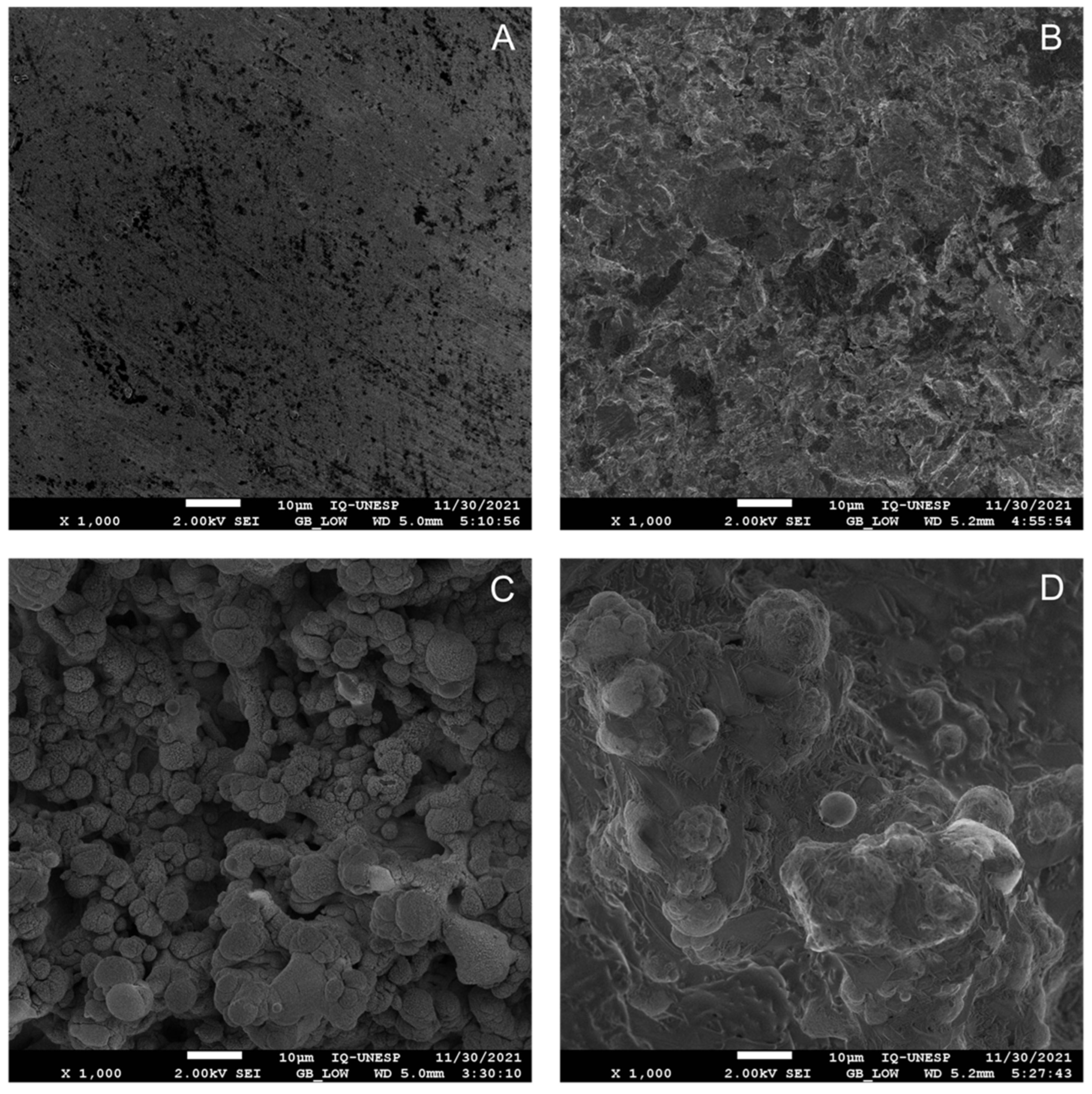
3.1. HA Coating Affects the Surface Morphology Observed with Laser Treatment

To determine the roughness of each surface, SEM analysis was performed. MS showed a smooth topography with traces of the machining process (Figure 1A). SBAS exhibited a pattern of subtractive topography, leading to the formation of microcavities with varying depths and sizes (Figure 1B). The LS had a rough surface displaying a more consistent and uniform morphological pattern and the existence of spherical particles indicates the presence of nano-sized structures. (Figure 1C). The incorporation of HA into the surface modified by laser beam (LHS) presented a rough, homogeneous, and regular surface (Figure 1D).

3.2. Laser Treatment Enhances the Wettability of Titanium Discs

To evaluate the degree of wettability, measurements of the contact angle were performed. Three measurements were performed, and the means are expressed in Table 1. All surfaces were hydrophilic with a contact angle of less than 90°; however, the high contact angle for MS and SBAS suggests that both surfaces present a moderate degree of wettability.

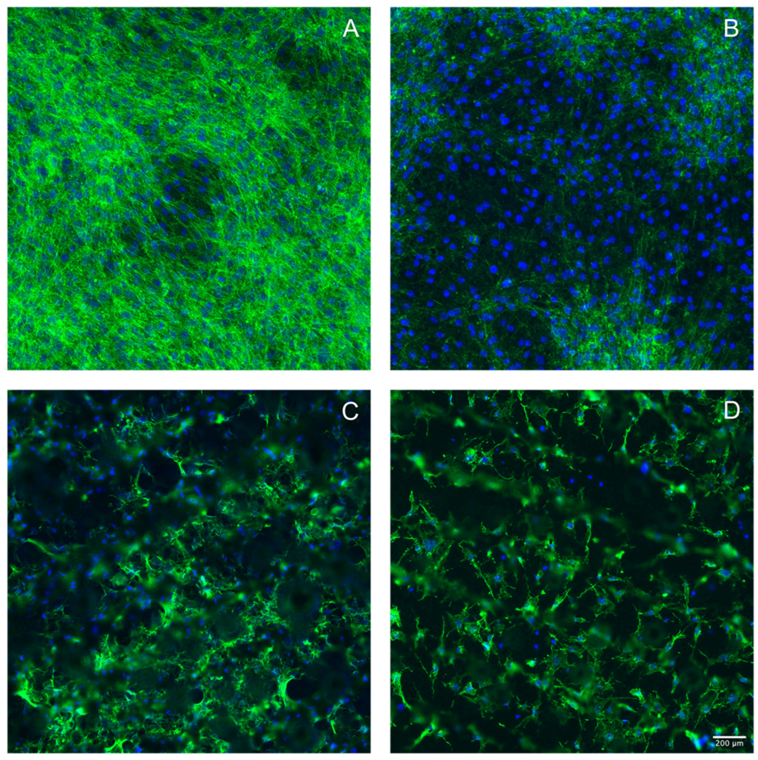
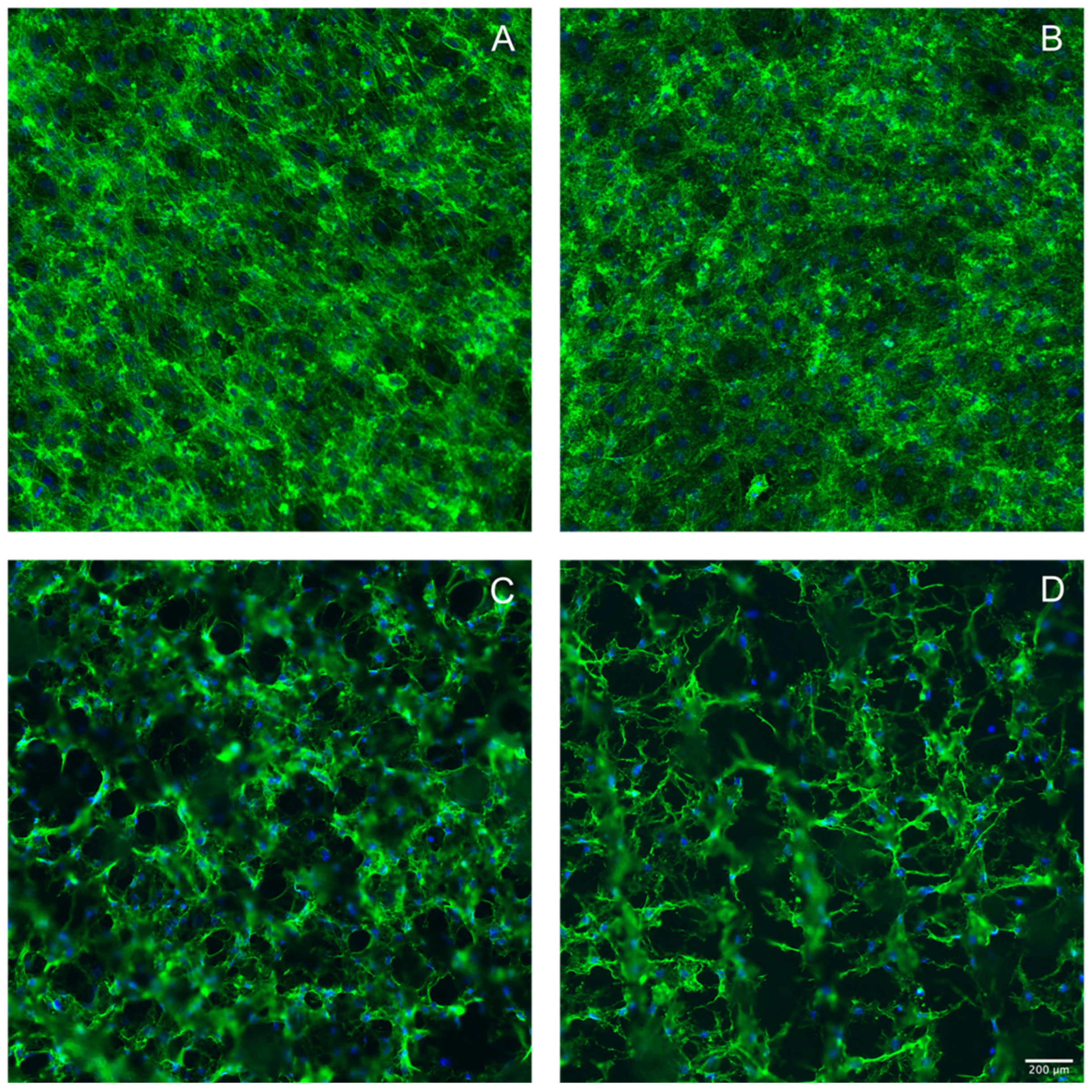
3.3. Laser Treatment of Titanium Discs Altered the Morphology of MC3T3-E1 and ST2 Cells

For the evaluation of cell morphology, phalloidin staining was performed. When grown on the smooth MS and SBAS surfaces (Figure 2A,B), MC3T3-E1 cells maintained their fibroblastic morphology, expressing long actin filaments. In contrast, there was an irregular morphology when MC3T3-E1 cells were grown on the laser-treated surfaces (Figure 2C,D). Consistently, ST2 cells maintained their original morphology when grown on a smooth surface (Figure 3A,B) but changed their morphology to irregular when grown on the laser-treated surfaces (Figure 3C,D). The presence of HA further weakened the phalloidin staining intensity in MC3T3-E1 and ST2 cells (Figure 2D and Figure 3D).

3.4. Hydroxyapatite Coating Reduced Cell Viability in MC3T3-E1 and ST2 Cells

To understand the impact of surface modification on the cell viability, an MTT assay was performed. As indicated in Table 2, HA coating caused a significant lowering of formazan formation, while all other surface modifications were comparable to the machined surface.

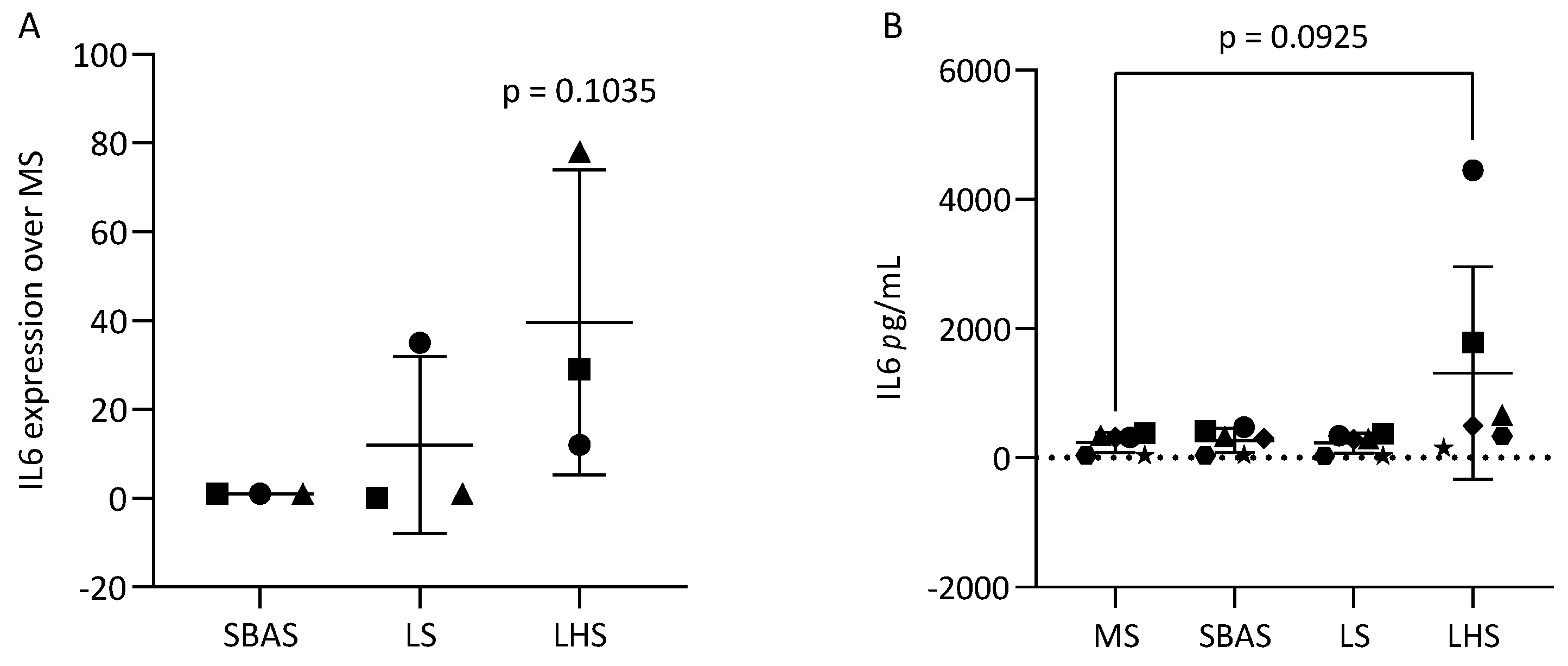
3.5. Hydroxyapatite Coating Increased IL6 in MC3T3-E1 and ST2 Cells

To determine the inflammatory response of MC3T3-E1 and ST2 to different surfaces, cells were grown on the discs for 48 h. The increased expression of IL6 was noted when MC3T3 cells were exposed to HA present on the surface LHS (Figure 4A). At the IL6 protein level, consistent results were observed (Figure 4B). LHS caused ST2 cells to express relatively more IL6 on the transcriptional (Figure 5A) and at the protein level (Figure 5B).

4. Discussion

The surface topography and roughness of dental implants exert a substantial influence on the behavior of osteogenic cells in vitro [24]. In the present study, we observed that the roughness patterns of the MS and SBAS surfaces were similar and showed moderate hydrophilicity. The laser-modified surface with and without HA, however, had a high roughness, with valleys, peaks and absolute hydrophilicity. These surface properties can affect how cells respond and may influence the rate and extent of bone deposition [4]. For a surface with moderate roughness, it is advantageous for cells to adhere stably, promoting their growth and subsequent proliferation; on the other hand, superhydrophilic and superhydrophobic surfaces are not conducive to cell attachment and growth [9]. In general, laser-modified surfaces can accelerate bone healing around implanted materials [4,36,37], through their ability to enhance osteoblast differentiation, increase matrix mineralization, and increase the expression of genes specific to bone formation [24]. Moreover, laser-treated implants with HA coating exhibit higher torque values and increased new bone volume compared to machined and acid-etched implants [4,5]. Nevertheless, there is concern regarding the HA coating of implant surfaces due to the risk of fragmentation. Fragmentation may lead to HA particles potentially triggering leukocyte activation and consequently provoking an inflammatory response [29]. However, the possible role of HA coating to affect the viability and the inflammatory status of mesenchymal cells is less clear.
The present study revealed that the laser-modified surface prompted alterations in the morphology of both MC3T3-E1 and ST2 cells, with an additional impact of HA coating. With HA coating, the cell shape was altered, and less cells with nuclear staining were noticed. Support for our findings comes from others showing that MC3T3-E1 cells were less spread and elongated on an HA surface compared to a tissue culture surface [32]. Consistently, HA coating reduced cell viability in MC3T3-E1 and ST2 cells, similar to the results from previous reports in a tissue culture dish serving as the controls [32].
Previous studies have established that the structure of HA nanoparticles significantly influences immediate inflammation, regardless of their chemical composition and charge [20]. HA nanoparticles have the capacity to enhance the release of pro-inflammatory agents such as IL1, along with chemotactic factors like IL8, macrophage inflammatory proteins, and matrix metalloproteinase 9. HA also activates polymorphonuclear neutrophils, possibly contributing to implant-related inflammation [29]. Furthermore, HA particles can trigger the production of IL18 in human monocytes due to their specific characteristics [38]. In light of these results, and to understand if the lower viability translates into an inflammatory response, the expression of IL6 was determined. Consistent with the lower viability, HA coating caused higher GAPDH values, reflecting lower cell numbers. Interestingly, HA coating caused an increased IL6 on the level of gene expression and protein in the supernatant of the MC3T3-E1 and ST2 cells. Taken together, these data suggest that while laser treatment seems safe and increases the wettability, the additional HA coating might cause some adverse reaction that affects the viability of mesenchymal cells and may even translate into a local inflammatory response, at least under in vitro conditions.
There are, however, certain limitations of this study that need to be discussed. Considering that in vitro experiments were performed, simulating patterns were created in order to simulate complex organisms, but they do not represent the clinical situation. There are several coating techniques with different tricalcium phosphate derivatives, and, in the present study, only HA coating without heat treatment and laser was tested. Further studies are necessary, evaluating different sizes and shapes of HA particles, as well as coating methods, titanium alloys, and evaluations of early and late periods, to understand under which conditions, specifically, HA is beneficial for cells and which methods should be avoided to reduce the local inflammatory process.
In summary, we can conclude that the laser treatment of titanium supports hydrophilicity, but adding hydroxyapatite may provoke an inflammatory response in vitro. These data may be important for future studies, which may use different HA coating techniques, as well as different particle sizes, aiming to reduce the expression of inflammatory mediators in short incubation periods, maintaining the advantages related to proliferation and differentiation in osteoblastic cells.

Author Contributions

Conceptualization, R.G. and F.Á.S.; methodology, R.O., A.C.G., T.P.Q., R.G. and F.Á.S.; software, L.P.; formal analysis, A.F.P.S. and L.P.; data curation, A.F.P.S., L.C.C.C. and L.P.; writing—original draft, A.F.P.S. and R.G. writing—review and editing, A.F.P.S., L.P., R.G. and F.Á.S.; visualization, A.F.P.S., T.P.Q., L.P. and F.Á.S.; supervision, L.P., R.G. and F.Á.S.; project administration, R.O., R.G. and F.Á.S.; funding acquisition, R.G. and F.Á.S. All authors have read and agreed to the published version of the manuscript.

Funding

Ana Flávia Piquera Santos received financial support through a scholarship from the Brazilian Federal Agency for Support and Evaluation of Graduate Education (CAPES), specifically through the CAPES-PrInt Program, process number 88887.576754/2020-00. The surfaces utilized in this article were also a part of a research project that received funding provided by the São Paulo Research Foundation—FAPESP, process number 2018/22108-1.

Institutional Review Board Statement

The study was carried out in accordance with the principles outlined in the Declaration of Helsinki and received approval from the Ethics Committee of the Medical University of Vienna (EK NR 631/2007).

Informed Consent Statement

Informed consent was obtained from all subjects involved in the study.

Data Availability Statement

The data presented in this study are openly available in UNESP Institutional Repository at http://hdl.handle.net/11449/244147 (accessed on 15 June 2023).

Acknowledgments

The authors extend their gratitude to Zahra Kargarpour, Azarakhsh Oladzad Abbasabadi, and Mariane Beatriz Sordi for their valuable technical and scientific contributions throughout the experimental phase of this study.

Conflicts of Interest

The authors declare no conflict of interest.

References

  1. Junker, R.; Dimakis, A.; Thoneick, M.; Jansen, J.A. Effects of Implant Surface Coatings and Composition on Bone Integration: A Systematic Review. Clin. Oral Implant. Res. 2009, 20 (Suppl. S4), 185–206. [Google Scholar] [CrossRef]
  2. Albrektsson, T.; Zarb, G.A. Current Interpretations of the Osseointegrated Response: Clinical Significance. Int. J. Prosthodont. 1993, 6, 95–105. [Google Scholar]
  3. Costa, D.O.; Prowse, P.D.H.; Chrones, T.; Sims, S.M.; Hamilton, D.W.; Rizkalla, A.S.; Dixon, S.J. The Differential Regulation of Osteoblast and Osteoclast Activity by Surface Topography of Hydroxyapatite Coatings. Biomaterials 2013, 34, 7215–7226. [Google Scholar] [CrossRef]
  4. Faeda, R.S.; Tavares, H.S.; Sartori, R.; Guastaldi, A.C.; Marcantonio, E. Biological Performance of Chemical Hydroxyapatite Coating Associated with Implant Surface Modification by Laser Beam: Biomechanical Study in Rabbit Tibias. J. Oral Maxillofac. Surg. 2009, 67, 1706–1715. [Google Scholar] [CrossRef]
  5. Sisti, K.E.; de Rossi, R.; Antoniolli, A.M.B.; Aydos, R.D.; Guastaldi, A.C.; Queiroz, T.P.; Garcia, I.R.; Piattelli, A.; Tavares, H.S. Surface and Biomechanical Study of Titanium Implants Modified by Laser with and without Hydroxyapatite Coating, in Rabbits. J. Oral Implantol. 2012, 38, 231–237. [Google Scholar] [CrossRef]
  6. Albrektsson, T.; Wennerberg, A. Oral implant surfaces: Part 1—Review focusing on topographic and chemical properties of different surfaces and in vivo responses to them. Int. J. Prosthodont. 2004, 17, 536–543. [Google Scholar] [PubMed]
  7. Smeets, R.; Stadlinger, B.; Schwarz, F.; Beck-Broichsitter, B.; Jung, O.; Precht, C.; Kloss, F.; Gröbe, A.; Heiland, M.; Ebker, T. Impact of Dental Implant Surface Modifications on Osseointegration. BioMed Res. Int. 2016, 2016, 6285620. [Google Scholar] [CrossRef]
  8. Coelho, P.G.; Jimbo, R.; Tovar, N.; Bonfante, E.A. Osseointegration: Hierarchical Designing Encompassing the Macrometer, Micrometer, and Nanometer Length Scales. Dent. Mater. 2015, 31, 37–52. [Google Scholar] [CrossRef]
  9. Majhy, B.; Priyadarshini, P.; Sen, A.K. Effect of Surface Energy and Roughness on Cell Adhesion and Growth—Facile Surface Modification for Enhanced Cell Culture. RSC Adv. 2021, 11, 15467–15476. [Google Scholar] [CrossRef]
  10. Oue, H.; Doi, K.; Oki, Y.; Makihara, Y.; Kubo, T.; Perrotti, V.; Piattelli, A.; Akagawa, Y.; Tsuga, K. Influence of Implant Surface Topography on Primary Stability in a Standardized Osteoporosis Rabbit Model Study. J. Funct. Biomater. 2015, 6, 143–152. [Google Scholar] [CrossRef]
  11. Alghamdi, H.S. Methods to Improve Osseointegration of Dental Implants in Low Quality (Type-IV) Bone: An Overview. J. Funct. Biomater. 2018, 9, 7. [Google Scholar] [CrossRef]
  12. Gómez-de Diego, R.; del Rocío Mang-de la Rosa, M.; Romero-Pérez, M.-J.; Cutando-Soriano, A.; López-Valverde-Centeno, A. Indications and Contraindications of Dental Implants in Medically Compromised Patients: Update. Med. Oral Patol. Oral Cir. Bucal 2014, 19, e483–e489. [Google Scholar] [CrossRef]
  13. Zhou, W.; Tangl, S.; Reich, K.M.; Kirchweger, F.; Liu, Z.; Zechner, W.; Ulm, C.; Rausch-Fan, X. The Influence of Type 2 Diabetes Mellitus on the Osseointegration of Titanium Implants with Different Surface Modifications—A Histomorphometric Study in High-Fat Diet/Low-Dose Streptozotocin-Treated Rats. Implant. Dent. 2019, 28, 11–19. [Google Scholar] [CrossRef] [PubMed]
  14. Najeeb, S.; Zafar, M.S.; Khurshid, Z.; Zohaib, S.; Hasan, S.M.; Khan, R.S. Bisphosphonate Releasing Dental Implant Surface Coatings and Osseointegration: A Systematic Review. J. Taibah Univ. Med. Sci. 2017, 12, 369–375. [Google Scholar] [CrossRef] [PubMed]
  15. Le Guéhennec, L.; Soueidan, A.; Layrolle, P.; Amouriq, Y. Surface treatments of titanium dental implants for rapid osseointegration. Dent. Mater. 2007, 23, 844–854. [Google Scholar] [CrossRef] [PubMed]
  16. Joób-Fancsaly, A.; Divinyi, T.; Fazekas, A.; Petó, G.; Karacs, A. Surface Treatment of Dental Implants with High-Energy Laser Beam. Fogorv. Sz. 2000, 93, 169–180. [Google Scholar] [PubMed]
  17. Bereznai, M.; Pelsöczi, I.; Tóth, Z.; Turzó, K.; Radnai, M.; Bor, Z.; Fazekas, A. Surface Modifications Induced by Ns and Sub-Ps Excimer Laser Pulses on Titanium Implant Material. Biomaterials 2003, 24, 4197–4203. [Google Scholar] [CrossRef] [PubMed]
  18. Shah, F.A.; Johansson, M.L.; Omar, O.; Simonsson, H.; Palmquist, A.; Thomsen, P. Laser-Modified Surface Enhances Osseointegration and Biomechanical Anchorage of Commercially Pure Titanium Implants for Bone-Anchored Hearing Systems. PLoS ONE 2016, 11, e0157504. [Google Scholar] [CrossRef] [PubMed]
  19. Kang, S.H.; Cho, S.A. Comparison of removal torques for laser-treated titanium implants with anodized implants. J. Craniofac. Surg. 2011, 22, 1491–1495. [Google Scholar] [CrossRef]
  20. Pujari-Palmer, S.; Chen, S.; Rubino, S.; Weng, H.; Xia, W.; Engqvist, H.; Tang, L.; Ott, M.K. In Vivo and In Vitro Evaluation of Hydroxyapatite Nanoparticle Morphology on the Acute Inflammatory Response. Biomaterials 2016, 90, 1–11. [Google Scholar] [CrossRef]
  21. Lange, T.; Schilling, A.F.; Peters, F.; Haag, F.; Morlock, M.M.; Rueger, J.M.; Amling, M. Proinflammatory and Osteoclastogenic Effects of Beta-Tricalciumphosphate and Hydroxyapatite Particles on Human Mononuclear Cells In Vitro. Biomaterials 2009, 30, 5312–5318. [Google Scholar] [CrossRef]
  22. Rydén, L.; Omar, O.; Johansson, A.; Jimbo, R.; Palmquist, A.; Thomsen, P. Inflammatory Cell Response to Ultra-Thin Amorphous and Crystalline Hydroxyapatite Surfaces. J. Mater. Sci. Mater. Med. 2017, 28, 9. [Google Scholar] [CrossRef]
  23. Narducci, P.; Nicolin, V. Differentiation of Activated Monocytes into Osteoclast-like Cells on a Hydroxyapatite Substrate: An In Vitro Study. Ann. Anat. 2009, 191, 349–355. [Google Scholar] [CrossRef]
  24. Stich, T.; Alagboso, F.; Křenek, T.; Kovářík, T.; Alt, V.; Docheva, D. Implant-Bone-Interface: Reviewing the Impact of Titanium Surface Modifications on Osteogenic Processes In Vitro and In Vivo. Bioeng. Transl. Med. 2022, 7, e10239. [Google Scholar] [CrossRef]
  25. Lu, R.J.; Wang, X.; He, H.X.; E, L.L.; Li, Y.; Zhang, G.L.; Li, C.J.; Ning, C.Y.; Liu, H.C. Tantalum-incorporated hydroxyapatite coating on titanium implants: Its mechanical and in vitro osteogenic properties. J. Mater. Sci. Mater. Med. 2019, 30, 111. [Google Scholar] [CrossRef]
  26. Barrère, F.; van der Valk, C.M.; Meijer, G.; Dalmeijer, R.A.J.; de Groot, K.; Layrolle, P. Osteointegration of Biomimetic Apatite Coating Applied onto Dense and Porous Metal Implants in Femurs of Goats. J. Biomed. Mater. Res. B Appl. Biomater. 2003, 67, 655–665. [Google Scholar] [CrossRef]
  27. Velard, F.; Laurent-Maquin, D.; Braux, J.; Guillaume, C.; Bouthors, S.; Jallot, E.; Nedelec, J.-M.; Belaaouaj, A.; Laquerriere, P. The Effect of Zinc on Hydroxyapatite-Mediated Activation of Human Polymorphonuclear Neutrophils and Bone Implant-Associated Acute Inflammation. Biomaterials 2010, 31, 2001–2009. [Google Scholar] [CrossRef]
  28. Harada, Y.; Wang, J.T.; Doppalapudi, V.A.; Willis, A.A.; Jasty, M.; Harris, W.H.; Nagase, M.; Goldring, S.R. Differential Effects of Different Forms of Hydroxyapatite and Hydroxyapatite/Tricalcium Phosphate Particulates on Human Monocyte/Macrophages In Vitro. J. Biomed. Mater. Res. 1996, 31, 19–26. [Google Scholar] [CrossRef]
  29. Velard, F.; Laurent-Maquin, D.; Guillaume, C.; Bouthors, S.; Jallot, E.; Nedelec, J.-M.; Belaaouaj, A.; Laquerriere, P. Polymorphonuclear Neutrophil Response to Hydroxyapatite Particles, Implication in Acute Inflammatory Reaction. Acta Biomater. 2009, 5, 1708–1715. [Google Scholar] [CrossRef]
  30. Rydén, L.; Molnar, D.; Esposito, M.; Johansson, A.; Suska, F.; Palmquist, A.; Thomsen, P. Early Inflammatory Response in Soft Tissues Induced by Thin Calcium Phosphates. J. Biomed. Mater. Res. A 2013, 101, 2712–2717. [Google Scholar] [CrossRef]
  31. Ozawa, S.; Kasugai, S. Evaluation of Implant Materials (Hydroxyapatite, Glass-Ceramics, Titanium) in Rat Bone Marrow Stromal Cell Culture. Biomaterials 1996, 17, 23–29. [Google Scholar] [CrossRef]
  32. Goldberg, A.J.; Liu, Y.; Advincula, M.C.; Gronowicz, G.; Habibovic, P.; Kuhn, L.T. Fabrication and Characterization of Hydroxyapatite-Coated Polystyrene Disks for Use in Osteoprogenitor Cell Culture. J. Biomater. Sci. Polym. Ed. 2010, 21, 1371–1387. [Google Scholar] [CrossRef]
  33. Xu, W.; Xu, C.; Yi, J.; Dai, H. The Effect of Different Hydroxyapatite Microparticles on the Osteogenic Differentiation of MC3T3-E1 Preosteoblasts. J. Mater. Chem. B 2018, 6, 5234–5242. [Google Scholar] [CrossRef]
  34. Aparecida, A.H.; Fook, M.V.L.; Guastaldi, A.C. Biomimetic Apatite Formation on Ultra-High Molecular Weight Polyethylene (UHMWPE) Using Modified Biomimetic Solution. J. Mater. Sci. Mater. Med. 2009, 20, 1215–1222. [Google Scholar] [CrossRef]
  35. Panahipour, L.; Omerbasic, A.; Nasirzade, J.; Gruber, R. TGF-β Activity of a Demineralized Bone Matrix. Int. J. Mol. Sci. 2021, 22, 664. [Google Scholar] [CrossRef]
  36. Cho, S. A Removal Torque of the Laser-Treated Titanium Implants in Rabbit Tibia. Biomaterials 2003, 24, 4859–4863. [Google Scholar] [CrossRef]
  37. Gaggl, A.; Schultes, G.; Müller, W.D.; Kärcher, H. Scanning Electron Microscopical Analysis of Laser-Treated Titanium Implant Surfaces—A Comparative Study. Biomaterials 2000, 21, 1067–1073. [Google Scholar] [CrossRef]
  38. Grandjean-Laquerriere, A.; Laquerriere, P.; Laurent-Maquin, D.; Guenounou, M.; Phillips, T.M. The Effect of the Physical Characteristics of Hydroxyapatite Particles on Human Monocytes IL-18 Production In Vitro. Biomaterials 2004, 25, 5921–5927. [Google Scholar] [CrossRef]
Figure 1. Scanning electron microscopy (SEM). Images showing the roughness pattern present on machined commercially pure titanium surfaces (A); sandblasting followed by acid etching (B); laser treatment without HA coating (C) and with HA coating (D). The inset images provided a detailed view of the surface morphology at high resolution. 1000× magnification.
Applsci 13 12646 g001
Figure 2. Laser treatment of titanium discs altered the morphology of MC3T3-E1 cells. Light and fluorescence images of phalloidin staining of MC3T3-E1 cells seeded on (A) machined surface—MS, (B) sandblasting followed by acid etching—SBAS, (C) laser treatment without HA coating and (D) with HA coating and observed by confocal laser scanning microscope. Black spaces in LS and LHS correspond to surface roughness. The scale bar is 200 μm.
Applsci 13 12646 g002
Figure 3. Laser treatment of titanium discs altered the morphology of ST2 cells. Light and fluorescence images of phalloidin staining of ST2 cells seeded on (A) machined surface—MS, (B) sandblasting followed by acid etching—SBAS, (C) laser treatment without HA coating, and (D) with HA coating and observed by confocal laser scanning microscope. Black spaces in LS and LHS correspond to surface roughness. The scale bar is 200 μm.
Applsci 13 12646 g003
Figure 4. Effect of HA-mediated IL6 secretion in MC3T3-E1. (A) IL6 expression. The RT-PCR analysis was performed in accordance with the procedure described in the Materials and Methods section, and the data depict the expression levels of IL6 normalized to the control, represented by MS surface. (B) Immunoassay showed that HA increased the expression of IL6 in MC3T3-E1. Each format shape represents the levels of IL6 from individual experiments and are presented with mean and SD values. Statistical analysis was carried out through a one-way ANOVA, and corrected p-values were indicated.
Applsci 13 12646 g004
Figure 5. Effect of HA-mediated in IL6 secretion in ST2 cells. (A) IL6 expression. The RT-PCR analysis was performed in accordance with the procedure described in the Materials and Methods section, and the data depict the expression levels of IL6 normalized to the control, represented by MS surface. (B) Immunoassay showed that HA increased the expression of IL6 in ST2 cells. Each format shape represents the levels of IL6 from individual experiments and are presented with mean and SD values. Statistical analysis was carried out through a one-way ANOVA, and corrected p-values were indicated.
Applsci 13 12646 g005
Table 1. Variation in contact angle (θ) values across different surface modifications. The wetting properties of the discs were assessed in triplicate under room temperature conditions. These measurements are indicated by the contact angle with water. A lower contact angle indicates a higher level of surface hydrophilicity.
Surface/AngleMSSBASLSLHS
1st review68.9°48.8°00
2nd review81.2°37.5°00
3rd review72.9°22.9°00
Average74.3°36.4°00
Table 2. Cell viability decreased upon exposure to the HA present on the LHS surface. Cell viability is quantified by measuring formazan production and was expressed as a percentage relative to the control (MS). HA exposure resulted in a reduction in cell viability of approximately 30–50% in both MC3T3-E1 and ST2 cell lines.
MC3T3-E1ST2
SBAS133.5 ± 43.3122.1 ± 18.7
LS95.2 ± 14.1111.3 ± 32.8
LHS69.7 ± 13.353.5 ± 7.5
Disclaimer/Publisher’s Note: The statements, opinions and data contained in all publications are solely those of the individual author(s) and contributor(s) and not of MDPI and/or the editor(s). MDPI and/or the editor(s) disclaim responsibility for any injury to people or property resulting from any ideas, methods, instructions or products referred to in the content.

