Communication

Numerical Study of Tympanosclerosis Including Its Effect on Human Hearing

1
Escola Superior de Saúde—I.P. Porto, LAETA-INEGI, Clínica ORL-Dr. Eurico Almeida, Widex, 4200-072 Porto, Portugal
2
LAETA-INEGI, Faculty of Engineering of the University of Porto, FEUP, 4200-465 Porto, Portugal
3
LAETA-INEGI, Institute of Mechanical Engineering and Industrial Management, 4200-465 Porto, Portugal
*
Author to whom correspondence should be addressed.
Appl. Sci. 2023, 13(3), 1665; https://doi.org/10.3390/app13031665
Submission received: 30 November 2022 / Revised: 14 January 2023 / Accepted: 18 January 2023 / Published: 28 January 2023
(This article belongs to the Special Issue Recent Developments and Emerging Trends in Biomechanics)

Abstract

:
Tympanosclerosis is an abnormal disorder of the middle ear or only the eardrum (i.e., myringosclerosis) in which there are calcium deposits. Normally, it is caused by recurrent middle ear infections. In this work, a 3D finite element model of the ear was developed, simulating different cases of tympanosclerosis. Through this model, the magnitude and the phase angle of the umbo and stapes displacement were obtained. The middle ear sound transfer function was determined for a stimulus of 80, 90 and 100 dB SPL, in a frequency range between 100 Hz and 10 kHz, applied on the outer surface of the eardrum in the external auditory canal. Depending on the tympanosclerosis affected area, the main conclusion is that worse results (leading to hearing loss) occur when all of the ossicular chain is affected.

1. Introduction

Tympanosclerosis is characterized by the calcification of the tissues of the eardrum and middle ear ossicles. When this calcification only involves the eardrum, it is designated as myringosclerosis. Tympanosclerosis, itself, can also invade the malleus, the malleus and incus, or all the tympanic cavity of the middle ear. It is easy to identify a tympanosclerosis due to the typical white and opaque look of the eardrum. The affected area can vary considerably between cases [1].
Tympanosclerosis within the middle ear is similar to that occurring within the eardrum. When it occurs in the middle ear, it often leads to conductive hearing loss, caused by the ossicular fixation.
The eardrum can be divided into a “pars tensa” and “pars flaccida”. The “pars tensa” has three layers. The exterior layer is the epidermis that continues with the external auditory canal. The intermediate layer, the lamina propria, is mainly composed of collagen fibers (responsible for its mobility). The interior layer is the mucosal epithelium, which continues in the middle ear mucosa. Tympanosclerosis develops in the lamina propria.
Histologically, tympanosclerosis arises as an acellular hyalinization of the subepithelial connective tissue of the eardrum and the middle ear, with deposition of proteinaceous substances. In most instances, calcification is also present.
Tympanosclerosis occurs due to the fact of a degenerative process within the connective tissue. This leads to the degradation of the collagen fibers and originates dystrophic calcification. This degeneration may be a consequence of inflammation or an infection within the middle ear due to the fact of, for example, bacterial proteinases and collagenases. Another possible cause of tympanosclerosis is an autoimmune process [2].
A study by Ferri M et al. proposed that a correlation exists between the presence of atherosclerosis and tympanosclerosis, where people with atherosclerosis are more prone to having tympanosclerosis [3].
Tympanosclerosis or myringosclerosis can be caused by a prior history of tympanostomy tube placement and/or an associated history of otitis media. Unless the involvement of the eardrum is particularly severe, it is rare for tympanosclerosis to cause significant conductive hearing loss.
Tympanosclerosis is a less common pathology than myringosclerosis. Tympanosclerosis frequently develops after an acute or chronic otitis media. Children who have otitis media with effusion and ventilation tubes have a higher risk of developing tympanosclerosis, between 11 and 37% [4,5].
If the tympanosclerosis is extensive, it may affect hearing. Audiometry tests (tonal and vocal audiograms) should be conducted if there is hearing loss. In the vast majority of cases, the air–bone gap is smaller than 40 dB. Tympanometry produces flattened tympanograms, type B tracing, with a low static admittance due to the increased stiffness of the eardrum [6]. Computerized tomography (CT) can be used to determine if tympanosclerosis exists in the middle ear.
In a study by Wu (2006), 35.6% of patients with chronic otitis media, suppurative or not, had tympanosclerosis. In these patients, 77.8% had dry ear and conductive hearing loss [7].
Studies [8,9] have shown that tympanosclerosis plaques have a concentration of phosphate and calcium, with a Ca/P ratio similar to hydroxyapatite (HA). In the lamina propria, the crystals of carbonate apatite appear dispersed in the matrix of degenerated connective tissue [10].
Tympanoplasty and ossicular reconstruction can be performed in cases of tympanosclerosis. The risks of cochlear damage occur due to the extensive dissection that is required in tympanosclerotic ears and the coexistence of labyrinthine erosion.
Treatment is only necessary when hearing loss exists. When only myringosclerosis is present, hearing loss is rare. If this process continues to the middle ear, creating larger plates adherent to the ossicles, deafness occurs, the severity of which depends on the middle ear contribution and on how the ossicular chain is affected. Surgery comprises excision of the tympanosclerosis plaques and reconstruction of the ossicular chain. This surgery frequently results in significant hearing improvement, but occasionally the problem can return, and the best solution for hearing loss correction will be the usage of hearing aids.
When tympanosclerosis fixes the incus and malleus, a reconstruction of the ossicular chain is necessary, using manubrio-stapedioplasty surgery [11]. If only the malleus becomes fixed, remotion of the sclerotic plaques is needed by performing a canaloplasty [12].
A biomechanical study of myringosclerosis [13] showed that the transmission of sound to the stapes footplate depends on the calcification degree of the myringosclerosis plaques, as well as the localization and dimensions. Other results indicate that the dimension and localization of these plaques are more relevant for hearing loss than the degree of calcification [14].
Eardrum perforations occur following acute or chronic otitis media, where consequent myringosclerosis and tympanosclerosis can develop. A previous work [15] studding the mechanical behavior of the tympano-ossicular chain, comparing the effect of myringosclerosis and eardrum perforations on the mechanical properties of the ossicular chain. By using the finite element method, it is possible to verify that when a perforation of 0.6 mm is present in the eardrum, no differences occur relative to the displacement of the umbo and stapes footplate, when compared with a model that represents a normal eardrum. The same study concluded that from a hearing point of view, the differences between a normal and a myringosclerosis ear are reduced, which is in accordance with clinical practice. The largest differences occur when a large perforation in the inferior two quadrants of the eardrum (7.0 mm) is present, leading to a large decrease in the displacements for the low and middle frequencies (Figure 1).
In the present study, the finite element method was used to compare a model representing a healthy ear with three different models representing different tympanosclerosis cases (only in the eardrum; affecting the eardrum and malleus; and affecting the eardrum and the three ossicles).

2. Methods

The geometrical models used in this work were obtained from the project “The Visible Ear”. The images were acquired, using a cryosectioning procedure, from the temporal bone of an old woman. Finally, hand segmentation of the different tissues was performed [16]. The slice thickness was 25 μm, and high-resolution images were captured every 50 μm. A total of 597 images, with 24 bit RGB and a resolution of 50 μm/pixel, were obtained. Approximately 26 different organs of the middle ear were identified through the manual segmentation, performed by Mads Sølvsten Sørensen, of each image.
A 3D finite element model of the ear was built, and it included the external auditory canal, the eardrum, ossicles (malleus, incus and stapes), tympanic cavity, six ligaments (malleus: anterior, lateral and superior; incus: posterior and superior; stapes: stapedius annular ligament), two muscles (stapes: stapedius; malleus: tensor tympani), incudomalleolar and incudostapedial joints and the cochlear fluid [17]. The linear elastic properties were incorporated in all parts of the 3D finite element model.
The geometry was discretized into a finite element mesh, and numerical simulations were conducted using the Abaqus® Standard [18].
The finite element mesh was built using linear tetrahedral elements (C3D4) for the ossicles, linear tetrahedral acoustic elements (AC3D4) for the acoustic components and linear truss elements (T3D2) for the tendons and ligaments. The connection of the Eustachian tube with the nasopharynx was not modeled, since it was assumed closed.
The eardrum was considered as having homogeneous properties, with isotropic behavior in the “pars flaccida” and orthotropic behavior in “pars tensa” [17,19,20,21,22]. In the “pars tensa”, the radial and circumferential Young’s modulus was 32 and 20 MPa, respectively. For the “pars flaccida”, a Young’s modulus of 10 MPa was considered (Table 1).
A homogeneous and isotropic behavior was assumed for the ossicles, with a Young’s modulus of 14.1 GPa. The adopted values for the density are in accordance with the literature and are shown in Table 1 [20,21].
A Poisson’s ratio of 0.3 was considered for the entire model [20], except for the ear cartilage, where a value of 0.4 was assumed [23].
The Raleigh damping matrix, C, can be expressed as a combination of the stiffness and mass matrixes [17] and requires two parameters. In the middle ear components, the Raleigh’s proportional damping coefficients considered were α = 0 s−1 and β = 0.0001 s.
The ligaments suspending the ossicles and the muscles were assumed to behave in a linear elastic regime, and the mechanical properties (density and Young’s modulus) are shown in Table 2. Table 2 also contains the density and bulk modulus for the acoustic medium, including the air in the tympanic cavity and the fluids in the tympanic cavity and cochlea [20,21].
Fixed boundary conditions were applied to the tympanic annulus, surrounding the eardrum, which is attached to the temporal bone. The bone was assumed to have an isotropic behavior, with a Young’s modulus of 6.0 E4 Pa. The free extremities of the stapedius annular ligament (around the stapes), the three ligaments of the malleus and the two ligaments of the incus were also considered fixed, and the properties are shown in Table 2.
Acoustic elements, containing only pressure degrees of freedom, were used in order to simulate the existence of air inside the tympanic cavity, in the external auditory canal and the fluid in the cochlea. The constitutive behavior of the fluid was defined mathematically in the work of Bruno et al. [17].
According to Equation (1), sound pressure levels (SPLs) of 80, 90 and 100 dB SPL can be converted into pressure ( ρ ), respectively, as 0.200, 0.632 and 2.000 Pa. These acoustic pressures were applied in the external auditory canal (Figure 2).
dB   SPL = 20 × log 10 ( ρ / ρ 0 )
In Equation (1), ρ 0 = 20   μ Pa is the reference sound pressure, which corresponds to the audibility threshold.
In order to transmit the sound pressures from the acoustic elements into the structural elements, the Abaqus® Standard TIE command was used [18].
According to the work of Berdich et al. [13], the mechanical properties of the intermediate layer of the eardrum were modified, using the classical rule of mixtures for composites containing particles, assuming that the plaques were made of HA particles imbedded in a matrix of connective tissue. The HA plaque properties were obtained using the rule of mixtures for composite materials, with the fibers uniformly distributed [13,14]. The plaques were considered isotropic, and the particles of HA were assumed as having a uniform distribution throughout the plaque. The density considered for the HA was 3160 kg/m3 (Table 3) [24].
In a steady-state dynamic analysis, the equations of the steady harmonic motion of the system were solved directly. Such an analysis provides the steady-state amplitude and phase of the umbo and stapes footplate response due to the fact of harmonic excitation at a given frequency. The amplitude and phase of the response in the matrix form was obtained through Equation (2).
[ K NM Ω 2 M NM Ω ( C m NM + C k NM ) Ω ( C m NM + C k NM ) K NM + Ω 2 M NM ] { R ( u M ) I ( u M ) } = { R ( P N ) I ( P N ) }
where Ω is the circular frequency, K is the stiffness matrix, M is the mass matrix, C k is the stiffness proportional damping matrix, C m is the mass proportional damping matrix, u is the complex displacement ( R and I are the real and imaginary parts, respectively) and P is the complex force applied to the model. The acoustic–structural coupling is described by Equation (3).
1 ρ f p x · n + u ¨ · n = 0
where ρ f is the fluid density, p is the acoustic pressure, n is the normal vector pointing into the fluid and u ¨ is the structural acceleration.
The mesh size (number of nodes and elements) can be found in Table 4.
In this work, depending on the affected area, different cases of tympanosclerosis were considered: (1) tympanosclerosis only affecting the eardrum, also known as myringosclerosis; (2) tympanosclerosis affecting the eardrum and the malleus; (3) tympanosclerosis affecting the eardrum and the three ossicles (Figure 3).
The different results were compared with the results obtained with the model that was representative of a normal middle ear.

3. Results

To confirm the validity of the present model, the results were compared with the results of other works [21,22,25,26,27,28] and those published in [22]. The different results presented were obtained using a finite element model of the ear, which was validated by comparing the experimental and numerical results [17,29]. The validation of the model was achieved by comparing the responses obtained with the experimental results of Nishihara et al. [25] and Huber et al. [27].
All of the results in Figure 4, Figure 5, Figure 6, Figure 7, Figure 8, Figure 9, Figure 10 and Figure 11 were obtained by applying a pressure corresponding to 80 dB SPL in the external auditory canal.

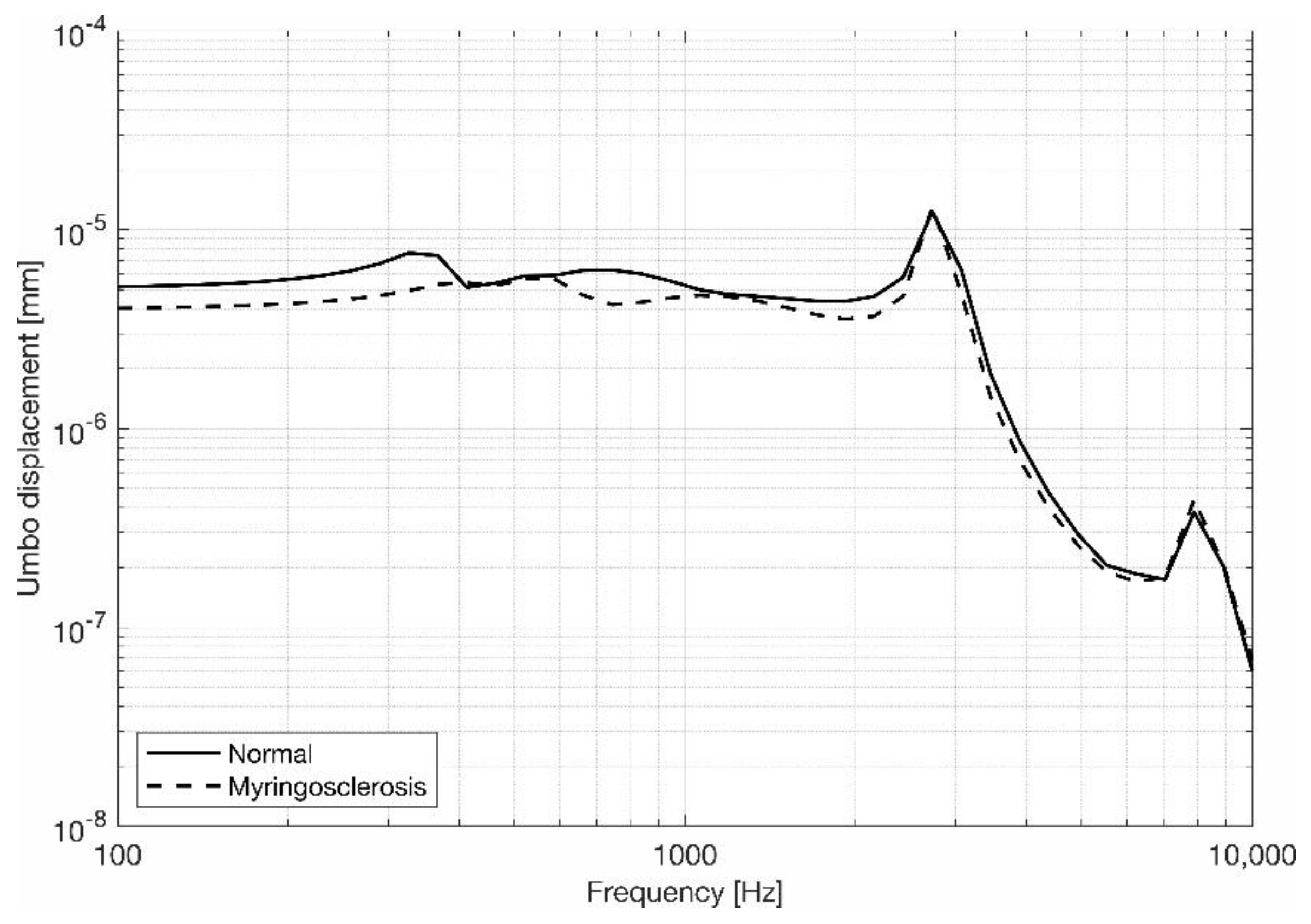
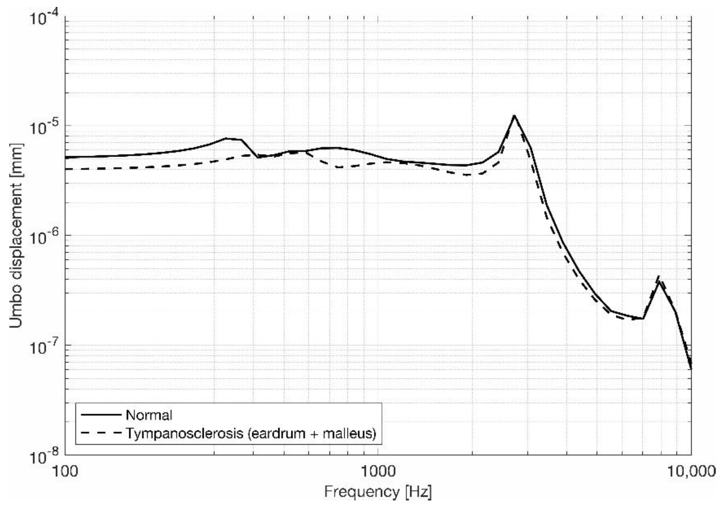
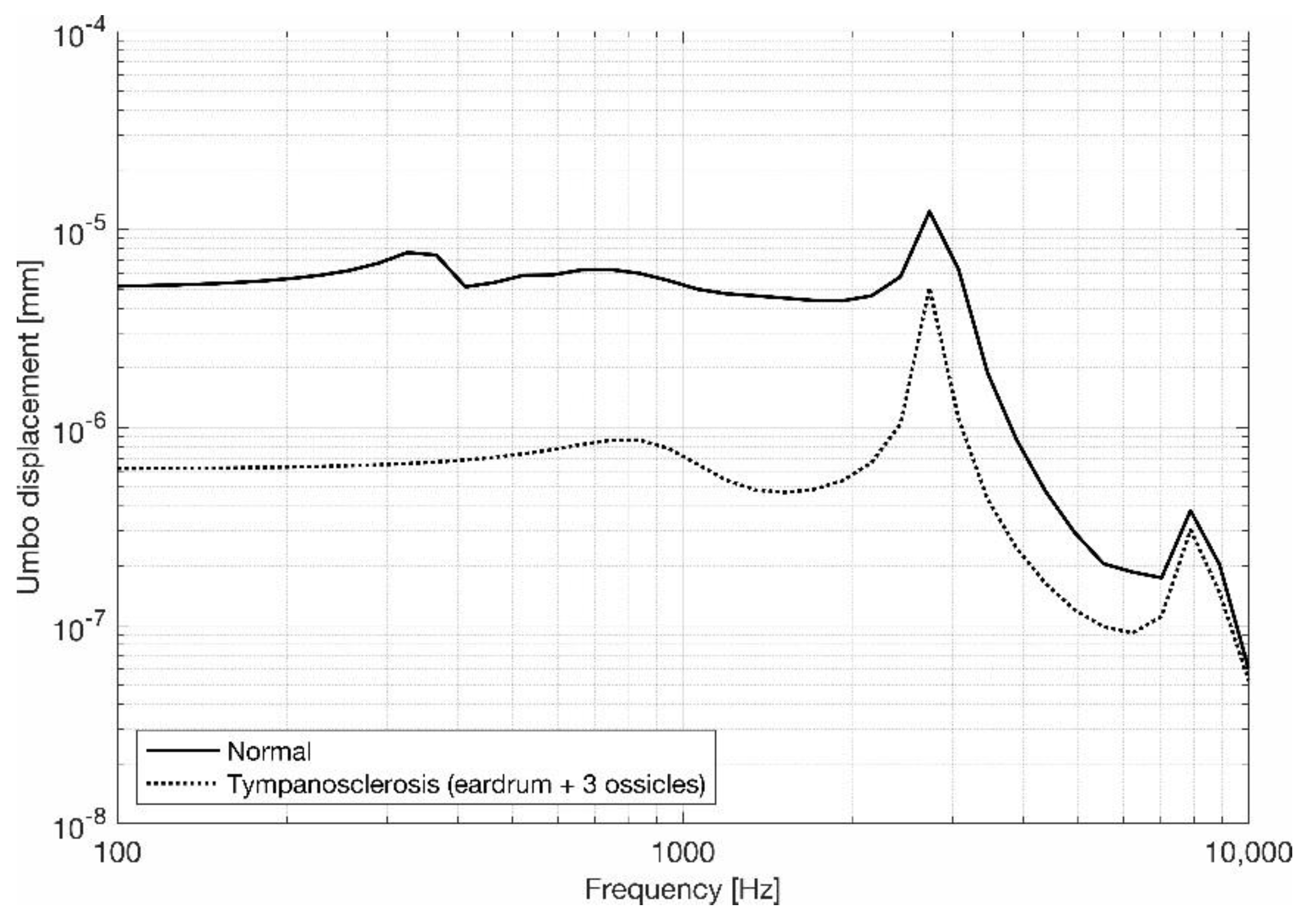
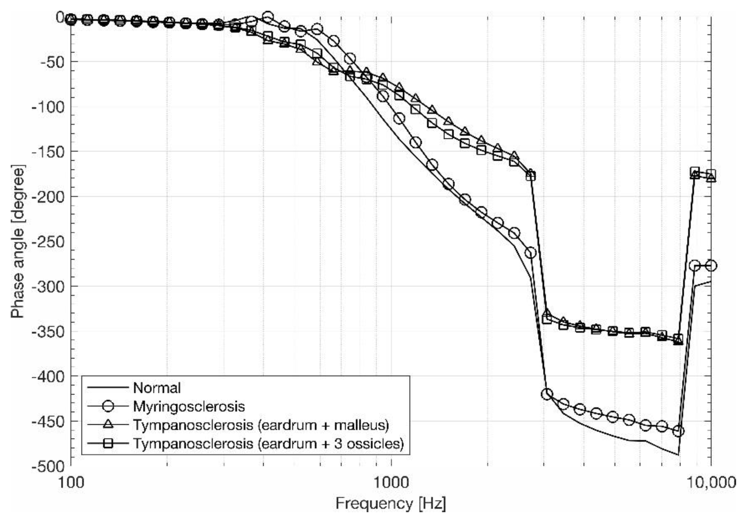
3.1. Eardrum Behavior

For a better study of the eardrum behavior, the umbo displacements and the phase angle were obtained and plotted. In terms of movement, the behavior of the eardrum was similar for all frequencies below 3 kHz (where the maximum point occurs), showing a decrease for the higher frequencies.
Figure 4 shows the magnitude of the umbo displacements, comparing the model that was representative of a normal ear and myringosclerosis. In Figure 5, we can observe the results where the tympanosclerosis affected the eardrum and the malleus, and in Figure 6, the results where the tympanosclerosis invaded the eardrum and the three ossicles.
Figure 4. Umbo displacements’ magnitude, comparing the model that was representative of a normal ear and myringosclerosis.
Applsci 13 01665 g004
Figure 5. Umbo displacements’ magnitude, comparing the model that was representative of a normal ear and tympanosclerosis affecting the eardrum and the malleus.
Applsci 13 01665 g005
Figure 6. Umbo displacements’ magnitude, comparing the model that was representative of a normal ear and tympanosclerosis affecting the eardrum and the three ossicles.
Applsci 13 01665 g006
We can see that if the tympanosclerosis only affected the eardrum or the eardrum and malleus, similar results were obtained, compared with the normal (healthy) model. If the tympanosclerosis touched the three ossicles, smaller displacements of the umbo were obtained. For low and middle frequencies, these results were more pronounced.
Figure 7 shows the umbo phase angle. A 180° phase shift near the 3 kHz frequency was encountered when the ossicles were affected. In a normal situation or in cases of myringosclerosis, this shift was nearly 260° in the same frequency (3 kHz). This shows an acoustic resonance in the external auditory canal, near that frequency.
The air into the external auditory meatus and into the tympanic cavity represents a significant gain in sound pressure near the tympanic membrane, as shown by Bruno et al. [25]. The phase shift of 180° near 3 and 8 kHz, supported by a rapid displacement increase at the umbo and stapes footplate, showed the presence of resonances in the external auditory meatus.
Figure 7. Phase angles for the umbo, comparing different situations.
Applsci 13 01665 g007

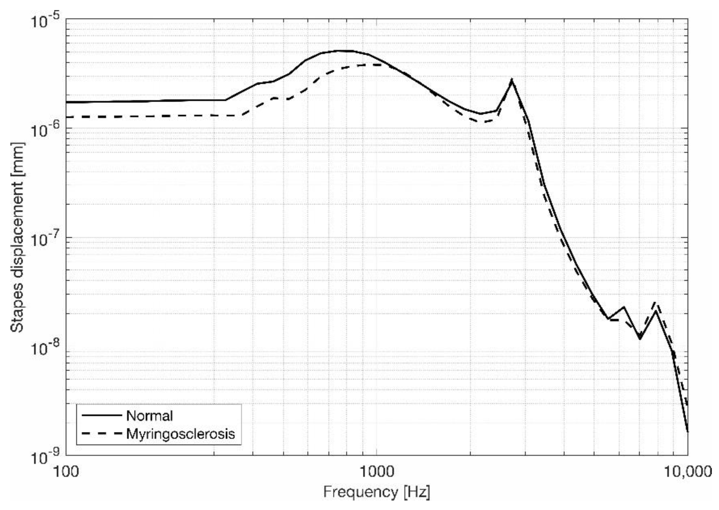
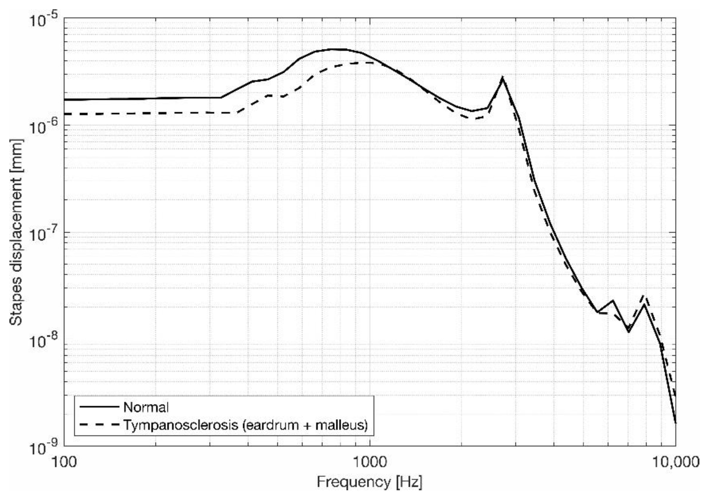
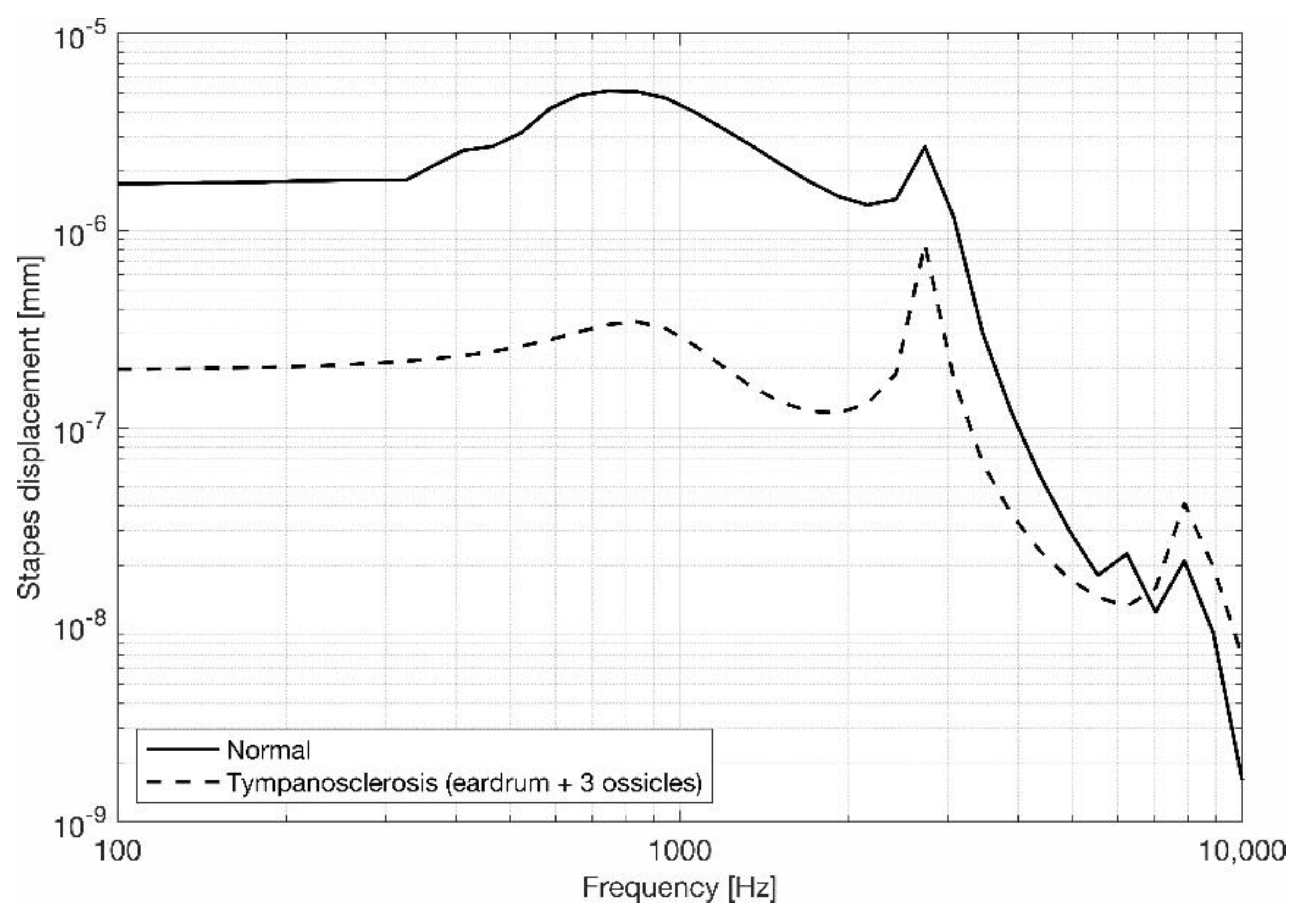
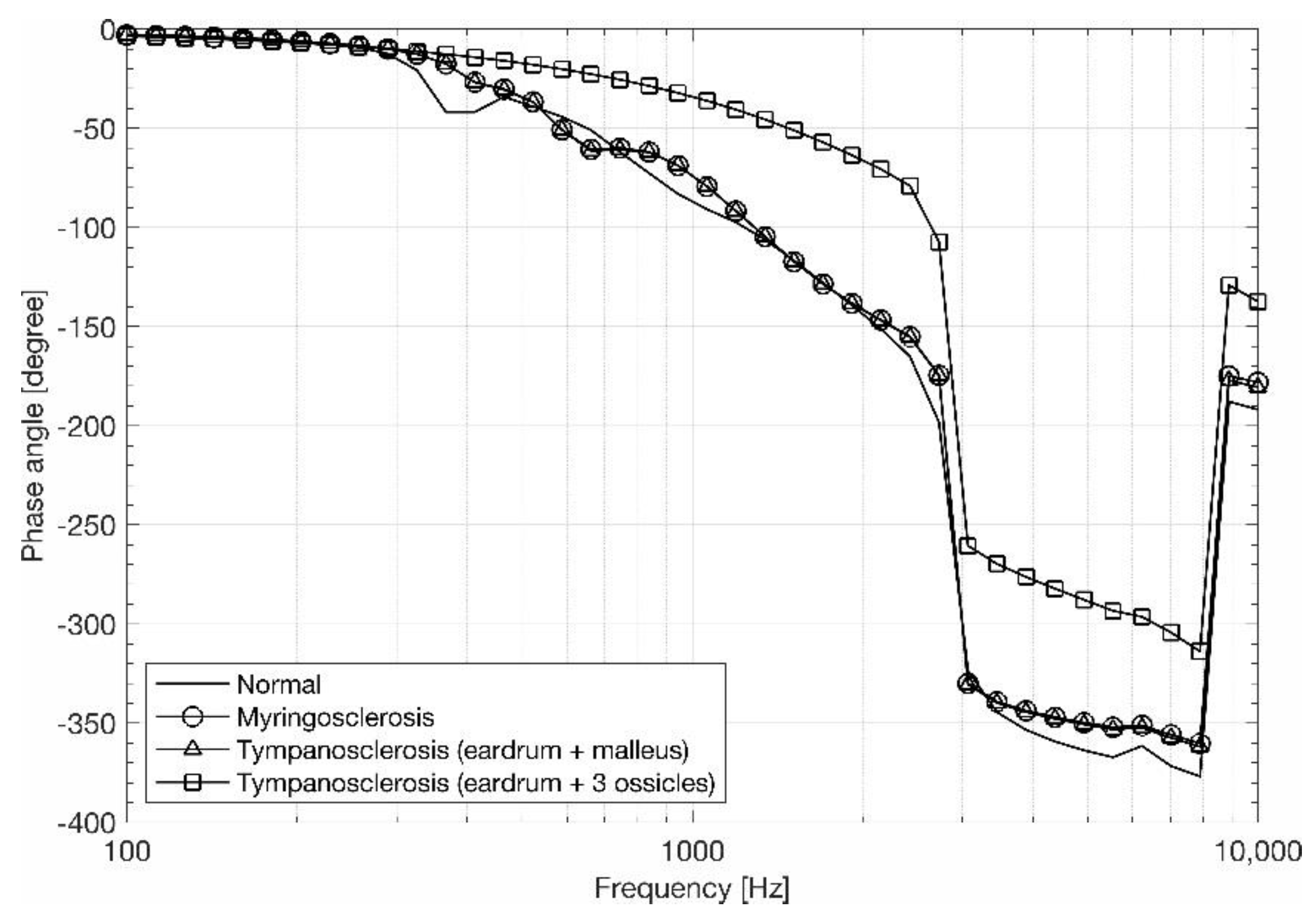
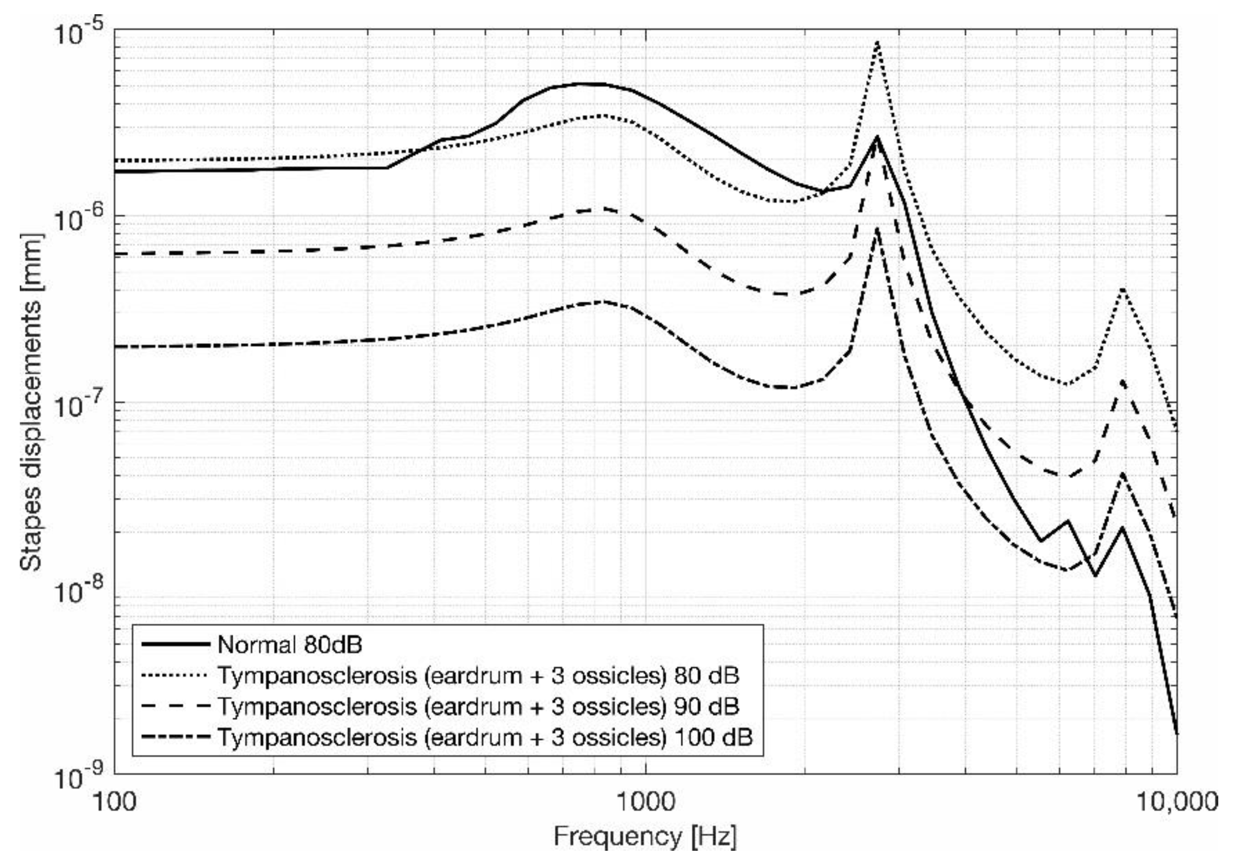
3.2. Stapes Behavior

The behavior of the displacements in the stapes footplate was similar for the middle frequencies, reaching a peak near a frequency of 1 kHz and a second one near a frequency of 3 kHz, with a decrease for the higher frequencies.
When comparing the results from the model that was representative of a normal ear with the model representing an ear with myringosclerosis, only a small difference was verified for frequencies below 1 kHz. For higher frequencies, no differences were verified (Figure 8). Even when the tympanosclerosis affected the malleus, the results were similar when compared with myringosclerosis (Figure 9). If the tympanosclerosis affected the three ossicles, smaller displacements were obtained for the entire frequency range, with higher differences in the lower and middle frequencies (Figure 10). These results are in accordance with clinical practice [30], since smaller displacements in the stapes footplate reflect a hearing loss.
Figure 8. Stapes footplate displacements’ magnitude, comparing the model that was representative of a normal ear and myringosclerosis.
Applsci 13 01665 g008
Figure 9. Stapes footplate displacements’ magnitude, comparing the model that was representative of a normal ear and tympanosclerosis affecting the eardrum and the malleus.
Applsci 13 01665 g009
Figure 10. Stapes footplate displacements’ magnitude, comparing the model that was representative of a normal ear and tympanosclerosis affecting the eardrum and the three ossicles.
Applsci 13 01665 g010
In the stapes footplate, a phase shift of 260° occurred for a frequency of 3 kHz (similar to the one encountered for the umbo) for the three models: the normal one, with myringosclerosis, and with tympanosclerosis in the eardrum and malleus. The phase shift value decreased to 79° when the entire ossicular chain had tympanosclerosis (Figure 11).
Figure 11. Phase angles for the stapes footplate, comparing different situations.
Applsci 13 01665 g011
To better understand how tympanosclerosis affects hearing capacity, two different simulations were conducted: one considering a 90 dB SPL and the other with a 100 dB SPL for the model where tympanosclerosis invaded the entire ossicular chain. For this simulation, a difference of 20 dB for the lower and middle frequencies was obtained, and a difference of 10 dB was obtained for the higher frequencies (Figure 12). These results are similar with those encountered in clinical practice [30]. This is the equivalent of saying that in cases of tympanosclerosis, the most common hearing loss is reflected in the lower and middle frequencies, with the higher frequencies being more preserved.
Figure 12. Stapes footplate displacements, comparing the different sound pressure applied.
Applsci 13 01665 g012

4. Discussion and Conclusions

Biomechanical studies of tympanosclerosis can be of great aid to improve our understanding of the mechanisms of tympanosclerosis plaque formation and its biomechanical behavior. The finite element method can be a very powerful tool to simulate the tympanosclerosis effects.
Histologically, tympanosclerosis is a change in the subepithelial connective tissue of the eardrum and middle ear. Tympanosclerosis plaques (calcium phosphate concentration) occur due to the fact of a degenerative process of collagen fibers, causing calcification of the tissues. Several factors contribute to the formation of these calcium plaques, such as the eardrum healing after the removal of ventilation tubes that were previously placed for the drainage of fluid from the middle ear; otitis of the middle ear; infections that become chronic; and traumas of the eardrum. Clinically, myringosclerosis is usually asymptomatic without causing hearing loss. Tympanosclerosis, according to the area and affected ossicles, can cause hearing loss. It is very important to have a correct approach to treatment. The goal is auditory preservation without recourse to solutions that can leave sequelae.
The results have shown that the localization and dimension of the plaques are very important. When larger plaques were present, the mobility of the stapes decreased, as well as when the tympanic annulus and the area of the malleus handle were involved.
This work presents a comparative (and quantitative) study between the different affected parts of the tympanic cavity with tympanosclerosis. Three different cases of tympanosclerosis were considered: (1) myringosclerosis; (2) tympanosclerosis affecting the eardrum and the malleus; (3) tympanosclerosis affecting the eardrum and the three ossicles. A computational approach was applied to assess the importance of the tympanosclerosis effects and their correlation with hearing loss.
The 3D middle ear model used was validated by comparing the displacements of the umbo and the stapes with other data available in the literature. A sound pressure level of 80, 90 and 100 dB SPL was applied to the external auditory canal. The onset of tympanosclerosis can be symptomatic of acute middle ear infection and related to its structural change. The findings from this study are corroborated by clinical practice.
Improving the study of tympanosclerosis behavior can be of extreme importance, particularly in people who have had recurrent middle ear infections in the past, resulting in hearing loss or other complicated pathologies with unhealthier effects.

Author Contributions

Conceptualization, F.G., C.S. and R.N.J.; methodology, M.P., C.S. and B.A.; software, M.P. and B.A.; validation, F.G., M.P., C.S. and B.A.; formal analysis, C.S. and B.A.; investigation, R.N.J., F.G. and C.S.; resources, F.G. and C.S.; data curation, M.P. and B.A.; writing—original draft preparation, F.G.; writing—review and editing, M.P. and R.N.J.; visualization, F.G. and C.S.; supervision, R.N.J. All authors have read and agreed to the published version of the manuscript.

Funding

Funding provided by Ministério da Ciência, Tecnologia e Ensino Superior-Fundação para a Ciência e a Tecnologia (Portugal). This research was also supported by the Portuguese Foundation of Science and Technology, through LAETA, project UIDB/50022/2020.

Institutional Review Board Statement

Not applicable.

Informed Consent Statement

Not applicable.

Data Availability Statement

Not applicable.

Conflicts of Interest

The authors declare no conflict of interest.

References

  1. Dinç, A.E.; Cömert, F.; Damar, M.; Sevik, E.S.; Erdem, D.; Isik, H. Role of Chlamydia pneumoniae and Helicobacteria pylori in the development of tympanosclerosis. Eur. Arch. Otorhinolaryngol. 2016, 273, 889–892. [Google Scholar] [CrossRef] [PubMed]
  2. Flint, P.; Haughey, B.; Lund, V.; Niparko, J.; Robbins, K.; Thomas, J.R.; Lesperance, M. Cummings Otolaryngology–Head and Neck Surgery, 6th ed.; Elsevier: Amsterdam, The Netherlands, 2015. [Google Scholar]
  3. Ferri, M.; Faggioli, G.L.; Ferri, G.G.; Pirodda, A. Is carotid stenosis correlated with tympanosclerosis. Int. Angiol. J. Int. Union Angiol. 2004, 23, 144–146. [Google Scholar]
  4. Wallace, I.F.; Berkman, N.D.; Lohr, K.N.; Harrison, M.F.; Kimple, A.J.; Steiner, M.J. Surgical treatments for otitis media with effusion: A systematic review. Pediatrics 2014, 133, 296–311. [Google Scholar] [CrossRef] [Green Version]
  5. Kuo, C.L.; Tsao, Y.H.; Cheng, H.M.; Lien, C.F.; Hsu, C.H.; Huang, C.Y.; Shiao, A.S. Grommets for otitis media with effusion in children with cleft palate: A systematic review. Pediatrics 2014, 134, 983–994. [Google Scholar] [CrossRef] [PubMed] [Green Version]
  6. Onusko, E.M. Tympanometry. Am. Fam. Physician 2004, 70, 1713–1720. [Google Scholar] [PubMed]
  7. Wu, Y.; Yin, S.; Zhu, H.; Zhang, S. Tympanosclerosis incidence among patients with chronic suppurative otitis media. J. Clin. Otorhinolaryngol. 2006, 20, 1016–1017. [Google Scholar]
  8. Doner, F.; Yariktas, M.; Dogru, H.; Uzun, H.; Aydin, S.; Delibas, N. The biochemical analysis of tympanosclerotic plaques. Otolaryngol. Head Neck Surg. 2003, 128, 742–745. [Google Scholar] [CrossRef]
  9. Gibb, A.G.; Pang, Y.T. Current considerations in the etiology and diagnosis of tympanosclerosis. Eur. Arch. Oto Rhino L. 1994, 251, 439–451. [Google Scholar] [CrossRef]
  10. Selcuk, A.; Ensarı, S.; Sargın, A.K.; Can, B.; Dere, H. Histopathological classification of tympanosclerotic plaques. Eur. Arch. Oto Rhino L. 2008, 265, 409–413. [Google Scholar] [CrossRef]
  11. Sennaroglu, L.; Gungor, V.; Atay, G.; Ozer, S. Manubrio-stapedioplasty: New surgical technique for malleus and incus fixation due to tympanosclerosis. J. Laryngol. Otol. 2015, 129, 587–590. [Google Scholar] [CrossRef]
  12. Sakalli, E.; Celikyurt, C.; Guler, B.; Bişkin, S.; Tansuker, H.D.; Erdurak, S.C. Surgery of isolated malleus fixation due to tympanosclerosis. Eur. Arch. Oto Rhino Laryngol. 2014, 272, 3663–3667. [Google Scholar] [CrossRef] [PubMed]
  13. Berdich, K.; Gentil, F.; Parente, M.; Garbe, C.; Santos, C.; Paço, J.; Jorge, R.N.; Martins, P.; Faur, N. Finite element analysis of the transfer of sound in the myringosclerotic ear. Comput. Methods Biomech. Biomed. Eng. 2015, 19, 248–256. [Google Scholar] [CrossRef] [PubMed]
  14. Marin, K.C.; Berdich-Kun, K.N.; Gentil, F.; Parente, M.; Natal, R.J.; Marin, H.A.; Poenaru, M.; Popa, D.R. Application of a finite element model in the diagnosis process of middle ear pathologies. Rom. J. Morphol. Embryol. 2014, 55, 1511–1514. [Google Scholar] [PubMed]
  15. Gentil, F.; Garbe, C.; Parente, M.; Martins, P.; Ferreira, A.; Natal, R.; Santos, C.; Paço, J. Analysis of eardrum pathologies using the finite element method. J. Mech. Med. Biol. 2014, 14, 1450034. [Google Scholar] [CrossRef]
  16. Nielsen, A.H.; Mikkelsen, J.; Nissum, K.; Vesterbæk, J.B. The Visible Ear, Project Group CVG8-820 in Faculty of Engineering and Science Aalborg University; Institute of Electronic Systems: Aalborg, Denmark, 2005. [Google Scholar]
  17. Areias, B.; Parente, M.P.L.; Santos, C.; Gentil, F.; Jorge, R.M.N. The human otitis media with effusion: A numerical-based study. Comput. Methods Biomech. Biomed. Eng. 2017, 20, 958–966. [Google Scholar] [CrossRef] [PubMed]
  18. Hibbit, D.K.B.; Sorenson, P. ABAQUS Analysis User’s Manual Version 6.14-1; Dassault Systèmes: Vélizy-Villacoublay, France, 2014. [Google Scholar]
  19. Gentil, F.; Parente, M.; Martins, P.; Garbe, C.; Paço, J.; Ferreira, A.; Tavares, J.M.R.S.; Jorge, R.N. The influence of muscles activation on the dynamical behavior of the tympano-ossicular system of the middle ear. Comput. Methods Biomech. Biomed. Eng. 2013, 16, 392–402. [Google Scholar] [CrossRef] [Green Version]
  20. Sun, Q.; Gan, R.Z.; Chang, K.H.; Dormer, K.J. Computer-integrated finite element modeling of human middle ear. Biomech. Model. Mechanobiol. 2002, 1, 109–122. [Google Scholar] [CrossRef]
  21. Prendergast, P.J.; Ferris, P.; Rice, H.J.; Blayney, A.W. Vibro-acoustic modelling of the outer and middle ear using the finite-element method. Audiol. Neurootol. 1999, 4, 185–191. [Google Scholar] [CrossRef] [PubMed]
  22. Gentil, F.; Parente, M.; Martins, P.; Garbe, C.; Jorge, R.N.; Ferreira, A.; Tavares, J.M.R.S. The influence of mechanical behaviour of the middle ear ligaments: A finite element analysis. Proc. Inst. Mech. Eng. Part H J. Eng. Med. 2011, 225, 68–76. [Google Scholar] [CrossRef]
  23. Mukherjee, S.; Chawla, A.; Karthikeyan, B. A review of the mechanical properties of human body soft tissues in the head, neck and spine. J. Inst. Eng. 2006, 87, 10–24. [Google Scholar]
  24. Orlovskii, V.P.; Komlev, V.S.; Barinov, S.M. Hydroxyapatite and hydroxyapatite-based ceramics. Inorg Mater. 2002, 38, 973–984. [Google Scholar] [CrossRef]
  25. Nishihara, S.; Goode, R. Measurement of Tympanic Membrane Vibration in 99 Human Ears. In Middle Ear Mechanics in Research and Otosurgery; Huttenbrink, K.B., Ed.; Department of Oto-Rhino-Laryngology, Dresden University of Technology: Dresden, Germany, 1996; pp. 91–93. [Google Scholar]
  26. Gan, R.Z.; Feng, B.; Sun, Q. Three-dimensional finite element modeling of human ear for sound transmission. Ann. Biomed. Eng. 2004, 32, 847–859. [Google Scholar] [CrossRef] [PubMed]
  27. Huber, A.; Ball, G.; Asai, M.; Goode, R.L. The Vibration Pattern of the Tympanic Membrane after Placement of a Total Ossicular Replacement Prosthesis. In Proceeding of the International Workshop on Middle Ear Mechanics in Research and Otosurgery, Dresden, Germany, 19–22 September 1996; Department of Oto-Rhino-Laryngology, Univ. Hospital Carl Gustav Carus, University of Technology: Dresden, Germany, 1997; pp. 219–222. [Google Scholar]
  28. Lee, C.F.; Chen, P.-R.; Lee, W.-J.; Chen, J.H.; Liu, T.-C. Computer aided three-dimensional reconstruction and modeling of middle ear biomechanics by high-resolution computed tomography and finite element analysis. Biomed. Eng. Appl. Basis Commun. 2006, 18, 214–221. [Google Scholar] [CrossRef]
  29. Areias, B.; Santos, C.; Jorge, R.M.N.; Gentil, F.; Parente, M.P. Finite element modelling of sound transmission from outer to inner ear. Proc. Inst. Mech. Eng. Part H J. Eng. Med. 2016, 230, 999–1007. [Google Scholar] [CrossRef] [PubMed]
  30. Asiri, S.; Hasham, A.; Anazy, F.; Zakzouk, S.; Banjar, A. Tympanosclerosis: Review of literature and incidence among patients with middle-ear infection. J Laryngol Otol. 1999, 113, 1076–1080. [Google Scholar] [CrossRef]
Figure 1. Visualization of an eardrum perforation occupying its inferior quadrants (of 7 mm).
Applsci 13 01665 g001
Figure 2. Surface area of the external auditory canal, where the pressure was applied.
Applsci 13 01665 g002
Figure 3. Eardrum and ossicles evidencing the tympanosclerotic plaques (in grey): (a) only in the eardrum; (b) in the eardrum and malleus; (c) in the eardrum and 3 ossicles.
Applsci 13 01665 g003
Table 1. Material properties of the eardrum, ossicles and joints [20,21].
Ear ComponentsDensity ρ (kg/m3)Young’s Modulus E (Pa)
EardrumPars tensa1.2 × 1033.2 × 107 (radial)
2.0 × 10⁷ (circumferential)
Pars flaccida1.0 × 107
Malleus2.55 × 103 (Head)1.41 × 10¹⁰
4.53 × 10³ (Neck)
3.70 × 10³ (Handle)
Incus2.36 × 103 (Body)
2.26 × 10³ (Short process)
5.08 × 10³ (Long process)
Stapes2.2 × 103
Incudomalleolar joint3.2 × 103
Incudostapedial joint1.2 × 1036.0 × 105
Table 2. Material properties of the ligaments, muscles and acoustic medium [20,21].
Ear ComponentsDensity ρ (kg/m3)Young’s Modulus E (Pa)Bulk Modulus B (Pa)
Superior malleolar ligament2.5 × 1034.9 × 104
Lateral malleolar ligament6.7 × 104
Anterior malleolar ligament2.1 × 106
Superior incudal ligament4.9 × 104
Posterior incudal ligament6.5 × 105
Tensor tympani tendon2.6 × 106
Stapedius tendon5.2 × 105
Stapedius annular ligament2.0 × 104
Tympanic annulus1.2 × 1036.0 × 104
Tympanic cavity air1.164 1.01 × 10⁵
Tympanic cavity fluid1000-2.2 × 10⁹
Cochlear fluid
Table 3. Material properties of the tympanosclerosis plaques [13,14,24].
Young’s ModulusShear’s ModulusPoisson’s RatioDensity
(N/m2)(N/m2)-Kg/m3
120.6 × 10947.77 × 1090.2623160
Table 4. Number of nodes and elements and their respective element types.
NodesElementsElement Type
Eardrum 5455 19,495 C3D4
Tympanic annulus 914 1916 C3D4
Malleus 3485 8111 C3D4
Incudomalleolar joint 336 988 C3D4
Incus 3966 18,749 C3D4
Incudostapedial joint 320 1057 C3D4
Stapes 3995 17,692 C3D4
Incudostapedial joint 320 1057 C3D4
Stapedius annular ligament 314 641 C3D4
Cochlear fluid 8944 41,292 AC3D4
Bone 96,273 499,891 C3D4
Jaw 6826 32,911 C3D4
Soft tissues 107,461 518,248 C3D4
Ear cartilage 15,229 56,310 C3D4
External auditory canal 7709 34,607 AC3D4
Tympanic cavity 17,664 83,230 AC3D4
Superior malleolar ligament 2 1 T3D2
Lateral malleolar ligament 2 1 T3D2
Anterior malleolar ligament 2 1 T3D2
Superior incudal ligament 2 1 T3D2
Posterior incudal ligament 2 1 T3D2
Tensor tympani tendon 2 1 T3D2
Stapedius tendon 2 1 T3D2
Disclaimer/Publisher’s Note: The statements, opinions and data contained in all publications are solely those of the individual author(s) and contributor(s) and not of MDPI and/or the editor(s). MDPI and/or the editor(s) disclaim responsibility for any injury to people or property resulting from any ideas, methods, instructions or products referred to in the content.

