Article

Influence of L-PRF Topical Application on Bone Tissue Healing after Surgical Extraction of Impacted Mandibular Third Molars: Randomized Split-Mouth Clinical Study

1
Department of Dental Medicine, Faculty of Medicine, University of Novi Sad, 21000 Novi Sad, Serbia
2
Department of Production Engineering, Faculty of Technical Sciences, University of Novi Sad, 21000 Novi Sad, Serbia
*
Author to whom correspondence should be addressed.
Appl. Sci. 2023, 13(8), 4823; https://doi.org/10.3390/app13084823
Submission received: 18 March 2023 / Revised: 7 April 2023 / Accepted: 8 April 2023 / Published: 12 April 2023

Abstract

:
The beneficial effect of autologous blood products in the post-extraction period has been proven regarding acceleration of soft tissue healing, pain reduction, swelling and trismus, but data concerning bone healing are contradictory. The objective of this study was to evaluate the effect of L-PRF on bone tissue healing after third mandibular molar extraction. Extractions of bilateral, symmetrical, impacted mandibular molars were performed in 30 patients, in a prospective split-mouth, randomized, double-blind clinical trial. L-PRF was applied to one alveolus, while the other alveolus was left to heal spontaneously. A sample of 60 extraction alveoli (the control and experimental groups, with 30 alveoli each) was analyzed. Two CBCT images were performed immediately after the surgery and eight weeks postoperatively to reconstruct the healing site and analyze the volume of the extraction defect and bone density. The depth of the periodontal pocket on the adjacent tooth was clinically measured 7 days and 8 weeks after extraction. The results show that the minimum and maximum values of the monitored parameters in the alveoli in both the control and experimental groups are within the expected range. Based on p = 0.826 (MANOVA; for I measurement) and p = 0.499 (MANOVA; for II measurement), it was concluded that no significant difference and clearly defined boundary between the groups were observed. Considering that p > 0.1 (VOL, bone density, periodontal pocket) is for both I and II measurement, no significant difference was observed between the groups regarding the wound volume, bone density and periodontal pocket.

1. Introduction

Despite the evident progress of science and technology and the development of numerous preventive and therapeutic dental procedures aimed at preserving the tooth, its extraction is often unavoidable. In modern dental medicine, the healing process of an extraction wound is still a current topic of research and scientific discussion [1]. Many events in the extraction alveolus have been documented for a long time, but there are still clinical studies dealing with this topic [2,3]. Only if we fully understand the events occurring in soft and hard tissues after exodontia will we be able to prevent and/or direct them to enable the subsequent easier aesthetic and functional rehabilitation of patients.
More recently, studies have used CBCT images to visualize the state of the bone after tooth extraction [4,5,6,7,8]. In recent years, CBCT has become a routinely used diagnostic tool in oral surgery and implantology, but also in other areas of dental medicine. New technologies enable very precise 3D visualization, using a small field of view (FOV), good image quality and low radiation dose [9]. For this reason, the use of CBCT images enables the visualization of high-contrast structures of the oral region (bone, teeth, air cavities) with high resolution and less radiation than with other three-dimensional radiographic methods (CT) [5,6,7].
Extractions of the mandibular third molar are among the most common interventions that oral and maxillofacial surgeons encounter in their daily work. Surgical extraction of the mandibular third molar is associated with postoperative effects that greatly affect the patient’s quality of life, such as pain, trismus, oedema, infection, alveolitis and periodontal pockets on the second permanent molar [10,11]. Evidence has shown an increased incidence of periodontal breakdown or other dental morbidities on the adjacent second molars when third molars were present or impacted; the prevalence rises as the patient ages [12].
Atrophy of the alveolar ridge after extraction of all teeth has been a well-known physiological phenomenon for a long time [10,13,14]. It has been shown in the literature that these changes can result in the loss of residual ridge dimensions of 40–60% of its height and width [15,16] and reduction of the width of keratinized gingiva, as well as a reduction in the thickness of soft tissues [17]. Resorption of the alveolar ridge is the highest in the first year, especially in the first three months. About 60% of ridge dimensions are reduced in the first 2–3 years after extraction, and then this resorption continues at a rate of 0.25–0.5% per year and lasts for life [18,19]. Bone loss is affected by many factors, such as the patient’s age, jaw region, systemic diseases and local pathology within the bone.
The phenomenon of residual alveolar ridge atrophy is a progressive and irreversible process. Aiming to reduce the loss of alveolar bone after tooth extraction, several techniques for its preservation have been proposed [20,21,22], such as minimally invasive tooth extraction; immediate alveolar grafting with different artificial materials, animal, cadaverous or with autologous bone; GBR (“guided bone regeneration”) techniques [23]; the use of various preparations that have a stimulating effect on bone, such as “enamel matrix derivatives”; recombinant growth and differentiation factors; autologous preparations of platelet concentrates; stem cells [24]; and techniques of primary alveolar closure with the help of various artificial materials or soft tissue grafts [2].
None of these methods has given the desired results; they are often very expensive and technically complicated, and so it is not surprising that there is a need to find simpler and more efficient solutions.
Many preparations of platelet concentrates are currently used in many medical fields, especially in oral and maxillofacial surgery [25,26], plastic surgery, orthopedics and sports medicine [27]. The basic idea that unites this extremely heterogeneous group of preparations is that by centrifuging the blood, all the elements that will promote the healing and tissue regeneration process are extracted, especially platelets (as a source of growth factors), fibrin (as a matrix), and in some cases, leukocytes [28]. Oral implantology, maxillofacial reconstruction, orthodontic therapy and regenerative periodontal procedures are all directly dependent on the success of the bone regeneration process [29]. Many studies have demonstrated the effectiveness of growth factors in promoting cell proliferation, differentiation, chemotaxis and extracellular matrix synthesis during tissue healing [30].
Autologous blood products are classified into four basic categories, based on cellular content and fibrin-fiber architecture, namely, pure platelet-rich plasma (P-PRP), leukocyte and platelet-rich plasma (L-PRP), pure platelet-rich fibrin (P-PRF) and leukocyte and platelet rich fibrin (L-PRF) [31]. To prepare these PRF products, blood is collected in test tubes without anticoagulants and immediately centrifuged. Unlike PRP, PRF does not degrade rapidly after application; on the contrary, its strong fibrin matrix is easily remodeled, like a natural blood clot. The number of platelets and leukocytes in these preparations is high, and it is important to note that they remain preserved during the procedure of preparing the product. Platelets are activated during the preparation process, which leads to a significant release of growth factors from them and from leukocytes. Its composition allows for the slow and continuous release of many growth factors (PDGF, TGF, VEGF, IGF) over a period of 7–14 days [32,33,34].
The fibrin L-PRF clot is often described as an “optimized” blood clot. L-PRF preparations are the right answer to the requirements of oral and maxillofacial surgery, because they are easily combined with the largest number of surgical techniques (as an augmentation material, as a combination with another material or as a protective membrane for healing) [35,36,37,38].
Blood preparations containing concentrated growth factors are widely used today, with the aim of shortening the interval between tooth extraction and implantation [39] and bone augmentation in the maxillary sinus [40], as well as improving the success of implant therapy. The beneficial effect of these preparations in the post-extraction period is evident in terms of accelerated healing of soft tissues; reduction of pain, swelling and trismus; and stimulating formation of bone tissue [41,42,43].
Based on the assumption that the reduction of G-force will lead to an increase in the number of leukocytes in the PRF matrix, new protocols have been developed for the preparation of advanced platelet-rich fibrin (A-PRF and A-PRF+), injectable platelet-rich fibrin (i-PRF) and concentrated platelet-rich fibrin (C-PRF), products that have also found their clinical application [33,44,45]. All these preparations, because of different preparation protocols, have different characteristics.
There is a lot of contradictory information in the literature about the effectiveness of different autologous platelet preparations regarding healing of soft and bone tissues after tooth extraction, which necessitates the need to conduct controlled clinical studies. There is evidence that the application of the autologous blood preparations can, to a certain extent, improve the tissue healing process and reduce the unwanted side effects of surgical intervention after the extraction of the mandibular third molar [46,47,48,49,50,51].
Since surgical extraction of the third mandibular molar is the most frequent intervention done by oral and maxillofacial surgeons, complications and sequelae of this intervention often represent a great clinical problem. L-PRF is simple to produce and apply; it is an autologous product, so the danger of adverse reactions and transmitting diseases is eliminated. It is a reservoir of growth factors, so many clinicians use it to minimize postoperative complications. Beneficial effects of these products are well documented in the literature regarding soft tissue healing, pain, swelling and trismus, but it is still unclear if they have a positive effect on bone tissue healing [46,47,48,49,50,51].
The main objective of this clinical study was to determine whether the application of L-PRF after the extraction of the third mandibular molar promotes bone healing, in terms of the new bone formation’s quantity and density. The depth of the distal periodontal pocket of the second mandibular molar was also measured.

2. Materials and Methods

The research has been conducted as a prospective randomized, double-blind, split-mouth study. The study was fully conducted at the Dentistry Clinic of Vojvodina in Novi Sad, in the Department of Oral Surgery; this clinic is the teaching base of the Faculty of Medicine of the University of Novi Sad. The methodology of this study was developed in accordance with the Declaration of Helsinki regarding medical and ethical protocols. The Ethics Committee of the Dentistry Clinic of Vojvodina, within which the study was carried out, as well as the Ethics Commission of the Faculty of Medicine of the University of Novi Sad, gave their consent for the study to be conducted. Participants were recruited starting from January 2016 to June 2018. A multidisciplinary team was involved in this study, and it consisted of both engineers and medical staff, in order to perform all necessary analyses and work that was involved in this research.
The research methodology is shown in Figure 1. In the first step, patients were selected and randomized. After that, the preparation of leukocyte- and platelet-rich fibrin (L-PRF) was performed, followed by tooth extraction surgery. Surgical extraction of both mandibular molars was performed simultaneously. One extraction alveolus was assigned to the control group (nothing applied), while the extraction alveolus on the opposite side was assigned to the experimental group (L-PRF application). After the surgical procedure, patients received instructions for the postoperative period and control check-ups. Two CBCT images were made, immediately after surgery and eight weeks postoperatively, with the objective of collecting data and reconstructing a 3D model of those regions. Periodontal probing at the distal of the second permanent mandibular molar was measured twice, 7 days and 8 weeks postoperatively. Evaluators did not have access to information about which alveolus was assigned to the control or to the experimental group. In accordance with the obtained data, statistical analysis and data processing was performed.

2.1. Patient Selection and Randomization

The study included 30 patients over the age of 18 who required surgical extraction of both impacted mandibular third molars. Inclusion criteria were the indication for bilateral third mandibular molar extraction, impacted mandibular third molars and bilateral similarity between the third molar positions according to Winter’s classification. Non-inclusion/exclusion criteria were acute pericoronitis in the region of the third mandibular molar, acute infection in the oral cavity, systemic diseases, the use of drugs that may affect wound healing (such as corticosteroids, immunosuppressive agents, chemotherapeutics), pregnancy and lactation, smoking, postoperative infection and dry socket. The present research was a prospective study with strict inclusion and exclusion criteria. The sample size for this study was determined according to [52].
The indication for tooth extraction was established after taking a medical history, clinical examination and analysis of the OPT image of the patients. After establishing the indication for bilateral third mandibular molar tooth extraction and an informative interview, the participants signed an informed consent form for enrolling in the study. Enrolment of the patients and randomization of the alveoli was performed by a single doctor, who did not perform surgery and postoperative measurements.
Randomization was carried out by coin flipping. It was determined that the first patient included in the study would have the preparation of L-PRF applied to the alveolus of the right lower third molar; the next patient enrolled had the preparation of L-PRF applied to the alveolus of the left lower third molar, and this pattern was used throughout the research. L-PRF was alternately placed in the region of alveolus 38 or 48, as participants were included in the study. Both the dentist who performed the periodontal pocket measurement and the researcher who analyzed CBCT scans were blind regarding the site of the L-PRF application.
The control group in the study consisted of 30 dental alveoli in which the preparation of L-PRF was not applied, while the experimental group included 30 alveoli in which the preparation of L-PRF was applied after tooth extraction.

2.2. Preparation of Leukocyte- and Platelet-Rich Fibrin

The preparation of the L-PRF was performed by taking venous blood from the patient in sterile conditions in 2 test tubes without added anticoagulant, with a volume of 10 cm3, immediately before the surgical intervention of tooth extraction. After that, the test tubes were immediately placed in a centrifuge, where they were centrifuged according to the protocol at 2700 rpm (approximately 400 g) for 12 min.
After the end of the centrifugation cycle, the test tubes were taken out of the rack. The produced product consists of three layers: acellular plasma at the top, fibrin cloth as the middle layer and red blood cells as the base layer. L-PRF clots were collected from the tubes, separated from the red blood cells layer and placed in sterile glass containers (Figure 2).

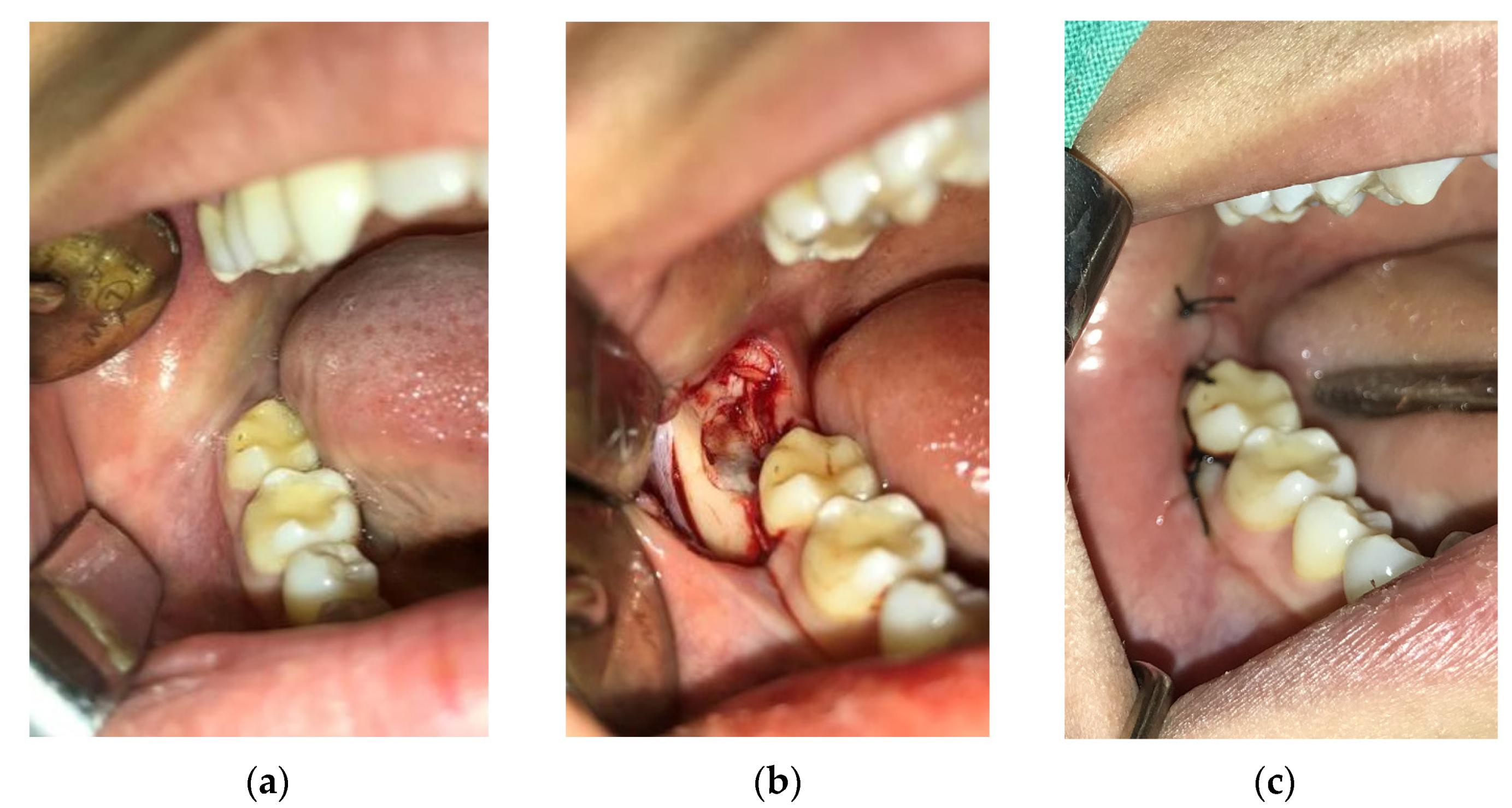
2.3. Surgical Procedure

This phase was performed according to the same protocol for all patients. Patients rinsed their mouth preoperatively with 15 mL of 0.12% chlorhexidine gluconate solution (Curasept ADS212®, CURADEN AG, Kriens, Switzerland), and the skin around the mouth was cleaned with Octanisept solution (Octanisept, Schülke&Mayr GmbH, Norderstedt, Germany). A sterile surgical drape, with an aperture for the nose and mouth, was placed over the face. This way, the risk of surgical site infection was reduced, and patient blinding was obtained (the patient should not know which alveolus L-PRF was applied, according to the study design, until the end of the trial). Local inferior alveolar nerve block anesthesia was obtained with 2% lidocaine with 1:100,000 adrenaline (Lidocaine 2%-adrenaline, Galenika AD, Belgrade, Serbia). Figure 3a shows the site of the surgical intervention. A modified Ward’s incision has been made, followed by a reflecting mucoperiosteal flap. Removal of the alveolar bone around the tooth was carried out to the necessary extent, as well as eventual separation of the tooth with a rotating burr, with continuous irrigation with saline solution. After the extraction of the tooth, all remaining fragments of bone, tooth and soft tissue debris were removed with a curette, and the edges of the alveolus were smoothed. The alveolus was rinsed with saline solution, and, in the case of the control group, the mucoperiosteal flap was repositioned and sutured with single sutures with 3–0 silk.
In the alveoli of the experimental group, before suturing, two L-PRF plugs were applied after irrigation of the alveoli (Figure 3b). After the flap was repositioned, suturing was performed in the same way as in the control group (Figure 3c).
All surgical procedures were performed by a single surgeon.

2.4. Postoperative Period and Control Check-Ups of Patients

After the surgical procedure, the patients were given written instructions on the behavior in the postoperative period (hygienic and dietary regime and antibiotics prescription). The design of the study provided for the following scheme of antibiotics: Amoxicillin 500 mg per os, every 8 h for 7 days, and in the case of a penicillin allergy, the prescription of Hemomycin 500 mg, every 24 h for 3 days, was planned. However, since none of the subjects who were included in the study had a penicillin allergy, all patients were given Amoxicillin after extraction.
Patients were scheduled for a control check-up 7 days after the surgical procedure. At the control check-up, the sutures were removed, and a clinical examination of the wound was performed. The depth of the periodontal pocket on the distal surface of the second molar was measured in three positions, namely, on the distobuccal edge, in the middle of the distal surface and on the distolingual edge of the distal surface of the tooth. A control clinical check-up, during which the depth of the periodontal pocket was measured again in the same way, was also scheduled 8 weeks after the tooth extraction.
The control check-up and measurement of the depth of the periodontal pocket was performed by a doctor who did not operate on any of the patients involved in this study and did not have information on which alveolus L-PRF was placed.
Two CBCT image datasets were obtained, the first image immediately after tooth extraction, and the second CBCT imaging was made eight weeks after the surgical procedure. In order to ensure the data quality and reliability of the presented study, all follow-up CBCT imaging was performed on the same CBCT scanner, using the same parameters, and by the same operator. CBCT imaging was performed on a Cranex 3D system (Soredex, Finland), with the following acquisition parameters: field of view (FOV)—60 × 40 mm, voxel size—0.2 mm3, focal point—0.15 × 0.5 mm and image resolution—200 × 200 pix.

2.5. Analysis and Processing of Data from CBCT Images

After the imaging and collection of CBCT images from both control check-ups, the reconstruction of surface 3D cavity models at the site of the third molar was performed. All CBCT processing and reconstruction of 3D models was performed by the same person in order to ensure the data quality and reliability of the presented study. The goal of this step was to collect data on the volume of the alveolar cavity that remained after tooth extraction and the density of bone tissue. These are the parameters on which the evaluation of the healing process of bone tissue after the surgical procedure were based. In order to approach the determination of the volume of the cavity, it was necessary to first isolate the zone where the tooth was extracted. For this step, the software 3D-DOCTOR (Able Software Corp) was used. In order to monitor the bone tissue healing process and to measure the density of newly formed bone inside the cavities, we used the software OnDemand3D (CYBERMED INC.), which includes a module for placing implants, and it was also used to check the newly formed bone density. Implants of certain dimensions and shapes were defined, i.e., (Figure 4a) 10 × 15 mm, (Figure 4b) 5 × 6 mm and (Figure 4c) 9 × 7 mm; their size depended on the size of the cavity. They were then placed inside the cavity in order to perform bone density measurements.

2.6. Statistical Analysis

A statistical analysis was performed for the obtained measurement results. A sample of 60 dental alveoli was analyzed and divided into 2 groups, namely, the control group and the experimental group, with 30 dental alveoli in each group. Descriptive parameters, mean value, standard deviation, minimum and maximum values, coefficient of variation, confidence interval, measures of asymmetry, measures of externality and MANOVA were used in the statistical analysis.

3. Results

Thirty-two patients were selected to participate in the present study; two patients did not return at the proposed time for the CBCT exam. In total, 30 patients (15 males and 15 females) were included in the final analysis. The participants ranged in age from 18 to 40 years (mean 23.33 years old). Overall, 4 patients had bilaterally impacted mandibular third molars in the vertical position, 7 in the horizontal, 16 in the mesioangular position and 3 in the distoangular position. All extractions included an osteotomy, and the operating time ranged from 25 to 65 min (mean 44.53 min) in the control group and from 27 to 65 min (mean 42.87 min) in the experimental group.

3.1. Reconstruction of Surface 3D Models of Cavites Based on CBCT Data

For the segmentation of CBCT images, two parameters were defined, namely, the minimum and maximum pixel intensity. Since the algorithm within the 3D-DOCTOR software is based on the Otsu thresholding method for segmentation, defining these two parameters defines the range of pixel intensity on the resulting CBCT images for segmentation (Figure 5a). This way, all images were segmented in their entirety, and after, those unnecessary data were removed in order to isolate the area where the tooth was extracted. As a result, a polygonal 3D model of the jaw was obtained (Figure 5b).

3.2. Extraction of Surface 3D Cavity Model

In order to properly perform segmentation of the interior of the cavity and reconstruct its surface 3D model, it was necessary to increase the segmentation parameters in order to successfully segment all bone structure around the alveolar cavity of the extracted tooth. After this, surface 3D models of the jaw were reconstructed (Figure 6a), and any additional noise was subsequently removed. The cavity of the alveolus of the extracted tooth was then isolated (Figure 6b).
In order to reconstruct a completely enclosed surface 3D model in order to measure its volume, it was necessary to close the top of the extracted cavity. Since that upper edge has an irregular geometry, the flat fill method was used. This method is based on closing the openings by using the least number of polygons in the 3D mesh model. In this way, when closing the holes, their influence on the change in the shape of the upper surface is eliminated, and therefore, their influence on the increase of the total volume of the cavity is also eliminated. Figure 7a,b show the reconstructed polygonal 3D models from the first and second CBCT imaging, based on which the differences in their size and shape can be observed. The volumes of the samples from both the control and experimental groups were then measured.
The difference in the size and shape of the cavity of the observed alveolus during the 8 weeks of the healing process was also visible when 2 surface 3D models of the first and second measurement were overlapped (Figure 7c). This overlap was performed manually due to the size and shape difference between the first and second CBCT imaging.

3.3. Measurement of Bone Density in Cavities

In OnDemand3D software, it was necessary to position the selected implant in the place where the bone density needed to be measured (Figure 8). On the left side, the 2D image shows the placement of the implant, and in the right image, the overall position of the implant inside the socket can be seen. For the purposes of this research, the parameter of the mean value of the bone density inside the implant was used. In this way, the dimensions of the implant, within which the bone density was measured, were dictated by the size of the cavity.
In addition to this, standard deviation of the bone density within the implant was also measured. These data were indicators for the correct positioning of the implant inside the cavity, because if the value of the standard deviation starts to rise sharply while positioning the implant, it is an indicator that the implant overlaps with the surrounding denser bone tissue next to the newly formed tissue, which requires its further positioning. This transformation is also displayed visually within the software itself, where the bone density on the implant is displayed with a certain range of colors. It can be seen that the colors range from blue to red, presenting the different densities of bone inside those implants (Figure 9).
This measurement procedure for bone density was performed for each patient individually, for a total of 60 dental alveoli for the control group and the experimental group (30 dental alveoli in each group).
Table 1 shows, in the example of one patient, the results of measurements of the left and right sides of bone density, cavity volume and depth of the periodontal pocket in the first and second measurements.

3.4. Statistical Analysis

For statistical analysis, the monitored parameters in the extraction alveoli were analyzed in I measurement by groups. The analysis was conducted on 3 parameters, namely, wound volume, bone density and periodontal pocket, on a sample of 60 extraction alveoli, which make up the control and experimental groups, with 30 alveoli in each.

3.4.1. Analysis of Parameters in the I Measurement

The results of I measurement for both the control and experimental groups are shown in Table 2.
The minimum (min) and maximum (max) values of the monitored parameters in I measurement in the alveoli of the control group indicate that the values are within the expected range. Higher values of the coefficient of variation (c.var) indicate the heterogeneity of the control group in terms of wound volume (I), bone density (I) and periodontal pocket (I). Increased values of Skewness (sk) indicate that the distribution is negatively asymmetric. This means that the curve of the distribution of the results tends towards higher values, or that there are more higher values compared to the normal distribution, in the case of periodontal pocket (I). Reduced values of Skewness (sk) indicate that the distribution is positively asymmetric. This means that the distribution curve of the results tends towards lower values, or that there are more lower values compared to the normal distribution, for wound volume (I) and bone density (I). Higher values of Kurtosis (ku) indicate that the curve is elongated for bone density (I). Negative values of Kurtosis (ku) indicate that the curve is flattened for wound volume (I) and periodontal pocket (I). The distribution of values is mostly within the normal distribution (p) of all three parameters, namely, wound volume (I), bone density (I) and periodontal pocket (I).
The minimum (min) and maximum (max) values of the monitored parameters in the alveoli of the experimental group in I measurement indicate that the values are within the expected range. Higher values of the coefficient of variation (c.var) indicate the heterogeneity of the experimental group in terms of all three monitored parameters: wound volume (I), bone density (I) and periodontal pocket (I). Increased values of Skewness (sk) indicate that the distribution is negatively asymmetric. This means that the curve of the distribution of the results tends towards higher values, or that there are more higher values compared to the normal distribution, in the case of bone density (I) and periodontal pocket (I). Reduced values of Skewness (sk) indicate that the distribution is positively asymmetric. This means that the curve of the distribution of the results tends towards lower values, or that there are more lower values compared to the normal distribution of the wound volume (I). Negative values of Kurtosis (ku) indicate that the curve is flattened for wound volume (I), bone density (I) and periodontal pocket (I). The distribution of values is generally within the normal distribution (p) for wound volume (I), bone density (I) and periodontal pocket (I).
Table 3 shows the results of the significance of the difference between the groups, in regards to the monitored parameters in the I measurement.
Based on the value of p = 0.826 (MANOVA), it can be concluded that no significant difference and a clearly defined boundary between the groups were observed. Further, as the value is p > 0.1 (VOL, density, periodontal pocket), it means that no significant difference was observed between the groups in terms of wound volume (I), bone density (I) and periodontal pocket (I).

3.4.2. Analysis of Parameters in the II Measurement

The results of II measurement for the control and experimental groups is shown in Table 4.
The minimum (min) and maximum (max) measured values in the II measurement of the control group indicate that the values are within the expected range. Higher values of the coefficient of variation (c.var) indicate the heterogeneity of the control group in terms of extraction wound volume (VOL (II)), bone tissue density (density (II)) and periodontal pocket depth (pocket (II)). Increased values of Skewness (sk) indicate that the distribution is negatively asymmetric, which means that the curve of the distribution of the results tends to higher values, or that there are more higher values compared to the normal distribution, for all three observed features. Higher values of Kurtosis (ku) indicate that the curve is elongated for bone density (density (II)) and pocket (pocket (II)). Negative values of Kurtosis (ku) indicate that the curve is flattened for volume values (VOL (II)). The distribution of values generally moves within the normal distribution (p) for all three observed parameters.
The minimum (min) and maximum (max) parameters that were monitored in the II measurement in the experimental group indicate that the values are within the expected range. Higher values of the coefficient of variation (c.var) indicate experimental heterogeneity in terms of all three observed parameters. Increased values of Skewness (sk) indicate that the distribution is negatively asymmetric, which suggests that the curve of the distribution of the results tends to higher values, or that there are more higher values compared to the normal distribution for all observed features. Higher values of Kurtosis (ku) indicate that the curve is elongated for density (density (II)) and periodontal pocket (pocket (II)). Negative values of Kurtosis (ku) indicate that the curve is flattened for volume (VOL (II)). The distribution of values generally moves within the normal distribution (p) in the case of volume (VOL (II)) and density (density (II)). The distribution of values deviates from the normal distribution (p) in the case of a periodontal pocket (pocket (II)).
Table 5 shows the results of the significance of the difference between the groups, in regards to the monitored parameters in the II measurement.
Based on the value of p = 0.499 (MANOVA it can also be concluded that no significant difference and a clearly defined boundary between the groups were observed. Further, as the value is p > 0.1 (VOL, density, periodontal pocket), it means that no significant difference was observed between the groups in terms of wound volume (II), bone density (II) and periodontal pocket (II).

4. Discussion

Third mandibular molar surgery is the most common clinical procedure done by oral and maxillofacial surgeons. It can be associated with several postoperative complications, such as pain, swelling, trismus, alveolar osteitis or surgical site infection. Many studies have been conducted to determine procedures capable of reducing the incidence of these complications.
The idea of applying a platelet concentrate in the wound area foresees the synergistic action of various growth factors and the multiplication of their biological significance locally [30]. The production protocol of these products affects their composition and biological characteristics and the clinical effect of their application [53,54].
The healing time of the extraction alveolus varies significantly among individuals [13]. Various studies show that in the 4th week of socket healing, about 2/3 of the alveolus is filled with woven bone, and in the 10th week, it is completely filled. However, after this period, the intensive process of bone tissue remodeling continues with only partial replacement of woven bone with mature lamellar bone type, which lasts up to 6 months [14].
Some authors consider that a period of 2 or 3 months is too short to monitor the effects of these products on the healing of the extraction alveolus. On the other hand, there are authors who believe that a short follow-up period (up to 3 months) is more suitable for monitoring the effects of platelet preparations, because their beneficial effect is expected only in the initial stages of healing [55].
Several studies were published with methodology similar to our study [56,57]. They reported statistically significant better bone healing of alveoli in which L-PRF was applied. Our results partly coincide with this; the present study results suggest some positive effect of L-PRF application. The volume of new bone tissue and bone density were higher in the experimental group, and the depth of the periodontal pocket distal to the second molar was smaller in this group. Although neither of these differences were statistically significant, they undeniably exist.
Several studies evaluated the effect of L-PRF in promoting bone tissue regeneration, but in other clinical applications, such as in maxillary sinus floor elevation and various augmentation procedures [58], furcation defect treatment [26], endodontic therapy [59] and socket preservation [3].
There is no clear consensus in terms of the required quantity of L-PRF for the optimal effect. The content of platelets in platelet concentrates is 2 to 8.5 times higher than in blood, depending on the method of production. However, it is important for clinicians to note that the initial platelet count is not the same in all patients and that different individuals probably have a different critical concentration of platelets in these products required to achieve a significant biological effect. Further, the significance of the different content of other cellular elements of the blood remains unclear [27]. In the present study, two L-PRF plugs were placed in the experimental alveoli. We can assume that more of L-PRF is probably needed, especially in the alveoli of teeth with more than one root.
In 2017, MacBeth et al. published a systematic review of the literature on soft and hard tissue changes after alveolar ridge preservation interventions. For some studies included in this review, he determined that there was an extremely high degree of heterogeneity, and this could explain why in these studies, the differences between the control and experimental groups were not statistically significant [60].
The inability to bilaterally make defects of the same dimensions and shapes can potentially explain the results of the present study. However, this is a limitation faced by all clinical studies because it is not possible to create two identical defects, as it is possible in in vitro studies. A similar conclusion was reached by Del Fabro et al., who published a systematic review in 2018. The review included studies evaluating the effect of autologous platelet preparations on infrabony periodontal defects and concluded that most of them failed to prove that the difference between the control and experimental groups was statistically significant, given the great differences in the shape and size of the defects, primarily in terms of the number of bony walls that exist in the defect [61].
Research has shown that changes in the number of revolutions of the centrifuge (centrifugal force intensity) and the length of time of centrifugation affect the biological characteristics of the preparations obtained, such as the amount and type of growth factors they release and the dynamics of their release [32,62].
To minimize individual differences in their characteristics, it is necessary to standardize clinical protocols for their production. It is also necessary to determine the benefit from combining these preparations with other types of bone grafts [26] and allografts [4] in the treatment of bone defects. It can be concluded that the use of some grafting material and autologous blood preparations can, to a certain extent, reduce the resorption of bone tissue in the post-extraction period and prove to be an optimal procedure in therapy after tooth extraction [19].
Although it is evident that, today, the positive effect of the application of growth factor concentrates on tissue healing, and therefore on extraction wounds, the necessary biocompatibility, safety of their application, clinical reproducibility of the preparation methodology and “cost-effect” relationship must still be kept in mind during its administration. The use of a clearly defined, safe and controlled technique can be a powerful tool to achieve better tissue regeneration in many clinical applications.
Many studies evaluated the formation of bone tissue by analyzing two-dimensional X-ray images (periapical or OPT) [63,64], but in the present study, 3D CBCT images were used to obtain more accurate information, i.e., surface 3D models of cavities, due to higher spatial resolution. Moreover, application of the presented methodology for extraction and analysis on surface 3D models for the density, volume and periodontal pocket presents a new way to acquire reliable results on the basis of the reconstructed 3D geometry of the cavity.
This study has certain limitations, primarily in terms of the sample size and the fact that it was only conducted in the mandibular third molar region. The region of the third molar in the lower jaw shows certain peculiarities in relation to the healing of extraction wounds in other parts of the oral cavity. Further, the preoperative platelet and leukocyte count was not taken into consideration.

5. Conclusions

Based on the results of the study, it can be concluded that there is not enough evidence that the use of L-PRF after the third lower third molar extraction has a favorable effect on bone tissue healing and periodontal pocket reduction distally to the second molar.
Further research that includes a larger sample, longer observation period, precise protocols of autologous blood preparations production and identification of all the factors that may influence the composition and properties is necessary.

Author Contributions

Conceptualization, A.T.; Methodology, A.T.; Investigation, A.T., B.B. and I.M.; Validation, A.T. and B.B.; Formal analysis, J.M., K.V. and M.S.; Resources, B.B. and K.V.; Software, J.M. and M.S.; Visualization, I.M. and M.S.; Writing—original draft, A.T., I.M., J.M., K.V. and M.S.; Writing—review & editing, B.B. and D.V.; Supervision, D.V. All authors have read and agreed to the published version of the manuscript.

Funding

This research received no external funding.

Institutional Review Board Statement

This study was conducted in accordance with the Declaration of Helsinki and approved by the Ethics Committee of the Faculty of Medicine Novi Sad (protocol code 01-39/1) and by the Ethics Committee of the Dentistry Clinic of Vojvodina Novi Sad (protocol code 01-11/60-2015).

Informed Consent Statement

Informed consent was obtained from all subjects involved in the study.

Data Availability Statement

Not applicable.

Acknowledgments

This research was supported by the Ministry of Science, Technological Development and Innovation of the Republic of Serbia.

Conflicts of Interest

The authors declare no conflict of interest.

References

  1. Srinivas, B.; Das, P.; Rana, M.M.; Qureshi, A.Q.; Vaidya, K.C.; Raziuddin, S.J.A. Wound healing and bone regeneration in postextraction sockets with and without platelet-rich fibrin. Ann. Maxillo-Facial Surg. 2018, 8, 28–34. [Google Scholar]
  2. Araújo, M.G.; da Silva, J.C.C.; de Mendonça, A.F.; Lindhe, J. Ridge alterations following grafting of fresh extraction sockets in man. A randomized clinical trial. Clin. Oral Implants Res. 2015, 26, 407–412. [Google Scholar] [CrossRef]
  3. Ivanova, V.; Chenchev, I.; Zlatev, S.; Mijiritsky, E. Comparison Study of the Histomorphometric Results after Socket Preservation with PRF and Allograft Used for Socket Preservation—Randomized Controlled Trials. Int. J. Environ. Res. Public Health 2021, 18, 7451. [Google Scholar] [CrossRef] [PubMed]
  4. Crespi, R.; Capparè, P.; Gherlone, E. Bone Recontouring in Fresh Sockets with Buccal Bone Loss: A Cone Beam Computed Tomography Study. Int. J. Oral Maxillofac. Implants 2014, 29, 863–868. [Google Scholar] [CrossRef] [Green Version]
  5. Omran, M.; Min, S.; Abdelhamid, A.; Liu, Y.; Zadeh, H.H. Alveolar ridge dimensional changes following ridge preservation procedure: Part-2—CBCT 3D analysis in non-human primate model. Clin. Oral Implants Res. 2016, 27, 859–866. [Google Scholar] [CrossRef] [PubMed]
  6. Bornstein, M.; Scarfe, W.; Vaughn, V.; Jacobs, R. Cone Beam Computed Tomography in Implant Dentistry: A Systematic Review Focusing on Guidelines, Indications, and Radiation Dose Risks. Int. J. Oral Maxillofac. Implants 2014, 29, 55–77. [Google Scholar] [CrossRef]
  7. Chen, Y.W.; Finkelman, M.; Papaspirisdakos, P.; César-Neto, J.B.; Weber, H.P.; de Souza, A.B. Comparative analysis of dimensional alterations following extraction of maxillary molars using three-dimensional images’ superimposition: A CBCT study. Odontology 2021, 109, 514–523. [Google Scholar] [CrossRef] [PubMed]
  8. Sokac, M.; Budak, I.; Puskar, T.; Mirkovic, S.; Santosi, Z.; Kuzmanovic, M.; Vukelic, D. Investigation of radiation level and assessment of dimensional accuracy of acquired CBCT images. Measurement 2020, 155, 107551. [Google Scholar] [CrossRef]
  9. Chappuis, V.; Engel, O.; Reyes, M.; Shahim, K.; Nolte, L.P.; Buser, D. Ridge Alterations Post-extraction in the Esthetic Zone. J. Dent. Res. 2013, 92, 195S–201S. [Google Scholar] [CrossRef] [Green Version]
  10. Schropp, L.; Wenzel, A.; Kostopoulos, L.; Karring, T. Bone healing and soft tissue contour changes following single-tooth extraction: A clinical and radiographic 12-month prospective study. J. Prosthet. Dent. 2003, 23, 313–323. [Google Scholar]
  11. Araújo, M.G.; Silva, C.O.; Misawa, M.; Sukekava, F. Alveolar socket healing: What can we learn? Periodontology 2000, 68, 122–134. [Google Scholar] [CrossRef]
  12. Chen, Y.W.; Chi, L.Y.; Lee, O.K. Revisit incidence of complications after impacted mandibular third molar extraction: A nationwide population-based cohort study. PLoS ONE 2021, 22, e0246625. [Google Scholar] [CrossRef] [PubMed]
  13. Couso-Queiruga, E.; Stuhr, S.; Tattan, M.; Chambrone, L.; Avila-Ortiz, G. Post-extraction dimensional changes: A systematic review and meta-analysis. J. Clin. Periodontol. 2021, 48, 127–145. [Google Scholar] [CrossRef]
  14. Leblebicioglu, B.; Hegde, R.; Yildiz, V.O.; Tatakis, D.N. Immediate effects of tooth extraction on ridge integrity and dimensions. Clin. Oral Investig. 2015, 19, 1777–1784. [Google Scholar] [CrossRef]
  15. Farmer, M.; Darby, I. Ridge dimensional changes following single-tooth extraction in the aesthetic zone. Clin. Oral Implants Res. 2014, 25, 272–277. [Google Scholar] [CrossRef] [PubMed]
  16. Hämmerle, C.H.F.; Araújo, M.G.; Simion, M. Evidence-based knowledge on the biology and treatment of extraction sockets. Clin. Oral Implants Res. 2012, 23, 80–82. [Google Scholar] [CrossRef]
  17. Thoma, D.S.; Benić, G.I.; Zwahlen, M.; Hämmerle, C.H.F.; Jung, R.E. A systematic review assessing soft tissue augmentation techniques. Clin. Oral Implants Res. 2009, 20, 146–165. [Google Scholar] [CrossRef] [PubMed] [Green Version]
  18. Del Fabbro, M.; Bucchi, C.; Lolato, A.; Corbella, S.; Testori, T.; Taschieri, S. Healing of Postextraction Sockets Preserved with Autologous Platelet Concentrates. A Systematic Review and Meta-Analysis. J. Oral Maxillofac. Surg. 2017, 75, 1601–1615. [Google Scholar] [CrossRef]
  19. Girish Kumar, N.; Chaudhary, R.; Kumar, I.; Arora, S.S.; Kumar, N.; Singh, H. To assess the efficacy of socket plug technique using platelet rich fibrin with or without the use of bone substitute in alveolar ridge preservation: A prospective randomised controlled study. Oral Maxillofac. Surg. 2018, 22, 135–142. [Google Scholar] [CrossRef] [PubMed]
  20. Cardaropoli, D.; Tamagnone, L.; Roffredo, A.; Gaveglio, L. Relationship between the Buccal Bone Plate Thickness and the Healing of Postextraction Sockets with/without Ridge Preservation. Int. J. Periodontics Restor. Dent. 2014, 34, 211–217. [Google Scholar] [CrossRef] [PubMed]
  21. Kotsakis, G.A.; Salama, M.; Chrepa, V.; Hinrichs, J.E.; Gaillard, P. A Randomized, Blinded, Controlled Clinical Study of Particulate Anorganic Bovine Bone Mineral and Calcium Phosphosilicate Putty Bone Substitutes for Socket Preservation. Int. J. Oral Maxillofac. Implants 2014, 29, 141–151. [Google Scholar] [CrossRef] [PubMed] [Green Version]
  22. Pohl, S.; Binderman, I.; Tomac, J. Maintenance of Alveolar Ridge Dimensions Utilizing an Extracted Tooth Dentin Particulate Autograft and Platelet-Rich fibrin: A Retrospective Radiographic Cone-Beam Computed Tomography Study. Materials 2020, 13, 1083. [Google Scholar] [CrossRef] [PubMed] [Green Version]
  23. Urban, I.A.; Monje, A. Guided Bone Regeneration in Alveolar Bone Reconstruction. Oral Maxillofac. Surg. Clin. N. Am. 2019, 31, 331–338. [Google Scholar] [CrossRef] [PubMed]
  24. Suárez-López del Amo, F.; Monje, A. Efficacy of biologics for alveolar ridge preservation/reconstruction and implant site development: An American Academy of Periodontology best evidence systematic review. J. Periodontol. 2022, 93, 1827–1847. [Google Scholar] [CrossRef]
  25. Yoshpe, M.; Ruparel, N.; Einy, S.; Ganatra, S.; Kaufman, A.Y. Treatment of Necrotic Anterior and Posterior Teeth with Regenerative Endodontic Procedures Using PRF as a Scaffold: A Retrospective Study. Appl. Sci. 2022, 12, 6774. [Google Scholar] [CrossRef]
  26. Nair, U.P.; Shivamurthy, R.; Nagate, R.R.; Chaturvedi, S.; Al-Qahtani, S.M.; Magbol, M.A.; Gokhale, S.T.; Tikare, S.; Chaturvedi, M. Effect of Injectable Platelet-Rich Fibrin with a Nano-Hydroxyapatite Bone Graft on the Treatment of a Grade II Furcation. Defect. Bioeng. 2022, 9, 602. [Google Scholar] [CrossRef]
  27. Agrawal, A.A. Evolution, current status and advances in application of platelet concentrate in periodontics and implantology. World J. Clin. Cases 2017, 5, 159. [Google Scholar] [CrossRef]
  28. Hwan Jung, M.; Hun Lee, J.; Wadhwa, P.; Bo Jiang, H.; Jang, H.S.; Seok Lee, E. Bone Regeneration in Peri-Implant Defect Using Autogenous Tooth Biomaterial Enriched with Platelet-Rich Fibrin in Animal Model. Appl. Sci. 2020, 10, 1939. [Google Scholar] [CrossRef] [Green Version]
  29. Francisco, I.; Fernandes, M.H.; Vale, F. Platelet-Rich Fibrin in Bone Regenerative Strategies in Orthodontics: A Systematic Review. Materials 2020, 13, 1866. [Google Scholar] [CrossRef] [Green Version]
  30. Anitua, E.; Tejero, R.; Zalduendo, M.M.; Orive, G. Plasma Rich in Growth Factors Promotes Bone Tissue Regeneration by Stimulating Proliferation, Migration, and Autocrine Secretion in Primary Human Osteoblasts. J. Periodontol. 2013, 84, 1180–1190. [Google Scholar] [CrossRef] [PubMed]
  31. Dohan Ehrenfest, D.M.; Rasmusson, L.; Albrektsson, T. Classification of platelet concentrates: From pure platelet-rich plasma (P-PRP) to leucocyte- and platelet-rich fibrin (L-PRF). Trends Biotechnol. 2009, 27, 158–167. [Google Scholar] [CrossRef] [PubMed]
  32. Dohan, D.M.; Choukroun, J.; Diss, A.; Dohan, S.L.; Dohan, A.J.J.; Mouhyi, J.; Gogly, L. Platelet-rich fibrin (PRF): A second-generation platelet concentrate. Part II: Platelet-related biologic features. Oral Surg. Oral Med. Oral Pathol. Oral Radiol. Endod. 2006, 101, e45–e50. [Google Scholar] [CrossRef] [PubMed]
  33. Fujioka-Kobayashi, M.; Katagiri, H.; Kono, M.; Schaller, B.; Zhang, Y.; Sculean, A.; Miron, J.R. Improved growth factor delivery and cellular activity using concentrated platelet-rich fibrin (C-PRF) when compared with traditional injectable (i-PRF) protocols. Clin. Oral Investig. 2020, 24, 4373–4383. [Google Scholar] [CrossRef]
  34. Baca-Gonzalez, L.; Serrano Zamora, R.; Rancan, L.; González Fernández-Tresguerres, F.; Fernández-Tresguerres, I.; López-Pintor, R.M.; López-Quiles, J.; Leco, I.; Jesús Torres, J. Plasma rich in growth factors (PRGF) and leukocyte-platelet rich fibrin (L-PRF): Comparative release of growth factors and biological effect on osteoblasts. Int. J. Implant Dent. 2022, 8, 39. [Google Scholar] [CrossRef]
  35. Tenore, G.; Zimbalatti, A.; Rocchetti, F.; Graniero, F.; Gaglioti, D.; Mohsen, A.; Caputo, M.; Lollobrigida, M.; Lamazza, L.; De Biase, A.; et al. Management of Medication-Related Osteonecrosis of the Jaw (MRONJ) Using Leukocyte- and Platelet-Rich Fibrin (L-PRF) and Photobiomodulation: A Retrospective Study. J. Clin. Med. 2020, 9, 3505. [Google Scholar] [CrossRef] [PubMed]
  36. Santos, J.M.; Marques, J.A.; Esteves, M.; Sousa, V.; Palma, P.J.; Matos, S. Intentional Replantation as a Starting Approach for a Multidisciplinary Treatment of a Mandibular Second Molar: A Case Report. J. Clin. Med. 2022, 11, 5111. [Google Scholar] [CrossRef]
  37. Jo, Y.Y.; Oh, J.H. New Resorbable Membrane Materials for Guided Bone Regeneration. Appl. Sci. 2018, 8, 2157. [Google Scholar] [CrossRef] [Green Version]
  38. Mozzati, M.; Gallesio, G.; Tumedei, M.; Del Fabbro, M. Concentrated Growth Factors vs. Leukocyte-and-Platelet-Rich Fibrin for Enhancing Postextraction Socket Healing. A Longitudinal Comparative Study. Appl. Sci. 2020, 10, 8256. [Google Scholar] [CrossRef]
  39. Wang, M.; Zhang, X.; Li, Y.; Mo, A. Lateral Ridge Augmentation with Guided Bone Regeneration Using Particulate Bone Substitutes and Injectable Platelet-Rich Fibrin in a Digital Workflow: 6 Month Results of a Prospective Cohort Study Based on Cone-Beam Computed Tomography Data. Materials 2021, 14, 6430. [Google Scholar] [CrossRef] [PubMed]
  40. Trimmel, B.; Gyulai-Gaál, S.; Kivovics, M.; Jákob, N.P.; Hegedűs, C.; Szabó, B.T.; Dobó-Nagy, C.; Szabó, G. Evaluation of the Histomorphometric and Micromorphometric Performance of a Serum Albumin-Coated Bone Allograft Combined with A-PRF for Early and Conventional Healing Protocols after Maxillary Sinus Augmentation: A Randomized Clinical Trial. Materials 2021, 14, 1810. [Google Scholar] [CrossRef]
  41. Pirpir, C.; Yilmaz, O.; Candirli, C.; Balaban, E. Evaluation of effectiveness of concentrated growth factor on osseointegration. Int. J. Implant Dent. 2017, 3, 7. [Google Scholar] [CrossRef] [PubMed] [Green Version]
  42. Trybek, G.; Rydlińska, J.; Aniko-Włodarczyk, M.; Jaroń, A. Effect of Platelet-Rich Fibrin Application on Non-Infectious Complications after Surgical Extraction of Impacted Mandibular Third Molars. Int. J. Environ. Res. Public Health 2021, 18, 8249. [Google Scholar] [CrossRef] [PubMed]
  43. Da Silva, M.T.; de Almeida Barros Mourão, C.F.; Mello-Machado, R.C.; Montemezzi, P.; de Lima Barbosa, R.; Sartoretto, S.C.; Leite, P.E.C.; Javid, K.; Kawase, T.; Alves, G.G.; et al. Effects of Leukocyte-Platelet-Rich Fibrin (L–PRF) on Pain, Soft Tissue Healing, Growth Factors, and Cytokines after Third Molar Extraction: A Randomized, Split-Mouth, Double-Blinded Clinical Trial. Appl. Sci. 2021, 11, 1666. [Google Scholar] [CrossRef]
  44. Ghanaati, S.; Booms, P.; Orlowska, A.; Kubesch, A.; Lorenz, J.; Rutkowski, J.; Landes, C.; Sader, R.; Kirkpatrick, C.; Choukroun, J. Advanced Platelet-Rich Fibrin: A New Concept for Cell-Based Tissue Engineering by Means of Inflammatory Cells. J. Oral Implantol. 2014, 40, 679–689. [Google Scholar] [CrossRef]
  45. Machut, K.; Żółtowska, A. Plasma Rich in Growth Factors in the Treatment of Endodontic Periapical Lesions in Adult Patients: 3-Dimensional Analysis Using Cone-Beam Computed Tomography on the Outcomes of Non-Surgical Endodontic Treatment Using A-PRF+ and Calcium Hydroxide: A Retrospective Cohort Study. J. Clin. Med. 2022, 11, 6092. [Google Scholar] [PubMed]
  46. Al-Hamed, F.S.; Tawfik, M.A.M.; Abdelfadil, E.; Al-Saleh, M.A.Q. Efficacy of Platelet-Rich Fibrin After Mandibular Third Molar Extraction: A Systematic Review and Meta-Analysis. J. Oral Maxillofac. Surg. 2017, 75, 1124–1135. [Google Scholar] [CrossRef]
  47. Yu, H.Y.; Chang, Y.C. A Bibliometric Analysis of Platelet-Rich Fibrin in Dentistry. Int. J. Environ. Res. Public Health 2022, 19, 12545. [Google Scholar] [CrossRef] [PubMed]
  48. Sun, J.; Hu, Y.; Fu, Y.; Zou, D.; Lu, J.; Lyu, C. Emerging roles of platelet concentrates and platelet-derived extracellular vesicles in regenerative periodontology and implant dentistry. APL Bioeng. 2022, 6, 031503. [Google Scholar] [CrossRef]
  49. Zhu, J.; Zhang, S.; Yuan, X.; He, T.; Liu, H.; Wang, J.; Xu, B. Effect of platelet-rich fibrin on the control of alveolar osteitis, pain, trismus, soft tissue healing, and swelling following mandibular third molar surgery: An updated systematic review and meta-analysis. Int. J. Oral Maxillofac. Surg. 2021, 50, 398–406. [Google Scholar] [CrossRef]
  50. Xiang, X.; Shi, P.; Zhang, P.; Shen, J.; Kang, J. Impact of platelet-rich fibrin on mandibular third molar surgery recovery: A systematic review and meta-analysis. BMC Oral Health 2019, 19, 163. [Google Scholar] [CrossRef] [Green Version]
  51. Starzyńska, A.; Kaczoruk-Wieremczuk, M.; Lopez, M.A.; Passarelli, P.C.; Adamska, P. The Growth Factors in Advanced Platelet-Rich Fibrin (A-PRF) Reduce Postoperative Complications after Mandibular Third Molar Odontectomy. Int. J. Environ. Res. Public Health 2021, 18, 13343. [Google Scholar] [CrossRef]
  52. Kwak, S.G.; Kim, J.H. Central limit theorem: The cornerstone of modern statistics. Korean J. Anesthesiol. 2017, 70, 144–156. [Google Scholar] [CrossRef] [Green Version]
  53. Wang, X.; Zhang, Y.; Choukroun, J.; Ghanaati, S.; Miron, R.J. Effects of an injectable platelet-rich fibrin on osteoblast behavior and bone tissue formation in comparison to platelet-rich plasma. Platelets 2018, 29, 48–55. [Google Scholar] [CrossRef]
  54. Miron, R.J.; Fujioka-Kobayashi, M.; Hernandez, M.; Kandalam, U.; Zhang, Y.; Ghanaati, S.; Choukroun, J. Injectable platelet rich fibrin (i-PRF): Opportunities in regenerative dentistry? Clin. Oral Investig. 2017, 21, 2619–2627. [Google Scholar] [CrossRef]
  55. Plachokova, A.S.; Nikolidakis, D.; Mulder, J.; Jansen, J.A.; Creugers, N.H.J. Effect of platelet-rich plasma on bone regeneration in dentistry: A systematic review. Clin. Oral Implants Res. 2008, 19, 539–545. [Google Scholar] [CrossRef] [PubMed]
  56. Pimentel, T.; Ritto, F.; Canellas, J.V.; Junger, B.; Cruz, M.; Medeiros, P.J. Re: Randomized double-blind clinical trial evaluation of bone healing after third molar surgery with the use of leukocyte- and platelet-rich fibrin. Int. J. Oral Maxillofac. Surg. 2020, 49, 692. [Google Scholar] [CrossRef] [PubMed]
  57. Castro, A.B.; Meschi, N.; Temmerman, A.; Pinto, N.; Lambrechts, P.; Teughels, W.; Quirynen, M. Regenerative potential of leucocyte- and platelet-rich fibrin. Part B: Sinus floor elevation, alveolar ridge preservation and implant therapy. A systematic review. J. Clin. Periodontol. 2017, 44, 225–234. [Google Scholar] [CrossRef] [Green Version]
  58. Machut, K.; Zoltowska, A.; Pawlowska, E.; Derwich, M. Plasma Rich in Growth Factors in the Treatment of Endodontic Periapical Lesions in Adult Patients: Case Reports. Int. J. Mol. Sci. 2021, 22, 9458. [Google Scholar] [CrossRef] [PubMed]
  59. MacBeth, N.; Trullenque-Eriksson, A.; Donos, N.; Mardas, N. Hard and soft tissue changes following alveolar ridge preservation: A systematic review. Clin. Oral Implants Res. 2016, 28, 982–1004. [Google Scholar] [CrossRef]
  60. Del Fabbro, M.; Karanxha, L.; Panda, S.; Bucchi, C.; Nadathur Doraiswamy, J.; Sankari, M.; Ramamoorthi, S.; Varghese, S.; Taschieri, S. Autologous platelet concentrates for treating periodontal infrabony defects. Cochrane Database Syst. Rev. 2018, 11, CD011423. [Google Scholar] [CrossRef]
  61. Zwittnig, K.; Kirnbauer, B.; Jakse, N.; Schlenke, P.; Mischak, I.; Ghanaati, S.; Al-Maawi, S.; Végh, D.; Payer, M.; Zrnc, T.A. Growth Factor Release within Liquid and Solid PRF. J. Clin. Med. 2022, 11, 5070. [Google Scholar] [CrossRef] [PubMed]
  62. Njokanma, A.R.; Fatusi, O.A.; Ogundipe, O.K.; Arije, O.O.; Akomolafe, A.G.; Kuye, O.F. Does platelet-rich fibrin increase bone regeneration in mandibular third molar extraction sockets? J. Korean Assoc. Oral Maxillofac. Surg. 2022, 48, 371–381. [Google Scholar] [CrossRef] [PubMed]
  63. Alam, S.; Khare, G.; Arun Kumar, K.V. A Comparative Study of Platelet-Rich Fibrin and Platelet-Rich Fibrin with Hydroxyapatite to Promote Healing of Impacted Mandibular Third Molar Socket. J. Oral Maxillofac. Surg. 2020, 21, 608–615. [Google Scholar] [CrossRef] [PubMed]
  64. Niedzielska, I.; Ciapiński, D.; Bąk, M.; Niedzielski, D. The Assessment of the Usefulness of Platelet-Rich Fibrin in the Healing Process Bone Resorption. Coatings 2022, 12, 247. [Google Scholar] [CrossRef]
Figure 1. Methodology workflow.
Applsci 13 04823 g001
Figure 2. L-PRF taken out of the test tube and placed inside a sterile glass container.
Applsci 13 04823 g002
Figure 3. Surgical procedure in the experimental group (a) surgical procedure site; (b) application of two preparations of concentrated growth factors after irrigation of the alveolus; (c) suturing of the wound.
Applsci 13 04823 g003
Figure 4. Implant shapes used in OnDemand3D software for bone density measurements: (a) 10 × 15 mm, (b) 5 × 6 mm and (c) 9 × 7 mm.
Applsci 13 04823 g004
Figure 5. (a) Segmentation on the CBCT image and (b) reconstructed surface 3D model of the jaw.
Applsci 13 04823 g005
Figure 6. Extracted volume of alveolar cavity (a) before mesh processing and (b) after mesh processing where the cavity of the alveolus of the extracted tooth is isolated.
Applsci 13 04823 g006
Figure 7. Surface 3D models of reconstructed cavities (a) after the first CBCT imaging and (b) after the second CBCT imaging and (c) overlap of surface 3D models of the first (bluish transparent color) and second CBCT measurement (green color) of the same patient.
Applsci 13 04823 g007
Figure 8. Implant positioning and bone density measurement in OnDemand3D software.
Applsci 13 04823 g008
Figure 9. Bone density measurements inside the cavity using a defined implant.
Applsci 13 04823 g009
Table 1. Measurement results for bone density and volume of both left and right cavity for patient no. 1.
Bone Density
(HU)
Volume
(mm3)
Periodontal Pocket
(mm)
LeftRightLeftRightLeftRight
I Measurement−297.76−163.36408.49397.926.346.23
II Measurement130.5758.25319.44346.336.726.69
Table 2. Results of I measurement in the control and experimental groups.
Mean Valuestd.dMinMaxc.varconf.inter.skkup
Control GroupVOL (I)430.9792.88251.5608.721.55396.28465.66−0.05−0.670.823
density
(I)
−89.2371.86−297.844.980.53−116.07−62.39−0.480.870.977
pocket (I)6.272.213.011.035.295.447.090.49−0.880.510
Experimental GroupVOL (I)417.5793.77197.8574.522.45382.55452.59−0.38−0.431.000
density
(I)
−94.0071.36−251.276.075.92−120.65−67.340.25−0.100.783
pocket (I)6.672.523.013.037.855.727.610.58−0.320.622
Table 3. Significance of the difference between the groups, in regard to the monitored parameters in the I measurement.
AnalysisFp
MANOVA0.2990.826
VOL (I)0.3090.580
Density (I)0.0660.798
Periodontal pocket (I)0.4260.516
Table 4. Results of II measurement in the control and experimental groups.
Mean Valuestd.dMinMaxc.varconf.inter.skkup
Control GroupVOL (II)296.02115.73112.2538.739.09252.80339.240.44−0.820.574
density (II)134.6963.5923.9310.247.22110.93158.440.670.920.565
pocket (II)4.321.761.09.040.873.664.980.690.480.516
Experimental GroupVOL (II)261.4790.31120.8460.434.54227.74295.200.39−0.370.641
density (II)131.3374.96−55.2319.557.08103.33159.330.241.170.778
pocket (II)3.831.601.08.041.723.244.430.840.510.095
Table 5. Significance of the difference between the groups, in regards to the monitored parameters in the II measurement.
AnalysisFp
MANOVA0.8000.499
VOL (I)1.6620.203
Density (I)0.0350.852
Periodontal pocket (I)0.4260.516
Disclaimer/Publisher’s Note: The statements, opinions and data contained in all publications are solely those of the individual author(s) and contributor(s) and not of MDPI and/or the editor(s). MDPI and/or the editor(s) disclaim responsibility for any injury to people or property resulting from any ideas, methods, instructions or products referred to in the content.

