Article

Artificial Intelligence-Based Patient Selection for Deep Inspiration Breath-Hold Breast Radiotherapy from Respiratory Signals

1
Medical Physics Department, Centro di Riferimento Oncologico di Aviano (CRO) IRCCS, via F. Gallini 2, 33081 Aviano, Italy
2
Radiation Oncology Department, Centro di Riferimento Oncologico di Aviano (CRO) IRCCS, via F. Gallini 2, 33081 Aviano, Italy
3
Institute of Nuclear Medical Physics (INMP), Bangladesh Atomic Energy Commission (BAEC), Dhaka 1349, Bangladesh
*
Author to whom correspondence should be addressed.
Appl. Sci. 2023, 13(8), 4962; https://doi.org/10.3390/app13084962
Submission received: 31 January 2023 / Revised: 7 April 2023 / Accepted: 10 April 2023 / Published: 14 April 2023
(This article belongs to the Special Issue Applications of Radiomics and Deep Learning in Medical Image Analysis)

Abstract

:
Purpose: to predict eligibility for deep inspiration breath-hold (DIBH) radiotherapy (RT) treatment of patients with left breast cancer from analysis of respiratory signal, using Deep Bidirectional Long Short-Term Memory (BLSTM) recurrent neural networks. Methods: The respiratory traces from 36 patients who underwent DIBH RT were collected. The patients’ RT treatment plans were generated for both DIBH and free-breathing (FB) modalities. The patients were divided into two classes (patient eligible or not), based on the decrease of maximum dose to the left anterior descending (LAD) artery achieved with DIBH, compared to that achieved with FB and ΔDL. Patients with ΔDL > median value of ΔDL within the patient cohort were assumed to be those selected for DIBH. A BLSTM-RNN was trained for classification of patients eligible for DIBH by analysis of their respiratory signals, as acquired during acquisition of the pre-treatment computed tomography (CT), for selecting the window for DIBH. The dataset was split into training (60%) and test groups (40%), and the hyper-parameters, including the number of hidden layers, the optimizer, the learning rate, and the number of epochs, were selected for optimising model performance. The BLSTM included 2 layers of 100 neural units, each followed by a dropout layer with 20% dropout, and was trained in 35 epochs using the Adam optimizer, with an initial learning rate of 0.0003. Results: The system achieved accuracy, specificity, and sensitivity of, F1 score and area under the receiving operating characteristic curve (AUC) of 71.4%, 66.7%, 80.1%, 72.4%, and 69.4% in the test dataset, respectively. Conclusions: The proposed BLSTM-RNN classified patients in the test set eligible for DIBH with good accuracy. These results look promising for building an accurate and robust decision system to provide automated assistance to the radiotherapy team in assigning patients to DIBH.

1. Introduction

Breast cancer, the most frequent neoplasm and the primary cause of death of women worldwide [1,2], is usually treated with surgery followed by chemotherapy, radiotherapy (RT), or both [3,4,5]. RT has been shown to reduce the rate of breast cancer recurrence [6], but has also been associated with risk of cardiac mortality [3,7,8], which increases with the mean dose to the heart [9]. The risk for a cardiac event starts to increase within the first 5 years after exposure, and continues for 20 years [9]. Cardiac doses over 20 Gray units (Gy) were correlated with the long-term risk of ischemic cardiac disease [9,10]. In left breast cancer radiotherapy, the dose to the heart can be reduced by the deep inspiration breath-hold (DIBH) RT technique, where radiation is delivered while the patient reaches and holds a predetermined level of inspiration, in order to maximise the distance between the heart and the radiation field [11,12,13]. A RT linear accelerator with DIBH capability is equipped with a system which tracks the respiratory cycle, by measuring the position of a marker on the chest. The system automatically triggers beam-hold when the chest is outside a specified window of DIBH [14]. The threshold for DIBH is chosen by the radiation oncologist, using the respiratory track acquired during acquisition of the pre-treatment computed tomography (CT).
DIBH has been shown to effectively reduce the dose to structures that are correlated with cardiovascular side effects and, therefore, DIBH could be become a routine clinical practice for left-sided patients. Aside from decreasing the dose to the heart, DIBH also reduces the dose to the left anterior descending artery (LAD), which is the cardiovascular structure at risk of receiving the highest dose because of its proximity to the target tissue [15,16]. Moreover, by minimizing motion due to patient breathing [11,17,18], DIBH reduces the intra-fraction dosimetric uncertainty [19,20].
Not all of the patients, however, comply with DIBH, because it requires training the patients to hold their breath reproducibly, and it also increases the time required for delivering RT. Moreover, the benefit, in terms of decreased dose to organs at risk, may be reduced for patients with limited expansion of the lung. Rochet and co-authors [21] found that para-sagittal cardiac contact distance correlated well with mean heart dose, and may potentially be used for patient selection.
A thorough assessment of the dosimetric benefit of DIBH requires comparing treatment plans, with or without DIBH, in terms of dose to the organs at risk, in particular those related to cardiovascular side effects [15]. Wang et al. [22] used a rapid planning method to select patients with unfavourable cardiac anatomy based on dosimetry. Patients underwent a FB CT scan. A plan was produced within 9 min, using an automated script in the Pinnacle3 (Philips Healthcare, Best, The Netherlands) planning system. If the FB plan resulted in a heart V50% >10 cm3, then the patient underwent a DIBH procedure. Although this method is able to select patients for DIBH, it requires the acquisition of an FB CT scan and the generating an FB RT plan, in order to compare dose to the cardiovascular structures between DIBH and FB RT treatments. As a consequence, more efficient selection criteria are needed, in order to predict which patients will benefit from DIBH. A promising tool for fast patient treatment recommendation is Artificial intelligence (AI), the computer science devoted bestow upon machines the ability to perform tasks requiring human intelligence [23,24,25]. AI has been shown to perform a wide array of tasks that can support clinical decisions, such as patient selection, risk prediction, and disease detection [26,27,28,29]. Lin and co-authors [30] applied a machine learning technique to develop an automated patient selection for DIBH for breast cancer, based on variables such as the volumes of the heart and ipsilateral breast, or the lung and breast volumes.
One of the most promising subfields of AI is that of multilayer neural networks, proposed in the 1980s [31] for image recognition [32], usually based on multiple convolutional layers. These structures are usually termed “deep learning” neural networks, as coined by R. Dechter [33].
DL networks can describe rich and comprehensive information, thus jointly performing data representation and prediction [34]. For these reasons, they became popular for performing automated detection of cancer [35] and other diseases [36,37,38]. They showed to be so versatile and powerful, that they could also perform image reconstruction [39], and rigid, [40] as well as deformable, image registration [41].
Recurrent neural networks (RNN) are a class of deep neural networks that can process a sequence of data in order to perform regression or classification [42]. Popular types of RNN include long short-term memory (LSTM) [43], and gated recurrent units.
These types of neural networks have been exploited, for instance, for automated cardiovascular disease diagnosis, by detecting heart arrhythmia from an electrocardiogram [44]. In the field of radiotherapy, LSTM could predict tumour motion, due to respiratory cycle from time sequences of markers’ positions [45].
DIBH radiotherapy could be a hotbed for the application of LSTM, as the patient data include respiratory motion data acquired during patient preparation [46]. Neural network-based classification of respiratory signals of patients acquired for DIBH, could allow the identification of patients that are eligible for DIBH. Such an AI-based analysis could provide early treatment recommendations from the DIBH signal acquired when preparing the patient for the treatment, removing the need to plan the treatments of DIBH and FB for dosimetric comparison.
The purpose of the present study was to train an artificial intelligence binary classifier to select patients for DIBH RT, from the patient’s respiratory waveforms. For this purpose, we used bidirectional LSTM (BLSTM), a deep learning recurrent neural network that specializes in analysis of time series data. The BLSTM network is trained on a retrospective series of patients, and validated using random training/test splitting. This is, to the very best of our knowledge, the first study to apply AI-based classification of respiratory tracks for radiotherapy treatment recommendation.

2. Methods

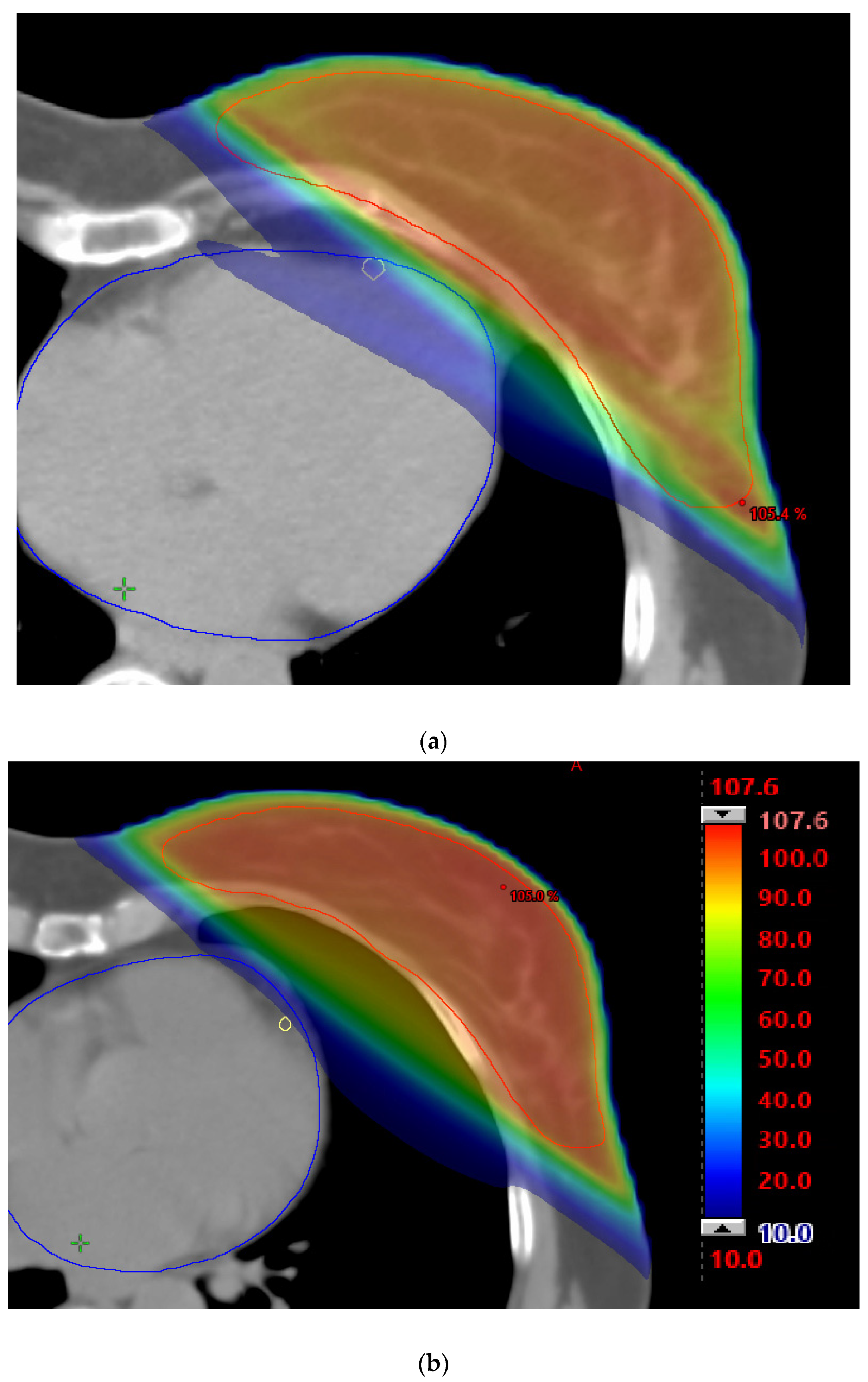
Figure 1 summarizes the flowchart of the study, from data acquisition to model development. The RT treatment data from 36 patients who received intensity modulated radiotherapy (IMRT) for cancer of the left breast in DIBH, whose characteristics are summarized in Table 1, were analysed. The treatment delivered a prescribed dose of 40.05 Gy in 15 fractions, using mostly 2 or 3 (average 2.6, 95% confidence intervals (CI) 2–4) intensity modulated tangential photon beams of 6 MV, with a dose rate of 600 monitor units per minute. All patients were imaged with a non-contrast-enhanced pre-treatment CT using a 32-slice scanner (Toshiba Aquilion LB, Toshiba Medical Systems Europe, Zoetermeer, the Netherlands). Both an FB and a DIBH CT planning scan were acquired, in order to compare the treatment plans generated on each modality (Figure 2). CT scans were contoured and planned using Varian Eclipse version 16.1 (Varian Medical System, Palo Alto, CA, USA), with a 2.5 mm grid size. The contoured regions of interest included the combined lungs, left lung, heart, contralateral (right) breast, and LAD. Dose distributions were calculated using the Anisotropic Analytical Algorithm. The DIBH was performed by the Real-time Position Management (RPM) system installed on a Varian Truebeam linear accelerator. The RPM consists of an infrared camera mounted on the wall of the treatment unit, infrared lights, and a marker box with infrared reflective markers placed on the patient’s chest. During the treatment, the reflective marker box is placed near to the xiphoid process, and the patient’s respiratory cycle is measured using the RPM during acquisition of the planning CT. The infrared camera measures the position of the infrared markers during this time, as a surrogate for the patient’s breathing.
During delivery of DIBH RT, the RPM system triggers beam-hold to the linear accelerator, if the patient’s breathing is outside the inspiration phase. [46]. The window in amplitude of the inspiration phase, which typically is of 2–8 mm, is selected during treatment planning, based on the respiratory signal acquired during the acquisition of pre-treatment CT, and is used for automated triggering of beam-on and beam-off during treatment.
In order to assess the dosimetric benefit of DIBH in terms of dose to the organs at risk for cardiovascular disease, for each DIBH patient a treatment plan was also calculated on the CT acquired in the FB modality, as shown in the Figure 2. The decrease in the maximum dose to the LAD, ΔDL, and between the DIBH and FB plan, was calculated, and its median value, med(ΔDL) among the patient cohort was used to split the patients into two classes with lower/higher ΔDL. The patients with ΔDL higher than med(ΔDL), were considered eligible for DIBH. For better dosimetric comparison, we also collected dose indexes of the planning target volume (PTV) and organs at risk. These included the minimum dose covering at least 95% of the PTV, V95%, and, for the organs at risk, the V15%, the hottest dose level covering at least 15% of the organ for the lung, and the maximum dose, Dmax, to the heart and contralateral breast.
The respiratory waveforms in the superior-inferior and anterior-posterior directions acquired for selection of the DIBH window, were exported for each patient, in the digital imaging and communications in medicine (DICOM) format. The statistical analysis and the implementation and evaluation of the BLSTM neural network, was performed in the MATLAB R2022b (The MathWorks, Inc., Apple Hill Drive, Natick, MA, USA) environment.

2.1. Neural Network

For predicting which patients will benefit from DIBH in terms of dose to the heart, a supervised machine learning algorithm was trained for binary classification from respiratory time series data.
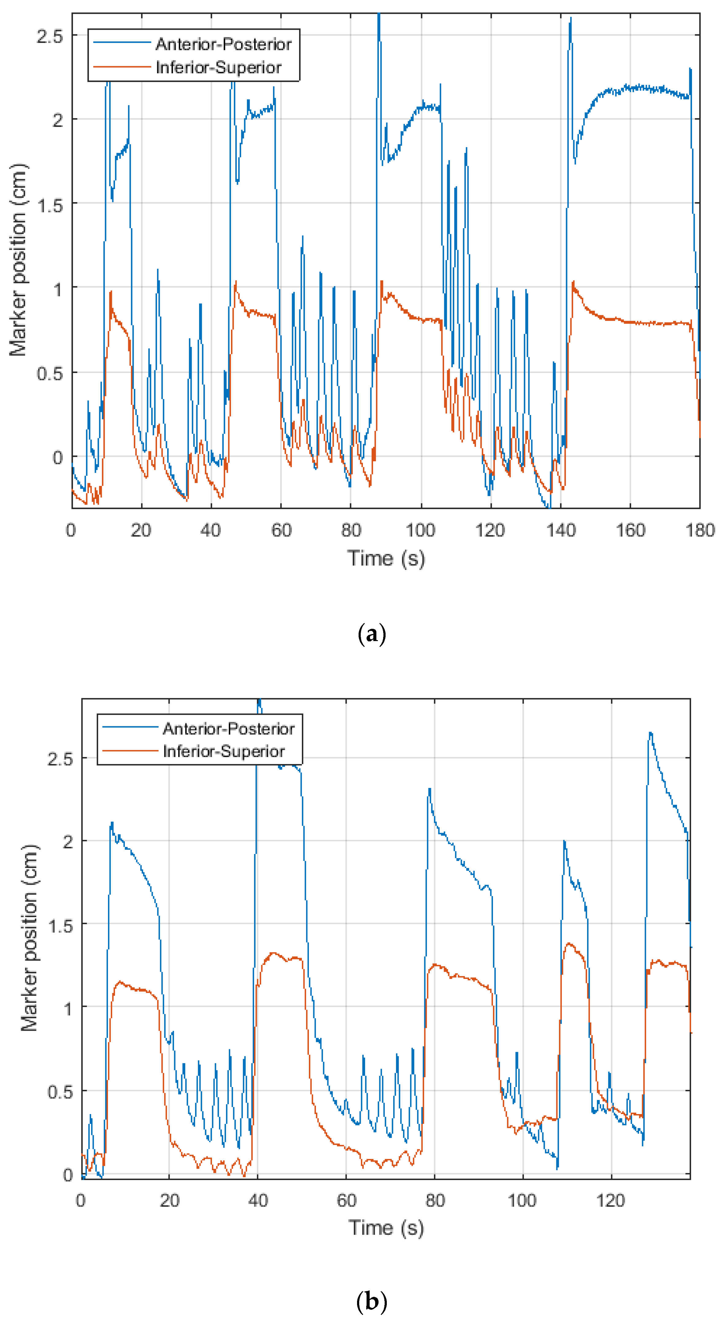
The respiratory time-series acquired for treatment preparation and gating window selection immediately after pre-treatment CT acquisition in the superior-inferior and anterior-posterior directions, were exported in a text file, which was converted into an excel spreadsheet for analysis. They usually include a series of cycles of free-breathing, followed by one or more deep inspiration in breath-holds, as shown in Figure 3a,b.
The algorithm of choice for machine learning was a bidirectional LSTM (BLSTM) neural network, which specializes in analysing time series data [47]. An LSTM is a type of deep neural network whose units have feedback connection, as in a recurrent neural network (RNN), in order to perform classification or regression from time series data. LSTM includes a cell and different types of gates. At the beginning, it included only input and output gates, but later it was modified to include a forget gate [48]. The cell decides which information should be stored based on the open and close operations at the gates, and the forget gate decides which information is kept from the previous time step, or if the LSTM should reset its state. In this way, LSTM can learn long-term dependencies between time steps, and eliminate the exploding or vanishing gradient problem [43].
BLSTM includes two distinct and interconnected neural networks, connected to the same output layer, that perform direct and reverse transformations, thus reading the training data in two different time directions. By taking both the left and right context information when training, it can yield higher predictive performance, compared to unidirectional LSTM architecture [49,50,51,52].
A neural network based on BLSTM was trained to classify the respiratory waveforms, and assign a positive binary outcome if the waveform belonged to a patient eligible for DIBH. In our implementation (Figure 4), the network included the sequence input layer, followed by two BLSTM layers. In order to decrease the overfitting, dropout layers were inserted after each of the two BLSTM layers. Dropout layers randomly turned off some neurons with a predefined probability during the training phase, thus reducing the number of parameters in the network and, as a consequence, overfitting [53]. The dropout probability was set at 20%.
Each cell in the BLSTM has four components: the cell weight, the input gate, the forget gate, and the output gate. As our network has 50 units and 2 dimensional (AP and SI) inputs, each BLSTM included 400 learnable weights. The BLSTM were followed by a fully connected layer, a softmax layer, and the classification layer. In total, the network had 81.8 thousand learnable properties.

2.2. Training of Classification Model

In order to train the BLSTM neural network, the dataset was split into training (60%, corresponding to 22 patients), and testing (40%, 14 patients) data, where the test set was used to verify the performance of the model developed on the training dataset. For network training and testing, the respiratory (anterior-posterior and superior-inferior) signals and the patient labels were imported in the MATLAB R2022b workspace. The dichotomic labels for supervised learning were: 1, for the patients with ΔDL > med(ΔDL), who benefit more from the DIBH’s treatment in terms of dose to the LAD; and 0, for the others.
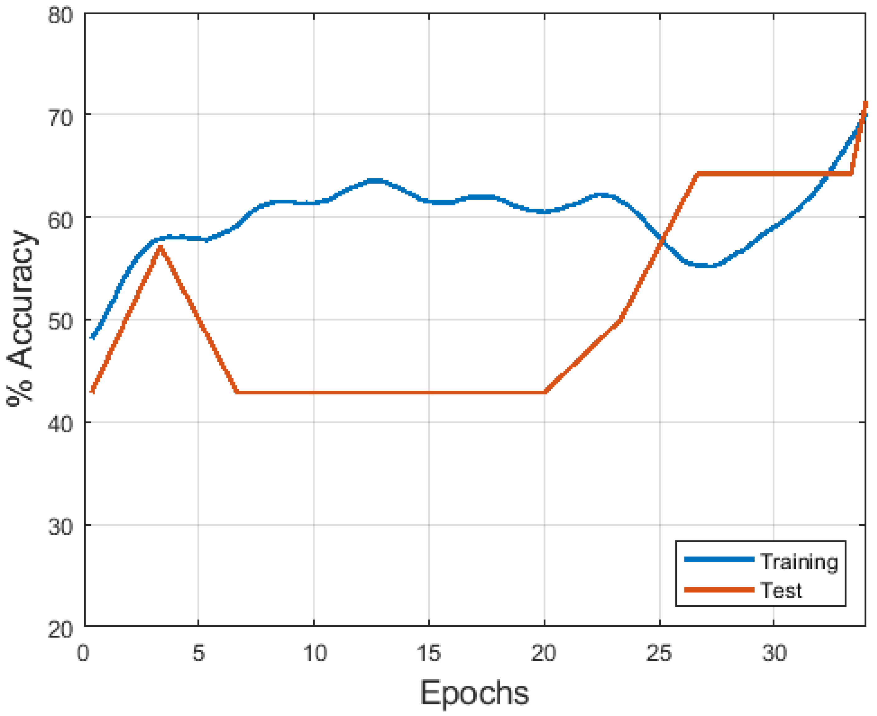
The training process was performed using the Adam optimizer with a batch size of 5 patients, resulting in 4 iterations for each epoch, for 35 epochs in total (Figure 4). The initial learning rate was 0.0003, which was reduced by a factor of 0.1, every 10 iterations. Every 10 iterations, the accuracy of the neural network was measured on the test set, in order to measure out-of-sample performance. The performance of the machine learning classifier was also measured by the area under the operating characteristic curve (AUC) of the test set.

3. Results

The results of the comparison between the FB and the DIBH treatment plans are shown in Table 2. Among the 36 patients, the DIBH treatment plans which were delivered to the patients, resulted in better dose coverage to the target, in terms of V95% to the PTV (Wilcoxon signed rank test p = 0.002), compared with the FB plans which were calculated for comparison. Interestingly, the maximum PTV dose showed a decrease with DIBH which was borderline significant (p = 0.051).
At the same time, the average maximum dose to the LAD was reduced to 0.56 (95% CI: 0.28–0.92) Gy in the DIBH treatment. In FB, the dose to the LAD was 11.11 (0.49–18.90) Gy. The difference was statistically significant (p << 0.001), and the reduction in maximum dose to the LAD, ΔDL, was 0.55 (95% CI: 0.07–11.99) Gy. This was largely due to the DIBH technique increasing the distance of LAD from the target region, from 0.08 cm to 2.19 cm (p << 0.001). The maximum dose to the heart was also reduced (p << 0.001). These results confirm the reduction of dose to the cardiovascular structures with DIBH. The value of med(ΔDL,) used to split patients into eligible or not eligible for DIBH, was 0.46 Gy.
Regarding dose metrics to the other main organs at risk, contralateral (right) breast and ipsilateral (left) lung, no significant differences were found between FB and DIBH, with Wilcoxon signed rank test p of 0.31 and 0.62, for lung and breast maximum doses, respectively.
Figure 5 shows the model accuracy achieved during training in both data subsamples. The accuracy, specificity, sensitivity, F1 score, and area under the receiving operating characteristic curve (AUC), were 71.4%, 66.7%, 80.1%, 72.4%, and 69.4%, respectively, in the test dataset, after 35 epochs of training.

4. Discussion

In our results, the comparison of DIBH against FB, confirmed that DIBH is effective in reducing dose to cardiac structures. This result is in agreement with previous investigators. Hayden and co-authors found that the mean dose to LAD planning risk volume was reduced, from 31.7 to 21.9 Gy, because of DIBH [54]. More recently the LAD maximum dose reduction was from 31.9 Gy to 25.8 Gy [55]. The lower LAD doses obtained in our study (from 11.11 Gy in FB to 0.56 Gy in DIBH) could be explained with the use of IMRT in all of our patients, compared with 3D-conformal RT (3D-CRT) in the aforementioned study, which allows better dose distribution optimization [55]. Borst and coauthors, using a hybrid 3D-CRT (80%) and IMRT(20%) technique, reduced the mean dose to the LAD from 11.4 to 5.5 Gy, which is more similar to our findings [56].
A more surprising result of our study, was the improvement in coverage of PTV with DIBH, perhaps also related to better sparing of organs at risk, which allows us to increase the dose to the target during inverse planning. We did not find a significant change in dose to the lung, whereas some previous studies experienced a significant increase in irradiated lung volume with DIBH, due to lung expansion within the tangential treatment fields [57]. By increasing the spatial separation between the target and the heart, DIBH can reduce the risk of heart disease [11], an event that can develop many years after the end of the treatment and result in increased rates of mortality [58], and therefore is a promising RT treatment option for left-sided breast cancer patients. DIBH also reduces the blurring of the static dose distribution caused by intra-fraction motion, which can result in a deviation between the calculated and the delivery dose distributions.
However, not all patients are eligible for DIBH, due to difficulty in breath holding, or lack of dosimetric benefit. To date, there are no clear selection criteria to predict which patients will benefit most from the DIBH technique, other than left breast laterality. The dosimetric benefit of DIBH seems to be related to the change in lung volume achieved by the patient between FB and DIBH scans, and to the shape of the breast [46]. For selection criteria, it was proposed to use the distance between the heart and the treatment field, as it was related to the mean heart dose [59]. In the study of Malone and co-workers [60] it was reported that, for left-sided breast cancer patients, the mean heart dose was related to the distance between the heart and the treatment field. In another study, the association of patient-related parameters, such as body mass index, age, PTV, cardiac contact distance, and lung volume at FB fully, with improved dosimetric indexes such as mean heart dose of 20%, were studied, but no significant correlations were found [61].
In order to select patients eligible for DIBH, a machine learning model which uses input variables such as heart and breast volumes, and distances, was proposed [30], resulting in accuracy of 0.88, and AUC 0.80 in cross validation. In our study, we adopted a random train –split validation, which is considered to be a stricter validation method, according to The Transparent Reporting of a Multivariable Model for Individual Prognosis or Diagnosis (TRIPOD) guidelines [62], which may explain our apparently lower performance.
Methods based on anatomical descriptors such as organ volumes and distances, suffer from limitations, as they do not account for the patient’s ability in maintaining breath hold. The respiratory signal acquired during the take-over of the pre-treatment CT, can provide a surrogate for this information. The importance of breathing signal was confirmed by the work of Ledsom et al. [63], wherein the increase in amplitude from FB to DIBH, was correlated with the reduction in cardiac dose.
For these reasons, in the present work we focused on the analysis of respiratory tracks for building a decision support system which could identify left breast cancer patient benefitting from the DIBH radiotherapy treatment. Although the introduction of AI in radiotherapy is still at an early stage [64,65], its most promising applications in this context include treatment recommendation, and prioritisation. A machine learning tool was developed which could recommend proton or photon treatment, by estimating which treatment will result in better quality of life for the patient [66,67,68]. LSTM looks promising for analysis of signals in radiotherapy. Lombardo et al. described the prediction of respiratory motion at different time steps of 250 ms, 500 ms, and 750 ms, for precise gated targeting in MR-guided radiotherapy [45]. Ma et al. applied LSTM to prediction of daily quality controls on a linear accelerator for radiotherapy [69].
Training an AI tool requires choice of a proper training dataset, as the quality of an AI tool is affected by the quality of the training dataset, a principle known as “garbage in, garbage out”. In our work, the strategy of choosing the median value of med(ΔDL), results in a perfectly balanced dataset, thus circumventing the difficulties related to dealing with data imbalance. Data imbalance occurs when data distribution is skewed, e.g., when the number of positives is much lower than the number of negatives, a common situation for screening of a rare disease [70]. Models developed from this type of imbalanced dataset would result in poor performance in the minority class, such as poor sensitivity. Because of the well-balanced dataset, in our result we had acceptable values of both sensitivity and specificity. The proposed AI tool based on BLSTM, could support the clinician’s decision early in the radiotherapy workflow in selecting patients for DIBH.
The clinical implementation of artificial intelligence algorithms is slow because they are considered by physicians as “black boxes”, meaning that the process that leads to their decisions is difficult to understand [71]. This issue usually makes implementation of AI in the clinical practice more difficult, as clinicians do not trust tools which are opaque in their decisions [23,72]. One of the strategies for improving interpretability, is to identify features in the data that are used by the AI in reaching its decision. By comparing respiratory tracks of patients classified as negative or positive, we can derive the characteristics used by the neural network to classify the patients. The respiratory signal of a patient considered eligible for DIBH treatment (Figure 2a) is characterised by longer deep inspiration phases, compared with the patient respiratory signal in Figure 1b, which is classified as negative.
At present, identifying patients with higher dosimetric benefit to the LAD due to DIBH, requires the acquisition of both FB and DIBH CTs, planning of treatments in FB and DIBH modalities, and evaluation of dose metrics to the LAD. The AI tool developed in the present study allows us to circumvent this cumbersome process, by analysing the respiratory tracks acquired in the same session as the pre-treatment CT acquisition. As such, the tool is useful in automating the choice of treatment approach for left-breast cancer patients. Due to its retrospective nature and the relatively small dataset, the findings in the present study should be confirmed in a larger, independent cohort, prior to being incorporated into clinical practice. Also, it remains to be investigated in a comparison of signal analysis among models; the more recent convolutional or residual LSTM [73], for instance, could improve the performance of the presented model, and this could be the subject of future investigations. A further difficulty is that our work did not consider the patient’s ability to tolerate DIBH. Finally, the integration in the model, of clinical and demographical patient data of interest, could be investigated, in order to improve the model’s performance.
Despite its limitations, this study provides proof of concept of AI-based prediction of the dosimetric benefit offered by DIBH from respiratory signals, and shows that a computerized decision support system can determine patient eligibility for DIBH RT. This implies that respiratory signals acquired for DIBH, are relevant, together with other data sources such as imaging, dose distribution [28,74], and electronic reports [75], in the era of AI-based patient data mining for automated disease detection and treatment recommendation.

5. Conclusions

In the present work, a deep neural network was implemented in order to predict which patients will benefit from DIBH RT from the respiratory tracks acquired early in the treatment workflow. The proposed system was the first method based on AI analysis to incorporate deep neural networks for selecting patients eligible for DIBH radiotherapy based on their respiratory tracks. Through automated analysis of respiratory signals, the developed system will potentially improve patient treatment recommendations, and speed-up the workflow in the radiotherapy department. Further validation of the presented concept in a larger, prospectively collected patient cohort, as well as model refinement through integration with other patient data, are warranted, in order to consolidate these findings.

Author Contributions

Conceptualization, M.A. and A.V.; Methodology, A.V., A.C., C.C. and M.A.; Validation, L.V. and L.B.; Investigation, A.V., L.V., C.C., M.P., P.C. and G.P.; Data curation, M.P., A.C., C.C. and L.B.; Supervision, A.D. All authors have read and agreed to the published version of the manuscript.

Funding

This research was funded by grant from the Italian Ministry of Health “5x1000 Ricerca Corrente” (code J34I19003280007).

Institutional Review Board Statement

The study was conducted in accordance with the Declaration of Helsinki, and approved by the Ethic Committee Comitato Etico Unico Regionale (CEUR) Friuli Venezia Giulia (Italy) protocol code CRO-2022-29 and date of approval 2 August 2022.

Informed Consent Statement

Informed consent was obtained from all subjects involved in the study.

Data Availability Statement

Data available upon request.

Conflicts of Interest

The authors declare no conflict of interest.

References

  1. Torre, L.A.; Siegel, R.L.; Ward, E.M.; Jemal, A. Global Cancer Incidence and Mortality Rates and Trends—An Update. Cancer Epidemiol. Biomark. Prev. 2016, 25, 16–27. [Google Scholar] [CrossRef] [Green Version]
  2. Ferlay, J.; Colombet, M.; Soerjomataram, I.; Dyba, T.; Randi, G.; Bettio, M.; Gavin, A.; Visser, O.; Bray, F. Cancer Incidence and Mortality Patterns in Europe: Estimates for 40 Countries and 25 Major Cancers in 2018. Eur. J. Cancer 2018, 103, 356–387. [Google Scholar] [CrossRef] [PubMed]
  3. Sardaro, A.; Petruzzelli, M.F.; D’Errico, M.P.; Grimaldi, L.; Pili, G.; Portaluri, M. Radiation-Induced Cardiac Damage in Early Left Breast Cancer Patients: Risk Factors, Biological Mechanisms, Radiobiology, and Dosimetric Constraints. Radiother. Oncol. 2012, 103, 133–142. [Google Scholar] [CrossRef] [Green Version]
  4. Kabel, A.M.; Baali, F.H. Breast Cancer: Insights into Risk Factors, Pathogenesis, Diagnosis and Management. J. Cancer Res. Treat. 2015, 3, 28–33. [Google Scholar] [CrossRef]
  5. Bleicher, R.J. Timing and Delays in Breast Cancer Evaluation and Treatment. Ann. Surg. Oncol. 2018, 25, 2829–2838. [Google Scholar] [CrossRef]
  6. Darby, S.; McGale, P.; Correa, C.; Taylor, C.; Arriagada, R.; Clarke, M.; Cutter, D.; Davies, C.; Ewertz, M.; Godwin, J.; et al. Effect of Radiotherapy after Breast-Conserving Surgery on 10-Year Recurrence and 15-Year Breast Cancer Death: Meta-Analysis of Individual Patient Data for 10 801 Women in 17 Randomised Trials. Lancet 2011, 378, 1707–1716. [Google Scholar] [CrossRef] [PubMed] [Green Version]
  7. Carlson, L.E.; Watt, G.P.; Tonorezos, E.S.; Chow, E.J.; Yu, A.F.; Woods, M.; Lynch, C.F.; John, E.M.; Mellemkjær, L.; Brooks, J.D.; et al. Coronary Artery Disease in Young Women After Radiation Therapy for Breast Cancer: The WECARE Study. JACC CardioOncology 2021, 3, 381–392. [Google Scholar] [CrossRef]
  8. Milo, M.L.H.; Thorsen, L.B.J.; Johnsen, S.P.; Nielsen, K.M.; Valentin, J.B.; Alsner, J.; Offersen, B.V. Risk of Coronary Artery Disease after Adjuvant Radiotherapy in 29,662 Early Breast Cancer Patients: A Population-Based Danish Breast Cancer Group Study. Radiother. Oncol. 2021, 157, 106–113. [Google Scholar] [CrossRef] [PubMed]
  9. Darby, S.C.; McGale, P.; Taylor, C.W.; Peto, R. Long-Term Mortality from Heart Disease and Lung Cancer after Radiotherapy for Early Breast Cancer: Prospective Cohort Study of about 300 000 Women in US SEER Cancer Registries. Lancet Oncol. 2005, 6, 557–565. [Google Scholar] [CrossRef]
  10. Aznar, M.; Korreman, S.S.; Pedersen, A.N.; Persson, G.F.; Josipovic, M.; Specht, L. Evaluation of Dose to Cardiac Structures during Breast Irradiation. Br. J. Radiol. 2011, 84, 743–746. [Google Scholar] [CrossRef] [PubMed]
  11. Keall, P.J.; Mageras, G.S.; Balter, J.M.; Emery, R.S.; Forster, K.M.; Jiang, S.B.; Kapatoes, J.M.; Low, D.A.; Murphy, M.J.; Murray, B.R.; et al. The Management of Respiratory Motion in Radiation Oncology Report of AAPM Task Group 76. Med. Phys. 2006, 33, 3874–3900. [Google Scholar] [CrossRef]
  12. Oechsner, M.; Düsberg, M.; Borm, K.J.; Combs, S.E.; Wilkens, J.J.; Duma, M.N. Deep Inspiration Breath-Hold for Left-Sided Breast Irradiation: Analysis of Dose-Mass Histograms and the Impact of Lung Expansion. Radiat. Oncol. 2019, 14, 1–10. [Google Scholar] [CrossRef]
  13. Rice, L.; Goldsmith, C.; Green, M.M.L.; Cleator, S.; Price, P.M. An Effective Deep-Inspiration Breath-Hold Radiotherapy Technique for Left-Breast Cancer: Impact of Post-Mastectomy Treatment, Nodal Coverage, and Dose Schedule on Organs at Risk. Breast Cancer Targets Ther. 2017, 9, 437–446. [Google Scholar] [CrossRef] [PubMed] [Green Version]
  14. Latty, D.; Stuart, K.E.; Wang, W.; Ahern, V. Review of Deep Inspiration Breath-Hold Techniques for the Treatment of Breast Cancer. J. Med. Radiat. Sci. 2015, 62, 74–81. [Google Scholar] [CrossRef]
  15. Mast, M.E.; Van Kempen-Harteveld, L.; Heijenbrok, M.W.; Kalidien, Y.; Rozema, H.; Jansen, W.P.A.; Petoukhova, A.L.; Struikmans, H. Left-Sided Breast Cancer Radiotherapy with and without Breath-Hold: Does IMRT Reduce the Cardiac Dose Even Further? Radiother. Oncol. 2013, 108, 248–253. [Google Scholar] [CrossRef] [PubMed]
  16. Nilsson, G.; Holmberg, L.; Garmo, H.; Duvernoy, O.; Sjögren, I.; Lagerqvist, B.; Blomqvist, C. Distribution of Coronary Artery Stenosis after Radiation for Breast Cancer. J. Clin. Oncol. 2012, 30, 380–386. [Google Scholar] [CrossRef]
  17. Maraldo, M.V.; Brodin, N.P.; Aznar, M.C.; Vogelius, I.R.; Munck af Rosenschöld, P.; Petersen, P.M.; Specht, L. Estimated Risk of Cardiovascular Disease and Secondary Cancers with Modern Highly Conformal Radiotherapy for Early-Stage Mediastinal Hodgkin Lymphoma. Ann. Oncol. 2013, 24, 2113–2118. [Google Scholar] [CrossRef]
  18. Yamauchi, R.; Mizuno, N.; Itazawa, T.; Saitoh, H.; Kawamori, J. Dosimetric Evaluation of Deep Inspiration Breath Hold for Left-Sided Breast Cancer: Analysis of Patient-Specific Parameters Related to Heart Dose Reduction. J. Radiat. Res. 2020, 61, 447–456. [Google Scholar] [CrossRef] [PubMed] [Green Version]
  19. Bertholet, J.; Knopf, A.; Eiben, B.; McClelland, J.; Grimwood, A.; Harris, E.; Menten, M.; Poulsen, P.; Nguyen, D.T.; Keall, P.; et al. Real-Time Intrafraction Motion Monitoring in External Beam Radiotherapy. Phys. Med. Biol. 2019, 64, 15TR01. [Google Scholar] [CrossRef]
  20. Brandner, E.D.; Chetty, I.J.; Giaddui, T.G.; Xiao, Y.; Huq, M.S. Motion Management Strategies and Technical Issues Associated with Stereotactic Body Radiotherapy of Thoracic and Upper Abdominal Tumors: A Review from NRG Oncology. Med. Phys. 2017, 44, 2595–2612. [Google Scholar] [CrossRef]
  21. Rochet, N.; Drake, J.I.; Harrington, K.; Wolfgang, J.A.; Napolitano, B.; Sadek, B.T.; Shenouda, M.N.; Keruakous, A.R.; Niemierko, A.; Taghian, A.G. Deep Inspiration Breath-Hold Technique in Left-Sided Breast Cancer Radiation Therapy: Evaluating Cardiac Contact Distance as a Predictor of Cardiac Exposure for Patient Selection. Pract. Radiat. Oncol. 2015, 5, e127–e134. [Google Scholar] [CrossRef]
  22. Wang, W.; Purdie, T.G.; Rahman, M.; Marshall, A.; Liu, F.F.; Fyles, A. Rapid Automated Treatment Planning Process to Select Breast Cancer Patients for Active Breathing Control to Achieve Cardiac Dose Reduction. Int. J. Radiat. Oncol. Biol. Phys. 2012, 82, 386–393. [Google Scholar] [CrossRef]
  23. Zanca, F.; Hernandez-Giron, I.; Avanzo, M.; Guidi, G.; Crijns, W.; Diaz, O.; Kagadis, G.C.; Rampado, O.; Lønne, P.I.; Ken, S.; et al. Expanding the Medical Physicist Curricular and Professional Programme to Include Artificial Intelligence. Phys. Med. 2021, 83, 174–183. [Google Scholar] [CrossRef] [PubMed]
  24. Avanzo, M.; Gagliardi, V.; Stancanello, J.; Blanck, O.; Pirrone, G.; El Naqa, I.; Revelant, A.; Sartor, G. Combining Computed Tomography and Biologically Effective Dose in Radiomics and Deep Learning Improves Prediction of Tumor Response to Robotic Lung Stereotactic Body Radiation Therapy. Med. Phys. 2021, 48, 6257–6269. [Google Scholar] [CrossRef]
  25. Zanca, F.; Brusasco, C.; Pesapane, F.; Kwade, Z.; Beckers, R.; Avanzo, M. Regulatory Aspects of the Use of Artificial Intelligence Medical Software. Semin. Radiat. Oncol. 2022, 32, 432–441. [Google Scholar] [CrossRef] [PubMed]
  26. Mancosu, P.; Lambri, N.; Castiglioni, I.; Dei, D.; Iori, M.; Loiacono, D.; Russo, S.; Talamonti, C.; Villaggi, E.; Scorsetti, M.; et al. Applications of Artificial Intelligence in Stereotactic Body Radiation Therapy. Phys. Med. Biol. 2022, 67, 16TR01. [Google Scholar] [CrossRef] [PubMed]
  27. Avanzo, M.; Trianni, A.; Botta, F.; Talamonti, C.; Stasi, M.; Iori, M. Artificial Intelligence and the Medical Physicist: Welcome to the Machine. Appl. Sci. 2021, 11, 1691. [Google Scholar] [CrossRef]
  28. Avanzo, M.; Pirrone, G.; Vinante, L.; Caroli, A.; Stancanello, J.; Drigo, A.; Massarut, S.; Mileto, M.; Urbani, M.; Trovo, M.; et al. Electron Density and Biologically Effective Dose (BED) Radiomics-Based Machine Learning Models to Predict Late Radiation-Induced Subcutaneous Fibrosis. Front. Oncol. 2020, 10, 490. [Google Scholar] [CrossRef] [PubMed] [Green Version]
  29. Avanzo, M.; Pirrone, G.; Mileto, M.; Massarut, S.; Stancanello, J.; Baradaran-Ghahfarokhi, M.; Rink, A.; Barresi, L.; Vinante, L.; Piccoli, E.; et al. Prediction of Skin Dose in Low-KV Intraoperative Radiotherapy Using Machine Learning Models Trained on Results of in Vivo Dosimetry. Med. Phys. 2019, 46, 1447–1454. [Google Scholar] [CrossRef] [PubMed] [Green Version]
  30. Lin, H.; Liu, T.; Shi, C.; Petillion, S.; Kindts, I.; Weltens, C.; Depuydt, T.; Song, Y.; Saleh, Z.; Xu, X.G.; et al. Feasibility Study of Individualized Optimal Positioning Selection for Left-Sided Whole Breast Radiotherapy: DIBH or Prone. J. Appl. Clin. Med. Phys. 2018, 19, 218–229. [Google Scholar] [CrossRef]
  31. Fukushima, K. Neocognitron: A Self-Organizing Neural Network Model for a Mechanism of Pattern Recognition Unaffected by Shift in Position. Biol. Cybern. 1980, 36, 193–202. [Google Scholar] [CrossRef]
  32. Fukushima, K. Neocognitron: A Hierarchical Neural Network Capable of Visual Pattern Recognition. Neural Netw. 1988, 1, 119–130. [Google Scholar] [CrossRef]
  33. Dechter, R. Learning While Searching in Constraint-Satisfaction-Problems. In Proceedings of the Fifth AAAI National Conference on Artificial Intelligence, Philadelphia, PA, USA, 11–15 August 1986; pp. 178–183. [Google Scholar]
  34. Lombardo, E.; Kurz, C.; Marschner, S.; Avanzo, M.; Gagliardi, V.; Fanetti, G.; Franchin, G.; Stancanello, J.; Corradini, S.; Niyazi, M.; et al. Distant Metastasis Time to Event Analysis with CNNs in Independent Head and Neck Cancer Cohorts. Sci. Rep. 2021, 11, 6418. [Google Scholar] [CrossRef] [PubMed]
  35. Antropova, N.; Huynh, B.Q.; Giger, M.L. A Deep Feature Fusion Methodology for Breast Cancer Diagnosis Demonstrated on Three Imaging Modality Datasets. Med. Phys. 2017, 44, 5162–5171. [Google Scholar] [CrossRef] [PubMed] [Green Version]
  36. Esteva, A.; Robicquet, A.; Ramsundar, B.; Kuleshov, V.; DePristo, M.; Chou, K.; Cui, C.; Corrado, G.; Thrun, S.; Dean, J. A Guide to Deep Learning in Healthcare. Nat. Med. 2019, 25, 24–29. [Google Scholar] [CrossRef]
  37. Castiglioni, I.; Ippolito, D.; Interlenghi, M.; Monti, C.B.; Salvatore, C.; Schiaffino, S.; Polidori, A.; Gandola, D.; Messa, C.; Sardanelli, F. Artificial Intelligence Applied on Chest X-Ray Can Aid in the Diagnosis of COVID-19 Infection: A First Experience from Lombardy, Italy. Eur. Radiol. Exp. 2021, 5, 7. [Google Scholar] [CrossRef]
  38. Brunese, L.; Mercaldo, F.; Reginelli, A.; Santone, A. Explainable Deep Learning for Pulmonary Disease and Coronavirus COVID-19 Detection from X-Rays. Comput. Methods Programs Biomed. 2020, 196, 105608. [Google Scholar] [CrossRef]
  39. Eo, T.; Jun, Y.; Kim, T.; Jang, J.; Lee, H.-J.; Hwang, D. KIKI-Net: Cross-Domain Convolutional Neural Networks for Reconstructing Undersampled Magnetic Resonance Images. Magn. Reson. Med. 2018, 80, 2188–2201. [Google Scholar] [CrossRef] [PubMed]
  40. Sloan, J.M.; Goatman, K.A.; Siebert, J.P. Learning Rigid Image Registration—Utilizing Convolutional Neural Networks for Medical Image Registration. In Proceedings of the 5th International Conference on Bioimaging, Funchal, Portugal, 19–21 January 2018; pp. 89–99. [Google Scholar]
  41. Fourcade, C.; Ferrer, L.; Moreau, N.; Santini, G.; Brennan, A.; Rousseau, C.; Lacombe, M.; Fleury, V.; Colombié, M.; Jézéquel, P.; et al. Deformable Image Registration with Deep Network Priors: A Study on Longitudinal PET Images. Phys. Med. Biol. 2022, 67, 155011. [Google Scholar] [CrossRef]
  42. Yang, S.; Zhu, F.; Ling, X.; Liu, Q.; Zhao, P. Intelligent Health Care: Applications of Deep Learning in Computational Medicine. Front. Genet. 2021, 12, 444. [Google Scholar] [CrossRef] [PubMed]
  43. Hochreiter, S.; Schmidhuber, J. Long Short-Term Memory. Neural Comput. 1997, 9, 1735–1780. [Google Scholar] [CrossRef]
  44. Denysyuk, H.V.; Pinto, R.J.; Silva, P.M.; Duarte, R.P.; Marinho, F.A.; Pimenta, L.; Gouveia, A.J.; Gonçalves, N.J.; Coelho, P.J.; Zdravevski, E.; et al. Algorithms for Automated Diagnosis of Cardiovascular Diseases Based on ECG Data: A Comprehensive Systematic Review. Heliyon 2023, 9, e13601. [Google Scholar] [CrossRef]
  45. Lombardo, E.; Rabe, M.; Xiong, Y.; Nierer, L.; Cusumano, D.; Placidi, L.; Boldrini, L.; Corradini, S.; Niyazi, M.; Belka, C.; et al. Offline and Online LSTM Networks for Respiratory Motion Prediction in MR-Guided Radiotherapy. Phys. Med. Biol. 2022, 67, 095006. [Google Scholar] [CrossRef]
  46. Bergom, C.; Currey, A.; Desai, N.; Tai, A.; Strauss, J.B. Deep Inspiration Breath Hold: Techniques and Advantages for Cardiac Sparing During Breast Cancer Irradiation. Front. Oncol. 2018, 8, 87. [Google Scholar] [CrossRef] [PubMed] [Green Version]
  47. Van Houdt, G.; Mosquera, C.; Nápoles, G. A Review on the Long Short-Term Memory Model. Artif. Intell. Rev. 2020, 53, 5929–5955. [Google Scholar] [CrossRef]
  48. Gers, F.A.; Schmidhuber, J.; Cummins, F. Learning to Forget: Continual Prediction with LSTM. Neural Comput. 2000, 12, 2451–2471. [Google Scholar] [CrossRef] [PubMed]
  49. Li, C.; Bao, Z.; Li, L.; Zhao, Z. Exploring Temporal Representations by Leveraging Attention-Based Bidirectional LSTM-RNNs for Multi-Modal Emotion Recognition. Inf. Process. Manag. 2020, 57, 102185. [Google Scholar] [CrossRef]
  50. Graves, A.; Mohamed, A.R.; Hinton, G. Speech Recognition with Deep Recurrent Neural Networks. In Proceedings of the 2013 IEEE International Conference on Acoustics, Speech and Signal Processing, Vancouver, BC, Canada, 26–31 May 2013; Volume 3, pp. 45–49. [Google Scholar]
  51. Huang, Z.; Xu, W.; Yu, K. Bidirectional LSTM-CRF Models for Sequence Tagging. arXiv 2015, arXiv:1508.01991. [Google Scholar]
  52. Graves, A.; Schmidhuber, J. Framewise Phoneme Classification with Bidirectional LSTM and Other Neural Network Architectures. Neural Netw. 2005, 18, 602–610. [Google Scholar] [CrossRef] [PubMed]
  53. Shen, C.; Nguyen, D.; Zhou, Z.; Jiang, S.B.; Dong, B.; Jia, X. An Introduction to Deep Learning in Medical Physics: Advantages, Potential, and Challenges. Phys. Med. Biol. 2020, 65, 05TR01. [Google Scholar] [CrossRef]
  54. Hayden, A.J.; Rains, M.; Tiver, K. Deep Inspiration Breath Hold Technique Reduces Heart Dose from Radiotherapy for Left-Sided Breast Cancer. J. Med. Imaging Radiat. Oncol. 2012, 56, 464–472. [Google Scholar] [CrossRef] [PubMed]
  55. Ferdinand, S.; Mondal, M.; Mallik, S.; Goswami, J.; Das, S.; Manir, K.S.; Sen, A.; Palit, S.; Sarkar, P.; Mondal, S.; et al. Dosimetric Analysis of Deep Inspiratory Breath-Hold Technique (DIBH) in Left-Sided Breast Cancer Radiotherapy and Evaluation of Pre-Treatment Predictors of Cardiac Doses for Guiding Patient Selection for DIBH. Tech. Innov. Patient Support Radiat. Oncol. 2021, 17, 25–31. [Google Scholar] [CrossRef]
  56. Borst, G.R.; Sonke, J.-J.; den Hollander, S.; Betgen, A.; Remeijer, P.; van Giersbergen, A.; Russell, N.S.; Elkhuizen, P.H.M.; Bartelink, H.; van Vliet-Vroegindeweij, C. Clinical Results of Image-Guided Deep Inspiration Breath Hold Breast Irradiation. Int. J. Radiat. Oncol. *Biol. *Phys. 2010, 78, 1345–1351. [Google Scholar] [CrossRef]
  57. Pandeli, C.; Smyth, L.M.L.; David, S.; See, A.W. Dose Reduction to Organs at Risk with Deep-Inspiration Breath-Hold during Right Breast Radiotherapy: A Treatment Planning Study. Radiat. Oncol. 2019, 14, 223. [Google Scholar] [CrossRef] [Green Version]
  58. Darby, S.C.; Ewertz, M.; McGale, P.; Bennet, A.M.; Blom-Goldman, U.; Brnønum, D.; Correa, C.; Cutter, D.; Gagliardi, G.; Gigante, B.; et al. Risk of Ischemic Heart Disease in Women after Radiotherapy for Breast Cancer. N. Engl. J. Med. 2013, 368, 987–998. [Google Scholar] [CrossRef] [Green Version]
  59. Vuong, W.; Garg, R.; Bourgeois, D.J.; Yu, S.; Sehgal, V.; Daroui, P. Dosimetric Comparison of Deep-Inspiration Breath-Hold and Free-Breathing Treatment Delivery Techniques for Left-Sided Breast Cancer Using 3D Surface Tracking. Med. Dosim. 2019, 44, 193–198. [Google Scholar] [CrossRef] [PubMed]
  60. Malone, C.; Fennell, L.; Folliard, T.; Kelly, C. Using a Neural Network to Predict Deviations in Mean Heart Dose during the Treatment of Left-Sided Deep Inspiration Breath Hold Patients. Phys. Med. 2019, 65, 137–142. [Google Scholar] [CrossRef] [PubMed]
  61. Czeremszyńska, B.; Drozda, S.; Górzyński, M.; Kępka, L. Selection of Patients with Left Breast Cancer for Deep-Inspiration Breath-Hold Radiotherapy Technique: Results of a Prospective Study. Rep. Pract. Oncol. Radiother. 2017, 22, 341–348. [Google Scholar] [CrossRef]
  62. Collins, G.S.; Reitsma, J.B.; Altman, D.G.; Moons, K.G. Transparent Reporting of a Multivariable Prediction Model for Individual Prognosis or Diagnosis (TRIPOD): The TRIPOD Statement. BMC Med. 2015, 13, 1-014-0241-z. [Google Scholar] [CrossRef] [Green Version]
  63. Ledsom, D.; Reilly, A.J.; Probst, H. Assessment of Deep Inspiration Breath Hold (DIBH) Amplitude and Reduction in Cardiac Dose in Left Breast Cancer Patients. Radiography 2018, 24, 98–103. [Google Scholar] [CrossRef] [Green Version]
  64. Meyer, P.; Noblet, V.; Mazzara, C.; Lallement, A. Survey on Deep Learning for Radiotherapy. Comput. Biol. Med. 2018, 98, 126–146. [Google Scholar] [CrossRef] [PubMed]
  65. Siddique, S.; Chow, J.C.L. Artificial Intelligence in Radiotherapy. Rep. Pract. Oncol. Radiother. 2020, 25, 656–666. [Google Scholar] [CrossRef] [PubMed]
  66. Barragán-Montero, A.; Javaid, U.; Valdés, G.; Nguyen, D.; Desbordes, P.; Macq, B.; Willems, S.; Vandewinckele, L.; Holmström, M.; Löfman, F.; et al. Artificial Intelligence and Machine Learning for Medical Imaging: A Technology Review. Phys. Med. 2021, 83, 242–256. [Google Scholar] [CrossRef]
  67. Vandewinckele, L.; Claessens, M.; Dinkla, A.; Brouwer, C.; Crijns, W.; Verellen, D.; van Elmpt, W. Overview of Artificial Intelligence-Based Applications in Radiotherapy: Recommendations for Implementation and Quality Assurance. Radiother. Oncol. 2020, 153, 55–66. [Google Scholar] [CrossRef] [PubMed]
  68. Al, N.P.B. A Quantitative Clinical Decision-Support Strategy Identifying Which Oropharyngeal Head and Neck Cancer Patients May Benefit the Most from Proton Radiation Therapy. Int. J. Radiat. Oncol. Biol. Phys. 2019, 176, 139–148. [Google Scholar] [CrossRef]
  69. Ma, M.; Liu, C.; Wei, R.; Liang, B.; Dai, J. Predicting Machine’s Performance Record Using the Stacked Long Short-Term Memory (LSTM) Neural Networks. J. Appl. Clin. Med. Phys. 2022, 23, e13558. [Google Scholar] [CrossRef]
  70. Johnson, J.M.; Khoshgoftaar, T.M. Survey on Deep Learning with Class Imbalance. J. Big Data 2019, 6, 27. [Google Scholar] [CrossRef] [Green Version]
  71. He, J.; Baxter, S.L.; Xu, J.; Xu, J.; Zhou, X.; Zhang, K. The Practical Implementation of Artificial Intelligence Technologies in Medicine. Nat. Med. 2019, 25, 30–36. [Google Scholar] [CrossRef] [PubMed]
  72. Zihni, E.; Madai, V.I.; Livne, M.; Galinovic, I.; Khalil, A.A.; Fiebach, J.B.; Frey, D. Opening the Black Box of Artificial Intelligence for Clinical Decision Support: A Study Predicting Stroke Outcome. PLoS ONE 2020, 15, e0231166. [Google Scholar] [CrossRef] [Green Version]
  73. Wang, Z.; Zhang, T.; Shao, Y.; Ding, B. LSTM-Convolutional-BLSTM Encoder-Decoder Network for Minimum Mean-Square Error Approach to Speech Enhancement. Appl. Acoust. 2021, 172, 107647. [Google Scholar] [CrossRef]
  74. Pirrone, G.; Matrone, F.; Chiovati, P.; Manente, S.; Drigo, A.; Donofrio, A.; Cappelletto, C.; Borsatti, E.; Dassie, A.; Bortolus, R.; et al. Predicting Local Failure after Partial Prostate Re-Irradiation Using a Dosiomic-Based Machine Learning Model. J. Pers. Med. 2022, 12, 1491. [Google Scholar] [CrossRef] [PubMed]
  75. Mahadevaiah, G.; Rv, P.; Bermejo, I.; Jaffray, D.; Dekker, A.; Wee, L. Artificial Intelligence-Based Clinical Decision Support in Modern Medical Physics: Selection, Acceptance, Commissioning, and Quality Assurance. Med. Phys. 2020, 47, e228–e235. [Google Scholar] [CrossRef] [PubMed]
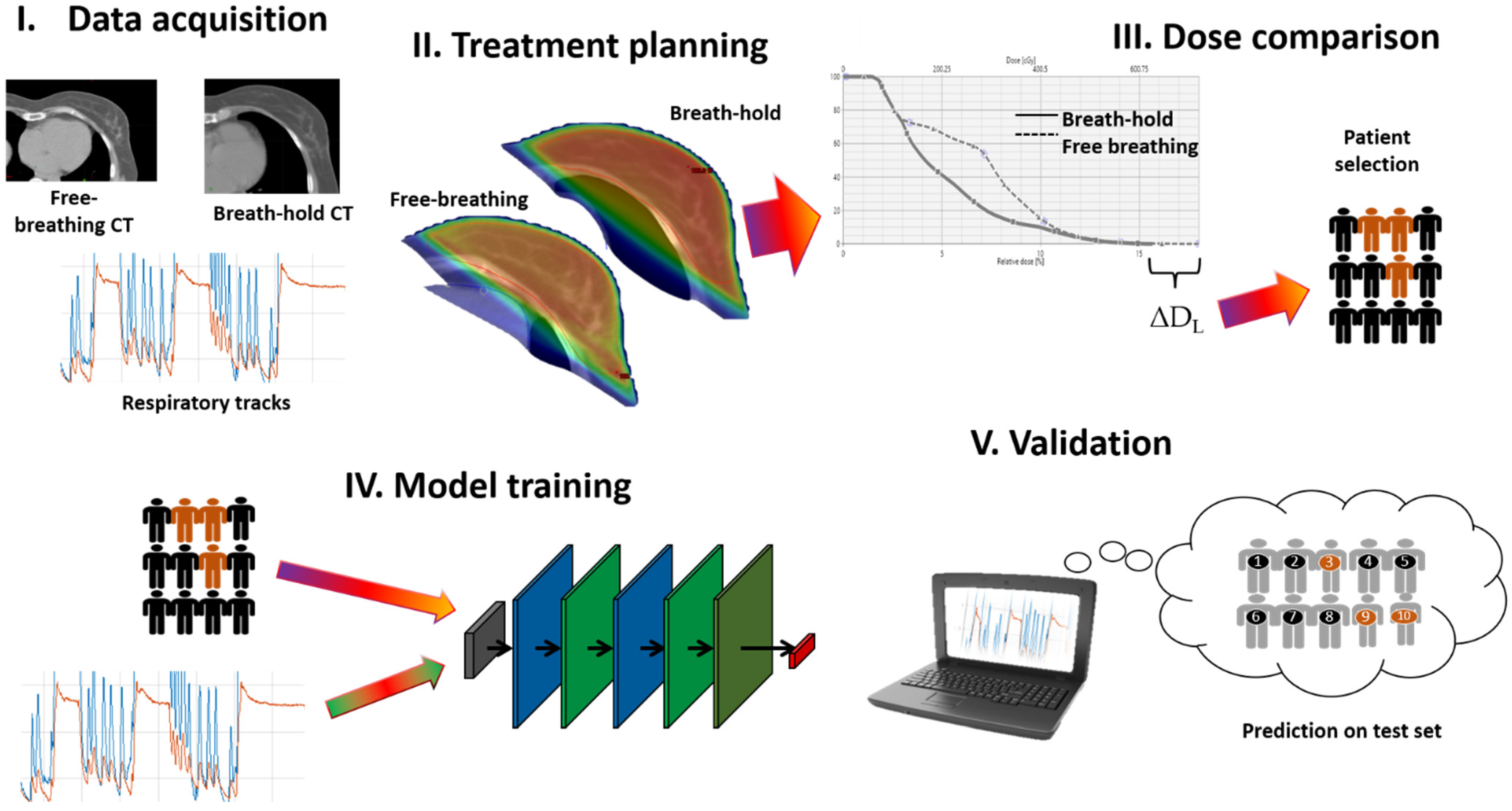
Figure 1. Flowchart which summarizes the main steps in the workflow of the present study.
Applsci 13 04962 g001
Figure 2. (a,b): Comparison of dose distributions from treatment plans of IMRT in free-breathing (a) and DIBH (b) planning scans of the same patient, where the increase in distance of the treated breast from the cardiovascular structures in the DIBH plan, resulted in reduced dose to the heart and LAD. The dose distributions are shown in colour-wash mode, which scale is in (b).
Applsci 13 04962 g002
Figure 3. (a,b): examples of respiratory tracks acquired for selecting DIBH threshold. (a) is an example of a track from a patient from the group considered eligible for DIBH due to higher decrease of maximum dose to the LAD from FB, while (b) is from a patient in the group with lower LAD dose reduction.
Applsci 13 04962 g003
Figure 4. Architecture of the deep neural network for classification of respiratory tracks based on BLSTM units.
Applsci 13 04962 g004
Figure 5. Graph showing the accuracy of the BLSTM neural network during the 250 epochs of training, both in the training (blue), and test sets (red).
Applsci 13 04962 g005
Table 1. Treatment and patient data of the cohort in the study.
VariableValue
SexFemale
Age mean (95% CI)52 (41, 69.5)
Prescribed dose40.05 Gy/15 fractions
Sequential boost18/36
Table 2. Results of comparison treatment plans in FB and DIBH modalities.
VariableMean Value (95% CI)Wilcoxon Signed Rank’s p
FBDIBH
Number of fields2.6 (2–5.3)2.6 (2–4)0.65
PTV V95% (%)95.0 (90.3–97.7)96.5 (93.9–98.7)0.002
PTV Dmax (Gy)42.9 (41.6–45.5)42.6 (41.6–43.7)0.051
Distance of LAD to PTV in BEV (cm)0.08 (−1.62,1.25)2.19 (1.44,3.75)<<0.001
LAD Dmax (Gy)11.11 (0.49–18.90)0.56 (0.28–0.92)<<0.001
Heart Dmax (Gy)19.66 (0.61–30.20)0.93 (0.39–17.00)<<0.001
Contralateral Breast Dmax (Gy)0.41 (0.11–0.87)0.50 (0.12–16.59)0.62
Lung V15% (Gy)0.44 (0.19–12.61)0.63 (0.33–12.26)0.31
Disclaimer/Publisher’s Note: The statements, opinions and data contained in all publications are solely those of the individual author(s) and contributor(s) and not of MDPI and/or the editor(s). MDPI and/or the editor(s) disclaim responsibility for any injury to people or property resulting from any ideas, methods, instructions or products referred to in the content.