Communication

Colorectal Cancer Cell Spheroids Co-Cultured with Molecularly Imprinted Fluorescent Particles Targeting Sialic Acid Show Preserved Cell Viability

by
Thomas Sjöberg
1,2,
Zahra El-Schich
1,2,
Knut Rurack
3 and
Anette Gjörloff Wingren
1,2,*
1
Department of Biomedical Sciences, Faculty of Health and Society, Malmö University, 205 06 Malmö, Sweden
2
Biofilms Research Center for Biointerfaces, Malmö University, 205 06 Malmö, Sweden
3
Federal Institute for Materials Research and Testing (BAM), 12205 Berlin, Germany
*
Author to whom correspondence should be addressed.
Appl. Sci. 2023, 13(9), 5330; https://doi.org/10.3390/app13095330
Submission received: 26 February 2023 / Revised: 18 April 2023 / Accepted: 23 April 2023 / Published: 24 April 2023
(This article belongs to the Special Issue Detection and Imaging of Tumor Cells in a 3D Environment)

Abstract

:
In vitro cultured 3D models of CRC have been demonstrated to hold considerable worth in drug discovery, drug resistance analysis, and in studying cell-cell and cell-matrix interactions that occur in the tumor microenvironment. The 3D models resemble the in vivo physiological microenvironment by replicating the cell type composition and tissue architecture. Molecularly imprinted polymers (MIPs) have been investigated for use instead of antibodies against small non-immunogenic structures, such as sialic acid (SA). Glyco-conjugates including SA are present on all cells, and often deregulated on cancer cells. Here, we present a novel approach for targeting and detecting colorectal cancer cells (CRC) by using in vitro cultured HT29 3D spheroids co-cultured in vitro with either fluorescent MIPs targeting SA, SA-MIPs, or the two lectins targeting SA, MAL I, and SNA. Both formaldehyde-fixed and viable HT29 3D spheroids with or without SA-MIPs were imaged in 3D by confocal microscopy. The results revealed a preserved cell morphology and viability of the HT29 3D spheroids co-cultured in vitro with SA-MIPs. However, the lectins MAL I and SNA targeting the α-2,3 or α-2,6 SA glycosidic linkages, respectively, affected the cell viability when co-cultured with the viable HT29 3D spheroids, and no living cells could be detected. Here, we have shown that the SA-MIPs could be used as a safe and low-cost diagnostic tool for targeting and detecting cancer cells in a physiologically relevant 3D cancer model in vitro.

1. Introduction

Improved diagnostics and treatment of aggressive cancer is one of the most challenging tasks in research today. The transformation from a normal cell into a tumor cell is a multistage process, typically involving progression from a pre-cancerous lesion to malignant tumors [1]. Colorectal cancer (CRC) is the third most common cause of cancer-related deaths worldwide, and the prognosis for patients with metastatic CRC (mCRC) is still poor [2]. The most common conventional method to culture cells today are monolayer 2D cell cultures, which are not accurately representing the cellular and micro-environmental interactions in vivo [3]. In vitro cultured 3D models of CRC have been shown to be valuable in drug discovery, drug resistance analysis, and studies of cell-cell and cell-matrix interactions that occur in the tumor microenvironment (TME) [4]. The development of safe and effective drugs is currently hampered by the poor predictive power of existing pre-clinical animal models that often lead to the failure of drug candidates in human trials [5]. Sialic acid (SA) is a monosaccharide present on proteins and lipids on almost all cells, but are often deregulated on cancer cells and, therefore, of interest as a cancer-related target molecule [6]. Previous studies have shown the diagnostic relevance of molecularly imprinted polymers (MIPs) recognizing SA, so called SA-MIPs [7,8,9,10]. MIPs are synthesized through a combination of functional monomers that strongly interact with a target molecule (template) and structural monomers and crosslinkers that covalently embed this complex in a polymer network [7,11,12]. The advantages of using MIPs include their low cost, quick preparation, chemical and physical stability, and reproducibility.
The combination of MIPs loaded with a drug and a moiety targeting a membrane protein on cancer cells were shown by Canfarotta et al. [13]. Using a double-imprinting method, the authors showed that the nanoMIPs could not only target the membrane receptor but also selectively deliver the drug to the corresponding cells. For targeting cells without a consequent drug delivery system, it was important to confirm that the treated cells remained viable. SA-MIPs have been shown to interact with phagocytic cells in vitro without causing any inflammatory or toxic response [14]. Hence, the next step was to use SA-MIPs to target and detect spheroid cancer cells grown in in vivo-like conditions and investigate if the in vitro SA-MIPs spheroid system was physiologically relevant.
SA would bind to the sialic acid-binding immunoglobulin-like lectin (Siglec), which provided a conserved mechanism for combatting pathogens [15]. The deregulation of SA and this pathway had been observed in diseases such as autoimmunity, neurodegeneration, allergy, and cancer. The importance of 3D in vitro systems and assays relied on the need to obtain reliable data regarding diagnostic tools and therapeutic candidates [16].
In this study, we present a novel approach for targeting and detecting spheroid cancer cells grown in in vivo-like conditions. SA-MIPs were targeted to the CRC cell line HT29 as viable spheroid cultures in vitro without affecting the cell viability. We hereby propose the use of SA-MIPs as a non-toxic and valuable diagnostic tool for detecting cancer.

2. Materials and Methods

2.1. Cell Culture

HT29 colorectal adenocarcinoma (HTB-38) cell line was obtained from the American Type Culture Collection (ATCC/LGC Standards, Teddington, UK). HT29 cells were cultured in McCoy’s 5A medium supplemented with 10% heat-inactivated fetal bovine serum (FBS; Thermo Fisher Scientific, Waltham, MA, USA). Cells were grown in T75 flasks and incubated at 37 °C and 5% CO2. The culture medium was changed regularly, and the cell passage was carried out at 70–80% confluency. Trypsin/EDTA (Thermo Fisher Scientific) was used to detach the cells.

2.2. SA-MIP Synthesis

The synthesis of SA-MIPs was performed as recently reported [8,17,18]. The SA-MIPs were equipped with nitrobenzoxadiazole (NBD) fluorescent reporter groups allowing environmentally sensitive fluorescence detection in green fluorescence.

2.3. HT29 Spheroid Cell Cultures

For HT29 spheroid cell culturing, 1 × 10⁴ HT29 cells were grown in 3 mL of Dulbecco’s Modified Eagle Medium F-12-1:1 Mixture containing 3.151 g/L glucose, L-Glutamine without Hepes (DMEM; Lonza, Basel, Switzerland) supplemented with B27 (Thermo Fisher Scientific), 20 ng/mL of both human epidermal growth factor (hEGF, Sigma-Aldrich, MO, USA), and human basic fibroblast growth factor (bFGF, Sigma-Aldrich, St. Louis, MO, USA). Spheroids were cultured in 35-mm non-treated culture dishes (Corning®, Corning, NY, USA). For co-culture, 40 µg/mL of SA-MIPs, lectins Maackia Amurensis Lectin I (MAL I) or Sambucus Nigra Lectin (SNA), 5 ug/mL (Vector Laboratories; Newark, CA, USA), respectively, were added to the HT29 spheroid medium, and the cells were left to grow for 8–10 days in 37 °C and 5% CO2.

2.4. Immunofluorescence Staining

HT29 spheroids grown without SA-MIPs or lectins were collected and washed twice with Dulbeccos Phosphate Buffered Saline (PBS) without Ca2+/Mg2+ (Thermo Fisher Scientific) followed by fixation in 4% formaldehyde (Thermo Fisher Scientific) for 30 min at room temperature (RT), washed once with PBS, followed by a 1-h incubation with 40 µg/mL SA-MIPs, or the biotin-labeled lectins MAL I and SNA, 5 ug/mL with agitation. Next, the spheroids were washed once with PBS, and streptavidin-fluorescein isothiocyanate (FITC) (Agilent Technologies, Santa Clara, CA, USA) was added to biotinylated-MAL I and SNA, respectively, and incubated for an additional 30 min at RT with agitation. Spheroids were then washed with PBS and permeabilized with 0.1% Triton X-100 (Sigma-Aldrich) in PBS for 30 min at RT, washed again with 0.1% Triton X-100 in PBS, and incubated with 1:100 rhodamine phalloidin (Sigma-Aldrich) for 1 h, at RT with agitation, followed by a wash with 0.1% Triton X-100 in PBS and finally incubated with 4′,6-diamidino-2-phenylindole (DAPI, Thermo Fisher Scientific) diluted in 0.1% Triton X-100/PBS for 5 min with agitation. Finally, the cells were washed with 0.1% Triton X-100/PBS and thereafter resuspended in PBS.

2.5. Confocal Imaging

For confocal imaging, HT29 spheroids were transferred to a µ-Slide 8 Well high Glass Bottom (Ibidi GmbH, Gräfelfing, Germany), and images were taken at an interval of 0.75 µm from top to bottom to confirm the presence of SA-MIPs or lectins. Fluorescence images were obtained using a Nikon A1plus equipped with a plan APO λ 20x/0.75 objective, 402 nm, 487 nm, and 566 nm lasers, and Nikon NIS-Elements software was used. All image stacks were acquired with comparable settings, at a resolution of 1024 × 1024 pixels, with z-step sizes between 0.75 to 0.925 μm. All experiments described under the section Materials and Methods were performed at least three times.

3. Results and Discussion

Here we present an important in vitro model for the evaluation of diagnostic tools and treatment protocols by combining HT29 spheroids grown with or without SA-MIPs. The first sets of HT29 spheroids were grown in conventional spheroid medium according to the Material and Methods section. After successful growth for up to 10 days, the spheroids were harvested, fixed with 4% formaldehyde, and stained with lectins MAL I, SNA, or SA-MIPs, respectively, as well as with DAPI (nuclei) and phalloidin (cytoskeleton) (Figure 1). The α-2,3- and α-2,6 SA expression of the HT29 cell line is determining the binding of MAL I and SNA, respectively, which was confirmed by flow cytometry analysis on single cell level.
MAL I and SNA staining indicated an intact expression of α-2,3- and α-2,6 SA on the outer layer of the HT29 spheroids (Figure 1). MAL I was homogeneously distributed across the outer layer of the HT29 spheroids, whereas SNA was limited to certain patches of the HT29 spheroids. SA-MIPs have previously been shown to display a binding pattern comparable to that of SA-targeting lectins [9]. Here we demonstrated a direct binding of the SA-MIPs to the formaldehyde-fixed cells across the HT29 spheroids (Figure 1).
Using a serum-free medium containing the appropriate growth factors, spheroids can be grown from established cell lines or from dissociated tissue cells. It is crucial that the cell environment is adjusted to mimic the in vivo conditions when performing 3D cell culture experiments in vitro. Spheroid cultures have become increasingly important tools, reproducing the in vivo cell environment and spheroid cultures can provide accurate data for various diseases [4]. Protocol development is particularly useful when working with more valuable patient samples in 3D, i.e., organoids. Additionally, by the utilization of spheroids and organoids, the reliance on animal models in drug discovery can be minimized offering more ethical and cost-effective alternatives for preclinical testing.
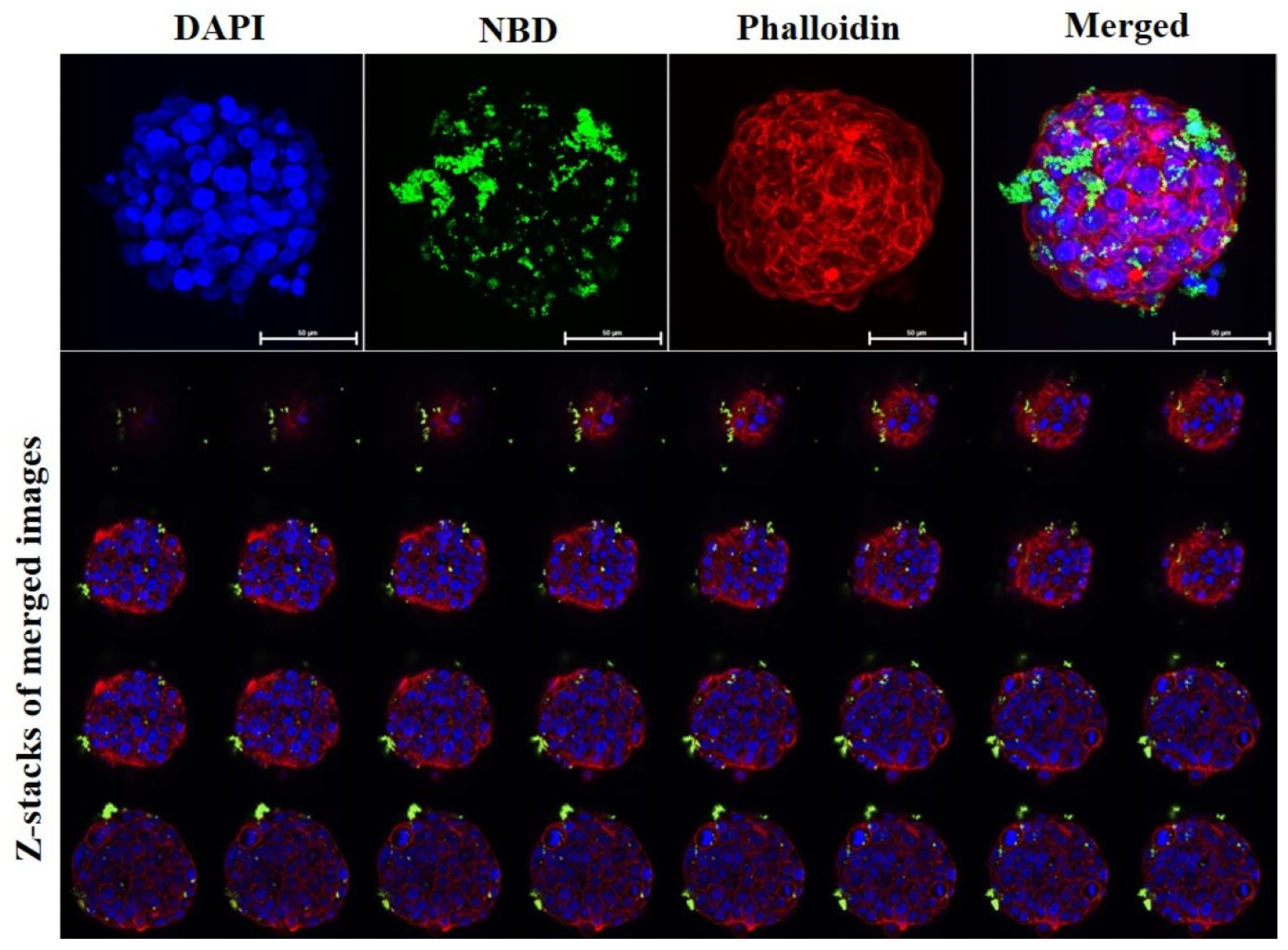
The addition of MAL I and SNA to viable, growing HT29 spheroid cell cultures, caused cell death leading to incomplete spheroid growth. Plant lectins have been reported to be effective in targeting cancer cells and inducing cell death [19]. Specifically, MAL I and SNA have been shown to induce apoptosis in non-small cell lung cancer cells [19] or in ovarian cancer cells [20], respectively. In contrast to formaldehyde-fixed spheroids, the addition of SA-MIPs to viable HT29 spheroid cell cultures, resulted in a more pronounced SA-MIP staining pattern, with no detected effect on cell viability (Figure 2). In addition, the Z-stack images of viable SA-MIP-HT29 spheroids demonstrated detailed binding pattern of SA-MIPs. Most importantly, SA-MIPs were co-cultured with HT29 spheroids for 8–10 days without any signs of toxicity or cell death.
Molecular imprinting relied on template-directed synthesis of polymers [7,8,11,12,21], and MIPs thus produced against different target molecules or molecular motifs had been used instead of antibodies against small non-immunogenic structures, including SA. MIPs are nowadays established in various applications and have proven to function well in drug delivery, cell signalling, sensors, assays, and imaging [11,21,22]. Tumor cells showed a de-regulated expression of SA as an immuno-suppressant to avoid recognition and elimination by the immune system, and hence it was important to target SA not only as a diagnostic tool [23]. Indeed, the importance of blocking SA in vivo in mice bearing metastatic lesions in the lung by using SA-blocking glycomimetics had been demonstrated [24]. Moreover, we had recently shown that SA-MIPs cultured in vitro with phagocytosing cells, macrophages, resulted in an attenuated induction of release of inflammatory cytokines [14].

4. Conclusions

Here, we demonstrate the successful co-culture of SA-MIPs with HT29 spheroids for the first time. The spheroids exhibit a high level of viability after 8–10 days of interaction with SA-MIPs in the in vitro cell cultures, suggesting the latter’s potential as a valuable diagnostic tool for detecting cancer and providing accurate models of the cell-cell interactions in vivo. These novel models hold promise for advancing our understanding of cancer targeting, diagnostics, drug delivery, and intercellular dynamics that are important in cancer research.

Author Contributions

T.S., Z.E.-S. and A.G.W. conceived and designed the study. T.S. and Z.E.-S. carried out the cell-based studies and performed experiments; K.R. designed and provided the polymer materials; T.S., Z.E.-S. and A.G.W. analysed the data; Z.E.-S. and K.R. provided advice and technical assistance; T.S. and A.G.W. wrote the manuscript with contributions from all other authors. All authors have read and agreed to the published version of the manuscript.

Funding

This study was funded by the European Union’s Horizon 2020 research and innovation program under the Marie Sklodowska-Curie grant agreement number 721297, the European Union’s Horizon 2020 research and innovation program grant agreement number 848098, the Malmö Cancer Center, Malmö, Sweden, Biofilms Research Center for Biointerfaces and Malmö University, Sweden.

Institutional Review Board Statement

Not applicable.

Informed Consent Statement

Not applicable.

Data Availability Statement

The data presented in this study are available in this article.

Acknowledgments

We thank Martha Kimani for support with MIP synthesis and Kornelia Gawlitza for fruitful discussions.

Conflicts of Interest

The authors declare no conflict of interest.

References

  1. Sung, H.; Ferlay, J.; Siegel, R.L.; Laversanne, M.; Soerjomataram, I.; Jemal, A.; Bray, F. Global cancer statistics 2020: GLOBOCAN estimates of incidence and mortality worldwide for 36 cancers in 185 countries. CA Cancer J. Clin. 2021, 71, 209–249. [Google Scholar] [CrossRef]
  2. Ciardiello, D.; Vitiello, P.P.; Cardone, C.; Martini, G.; Troiani, T.; Martinelli, E.; Ciardiello, F. Immunotherapy of colorectal cancer: Challenges for therapeutic efficacy. Cancer Treat. Rev. 2019, 76, 22–32. [Google Scholar] [CrossRef]
  3. Habanjar, O.; Diab-Assaf, M.; Caldefie-Chezet, F.; Delort, L. 3D cell culture systems: Tumor application, advantages, and disadvantages. Int. J. Mol. Sci. 2021, 22, 12200. [Google Scholar] [CrossRef]
  4. Jensen, C.; Teng, Y. Is it time to start transitioning from 2D to 3D cell culture? Front. Mol. Biosci. 2020, 7, 33. [Google Scholar] [CrossRef]
  5. Lazic, S.E.; Williams, D.P. Improving drug safety predictions by reducing poor analytical practices. Toxicol. Res. Appl. 2020, 4, 2397847320978633. [Google Scholar] [CrossRef]
  6. Zhou, X.; Yang, G.; Guan, F. Biological functions and analytical strategies of sialic acids in tumor. Cells 2020, 9, 273. [Google Scholar] [CrossRef]
  7. El-Schich, Z.; Zhang, Y.; Feith, M.; Beyer, S.; Sternbæk, L.; Ohlsson, L.; Stollenwerk, M.; Wingren, A.G. Molecularly imprinted polymers in biological applications. Biotechniques 2020, 69, 406–419. [Google Scholar] [CrossRef]
  8. Shinde, S.; El-Schich, Z.; Malakpour, A.; Wan, W.; Dizeyi, N.; Mohammadi, R.; Rurack, K.; Gjörloff Wingren, A.; Sellergren, B.R. Sialic acid-imprinted fluorescent core–shell particles for selective labeling of cell surface glycans. J. Am. Chem. Soc. 2015, 137, 13908–13912. [Google Scholar] [CrossRef]
  9. Beyer, S.; Kimani, M.; Zhang, Y.; Verhassel, A.; Sternbæk, L.; Wang, T.; Persson, J.L.; Härkönen, P.; Johansson, E.; Caraballo, R. Fluorescent Molecularly Imprinted Polymer Layers against Sialic Acid on Silica-Coated Polystyrene Cores—Assessment of the Binding Behavior to Cancer Cells. Cancers 2022, 14, 1875. [Google Scholar] [CrossRef]
  10. Kimani, M.; Beyer, S.; El-Schich, Z.; Gawlitza, K.; Gjorloff-Wingren, A.; Rurack, K. Imprinted particles for direct fluorescence detection of sialic acid in polar media and on cancer cells with enhanced control of nonspecific binding. ACS Appl. Polym. Mater. 2021, 3, 2363–2373. [Google Scholar] [CrossRef]
  11. Haupt, K.; Rangel, P.X.M.; Bui, B.T.S. Molecularly imprinted polymers: Antibody mimics for bioimaging and therapy. Chem. Rev. 2020, 120, 9554–9582. [Google Scholar] [CrossRef]
  12. Mustafa, Y.L.; Keirouz, A.; Leese, H.S. Molecularly imprinted polymers in diagnostics: Accessing analytes in biofluids. J. Mater. Chem. B 2022, 10, 7418–7449. [Google Scholar] [CrossRef]
  13. Canfarotta, F.; Lezina, L.; Guerreiro, A.; Czulak, J.; Petukhov, A.; Daks, A.; Smolinska-Kempisty, K.; Poma, A.; Piletsky, S.; Barlev, N.A. Specific drug delivery to cancer cells with double-imprinted nanoparticles against epidermal growth factor receptor. Nano Lett. 2018, 18, 4641–4646. [Google Scholar] [CrossRef]
  14. Sternbæk, L.; Kimani, M.; Gawlitza, K.; Rurack, K.; Janicke, B.; Alm, K.; Wingren, A.G.; Eriksson, H. Molecularly Imprinted Polymers Exhibit Low Cytotoxic and Inflammatory Properties in Macrophages In Vitro. Appl. Sci. 2022, 12, 6091. [Google Scholar] [CrossRef]
  15. Läubli, H.; Nalle, S.C.; Maslyar, D. Targeting the Siglec–Sialic Acid Immune Axis in Cancer: Current and Future Approaches. Cancer Immunol. Res. 2022, 10, 1423–1432. [Google Scholar]
  16. Badr-Eldin, S.M.; Aldawsari, H.M.; Kotta, S.; Deb, P.K.; Venugopala, K.N. Three-Dimensional In Vitro Cell Culture Models for Efficient Drug Discovery: Progress So Far and Future Prospects. Pharmaceuticals 2022, 15, 926. [Google Scholar] [CrossRef]
  17. El-Schich, Z.; Zhang, Y.; Göransson, T.; Dizeyi, N.; Persson, J.L.; Johansson, E.; Caraballo, R.; Elofsson, M.; Shinde, S.; Sellergren, B. Sialic acid as a biomarker studied in breast cancer cell lines in vitro using fluorescent molecularly imprinted polymers. Appl. Sci. 2021, 11, 3256. [Google Scholar]
  18. Patel, M.; Feith, M.; Janicke, B.; Alm, K.; El-Schich, Z. Evaluation of the impact of imprinted polymer particles on morphology and motility of breast cancer cells by using digital holographic cytometry. Appl. Sci. 2020, 10, 750. [Google Scholar] [CrossRef]
  19. Bhutia, S.K.; Panda, P.K.; Sinha, N.; Praharaj, P.P.; Bhol, C.S.; Panigrahi, D.P.; Mahapatra, K.K.; Saha, S.; Patra, S.; Mishra, S.R. Plant lectins in cancer therapeutics: Targeting apoptosis and autophagy-dependent cell death. Pharmacol. Res. 2019, 144, 8–18. [Google Scholar] [CrossRef]
  20. Chowdhury, S.R.; Ray, U.; Chatterjee, B.P.; Roy, S.S. Targeted apoptosis in ovarian cancer cells through mitochondrial dysfunction in response to Sambucus nigra agglutinin. Cell Death Dis. 2017, 8, e2762. [Google Scholar] [CrossRef]
  21. Vaneckova, T.; Bezdekova, J.; Han, G.; Adam, V.; Vaculovicova, M. Application of molecularly imprinted polymers as artificial receptors for imaging. Acta Biomater. 2020, 101, 444–458. [Google Scholar] [CrossRef]
  22. Refaat, D.; Aggour, M.G.; Farghali, A.A.; Mahajan, R.; Wiklander, J.G.; Nicholls, I.A.; Piletsky, S.A. Strategies for molecular imprinting and the evolution of MIP nanoparticles as plastic antibodies—Synthesis and applications. Int. J. Mol. Sci. 2019, 20, 6304. [Google Scholar] [CrossRef]
  23. Büll, C.; Heise, T.; Adema, G.J.; Boltje, T.J. Sialic acid mimetics to target the sialic acid–Siglec axis. Trends Biochem. Sci. 2016, 41, 519–531. [Google Scholar] [CrossRef]
  24. Bull, C.; Boltje, T.J.; van Dinther, E.A.; Peters, T.; de Graaf, A.M.; Leusen, J.H.; Kreutz, M.; Figdor, C.G.; den Brok, M.H.; Adema, G.J. Targeted delivery of a sialic acid-blocking glycomimetic to cancer cells inhibits metastatic spread. ACS Nano 2015, 9, 733–745. [Google Scholar] [CrossRef]
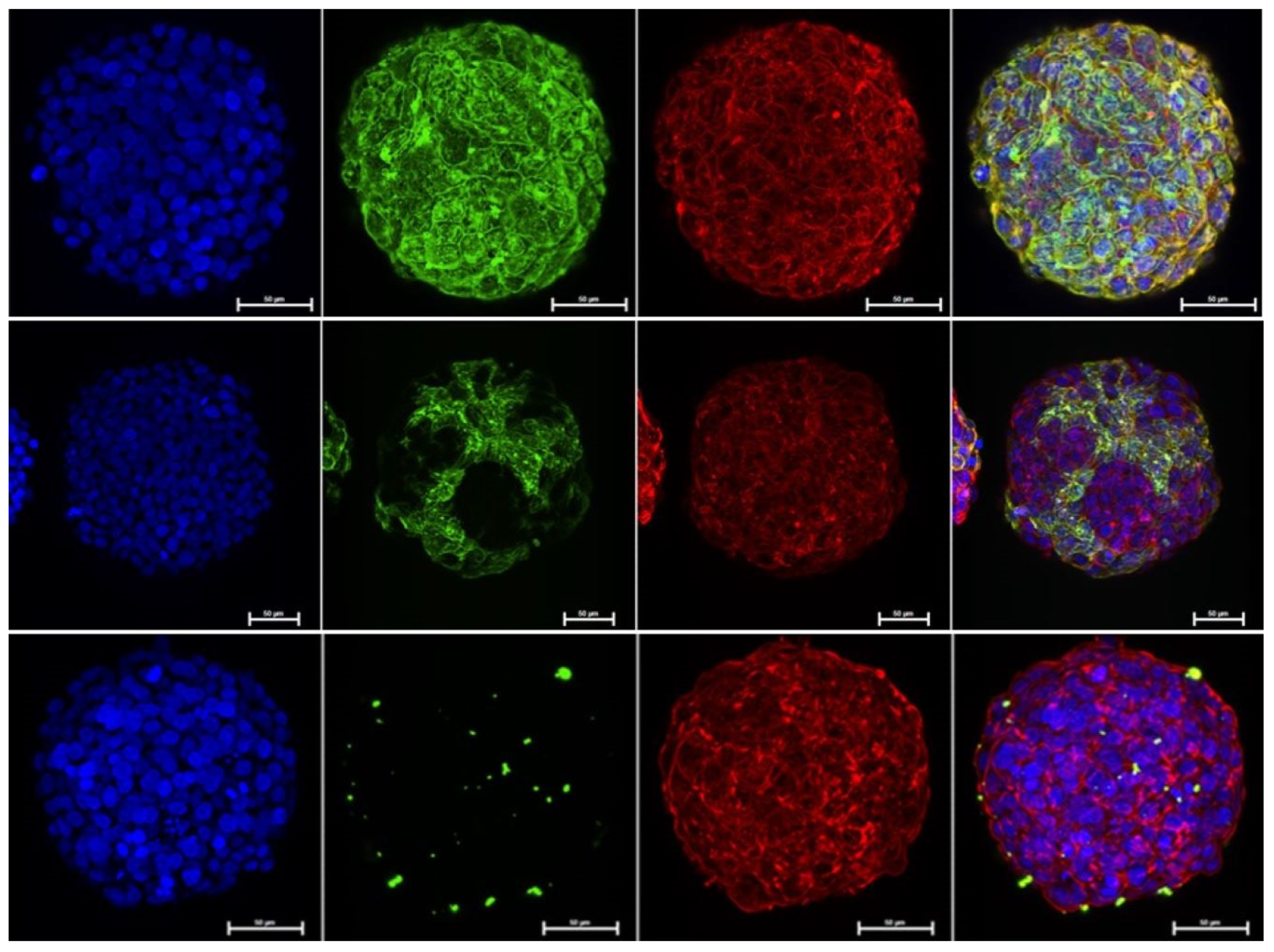
Figure 1. HT29 cells were grown according to the described protocol for spheroids and left for 8–10 days in 37 °C and 5% CO2. The spheroids were thereafter harvested, formaldehyde-fixed and the binding pattern of lectins MAL I, SNA, and the SA-MIPs, respectively, were investigated. Left row: DAPI (blue); Middle row: MAL I-FITC (upper green), SNA-FITC (middle green) and SA-MIPs-NBD (lower green); Right row: Phalloidin (red); merged (DAPI/green/Phalloidin. One representative experiment out of three performed is shown. Scale bar = 50 μm. NBD is nitrobenzoxadiazole.
Applsci 13 05330 g001
Figure 2. HT29 cells and the SA-MIPs were grown according to the described protocol for spheroids and left for 8–10 days in 37 °C and 5% CO2. The spheroids were thereafter harvested and the binding pattern of the SA-MIPs were investigated. Left to right: DAPI (blue); SA-MIPs-NBD (green); phalloidin (red); merged (DAPI/SA-MIPs/phalloidin. One representative experiment out of three performed is shown. Scale bar = 50 μm. NBD is nitrobenzoxadiazole.
Applsci 13 05330 g002
Disclaimer/Publisher’s Note: The statements, opinions and data contained in all publications are solely those of the individual author(s) and contributor(s) and not of MDPI and/or the editor(s). MDPI and/or the editor(s) disclaim responsibility for any injury to people or property resulting from any ideas, methods, instructions or products referred to in the content.