Article

Antimicrobial Activity of Human C-Type Lectin Domain Family 3 Member A (CLEC3A)

Institute for Clinical Chemistry, Medical Faculty, University of Cologne, Kerpener Str. 62, 50937 Cologne, Germany
*
Author to whom correspondence should be addressed.
Appl. Sci. 2024, 14(1), 184; https://doi.org/10.3390/app14010184
Submission received: 28 November 2023 / Revised: 21 December 2023 / Accepted: 22 December 2023 / Published: 25 December 2023
(This article belongs to the Special Issue Novel Interventions for Control of Human Pathogens)

Abstract

:
C-type lectins (CTLs) are a group of proteins that play a crucial role in immunological functions. With the rise of antibiotic-resistant bacteria, CTLs have emerged as a potential alternative to traditional antibiotics and antimicrobial peptides (AMPs), the latter exhibiting limited application due to their low biostability. In this study, we used viable count assays to investigate the antimicrobial activity of the human C-type Lectin Domain Family 3 Member A (CLEC3A) and its two protein domains, CLEC3A Ex23 and CLEC3A Ex3, against gram-positive and gram-negative bacteria. Additionally, using immunoblot analysis, we assessed the biostability of CLEC3A and its protein domains in bacterial supernatant and murine serum. Our findings demonstrate that CLEC3A, CLEC3A Ex23, and CLEC3A Ex3 possess antimicrobial activity against gram-positive Staphyloccocus aureus and gram-negative Pseudomonas aeruginosa. CLEC3A is more effective against P. aeruginosa than the well-investigated antimicrobial peptide LL-37. Furthermore, CLEC3A and its domains have low sensitivity to bacterial and serum proteases, making them more advantageous for systemic application than most AMPs. In conclusion, our research has demonstrated that CLEC3A is not only a precursor of AMPs but also an antimicrobial protein itself, with favorable characteristics for therapeutic applications.

1. Introduction

Over the past few decades, the number of antibiotic-resistant bacteria has steadily increased, and fewer new antibiotics have been approved. These factors pose significant challenges to the healthcare system, and experts warn of a possible post-antibiotic era [1]. In recent years, antimicrobial peptides (AMPs) have also been considered as an alternative treatment. By killing bacteria effectively but with a low risk of resistance development, AMPs are promising candidates as an alternative to antibiotics [2]. However, despite their potential benefits, AMPs have limitations that restrict their use in clinical practice. These limitations include low biostability and susceptibility to proteases [3]. To address the problem of antimicrobial resistance, alternative treatments to traditional antibiotics must be explored.
Another potential alternative to antibiotics is to be found in C-type lectins (CTLs) [4]. CTLs are a diverse group of proteins that make up a part of the innate immune system. They contain a carbohydrate recognition domain (CRD) that allows them to bind to glycans, but some can also bind to other organic and even inorganic molecules [5]. They have different immunological functions, such as pathogen recognition, opsonization, and they direct the killing of bacteria. Additionally, some CTLs are integral parts of the extracellular matrix of different tissues [6].
RegIIIα is a human CTL that shows an antimicrobial effect by pore-formation and thus permeabilizing bacterial membranes [7]. In addition to the many different antimicrobial CTLs of invertebrates [8,9,10,11,12,13], RegIIIα is an important example of a bacteria-killing, human CTL. However, RegIIIα is limited in its ability to kill gram-negative bacteria such as Escherichia coli (E. coli) [7]. Cartilage-specific human C-type Lectin Domain Family 3 Member A (CLEC3A) shows a comparable structure to human RegIIIα. It consists of 197 amino acids and has a molecular weight of 23 kDa. The full-length protein is encoded by three exons. The first exon encodes a signal peptide and a cationic protein segment, exon 2 encodes an alpha-helical oligomerization domain, and exon 3 a CRD [14] (Figure 1).
Two peptides derived from CLEC3A, Ex1, and Ex12, have been shown to exhibit antimicrobial activity [15]. The structural similarities between CLEC3A and human RegIIIα raise the question of whether CLEC3A, the CRD of CLEC3A (CLEC3A Ex3) or the oligomerization domain in combination with the CRD (CLEC3A Ex23) could also exert antimicrobial activities. This study examines the potential of CLEC3A and its protein domains to act as an antimicrobial agent. Additionally, we assess the biostability of CLEC3A and its protein domains in bacterial supernatant and murine serum to determine their potential use for therapeutic applications.

2. Materials and Methods

2.1. Recombinant Proteins

Human CLEC3A and its protein domains CLEC3A Ex23 and CLEC3A Ex3 (Figure 1) and the control proteins Matrilin-3 (Matn3), SARS-CoV-2 open reading frame 8 protein (ORF8), and Cartilage Oligomeric Matrix Protein (COMP) were recombinantly expressed as previously described [16,17,18]. Cell culture supernatant was centrifuged and underwent sterile vacuum filtration (Filtermax, 0.22 µM pore size; TPP, Trasadingen, Switzerland). Recombinant proteins, carrying a poly His-tag or Twin-Strep-tag®, were, respectively, purified by gravity flow column-based affinity purification following the protocol of TALON Superflow (Cytiva, Marlborough, MA, USA) (CLEC3A Ex23, CLEC3A Ex3, Matn3) or Strep-Tactin (IBA-Lifesciences, Göttingen, Germany) (CLEC3A, ORF8, COMP) columns. Protein concentration was determined using photometric analysis (wavelength 280 nm). Purity was checked by SDS-PAGE using Bolt Sample Reducing Agent (Thermo Fisher Scientific, Waltham, MA, USA), Bolt LDS Sample Buffer (Thermo Fisher Scientific, Waltham, MA, USA), 4–12% Bolt Bis-Tris gels (Thermo Fisher Scientific, Waltham, MA, USA), and Coomassie Brilliant Blue R 250 staining. Peptides (LL-37, CLEC3A Ex12, CLEC3A Ex2) (purity > 95%) were purchased from Genosphere biotechnologies (Boulogne-Billancourt, France).

2.2. Bacteria

We used three different bacterial strains in our viable count assays, bacterial cleavage, and biostability investigations: Pseudomonas aeruginosa (P. aeruginosa) ATCC-27853, Staphylococcus aureus (S. aureus) ATCC-29231, and methicillin-resistant Staphylococcus aureus (MRSA) ATCC-43000.

2.3. SDS-PAGE and Immunoblot

Proteins samples were prepared with Bolt Sample Reducing Agent (Thermo Fisher Scientific, USA) and Bolt LDS Sample Buffer (Thermo Fisher Scientific, USA). Samples were seperated on 4–12% Bolt Bis-Tris gels (Thermo Fisher Scientific, USA). Coomassie Brilliant Blue R 250 was used for staining.
Immunoblotting was performed by transferring the proteins to PVDF-membranes (Thermo Fisher Scientific, Waltham, MA, USA). Membranes were incubated with a polyclonal rabbit anti-human CLEC3A antibody (1:2000, overnight at 4 °C) (Pineda Antibody Service, Berlin, Germany) and with a polyclonal swine anti-rabbit-antibody (Agilent Technologies, Singapore). Signals were visualized with Western Blotting Detection Reagant (Cytiva Amersham, Amersham,, UK) by using a BIO-RAD ChemiDoc Imaging System (Hercules, CA, USA).

2.4. Antimicrobial Activity

To investigate the antimicrobial activity of CLEC3A and its protein domains, we used viable count assays. Bacterial strains were cultivated in tryptic soy broth (TSB) (Merck, Darmstadt, Germany) overnight at 37 °C. Incubation was stopped at mid-logarithmic phase (OD ≈ 0.5 (630 nm)) and bacterial culture was mixed with Tris-HCl-glucose-buffer (10 mM Tris-HCl, 5 mM glucose; pH = 7.4; TG-buffer) (S. aureus and P. aeruginosa) or with phosphate-glucose buffer (10 mM K2HPO4, 5 mM glucose; pH = 7.4; PG-buffer) (MRSA). These buffer conditions were optimized previously for P. aeruginosa, S. aureus, and MRSA [15,19]. After centrifugation (4500 g, 5 min), the supernatant was discarded, and the pellet resuspended with TG- or PG-buffer. 50 µL of bacterial solution (concentration: 2 × 10⁶ colony forming units (CFUs)/mL) were incubated with CLEC3A, CLEC3A Ex23, and CLEC3A Ex3 protein solutions, the well-investigated AMP LL-37 (positive control), and CLEC3A Ex2 (negative control) in 1:2 dilutions at 37 °C for 2 h. Double-distilled water and TG- or PG-buffer served as untreated control. Matn3, COMP, and ORF8 were further control proteins and were incubated with 50 µL of the respective bacterial solution in 1:2 dilutions at 37 °C for 2 h. Subsequently, solutions were diluted with a TG or PG-buffer, plated out onto pre-warmed TSB agar plates, and incubated overnight at 37 °C. Bacterial growth was determined by counting CFUs. For each protein concentration, the percentage of colonies compared with the untreated control was determined. Furthermore, minimal inhibiting concentrations (MIC50s), which are defined as the concentration of the antimicrobial proteins or peptides at which only 50% of colonies survived compared to the untreated control, were calculated. Experiments were performed three times for each protein and each bacterial strain.

2.5. Bacterial Cleavage

Immunoblots were used to detect a possible degradation of CLEC3A and CLEC3A protein domains during the 2-h incubation period of the viable count assays. For this, bacterial cultures were prepared as described above and mixed with 5 µM CLEC3A and CLEC3A Ex23 and 10 µM CLEC3A Ex3. After a 2-h incubation at 37 °C, samples were centrifugated and supernatants were taken. SDS-PAGE and immunoblots were performed as described above.

2.6. Biostability

To investigate the biostability of CLEC3A and CLEC3A protein domains, we incubated CLEC3A, CLEC3A Ex23, and CLEC3A Ex3 with bacterial supernatants of P. aeruginosa, S. aureus, and MRSA for different time periods. For this, bacterial strains were grown to mid-logarithmic phase (OD ≈ 0.5 at 630 nm), centrifugated (4500 g, 5 min), and the supernatant was collected. With regard to in vivo applications in a mouse model, we tested the biostability of CLEC3A and CLEC3A protein domains in mouse serum for different time periods. Blood from wildtype mice was gained immediately after death by cervical dislocation via cardiac puncture. The procedure was approved by the local government authority LANUV under permit no. UniKöln_Anzeige§4.22.001. Serum was separated from blood after incubation for 30 min at room temperature for blood clotting and subsequent centrifugation (21.1 g, 10 min). Bacterial supernatant or murine serum was mixed with 5 µM of CLEC3A, CLEC3A Ex23, and CLEC3A Ex3 and incubated at room temperature. Double-distilled water served as a control. After 15 min, 2 h, 6 h, and 24 h samples containing 1 µg of the respective protein and controls were taken, mixed with Bolt Sample Reducing Agent (Thermo Fisher Scientific, USA), Bolt LDS Sample Buffer (Thermo Fisher Scientific, USA), and incubated at 70 °C for 10 min. The immunoblots were performed as described above.

2.7. Statistical Analysis

The data for our study were obtained from three independent experiments. We used GraphPad Prism 9.3.1 (471) software to analyze the results of our viable count assays. Our values are presented as mean ± standard deviation. To test for statistical significance between the MIC50s of CLEC3A Ex12, CLEC3A Ex3, CLEC3A Ex23, and CLEC3A, and the MIC50 of LL-37, paired ANOVA were performed followed by multiple comparisons and Dunnett tests. Moreover, an unpaired t-test was used to test for statistical significance between the MIC50s of LL-37 and CLEC3A Ex12.

3. Results

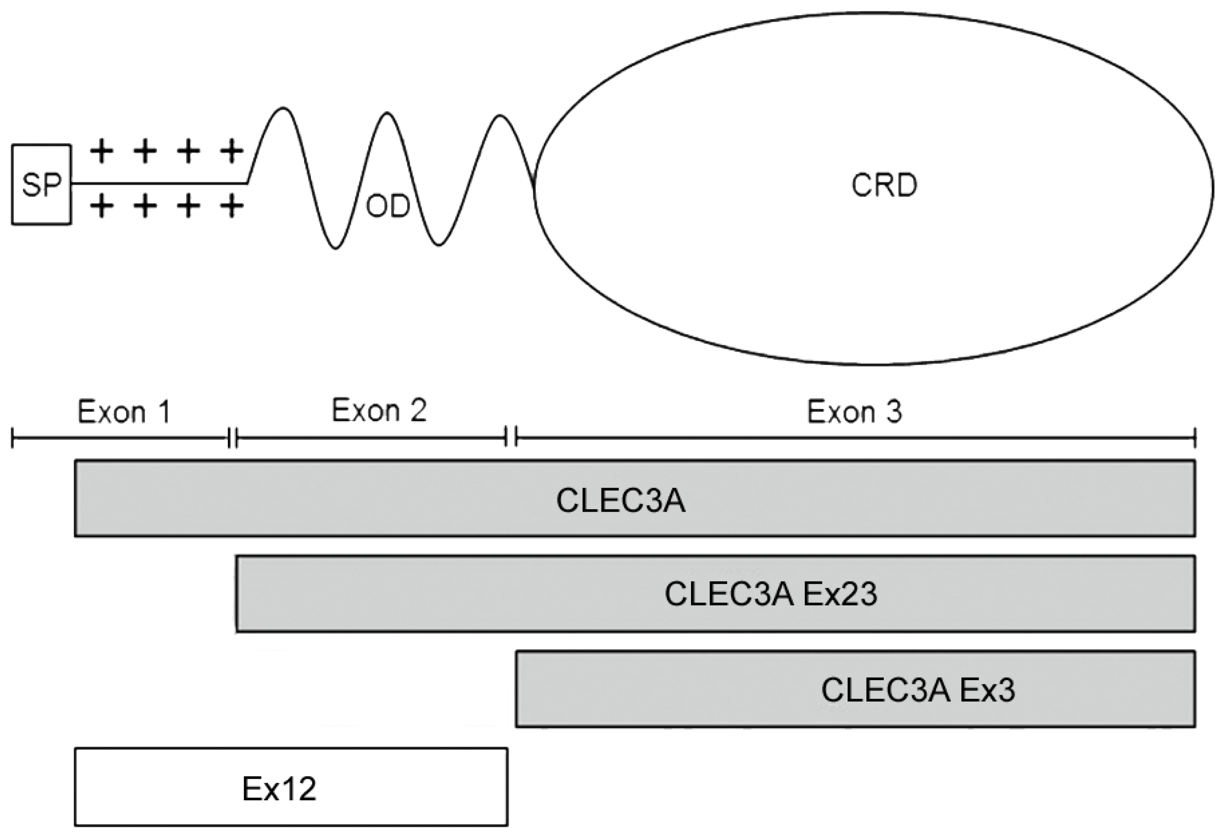
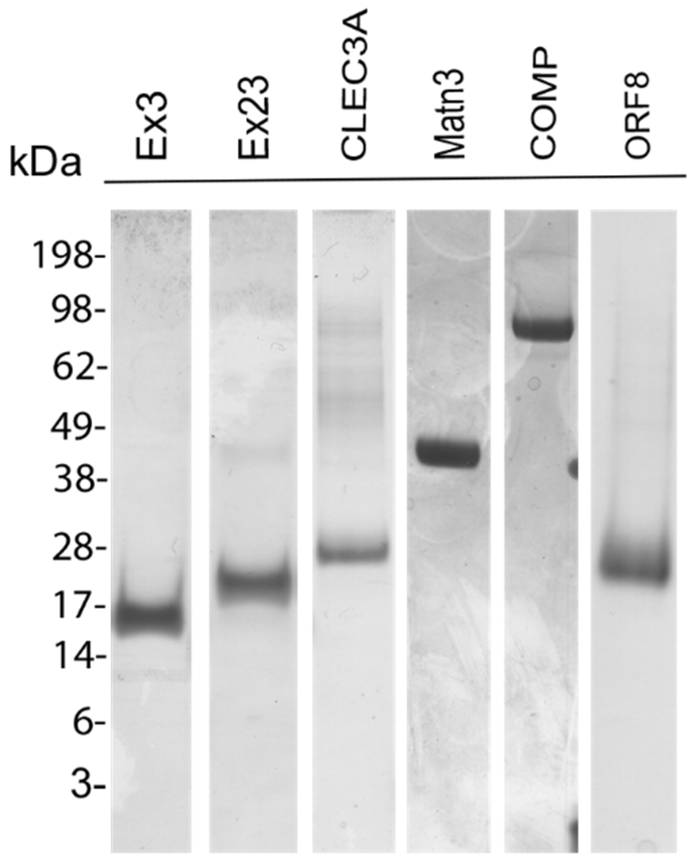
3.1. Recombinant Proteins

We recombinantly expressed CLEC3A and its protein domains CLEC3A Ex23 and CLEC3A Ex3 to investigate their antimicrobial activity. Protein integrity and purity were checked by SDS-PAGE and Coomassie Brilliant Blue staining (Figure 2). Protein bands of the expected size could be shown.

3.2. Antimicrobial Activity

The antimicrobial activity of CLEC3A, CLEC3A Ex23, and CLEC3A Ex3 was investigated using viable count assays. The gold standard AMP LL-37 and the CLEC3A-derived AMP Ex12 were used as positive controls. CLEC3A Ex2, which is solely the amphipathic alpha-helix of Ex12 without the positive charge of the N-terminus, was used as a negative control. We could show an outstanding reduction of P. aeruginosa colonies using CLEC3A. The MIC50 of CLEC3A is statistically significantly different to the MIC50 of the gold standard AMP LL-37 (p = 0.0192) and the CLEC3A-derived AMP CLEC3A Ex12 (p = 0.0254. CLEC3A Ex23 and CLEC3A Ex3 also demonstrated an antimicrobial activity against P. aeruginosa and S. aureus whereas none of the examined proteins showed an antimicrobial activity against MRSA (Figure 3). As CLEC3A carries a Twin-Strep-tag® and CLEC3A Ex23 and CLEC3A Ex3 carry a poly His-tag, we used COMP (with a Twin-Strep-tag®) and Matn3 (carrying a poly His-tag) as controls to rule out an effect of the tags. Matn3 did not show an antimicrobial effect against P. aeruginosa or S. aureus. COMP did not demonstrate an antimicrobial effect against S. aureus. However, we found a slight decrease in the bacterial colony count of P. aeruginosa. To validate that the tag has no effect, we used ORF8, another Twin-strep-tagged protein (Supplementary Figure S2).

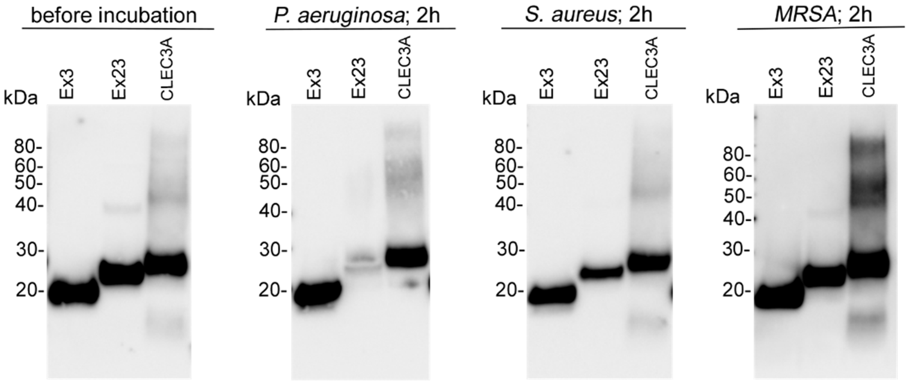
3.3. Bacterial Cleavage

Degradation of CLEC3A can lead to the generation of CLEC3A-derived AMPs [15]. Therefore, we investigated whether the antimicrobial activity of CLEC3A, CLEC3A Ex23, and CLEC3A Ex3 was caused by bacterial cleavage and generation of CLEC3A-derived AMPs during the 2-h incubation period of the viable count assay. For CLEC3A and CLEC3A Ex3, we did not observe bacterial cleavage after incubation with P. aeruginosa, S. aureus, or MRSA, thus we can exclude an association between degradation and antimicrobial activity. In contrast, we observed a cleavage of CLEC3A Ex23 when incubated with P. aeruginosa and S. aureus (Figure 4). We did not detect any additional bands with a lower weight, making it unlikely that the antimicrobial effect of CLEC3A Ex23 against P. aeruginosa or S. aureus was due to the generation of CLEC3A-derived AMPs. Nevertheless, we cannot entirely rule out this possibility.

3.4. Biostability of CLEC3A

To investigate the biostability of CLEC3A in bacterial supernatant, we incubated CLEC3A, CLEC3A Ex23, and CLEC3A Ex3 with bacterial supernatants for different time periods. In the case of S. aureus and MRSA (Supplementary Figures S4C,D), the proteins showed almost no susceptibility to the cleavage of bacterial proteases, even up to 24 h later. However, incubation of the proteins with the supernatant of P. aeruginosa led to a slight proteolytic cleavage of CLEC3A, CLEC3A Ex23, and CLEC3A Ex3 after 24 h (Figure 5). As CLEC3A and its protein domains did not inhibit the growth of MRSA in the viable count assay, biostability for this bacterial strain is only shown in the supplementary data.
Furthermore, we tested the biostability of CLEC3A, CLEC3A Ex23, and CLEC3A Ex3 in murine serum. We did not detect cleavage products of CLEC3A and CLEC3A Ex23 within 24 h, whereas CLEC3A Ex3 showed strong degradation beginning immediately after 15 min (Figure 6).

4. Discussion

Facing the global problem of increasing antibiotic resistance, many researchers are focusing on finding alternatives to the conventional antibiotic treatment of bacterial infections. CTLs have various immunological functions such as pathogen recognition, opsonization, and direct bacterial killing [6]. Mannan-binding lectin (MBL), a human CTL, was applied to decrease the susceptibility to bacterial infections of immune and MBL-deficient patients. Patients who received MBL-infusions did not show allergic reactions and MBL-antibodies were observed [20]. This example of MBL-infusions displays the potential of CTLs to be used in clinical practice [20,21].
For the problem of combatting the two common nosocomial bacteria S. aureus and P. aeruginosa, we investigated the antimicrobial activity of the CTL CLEC3A and its protein domains CLEC3A Ex23 and CLEC3A Ex3 against these bacteria. S. aureus is known to induce soft tissue infections in particular [22] while P. aeruginosa is frequently detected in infections of the respiratory tract [23]. In immune-deficient or critically ill patients, both S. aureus and P. aeruginosa can lead to lethal bloodstream infections [22,24]. Due to the clinical relevance of MRSA, we also tested CLEC3A and its protein domains against this bacterial strain. We could show that CLEC3A and its protein domains have an antimicrobial effect against P. aeruginosa. Notably, the antimicrobial activity of CLEC3A against P. aeruginosa is significantly stronger than that of the CLEC3A-derived AMP Ex12 and even than that of the AMP LL-37. For S. aureus, we were able to demonstrate that CLEC3A Ex23 and CLEC3A Ex3 possess antimicrobial activity. Recently CLEC3A has been shown as being highly susceptible to proteases [25] and is a precursor of AMPs [15]. The double function of CLEC3A being a precursor of AMPs and functioning as an antimicrobial CTL is exceptional.
RegIIIα is another human, antimicrobial CTL [7]. CLEC3A’s and RegIIIα’s antibacterial activity differ for gram-positive and gram-negative bacteria. RegIIIα shows bactericidal activity against gram-positive but not against gram-negative bacteria [7] whereas CLEC3A does not kill gram-positive but does kill gram-negative bacteria. CTLs recognize and bind bacteria using different membrane surface molecules. These vary for gram-positive and gram-negative bacteria, with peptidoglycans [8,12,26] and lipoteichoic acid (LTA) [27] frequently used in gram-positive, and lipopolysaccharides (LPS) in gram-negative bacteria [8,10,12,26]. RegIIIα binds to peptidoglycans, but the antibacterial, pore-forming activity was inhibited by LPS, which explains why RegIIIα could affect gram-positive bacteria only [7]. In contrast, the binding of CLEC3A to LPS has been shown [15], which could explain its strong effect against gram-negative P. aeruginosa. In addition, CLEC3A, in contrast to the CLEC3A-derived peptide Ex12, did not bind to LTA [15], which could explain its absent effect against S. aureus.
CLEC3A and RegIIIα share comparable structural characteristics as they consist of a CRD linked to a charged N-terminal segment, which is cationic for CLEC3A and anionic for RegIIIα [14,28]. RegIIIα bactericidal activity is suppressed by the N-terminal prosegment. When the N-terminal prosegment is removed, RegIIIα partially changes the conformation of one of its loops and can then form a hexameric pore in the membrane, which induces bacterial lysis [7]. We could demonstrate that CLEC3A, encompassing the N-terminal segment, kills P. aeruginosa and the charged N-terminal segment of CLEC3A does not have to be removed for its antimicrobial activity. In contrast, only the CLEC3A protein domains CLEC3A Ex23 and CLEC3A Ex3, which do not possess the charged N-terminal segment, exhibit an antibacterial effect against S. aureus. From this, it can be concluded that, similar to RegIIIα, the N-terminal charged segment of CLEC3A suppresses its antimicrobial activity against S. aureus.
Neither CLEC3A nor its protein domains CLEC3A Ex3 and CLEC3A Ex23 could affect the growth of MRSA. MRSA expresses a penicillin-binding protein that causes resistance to almost all ß-lactam antibiotics and other types of common antibiotics [29]. Thus, it is unknown whether the penicillin-binding protein or other components of MRSA are responsible for protecting against CLEC3A and its protein domains.
For therapeutic use, a long biostability is beneficial [6]. Bacteria are able to induce degradation of immunoglobulins and complement factors by secreting proteases [30]. Therefore, we tested the susceptibility of CLEC3A and its protein domains to bacterial proteases by incubating the proteins with bacterial supernatant. We could show that there was almost no susceptibility of the proteins in the supernatant of S. aureus and a low susceptibility in the supernatant of P. aeruginosa after 24 h. We also investigated the biostability of the proteins in murine serum. CLEC3A and CLEC3A Ex23 showed no signs of degradation by serum proteases within 24 h. Surprisingly, CLEC3A Ex3 showed strong degradation within just 15 min. Nevertheless, the biostability of CLEC3A and CLEC3A Ex23 in murine serum seems to be superior to that of the CLEC3A-derived peptide Ex12, for which a complete degradation could be shown in murine serum within 24 h [19]. Structural motifs of peptides or proteins can increase susceptibility, as they serve as recognition patterns for proteases, or decrease susceptibility, as they inhibit protease activation [31]. The structural differences between CLEC3A and CLEC3A Ex23 in comparison to CLEC3A Ex3 and CLEC3A-derived AMP Ex12 probably contribute to divergences of their susceptibility to serum proteases.
In conclusion, our study has shown that, besides being a precursor of AMPs, CLEC3A and its protein domains exhibit antimicrobial activity against P. aeruginosa and S. aureus. Interestingly, the antimicrobial effect of CLEC3A against P. aeruginosa was significantly stronger than the effect of the gold standard AMP LL-37 and the CLEC3A-derived peptide Ex12. In addition, CLEC3A and its protein domains show low sensitivity to bacterial and serum proteases, which is a major advantage for the systemic application of the proteins in contrast to most AMPs.

Supplementary Materials

The following supporting information can be downloaded at: https://www.mdpi.com/article/10.3390/app14010184/s1, Figure S1: Recombinant proteins. (A) 2 µg CLEC3A. (B) 2 µg CLEC3A Ex3 (Ex3). (C) 2 µg CLEC3A Ex23 (Ex23). (D) 2 µg Matrilin-3 (Matn3) and 2 µg Cartilage oligomeric matrix protein (COMP). (E) 4 µg SARS-CoV-2 open reading frame 8 protein (ORF8); Figure S2: Viable count assays. (A) Antimicrobial activity of LL-37, CLEC3A Ex2 (Ex2), Matrilin-3 (Mtn3 with poly His-Tag), and Cartilage Oligomeric Matrix Protein (COMP with Twin-Strep-Tag®) against P. aeruginosa. (B) Antimicrobial activity of LL-37, CLEC3A Ex2 (Ex2), and ORF8 (SARS-CoV-2) (with Twin-Strep-Tag®) against P. aeruginosa. (C) Antimicrobial activity of LL-37, CLEC3A Ex2 (Ex2), and Matrilin-3 (Mtn3 with poly His-Tag) against S. aureus. (D) Antimicrobial activity of LL-37, CLEC3A Ex2 (Ex2), and Cartilage Oligomeric Matrix Protein (COMP with Twin-Strep-Tag®) against S. aureus; Figure S3: Incubation of CLEC3A, CLEC3A Ex23 (Ex23) and, CLEC3A Ex3 (Ex3) with P. aeruginosa, S. aureus and MRSA. Full-sized immunoblots; Figure S4.A: Incubation of CLEC3A, CLEC3A Ex23 (Ex23) and, CLEC3A Ex3 (Ex3) with supernatants of P. aeruginosa and S. aureus for different periods. Secondary antibody-only (Controls); Figure S4.B: Incubation of CLEC3A, CLEC3A Ex23 (Ex23) and, CLEC3A Ex3 (Ex3) with supernatants of P. aeruginosa and S. aureus for different periods. Full-sized immunoblots (Originals); Figure S5: Incubation of CLEC3A, CLEC3A Ex23 (Ex23) and, CLEC3A Ex3 (Ex3) with murine serum for different periods. (A) Secondary antibody-only (Controls). (B) Full-sized immunoblots (Originals).

Author Contributions

Conceptualization, K.S.W., D.M., T.S. and A.R.K.; methodology, K.S.W., D.M. and G.H.; software, K.S.W. and D.M.; writing—original draft preparation, K.S.W. and A.R.K.; writing—review and editing, K.S.W., D.M., G.H., A.R., T.S. and A.R.K.; supervision, A.R.K.; project administration, K.S.W. and A.R.K.; funding acquisition, T.S. and A.R.K. All authors have read and agreed to the published version of the manuscript.

Funding

Funded by the Deutsche Forschungsgemeinschaft (DFG, German Research Foundation)—project number 527884186.

Institutional Review Board Statement

Not applicable.

Informed Consent Statement

Not applicable.

Data Availability Statement

All data generated in this study can be found in this article and the supplementary data.

Conflicts of Interest

The authors declare no conflict of interest.

References

  1. Medina, E.; Pieper, D.H. Tackling Threats and Future Problems of Multidrug-Resistant Bacteria. Curr. Top. Microbiol. Immunol. 2016, 398, 3–33. [Google Scholar] [CrossRef] [PubMed]
  2. Boparai, J.K.; Sharma, P.K. Mini Review on Antimicrobial Peptides, Sources, Mechanism and Recent Applications. Protein Pept. Lett. 2020, 27, 4–16. [Google Scholar] [CrossRef] [PubMed]
  3. Kumar, P.; Kizhakkedathu, J.N.; Straus, S.K. Antimicrobial Peptides: Diversity, Mechanism of Action and Strategies to Improve the Activity and Biocompatibility In Vivo. Biomolecules 2018, 8, 4. [Google Scholar] [CrossRef] [PubMed]
  4. Breitenbach Barroso Coelho, L.C.; Marcelino Dos Santos Silva, P.; Felix de Oliveira, W.; de Moura, M.C.; Viana Pontual, E.; Soares Gomes, F.; Guedes Paiva, P.M.; Napoleão, T.H.; Dos Santos Correia, M.T. Lectins as antimicrobial agents. J. Appl. Microbiol. 2018, 125, 1238–1252. [Google Scholar] [CrossRef] [PubMed]
  5. Zelensky, A.N.; Gready, J.E. The C-type lectin-like domain superfamily. FEBS J. 2005, 272, 6179–6217. [Google Scholar] [CrossRef] [PubMed]
  6. Brown, G.D.; Willment, J.A.; Whitehead, L. C-type lectins in immunity and homeostasis. Nat. Rev. Immunol. 2018, 18, 374–389. [Google Scholar] [CrossRef] [PubMed]
  7. Mukherjee, S.; Zheng, H.; Derebe, M.G.; Callenberg, K.M.; Partch, C.L.; Rollins, D.; Propheter, D.C.; Rizo, J.; Grabe, M.; Jiang, Q.X.; et al. Antibacterial membrane attack by a pore-forming intestinal C-type lectin. Nature 2014, 505, 103–107. [Google Scholar] [CrossRef]
  8. Li, J.; Bi, J.; Zhang, P.; Wang, Z.; Zhong, Y.; Xu, S.; Wang, L.; Li, B. Functions of a C-type lectin with a single carbohydrate-recognition domain in the innate immunity and movement of the red flour beetle, Tribolium castaneum. Insect. Mol. Biol. 2021, 30, 90–101. [Google Scholar] [CrossRef]
  9. Runsaeng, P.; Puengyam, P.; Utarabhand, P. A mannose-specific C-type lectin from Fenneropenaeus merguiensis exhibited antimicrobial activity to mediate shrimp innate immunity. Mol. Immunol. 2017, 92, 87–98. [Google Scholar] [CrossRef]
  10. Sun, Y.D.; Fu, L.D.; Jia, Y.P.; Du, X.J.; Wang, Q.; Wang, Y.H.; Zhao, X.F.; Yu, X.Q.; Wang, J.X. A hepatopancreas-specific C-type lectin from the Chinese shrimp Fenneropenaeus chinensis exhibits antimicrobial activity. Mol. Immunol. 2008, 45, 348–361. [Google Scholar] [CrossRef]
  11. Wei, X.; Wang, L.; Sun, W.; Zhang, M.; Ma, H.; Zhang, Y.; Zhang, X.; Li, S. C-type lectin B (SpCTL-B) regulates the expression of antimicrobial peptides and promotes phagocytosis in mud crab Scylla paramamosain. Dev. Comp. Immunol. 2018, 84, 213–229. [Google Scholar] [CrossRef] [PubMed]
  12. Zhao, L.L.; Wang, Y.Q.; Dai, Y.J.; Zhao, L.J.; Qin, Q.; Lin, L.; Ren, Q.; Lan, J.F. A novel C-type lectin with four CRDs is involved in the regulation of antimicrobial peptide gene expression in Hyriopsis cumingii. Fish Shellfish Immunol. 2016, 55, 339–347. [Google Scholar] [CrossRef] [PubMed]
  13. Zhu, Y.T.; Zhang, X.; Wang, S.C.; Li, W.W.; Wang, Q. Antimicrobial functions of EsLecH, a C-type lectin, via JNK pathway in the Chinese mitten crab, Eriocheir sinensis. Dev. Comp. Immunol. 2016, 61, 225–235. [Google Scholar] [CrossRef] [PubMed]
  14. Neame, P.J.; Tapp, H.; Grimm, D.R. The cartilage-derived, C-type lectin (CLECSF1): Structure of the gene and chromosomal location. Biochim. Biophys. Acta 1999, 1446, 193–202. [Google Scholar] [CrossRef] [PubMed]
  15. Elezagic, D.; Mörgelin, M.; Hermes, G.; Hamprecht, A.; Sengle, G.; Lau, D.; Höllriegl, S.; Wagener, R.; Paulsson, M.; Streichert, T.; et al. Antimicrobial peptides derived from the cartilage.-specific C-type Lectin Domain Family 3 Member A (CLEC3A)–potential in the prevention and treatment of septic arthritis. Osteoarthr. Cartil. 2019, 27, 1564–1573. [Google Scholar] [CrossRef]
  16. Lau, D.; Elezagic, D.; Hermes, G.; Mörgelin, M.; Wohl, A.P.; Koch, M.; Hartmann, U.; Höllriegl, S.; Wagener, R.; Paulsson, M.; et al. The cartilage-specific lectin C-type lectin domain family 3 member A (CLEC3A) enhances tissue plasminogen activator-mediated plasminogen activation. J. Biol. Chem. 2018, 293, 203–214. [Google Scholar] [CrossRef]
  17. Meinberger, D.; Koch, M.; Roth, A.; Hermes, G.; Stemler, J.; Cornely, O.A.; Streichert, T.; Klatt, A.R. Analysis of IgM, IgA, and IgG isotype antibodies Directed against SARS-CoV-2 spike glycoprotein and ORF8 in the course of COVID-19. Sci. Rep. 2021, 11, 8920. [Google Scholar] [CrossRef]
  18. Ruthard, J.; Hermes, G.; Hartmann, U.; Sengle, G.; Pongratz, G.; Ostendorf, B.; Schneider, M.; Höllriegl, S.; Zaucke, F.; Wagener, R.; et al. Identification of antibodies against extracellular matrix proteins in human osteoarthritis. Biochem. Biophys. Res. Commun. 2018, 503, 1273–1277. [Google Scholar] [CrossRef]
  19. Meinberger, D.; Drexelius, M.G.; Grabeck, J.; Hermes, G.; Roth, A.; Elezagic, D.; Neundorf, I.; Streichert, T.; Klatt, A.R. Modified CLEC3A-Derived Antimicrobial Peptides Lead to Enhanced Antimicrobial Activity against Drug-Resistant Bacteria. Antibiotics 2023, 12, 1532. [Google Scholar] [CrossRef]
  20. Valdimarsson, H.; Stefansson, M.; Vikingsdottir, T.; Arason, G.J.; Koch, C.; Thiel, S.; Jensenius, J.C. Reconstitution of opsonizing activity by infusion of mannan-binding lectin (MBL) to MBL-deficient humans. Scand J. Immunol. 1998, 48, 116–123. [Google Scholar] [CrossRef]
  21. Kilpatrick, D.C. Mannan-binding lectin: Clinical significance and applications. Biochim. Biophys. Acta 2002, 1572, 401–413. [Google Scholar] [CrossRef] [PubMed]
  22. Cheung, G.Y.C.; Bae, J.S.; Otto, M. Pathogenicity and virulence of Staphylococcus aureus. Virulence 2021, 12, 547–569. [Google Scholar] [CrossRef] [PubMed]
  23. Lyczak, J.B.; Cannon, C.L.; Pier, G.B. Establishment of Pseudomonas aeruginosa infection: Lessons from a versatile opportunist. Microbes Infect. 2000, 2, 1051–1060. [Google Scholar] [CrossRef] [PubMed]
  24. Heyland, D.K.; Cook, D.J.; Griffith, L.; Keenan, S.P.; Brun-Buisson, C. The attributable morbidity and mortality of ventilator-associated pneumonia in the critically ill patient. The Canadian Critical Trials Group. Am. J. Respir. Crit. Care Med. 1999, 159, 1249–1256. [Google Scholar] [CrossRef] [PubMed]
  25. Tsunezumi, J.; Higashi, S.; Miyazaki, K. Matrilysin (MMP-7) cleaves C-type lectin domain family 3 member A (CLEC3A) on tumor cell surface and modulates its cell adhesion activity. J. Cell. Biochem. 2009, 106, 693–702. [Google Scholar] [CrossRef]
  26. Zhang, X.W.; Yang, C.H.; Zhang, H.Q.; Pan, X.T.; Jin, Z.Y.; Zhang, H.W.; Xia, X.H. A C-type lectin with antibacterial activity in weather loach, Misgurnus anguillicaudatus. J. Fish Dis. 2020, 43, 1531–1539. [Google Scholar] [CrossRef] [PubMed]
  27. Zhu, Q.; Huo, H.; Fu, Q.; Yang, N.; Xue, T.; Zhuang, C.; Liu, X.; Wang, B.; Su, B.; Li, C. Identification and characterization of a C-type lectin in turbot (Scophthalmus maximus) which functioning as a pattern recognition receptor that binds and agglutinates various bacteria. Fish Shellfish Immunol. 2021, 115, 104–111. [Google Scholar] [CrossRef]
  28. Abergel, C.; Chenivesse, S.; Stinnakre, M.G.; Guasco, S.; Bréchot, C.; Claverie, J.M.; Devinoy, E.; Christa, L. Crystallization and preliminary crystallographic study of HIP/PAP, a human C-lectin overexpressed in primary liver cancers. Acta Crystallogr. D Biol. Crystallogr. 1999, 55, 1487–1489. [Google Scholar] [CrossRef]
  29. Peacock, S.J.; Paterson, G.K. Mechanisms of Methicillin Resistance in Staphylococcus aureus. Annu. Rev. Biochem. 2015, 84, 577–601. [Google Scholar] [CrossRef]
  30. Ramírez-Larrota, J.S.; Eckhard, U. An Introduction to Bacterial Biofilms and Their Proteases, and Their Roles in Host Infection and Immune Evasion. Biomolecules 2022, 12, 306. [Google Scholar] [CrossRef]
  31. Werle, M.; Bernkop-Schnürch, A. Strategies to improve plasma half life time of peptide and protein drugs. Amino Acids 2006, 30, 351–367. [Google Scholar] [CrossRef] [PubMed]
Figure 1. Schematic illustration of human CLEC3A. CLEC3A consists of a signal-peptide (SP) and a cationic sequence encoded by exon 1, an oligomerization domain (OD) encoded by exon 2, and a carbohydrate recognition domain (CRD) encoded by exon 3. Boxes shaded in grey represent CLEC3A and the CLEC3A protein domains CLEC3A Ex23 and CLEC3A Ex3. Ex12 is a CLEC3A-derived antimirobial peptide and is encoded by exon 1 and exon 2. Adapted from Elezagic et al., 2019 [15].
Applsci 14 00184 g001
Figure 2. Recombinant proteins. A 4–12% Bis-Tris Gel was loaded with 2 µg CLEC3A Ex3 (Ex3), CLEC3A Ex23 (Ex23), CLEC3A, Matrilin-3 (Matn3), Cartilage oligomeric matrix protein (COMP) and 4 µg SARS-CoV-2 open reading frame 8 protein (ORF8) under reducing conditions. Coomassie Brilliant Blue R 250 was used for staining. Cropped lanes are divided by white spaces. Full-sized gels are shown in Supplementary Figure S1.
Applsci 14 00184 g002
Figure 3. Viable count assays. (A) Antimicrobial activity of LL-37, CLEC3A Ex2 (Ex2), CLEC3A Ex12 (Ex12), CLEC3A Ex3 (Ex3) (** p = 0.0041), CLEC3A Ex23 (Ex23), and CLEC3A (* p = 0.0192) against P. aeruginosa. Concentrations shown on the horizontal axis were rounded for CLEC3A Ex3, CLEC3A Ex23 (maximal concentration = 9.6 µM), and CLEC3A (maximal concentration = 4.8 µM). (B) Antimicrobial activity of LL-37, CLEC3A Ex2 (Ex2), CLEC3A Ex12 (Ex12), CLEC3A Ex3 (Ex3), CLEC3A Ex23 (Ex23), and CLEC3A against S. aureus. (C) Antimicrobial activity of LL-37, CLEC3A Ex2 (Ex2), CLEC3A Ex12 (Ex12) (* p = 0.0389), CLEC3A Ex3 (Ex3), CLEC3A Ex23 (Ex23), and CLEC3A against MRSA. Each experiment was performed three times (n = 3). Averages and standard deviations are shown. The bar charts show the determined MIC50s. All MIC50s were compared to the MIC50 of the gold standard AMP LL-37 and the statistical significance was calculated using GraphPad Prism 9 and a paired ANOVA followed by multiple comparisons and Dunnett tests. Statistical significance between the MIC50 of LL-37 and CLEC3A Ex12 was calculated by using an unpaired t-test. ns = not significant. na = not applicable.
Applsci 14 00184 g003
Figure 4. Incubation of CLEC3A Ex3, CLEC3A Ex23, and CLEC3A with P. aeruginosa, S. aureus, and MRSA. Representative immunoblots showing 1 µg CLEC3A Ex3 (Ex3), CLEC3A Ex23 (Ex23), and CLEC3A in water (before incubation) and after 2 h of incubation (37 °C) with the respective bacterial strains. Proteins were detected by using an anti-human CLEC3A antibody. Full-sized immunoblots and controls are shown in Supplementary Figure S3. Immunoblots were performed three times for each bacterial strain.
Applsci 14 00184 g004
Figure 5. Incubation of CLEC3A Ex3, CLEC3A Ex23, and CLEC3A with supernatants of P. aeruginosa (A) and S. aureus (B) for different time periods. Representative immunoblots showing 1 µg CLEC3A Ex3 (Ex3), CLEC3A Ex23 (Ex23), and CLEC3A in water and after 15 min, 2 h, 6 h, and 24 h of incubation with bacterial supernatant (SN + protein) of the respective bacterial strains. Proteins were detected by using an anti-human CLEC3A antibody. Bacterial supernatant without protein (SN) is shown as a control at each time point. Full-sized immunoblots and secondary antibody-only controls are shown in Supplementary Figure S4. Immunoblots were performed three times for each bacterial strain.
Applsci 14 00184 g005
Figure 6. Incubation of CLEC3A Ex3, CLEC3A Ex23, and CLEC3A with murine serum for different time periods. Representative immunoblots showing 1 µg CLEC3A Ex3 (Ex3), CLEC3A Ex23 (Ex23), and CLEC3A in water and after 15 min, 2 h, 6 h, and 24 h of incubation at room temperature with murine serum (S + protein). Proteins were detected by using an anti-human CLEC3A antibody. Murine serum without protein (S) is shown as a control at each time point. Full-sized immunoblots and secondary antibody-only controls are shown in Supplementary Figure S5. Immunoblots were performed three times.
Applsci 14 00184 g006
Disclaimer/Publisher’s Note: The statements, opinions and data contained in all publications are solely those of the individual author(s) and contributor(s) and not of MDPI and/or the editor(s). MDPI and/or the editor(s) disclaim responsibility for any injury to people or property resulting from any ideas, methods, instructions or products referred to in the content.

