Article

Upper Airway Collapsibility during Sleep Endoscopy with a Titratable Mandibular Advancement Simulator in Obstructive Sleep Apnea Patients

by
Matteo Cameli
1,
Chiara Stipa
1,
Irene Pelligra
2,
Daniela Rita Ippolito
1,
Giovanni Sorrenti
3,
Giulio Alessandri-Bonetti
1 and
Serena Incerti Parenti
1,*
1
Unit of Orthodontics and Sleep Dentistry, Department of Biomedical and Neuromotor Sciences (DIBINEM), University of Bologna, Via San Vitale 59, 40125 Bologna, Italy
2
Department of Otolaryngology Head and Neck Surgery, Sant’Orsola-Malpighi Hospital, University of Bologna, 40138 Bologna, Italy
3
Private Practice, 40100 Bologna, Italy
*
Author to whom correspondence should be addressed.
Appl. Sci. 2024, 14(11), 4710; https://doi.org/10.3390/app14114710
Submission received: 1 May 2024 / Revised: 26 May 2024 / Accepted: 27 May 2024 / Published: 30 May 2024
(This article belongs to the Special Issue Challenges and Perspectives in Dental Sleep Medicine)

Abstract

:
Drug-induced sleep endoscopy (DISE) has been progressively used to determine the individual patient responsiveness to therapy with a mandibular advancement device (MAD) for obstructive sleep apnea (OSA). This retrospective cohort study compared the general and polygraphic characteristics, as well as the sites, degrees, and patterns of upper airway collapse, in patients who responded to advancement with a titratable mandibular advancement (TMA) simulator during DISE—referred to as responders—to those in non-responders. The sample included 335 OSA patients (307 males) with a mean age of 49.98 (SD = 9.88) years, and a mean AHI of 34.14 (SD = 18.61). Once the TMA simulator customized to the patient’s dental arches was inserted and the examination was performed at 0%, 25%, 50%, and 75% of the patient’s range of antero-posterior mandibular excursion, the simulator was removed and the upper airway behavior was studied in the baseline situation. Without TMA simulator non-responders had a higher percentage of oropharyngeal complete latero-lateral and complete concentric velopharyngeal collapse. With TMA simulators, there was a significant difference between responders and non-responders in individual obstructive sites at velopharyngeal, oropharyngeal, and epiglottis levels, while at the tongue level, responders and non-responders showed the same response tendency. If confirmed in future prospective studies, these results suggest that the presence of complete latero-lateral obstruction at the oropharynx level and complete circular obstruction at the velopharynx level could be adverse phenotypes for MAD treatment outcomes in OSA patients and MAD treatment should not be considered in these patients (at least as a single therapy).

1. Introduction

Obstructive sleep apnea (OSA) is a sleep-related breathing disorder characterized by recurrent partial or complete pharyngeal collapse during sleep, causing a significant reduction (hypopnea) or a cessation (apnea) of airflow, intermittent hypoxia, and sleep fragmentation [1,2]. The diagnosis and assessment of severity are currently defined by the apnea–hypopnea index (AHI), which is the number of episodes of complete or partial upper airway obstruction per hour of sleep [1]. Symptoms include excessive daytime sleepiness, decreased quality of life, and neurocognitive impairment. Moreover, OSA is associated with an increased likelihood of vehicular and occupational accidents, cardiovascular mortality, and metabolic dysfunction [1,2]. Due to factors such as aging and the global rise in obesity, OSA is an increasingly prevalent condition that greatly impacts public health and individual well-being. It is estimated that roughly one billion people between the ages of 30 and 69 years are affected all over the world [3]. Data from a large population-based sample of patients suggest a prevalence of moderate to severe OSA (AHI ≥ 15) in the general population aged ≥40 years of 23.4% in women and 49.7% in men [4]. Despite being acknowledged as a potentially life-threatening chronic condition with a high economic and social burden, OSA is still underdiagnosed and undertreated [5]. A recent epidemiological investigation focused on determining the societal and economic impacts of OSA within Italy concluded that approximately 56% of the adult population, ranging from 15 to 74 years of age, may suffer from OSA. However, the investigation revealed that merely 4% of these individuals have been officially diagnosed, and only 2% have received treatment [6].
OSA is recognized as a multifactorial disease, and an array of therapeutic interventions has been explored. Currently, continuous positive airway pressure (CPAP) constitutes the “gold standard” therapy for OSA [7]. It involves wearing a mask during sleep that is connected to a pump that delivers constant pressure to keep the airway open. Treatment with CPAP is characterized by high clinical efficacy, but therapeutic effectiveness is often limited by poor patient compliance [8]. Upper airway surgery, mandibular advancement device (MAD), and positional therapy are well-recognized non-CPAP treatment options for OSA. A proper patient selection is crucial given the endotypic variability associated with OSA [7]. However, the analysis of the four endotypic traits (i.e., pharyngeal airway collapsibility, upper airway neuromuscular compensation, loop gain, and arousal threshold) using detailed, physiology-based techniques is not commonly accessible in clinical practice and necessitates specialized equipment or methodologies that are technically challenging [7].
MAD is increasingly used for the treatment of patients with mild to moderate OSA or primary snoring and often represents an accepted therapy for patients with severe OSA who are unable or unwilling to tolerate CPAP [9,10]. It is contraindicated in the presence of certain conditions, specifically: (1) an inadequate number of teeth to serve as anchorage; (2) ongoing periodontal disease; (3) existing temporomandibular joint disorders; or (4) a limited capacity for mandibular protrusion, defined as less than 6 mm [11]. Overall, while MAD therapy is characterized by suboptimal efficacy, therapeutic effectiveness is favored by high patient compliance [8]. The mechanism of action of MAD is to keep the mandible forward, improving upper airway patency and decreasing pharyngeal collapsibility during sleep [12,13]. MADs may be employed as a standalone therapy or as an adjunct to other treatments that, in isolation, fail to achieve optimal therapeutic outcomes [12,13,14]. Additionally, MAD can be used to lessen the pressures during CPAP therapy, thereby enhancing patient compliance.
Drug-induced sleep endoscopy (DISE) has been progressively advocated as a useful assessment to help determine the most suitable treatment option for the individual patient seeking an alternative to CPAP therapy [15]. This procedure provides a dynamic assessment of the upper airway to determine the level and the pattern of upper airway collapse. This is achieved through the use of a flexible nasopharyngoscope while the patient is under sedation [16,17]. The evaluation of the upper airway obstruction levels is generally assessed by using the vote (velum, oropharynx, tongue base, epiglottis) classification that allows for the description of levels and structures that may be involved in the upper airway obstruction [15,18]. For each level, the degree of obstruction can also be reported as complete, partial, or none. Notably, there are no significant differences in respiratory parameters or the sites of upper airway obstruction when comparing natural sleep to drug-induced sleep [19,20]. Various mandibular advancement maneuvers have been documented in conjunction with DISE as a means to discern the individual patient’s responsiveness to MAD therapy and thereby contribute to a more targeted treatment plan. However, the need for standardization of the diagnostic maneuvers during DISE has been widely recognized [16]. The use of manual maneuvers is not recommended due to the risk of awakenings during DISE and, also, because they may not accurately resemble the real effect of a MAD. Simulation bites or provisional devices have also been proposed, but the use of titratable MAD simulators is desirable in order to perform an intra-operatory titration to observe the response of the upper airway at various stages of protrusion and to identify the less protruded position effective for each patient [21]. A recent study investigated the locations and degrees of upper airway collapse at various mandibular advancements during DISE using a selector advance mandibular device in 161 snoring and OSA patients; however, it failed to evaluate collapse patterns, which are important for identifying phenotypes. The study included both snoring and OSA patients [22].
The primary objective of this study was to compare the general, polygraphic, and endoscopic characteristics between responders to advancement with a titratable mandibular advancement (TMA) simulator during DISE and non-responders. A further aim was to describe the sites (velopharynx, oropharynx, tongue, epiglottis), degrees (none, partial, complete), and patterns (latero-lateral, anteroposterior, circular) of upper airway collapse in the baseline patient’s condition and with TMA simulator in situ.

2. Materials and Methods

2.1. Study Design and Data Collection

The study was designed as a retrospective cohort study. The study population included OSA patients referred to the Department of Otolaryngology, Head and Neck Surgery, Sant’Orsola-Malpighi Hospital, University of Bologna, Bologna, Italy, between March 2013 and December 2019. Inclusion criteria were as follows: white ancestry, age over 18 years, a diagnosis of OSA carried out based on polygraphy (i.e., AHI greater than or equal to 5 with symptoms/sequelae or AHI greater than or equal to 15 regardless of associated symptoms) [2], feasibility to use a TMA simulator (AB-Gauge and AB-Slide system, Cizeta Surgical, Bologna, Italy) (i.e., presence of a sufficient number of teeth), presence of VOTE scoring (Table 1) with and without the TMA simulator. Exclusion criteria were as follows: American Society of Anesthesiologists physical status classification IV, pregnancy, body mass index (BMI) equal or superior to 35 kg/m2, and missing data.
All data were obtained from the medical record review. The study was approved by the Ethics Committee of S. Orsola-Malpighi University Hospital, Bologna (number of approval: 3885/2017) and subjects gave voluntary informed consent to participate in the research.

2.2. Procedure

DISE was performed in an operating room in the presence of an anesthesiologist, an otolaryngologist, and a dentist experienced in dental sleep medicine. A specially designed TMA simulator was used in order to mimic the responsiveness to MAD therapy allowing to produce a controlled and progressive personalized mandibular advancement until reaching the optimal position in which obstructions are improved if the patient responds. Before sedation, the dentist took a dental impression in order to customize the TMA to the patient’s dental arches. The range of anteroposterior mandibular excursion was also measured for each patient starting from maximum voluntary retrusion (that is known to provide a more reliable measurement compared with habitual bite position [23]) through a gauge that allowed set the final advancement to assess at 75% of anteroposterior mandibular excursion. Sedation was induced by propofol with a target-controlled infusion system. Before the sedation reached the target, the TMA simulator was arranged. The patient underwent live monitoring by a portable polygraphy set (nasal airflow, oral airflow, thorax and abdomen traces, oxygen saturation, body position, snoring, heart rate, and oxygen saturation) and a simple tool to control the depth of sedation. Once the patient was sedated, the endoscopy usually began with a depth of sedation value from 50 to 70 and the TMA simulator fitted intraorally. The nasopharyngoscope entered from the nose, passed through the nasal cavity, and stopped in the nasopharyngeal region. The upper airway was assessed in the supine position using the VOTE classification described by Kezirian et al. [18]. In that phase, the otolaryngologist evaluated the patency of the pharyngeal lumen at each VOTE level in addition to the respiratory parameters. After an adequate observation had been carried out at 0%, 25%, 50%, and 75% of the patient’s range of anteroposterior mandibular excursion, the dentist removed the TMA simulator and the otolaryngologist made a new evaluation of the upper airway in the baseline patient’s condition. Based on the comparison between the observation with and without the TMA simulator the feasibility of MAD therapy was assessed. The locations, degrees, and patterns of upper airway obstruction were described. A good response to the TMA simulator was defined as the absence of obstructive events or dramatic improvement in airway patency for at least 5 min, associated with endoscopic evidence of improved airway patency at one or more levels of obstruction. In cases of success, subjects named “responders” to the TMA simulator were considered eligible to use a MAD, otherwise, they were considered “non-responders” to MAD and referred to CPAP therapy or surgery.

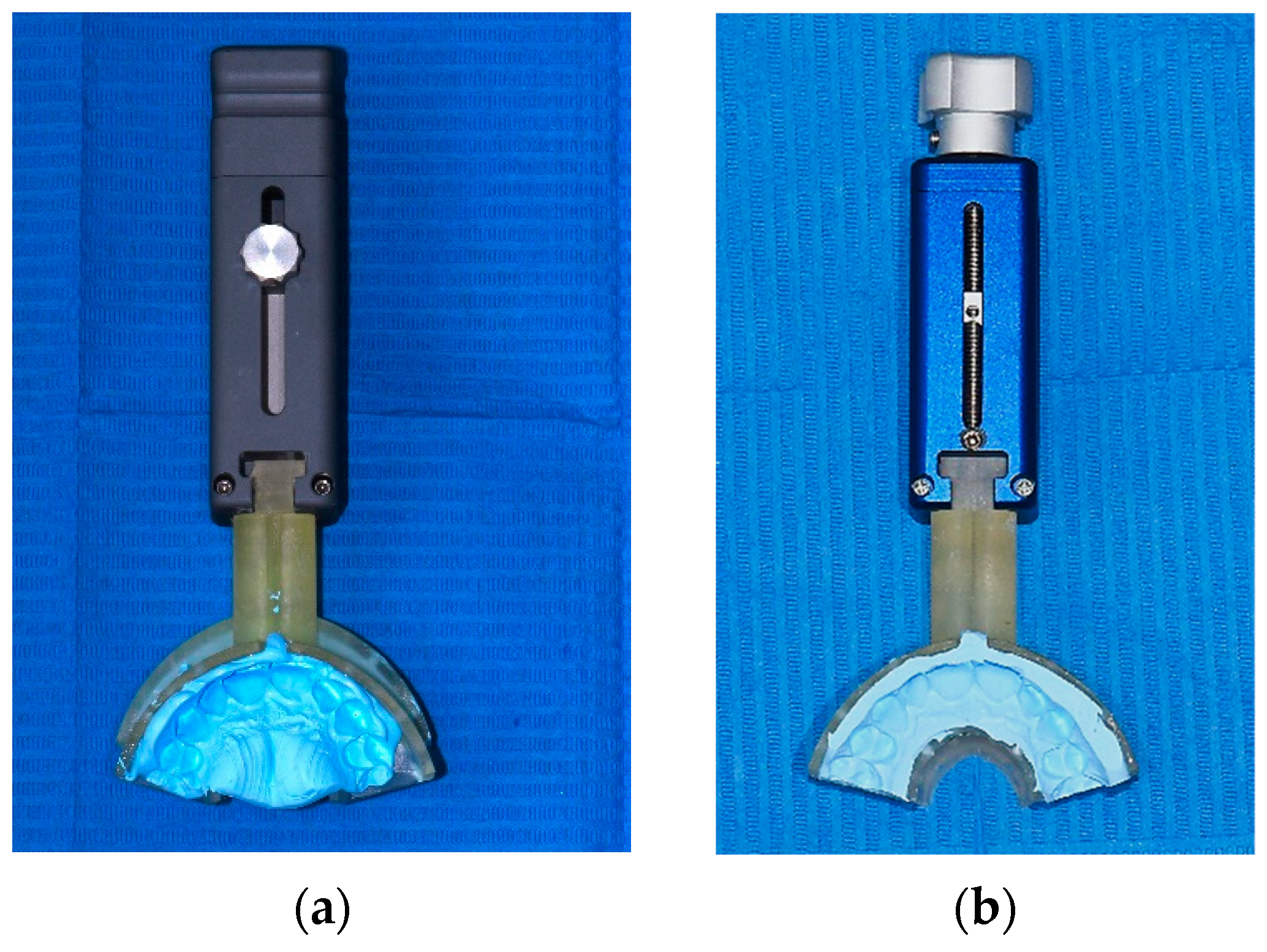
2.3. TMA Simulator Description

The TMA (Figure 1) consists of two devices both made up of a central body, in non-magnetic metal, characterized by a millimeter scale equipped with a pointer and an upper and lower tray holder, transferable from one device to another.
The gauge features a sliding rail system that allows the recording of the individual patient’s mandibular excursion. Once the impressions have been taken (only for the anterior sextant) in the pre-operative phase, the two trays are placed on the gauge using a precision joint (Figure 2). The device is positioned between the patient’s arches and the free mandibular excursion is recorded (from the position of maximum retrusion to the position of maximum advancement). From this measurement, it is possible to obtain the free excursion range for each patient, i.e., the millimetric value within which to make progressive advancement adjustments for observations in DISE.
The slide, instead, allows the possibility of advancing or retruding the mandibular position by using a screw system, having full control of the titration thanks to the indicator on the millimeter scale of the device itself.
Once the impression trays already used in the gauge have been transferred to the TMA slide system, the device is inserted into the patient’s mouth in the first stages of sedation and adjusted to the position of maximum retrusion previously recorded with the gauge. We then proceed with the DISE, initially visualizing the effect on the airways and respiratory parameters only with the occlusal rise determined by the device, without any mandibular advancement. After a stable observation phase of at least five minutes, a progressive mandibular advancement begins. This procedure is repeated with subsequent protrusion increments in order to identify the minimum advancement needed to avoid upper airway obstruction.
Thus, the detected value can be used, in case of indication for MAD therapy, for the construction of the device. In fact, this can be considered a pre-titration of the MAD. The identification of a minimum effective advancement value is a useful indication for the clinician, but also the patient.

2.4. Statistical Analysis

The main descriptive statistics were provided for each parameter (frequency distribution for nominal data, mean and standard deviation for continuous data). For the comparison between “responders” and “non-responders”, the following tests were used: chi-square test for nominal variables (sex, sites of obstruction, pattern of obstruction, VOTE) and t-test for independent samples for continuous variables (age, BMI, polygraphic parameters, number of obstructed sites). The sample size was calculated using the G*Power program (Version 3.1.9.6). Using the distribution of obstructive patterns, considering an alpha value of 0.05, a power of 0.95, and a medium effect size of 0.3 with 14 degrees of freedom, the minimum number of patients required for the study was 303. All statistical analyses were performed using the software SPSS (IBM Corp. 2013. IBM SPSS Statistics for Windows, Version 22.0. IBM Corp.: Armonk, NY, USA).

3. Results

The study sample includes a total of 335 patients. The general characteristics of the patients included in the study are shown in Table 2.

3.1. Between Group Comparison for General and Polygraphic Characteristics

The general characteristics of “responders” (males = 185; females = 28) and “non-responders” (males = 122; females = 6) are shown in Table 3.
“Non-responders” showed higher BMI (p = 0.007), whereas no statistically significant differences existed between the groups for age and gender.
As shown in Table 4, patients who showed a positive response to mandibular advancement during DISE had statistically significantly less severe baseline polygraphic parameters (p < 0.05), with the only exception of mean oxygen saturation, for which the difference between the groups was not statistically significant (p = 0.075).

3.2. Obstructive Sites without the TMA Simulator

We analyzed the individual obstructive sites without the TMA simulator and found that there was no difference in the percentage of velum obstruction (non-responders: 96.1%; responders: 97.6%; p = 0.515), of the tongue base (non-responders: 53.1%; responders: 58.5%; p = 0.365) and epiglottis (non-responders: 35.2%; responders: 40.6%; p = 0.356). On the contrary, responders showed a significantly lower percentage of obstruction of the oropharynx than non-responders (non-responders: 89.1%; responders: 80.2%; p = 0.034).
Concerning the number of obstructed sites, the two groups showed no significant differences (non-responders: 2.73 ± 0.84; responders: 2.77 ± 0.85; p = 0.686).
The Venn diagram in Figure 3 shows the distribution of obstructive patterns in patients who did not show a response to mandibular advancement in DISE (Figure 3a) and those who showed a response to mandibular advancement in DISE (Figure 3b). The two distributions did not differ statistically (p = 0.131).

3.3. VOTE Classification without the TMA Simulator

Table 5 shows the scores of the VOTE classification of non-responders and responders. The two groups did not show significant differences in the distribution of scores at the base of the tongue and epiglottis (p > 0.05). At the oropharynx level, non-responders showed a higher percentage of patients with complete latero-lateral obstruction while in responders the percentage of partial latero-lateral obstruction was higher (p = 0.003). At the level of the velum, the two groups showed a significant difference in the distribution of the VOTE scores (p = 0.008). In particular, in non-responders, there was a significantly higher proportion of complete circular obstruction while in responders there was a significantly higher proportion of partial and complete anterior-posterior collapse.

3.4. Obstructive Sites with TMA Simulator

Analyzing the single obstructive sites, the two groups after mandibular advancement in DISE differed significantly in the percentage of veil obstruction (non-responders: 91.4%; responders: 58.5%; p < 0.001), oropharynx (non-responders: 75.0%; responders: 31.9%; p < 0.001) and of the epiglottis (non-responders: 23.4%; responders: 8.2%; p < 0.001).
On the contrary, the two groups did not show statistically significant differences in the percentage of obstruction of the base of the tongue (non-responders: 28.1%; responders: 21.7%; p = 0.192).
The two groups differed significantly in the number of obstructed sites after mandibular advancement in DISE (non-responders: 2.18 ± 0.81; responders: 1.20 ± 1.01; p < 0.001).
The Venn diagram in Figure 4 shows the distribution of obstructive patterns in patients who did not show a response to mandibular advancement in DISE (Figure 4a) and those who showed a response to mandibular advancement in DISE (Figure 4b). The two distributions significantly differ (p < 0.001). In particular, responders showed a significantly higher percentage of patients with all unobstructed sites (31.4% vs. 1.6% of non-responders) and with only V-site obstruction (20.8% vs. 8.6% of non-responders). Conversely, non-responders had a higher percentage of patients with contextual obstruction of V and O (51.6% vs. 21.3%) and obstruction of all sites (VOTE; 7.8% vs. 0.5%).

3.5. VOTE Classification with TMA Simulator

Table 6 shows the scores of the VOTE classification of the non-responders and the responders. The two groups showed significant differences in the distribution of scores at the level of all sites (p < 0.05). At the velum site, we found significant differences in the obstructed site distribution except for the latero-lateral partial (V3) and circular partial (V5) types of obstruction. In the oropharynx we observed significant differences for no obstruction (O0) and complete latero-lateral obstruction (O2) but no statistical difference for l latero-lateral partial (O1).

4. Discussion

This retrospective study compared the general, polygraphic, and endoscopic characteristics between responders and non-responders to mandibular advancement during DISE. Particularly, the sites (velopharynx, oropharynx, tongue, epiglottis), degrees (none, partial, complete), and patterns (latero-lateral, anteroposterior, circular) of upper airway collapse were described at baseline and with TMA simulator placed intraorally in order to detect characteristics that, if confirmed in further prospective studies, will help in identifying beneficial and/or adverse phenotypes related to MAD treatment outcome. A previous prospective study investigated the locations and degree of upper airway collapse to different mandibular advancements during DISE using a selector advance mandibular device but failed to evaluate the collapse pattern which is an important variable for phenotypes identification and included both snoring and OSA patients [22]. Our study is the first to be conducted on a large sample undergoing DISE with a TMA simulator and including only patients with OSA, thus enhancing the generalizability of the results. A further strength of this study is the ability to evaluate the changes in upper airway collapsibility during DISE using a TMA simulator that is customized to the individual patient.
In the present study the average age of the two groups, responders (50.76 ± 9.83 years) and non-responders (48.77 ± 9.86 years) were comparable. Similar to the results of the prospective study by Petri et al. [24], in which age did not prove to be a success predictor for mandibular advancement device therapy, despite numerous retrospective studies indicating how younger patients are more likely to respond positively to MAD [8,9,25]. The discrepancy between the results of the different studies can likely be related to the variability of the samples analyzed. Concerning age, in our study patients should have good general health with few controlled comorbidities and a low risk for general anesthesia to justify a diagnostic DISE. Data from BMI evaluation showed results in agreement with previous findings [26], in fact, the BMI was significantly lower in the responder group (28.09 ± 3.21 kg/m2) compared to the non-responder group (29.14 ± 3.81 kg/m2).
The analysis of the polygraphic characteristics showed significant differences between the two groups, except for the average oxygen saturation (meanSat %). The baseline AHI was higher in the non-responder group, although the trend was toward a severe disease in both groups (mean AHI ≥ 30 ev/h). In accordance with the statement by the American Academy of Sleep Medicine in 2015 [13], our study indicates how there could be a positive response to MAD even in patients with severe OSA. Likewise, the AHIsup and AHInonsup also were significantly higher in the non-responder group than in the responder group. The evaluation of the respiratory parameters showed significant differences between the two groups regarding ODI: Satmin and CT90: the responder group showed lower values of the ODI and CT90 indices, while higher values of minimum saturation. Mean saturation was higher in the responder group, although not significantly. The findings of our study are consistent with those of Pitarch et al. [27], although their study is a prospective analysis based on a sample of only 41 patients.
The percentage of the obstructive sites without a TMA simulator showed significant differences only for the oropharynx, where the non-responders group presented a greater number of collapses (non-responders: 89.1%; responders: 80.2%; p = 0.034). This result could be explained by considering the nature of oropharynx obstructions. In fact, we also analyzed the distribution of the patterns and degrees of airway narrowing, with a qualitative evaluation as proposed by Kezirian [18]. At the oropharynx level, non-responders show a higher percentage of patients with complete latero-lateral obstruction while in responders the percentage of partial latero-lateral obstruction is higher (p = 0.003). Similarly, for VOTE classification the two groups differ at the velopharynx obstruction with a higher presence of complete circular obstruction in non-responders. These differences are peculiar. The complete obstruction of the latero-lateral oropharynx walls is the main indication for CPAP or pharyngeal surgery, especially in the last years with new techniques like functional expansion pharyngoplasty [28,29]. Differently, the complete circular obstruction is not a privileged therapy option. It is commonly observed in obese patients, where the presence of fat at the neck level is relevant, and/or in patients with loss of muscle responsiveness. Typically, there is no single therapy sufficient to address this condition. Instead, a combined therapeutic approach is preferred, with MAD considered an adjunct to other treatments that alone would fail to achieve optimal therapeutic outcomes [25]. These results appear to align with the finding by Op de Beeck et al. [25], who identified complete lateral oropharyngeal collapse as an adverse baseline DISE phenotype associated with MAD treatment outcomes.
Concerning the obstructive sites with the TMA simulator in situ, the two groups differ significantly for velopharynx, oropharynx, and epiglottis collapse. On the contrary, there was not a difference in the percentage of obstructions at the tongue level. As a trend the obstruction improved equally in both responders and non-responders, suggesting that MAD is equally effective at the tongue level. Therefore, we can hypothesize that the difference in response to mandibular advancement between the groups is due to obstructions present at other levels. Interestingly, tongue base collapse has been described as a beneficial baseline DISE phenotype associated with MAD treatment outcomes [25]. Moreover, without the TMA simulator, there was no difference between the groups in the velopharynx obstruction percentage. After mandibular advancement the difference is significant, suggesting that MAD therapy could be effective at the velopharynx level.
There is still considerable variation across studies on the recommended initial mandibular position for MAD therapy, but the use of a TMA simulator during DISE allows to determine a good candidate for treatment responsiveness and, also, a minimal protrusion from which clinicians could subsequently start MAD titration for each patient. The concept is to achieve the highest reduction in the AHI while reducing patient discomfort and potential side effects [30,31,32].
A limitation of the study is the need to perform a second-level examination that selected a non-representative sample of the entire OSA population, mainly including severe and/or difficult-to-treat patients. However, this limitation offers interesting data, since, despite the severity, almost 62% of patients responded positively to the TMA simulator.
Further well-designed prospective studies involving large cohorts of OSA patients are necessary to evaluate the clinical therapeutic response to MAD in individuals who responded positively to the TMA simulator during DISE in order to identify phenotypes that predict beneficial or adverse outcomes to MAD treatment. It would also be advisable to assess whether the percentage of mandibular advancement used during DISE with the TMA simulator corresponds to the minimum effective mandibular advancement in patients undergoing MAD treatment.

5. Conclusions

The individual obstructive sites of the two groups after mandibular advancement during DISE differ significantly in terms of the percentage of obstruction at the velopharynx, oropharynx, and epiglottis.
The use of a TMA simulator during DISE highlighted a trend toward a mandibular advancement efficacy against tongue collapse showing the same tendency in responders and non-responders. Among non-responders, the presence of complete latero-lateral obstruction at the oropharynx level and complete circular obstruction at the velopharynx was observed, indicating that MAD may not be effective in these cases, at least as a standalone therapy. Further prospective clinical studies focusing on OSA phenotype-specific therapy are recommended to improve the success rate of MAD therapy alone and to better understand the management of combined OSA therapies.

Author Contributions

Conceptualization, G.A.-B. and G.S.; methodology, C.S.; formal analysis, D.R.I.; investigation, M.C. and I.P.; writing—original draft preparation, M.C.; writing—review and editing, S.I.P. All authors have read and agreed to the published version of the manuscript.

Funding

This research received no external funding.

Institutional Review Board Statement

The study was conducted in accordance with the Declaration of Helsinki and approved by the Ethics Committee of S. Orsola-Malpighi University Hospital, Bologna (number of approval: 3885/2017).

Informed Consent Statement

Informed consent was obtained from all subjects involved in the study.

Data Availability Statement

The data presented in this study are available on request from the corresponding author.

Conflicts of Interest

The authors declare no conflicts of interest.

References

  1. AASM. American Academy of Sleep Medicine International Classification of Sleep Disorders; AASM: Westchester, IL, USA, 2005. [Google Scholar]
  2. Sateia, M.D.; Micheal, J. International Classification of Sleep Disorders—Third Edition Highlights and Modifications. Chest 2014, 146, 1384–1397. [Google Scholar] [CrossRef] [PubMed]
  3. Benjafield, A.V.; Ayas, N.T.; Eastwood, P.R.; Heinzer, R.; Ip, M.S.M.; Morrell, M.J.; Nunez, C.M.; Patel, S.R.; Penzel, T.; Pépin, J.-L.; et al. Estimation of the global prevalence and burden of obstructive sleep apnoea: A literature-based analysis. Lancet Respir. Med. 2019, 7, 687–698. [Google Scholar] [CrossRef] [PubMed]
  4. Heinzer, R.; Vat, S.; Marques-Vidal, P.; Marti-Soler, H.; Andries, D.; Tobback, N.; Mooser, V.; Preisig, M.; Malhotra, A.; Waeber, G.; et al. Prevalence of sleep-disordered breathing in the general population: The HypnoLaus study. Lancet Respir. Med. 2015, 3, 310–318. [Google Scholar] [CrossRef]
  5. Simmons, M.S.; Shapiro, C.M. The New DDS—“Dentists Diagnosing Sleep”. J. Dent. Sleep Med. 2020, 7. [Google Scholar] [CrossRef]
  6. Armeni, P.; Borsoi, L.; Costa, F.; Donin, G.; Gupta, A. Cost-of-Illness Study of Obstructive Sleep Apnea Syndrome (OSAS) in Italy; Università Bocconi: Milan, Italy, 2019. [Google Scholar]
  7. Bosi, M.; Incerti Parenti, S.; Sanna, A.; Plazzi, G.; De Vito, A.; Alessandri-Bonetti, G. Non-continuous positive airway pressure treatment options in obstructive sleep apnoea: A pathophysiological perspective. Sleep Med. Rev. 2021, 60, 101521. [Google Scholar] [CrossRef]
  8. Dieltjens, M.; Vanderveken, O. Oral Appliances in Obstructive Sleep Apnea. Healthcare 2019, 7, 141. [Google Scholar] [CrossRef]
  9. Marklund, M.; Braem, M.J.A.; Verbraecken, J. Update on oral appliance therapy. Eur. Respir. Rev. 2019, 28, 190083. [Google Scholar] [CrossRef] [PubMed]
  10. Mogell, K.; Blumenstock, N.; Mason, E.; Rohatgi, R.; Shah, S.; Schwartz, D. Definition of an Effective Oral Appliance for the Treatment of Obstructive Sleep Apnea and Snoring: An Update for 2019. J. Dent. Sleep Med. 2019, 6. [Google Scholar] [CrossRef]
  11. Incerti Parenti, S.; Bortolotti, F.; Alessandri-Bonetti, G. Oral appliances for obstructive sleep apnea. J. World Fed. Orthod. 2019, 8, 3–8. [Google Scholar] [CrossRef]
  12. Ferguson, K.A. The role of oral appliance therapy in the treatment of obstructive sleep apnea. Clin. Chest Med. 2003, 24, 355–364. [Google Scholar] [CrossRef]
  13. Ramar, K.; Dort, L.C.; Katz, S.G.; Lettieri, C.J.; Harrod, C.G.; Thomas, S.M.; Chervin, R.D. Clinical Practice Guideline for the Treatment of Obstructive Sleep Apnea and Snoring with Oral Appliance Therapy: An Update for 2015. J. Clin. Sleep Med. 2015, 11, 773–827. [Google Scholar] [CrossRef] [PubMed]
  14. Randerath, W.J.; Verbraecken, J.; Andreas, S.; Bettega, G.; Boudewyns, A.; Hamans, E.; Jalbert, F.; Paoli, J.R.; Sanner, B.; Smith, I.; et al. Non-CPAP therapies in obstructive sleep apnoea. Eur. Respir. J. 2011, 37, 1000–1028. [Google Scholar] [CrossRef] [PubMed]
  15. De Vito, A.; Carrasco Llatas, M.; Ravesloot, M.J.; Kotecha, B.; De Vries, N.; Hamans, E.; Maurer, J.; Bosi, M.; Blumen, M.; Heiser, C.; et al. European position paper on drug-induced sleep endoscopy: 2017 Update. Clin. Otolaryngol. 2018, 43, 1541–1552. [Google Scholar] [CrossRef] [PubMed]
  16. Vanderveken, O.M. Drug-induced sleep endoscopy (DISE) as a guide towards upper airway behavior and treatment outcome: The quest for a vigorous standardization of DISE. Sleep Breath. 2018, 22, 897–899. [Google Scholar] [CrossRef] [PubMed]
  17. Croft, C.B.; Pringle, M. Sleep nasendoscopy: A technique of assessment in snoring and obstructive sleep apnoea. Clin. Otolaryngol. Allied Sci. 1991, 16, 504–509. [Google Scholar] [CrossRef] [PubMed]
  18. Kezirian, E.J.; Hohenhorst, W.; de Vries, N. Drug-induced sleep endoscopy: The VOTE classification. Eur. Arch. Otorhinolaryngol. 2011, 268, 1233–1236. [Google Scholar] [CrossRef] [PubMed]
  19. Rabelo, F.A.; Küpper, D.S.; Sander, H.H.; Fernandes, R.M.; Valera, F.C. Polysomnographic evaluation of propofol-induced sleep in patients with respiratory sleep disorders and controls. Laryngoscope 2013, 123, 2300–2305. [Google Scholar] [CrossRef] [PubMed]
  20. Morrison, D.L.; Launois, S.H.; Isono, S.; Feroah, T.R.; Whitelaw, W.A.; Remmers, J.E. Pharyngeal narrowing and closing pressures in patients with obstructive sleep apnea. Am. Rev. Respir. Dis. 1993, 148, 606–611. [Google Scholar] [CrossRef] [PubMed]
  21. Fernández-Sanjuán, P.; Arrieta, J.J.; Sanabria, J.; Alcaraz, M.; Bosco, G.; Pérez-Martín, N.; Pérez, A.; Carrasco-Llatas, M.; Moreno-Hay, I.; Ríos-Lago, M.; et al. Optimizing Mandibular Advancement Maneuvers during Sleep Endoscopy with a Titratable Positioner: DISE-SAM Protocol. J. Clin. Med. 2022, 11, 658. [Google Scholar] [CrossRef]
  22. Fernández-Sanjuán, P.; Alcaraz, M.; Bosco, G.; Pérez-Martín, N.; Morato, M.; Lugo, R.; Arrieta, J.J.; Sanabria, J.; Ríos-Lago, M.; Plaza, G. Modifications in Upper Airway Collapsibility during Sleep Endoscopy with a Mandibular Positioner: Study in Snorers and Obstructive Sleep Apnea Patients. J. Clin. Med. 2024, 13, 1184. [Google Scholar] [CrossRef]
  23. Ippolito, D.R.; Stipa, C.; Cameli, M.; Sorrenti, G.; Pelligra, I.; Alessandri-Bonetti, G. Maximum voluntary retrusion or habitual bite position for mandibular advancement assessment in the treatment of obstructive sleep apnoea patients. J. Oral Rehabil. 2020, 47, 301–306. [Google Scholar] [CrossRef] [PubMed]
  24. Petri, N.; Christensen, I.J.; Svanholt, P.; Sonnesen, L.; Wildschiødtz, G.; Berg, S. Mandibular advancement device therapy for obstructive sleep apnea: A prospective study on predictors of treatment success. Sleep Med. 2019, 54, 187–194. [Google Scholar] [CrossRef] [PubMed]
  25. de Beeck, S.O.; Dieltjens, M.; Verbruggen, A.E.; Vroegop, A.V.; Wouters, K.; Hamans, E.; Willemen, M.; Verbraecken, J.; De Backer, W.A.; Van de Heyning, P.H.; et al. Phenotypic Labelling Using Drug-Induced Sleep Endoscopy Improves Patient Selection for Mandibular Advancement Device Outcome: A Prospective Study. J. Clin. Sleep Med. 2019, 15, 1089–1099. [Google Scholar] [CrossRef] [PubMed]
  26. Sutherland, K.; Takaya, H.; Qian, J.; Petocz, P.; Ng, A.T.; Cistulli, P.A. Oral Appliance Treatment Response and Polysomnographic Phenotypes of Obstructive Sleep Apnea. J. Clin. Sleep Med. 2015, 11, 861–868. [Google Scholar] [CrossRef] [PubMed]
  27. Marco Pitarch, R.; Selva García, M.; Puertas Cuesta, J.; Marco Algarra, J.; Fernández Julian, E.; Fons Font, A. Effectiveness of a mandibular advancement device in obstructive sleep apnea patients: A prospective clinical trial. Eur. Arch. Otorhinolaryngol. 2018, 275, 1903–1911. [Google Scholar] [CrossRef]
  28. Torre, C.; Liu, S.Y.; Kushida, C.A.; Nekhendzy, V.; Huon, L.K.; Capasso, R. Impact of continuous positive airway pressure in patients with obstructive sleep apnea during drug-induced sleep endoscopy. Clin. Otolaryngol. 2017, 42, 1218–1223. [Google Scholar] [CrossRef]
  29. Schwab, R.J.; Pack, A.I.; Gupta, K.B.; Metzger, L.J.; Oh, E.; E Getsy, J.; A Hoffman, E.; Gefter, W.B. Upper airway and soft tissue structural changes induced by CPAP in normal subjects. Am. J. Respir. Crit. Care Med. 1996, 154 Pt 1, 1106–1116. [Google Scholar] [CrossRef] [PubMed]
  30. Bartolucci, M.L.; Bortolotti, F.; Raffaelli, E.; D’Antò, V.; Michelotti, A.; Alessandri Bonetti, G. The effectiveness of different mandibular advancement amounts in OSA patients: A systematic review and meta-regression analysis. Sleep Breath. 2016, 20, 911–919. [Google Scholar] [CrossRef] [PubMed]
  31. Masse, J.F. On the role of dentists in the diagnosis and treatment of obstructive sleep apnea: Controversy and controversy. J. Dent. Sleep Med. 2018, 5, 29–30. [Google Scholar] [CrossRef]
  32. Ma, Y.; Yu, M.; Gao, X. The effect of gradually increased mandibular advancement on the efficacy of an oral appliance in the treatment of obstructive sleep apnea. J. Clin. Sleep Med. 2020, 16, 1369–1376. [Google Scholar] [CrossRef]
Figure 1. TMA simulator: AB-Gauge (a); AB-Slide (b).
Applsci 14 04710 g001
Figure 2. Tray impressions on the gauge (a) and on the slide (b).
Applsci 14 04710 g002
Figure 3. Obstructive sites in “non-responders” (a) and “responders” (b) without the TMA simulator; red = velopharynx, green = oropharynx, blue = tongue, yellow = epiglottis. The numbers indicate the numbers of patients.
Applsci 14 04710 g003
Figure 4. Obstructive sites in “non-responders” (a) and “responders” (b) with the TMA simulator; red = velopharynx, green = oropharynx, blue = tongue, yellow = epiglottis. The numbers indicate the numbers of patients.
Applsci 14 04710 g004
Table 1. Classification of obstructive pattern for each pharyngeal level.
0123456789
VNO OBSTRUCTIONPartial Antero-PosteriorComplete Antero PosteriorPartial Latero-lateralComplete Latero-lateralPartial CircularComplete Circular---
ONO OBSTRUCTIONPartial Latero-lateralComplete Latero-lateral-------
TNO OBSTRUCTIONPartial Antero PosteriorComplete Antero Posterior-------
ENO OBSTRUCTIONSecondary Partial Antero PosteriorSecondary Complete Antero PosteriorSecondary Partial Latero-lateralSecondary Complete Latero-lateralPrimary Partial Antero PosteriorPrimary Complete Antero PosteriorPrimary Partial Latero-lateralPrimary Complete Latero-lateralArytenoids And Glottis
V: velopharynx; O: oropharynx; T: tongue; E: epiglottis.
Table 2. Demographic and clinical characteristics of the patients included in the study.
Males [n (%)]307 (91.6%)
Age (years) [mean ± SD]49.98 ± 9.88
BMI (kg/m2) [mean ± SD]28.49 ± 3.48
AHI (ev/h) [mean ± SD]34.14 ± 18.61
OSAS mild (5 ≤ AHI < 15) [n (%)]54 (16.1%)
OSAS moderate (15 ≤ AHI < 30) [n (%)]101 (30.2%)
OSAS severe (AHI ≥ 30) [n (%)]180 (53.7%)
n: number; SD: standard deviation; BMI: body mass index; AHI: apnea–hypopnea index.
Table 3. Comparison between responders and non-responders: gender, age, and body mass index variables.
Non-Responders
[Mean ± SD]
Responders [Mean ± SD]Difference (CI)p (t-Test)
Age (years)48.77 ± 9.8650.76 ± 9.83−1.99 (−4.23; 0.25)0.082
BMI (kg/m2)29.14 ± 3.8128.09 ± 3.211.05 (0.29; 1.82)0.007
BMI: body mass index; SD: standard deviation.
Table 4. Comparison between responders and non-responders: polygraphic indices.
Non-Responders [Mean ± SD]Responders [Mean ± SD]Difference (CI)p (t-Test)
AHI (ev/h)39.50 ± 19.6530.82 ± 17.168.68 (4.66; 12.71)<0.001
AHIsup (ev/h)51.43 ± 21.5146.58 ± 21.014.85 (0.03; 9.67)0.049
AHInonsup (ev/h)25.71 ± 23.9216.12 ± 17.689.59 (4.86; 14.32)<0.001
ODI (ev/h)39.69 ± 21.1330.98 ± 19.668.71 (4.12; 13.30)<0.001
Satmed (%)92.89 ± 2.3493.31 ± 1.84−0.43 (−0.90; 0.04)0.075
Satmin (%)76.91 ± 8.1979.20 ± 8.29−2.28 (−4.19; −0.37)0.019
CT90 (%)14.51 ± 17.089.36 ± 11.255.15 (2.00; 8.30)0.001
ev: events; SD: standard deviation; CI: confidence interval; AHI: apnea–hypopnea index; AHIsup: apnea–hypopnea index in supine position; AHInonsup: apnea–hypopnea index in non-supine position; ODI: oxyhemoglobin desaturation index; Satmed: medium saturation; Satmin: minimum saturation; CT90: percentage of sleep time spent with saturation < 90%.
Table 5. Comparison between responders and non-responders: VOTE classification.
VOTE Classification
0123456789p
V
Non Resp.5 (3.9%)2 (1.6%)18 (14.1%)3 (2.3%)34 (26.6%)0 (0.0%)66 (51.6%) 0.008
Resp.5 (2.4%)13 (6.3%)53 (25.6%)5 (2.4%)62 (30.0%)1 (0.5%)68 (32.9%)
O
Non Resp.14 (10.9%)5 (3.9%)109 (85.2%) 0.003
Resp.41 (19.8%)23 (11.1%)143 (69.1%)
T
Non Resp.60 (46.9%)24 (18.8%)44 (34.4%) 0.103
Resp.86 (41.5%)27 (13.0%)94 (45.4%)
E
Non Resp.83 (64.8%)8 (6.3%)22 (17.2%)1 (0.8%)5 (3.9%)0 (0.0%)5 (3.9%)0 (0.0%)4 (3.1%)0 (0.0%)0.186
Resp.123 (59.4%)16 (7.7%)48 (23.2%)1 (0.5%)4 (1.9%)2 (1.0%)11 (5.3%)1 (0.5%)0 (0.0%)1 (0.5%)
Non Resp.: non-responders to mandibular advancement simulator during DISE; Resp.: responder to mandibular advancement simulator during DISE.
Table 6. Comparison between responders and non-responders after TMA simulator: VOTE classification.
VOTE Classification
0123456789p
V
Non Resp.11 (8.6%)23 (18.0%)22 (17.2%)16 (12.5%)26 (20.3%)2 (1.6%)28 (21.9%) <0.001
Resp.86 (41.5%)61 (29.5%)14 (6.8%)23 (11.1%)7 (3.4%)6 (2.9%)10 (4.8%)
O
Non Resp.32 (25.0%)35 (27.3%)61 (47.7%) <0.001
Resp.141 (68.1%)55 (26.6%)11 (5.3%)
T
Non Resp.92 (71.9%)24 (18.8%)12 (9.4%) 0.008
Resp.162 (78.3%)41 (19.8%)4 (1.9%)
E
Non Resp.98 (76.6%)10 (7.8%)4 (3.1%)2 (1.6%)1 (0.8%)4 (3.1%)5 (3.9%)2 (1.6%)2 (1.6%)0 (0.0%)0.005
Resp.190 (91.8%)6 (2.9%)0 (0.0%)0 (0.0%)0 (0.0%)5 (2.4%)3 (1.4%)2 (1.0%)0 (0.0%)1 (0.5%)
Non Resp.: non-responders to mandibular advancement simulator during DISE; Resp.: responder to mandibular advancement simulator during DISE.
Disclaimer/Publisher’s Note: The statements, opinions and data contained in all publications are solely those of the individual author(s) and contributor(s) and not of MDPI and/or the editor(s). MDPI and/or the editor(s) disclaim responsibility for any injury to people or property resulting from any ideas, methods, instructions or products referred to in the content.

