Article

Evaluation of the Suitability of Selecting a Faecal Microbiota Transplant: Bacterial Composition and Subsequent Long-Term Monitoring of the Viability of Its Frozen and Lyophilised Forms

1
Department of Microbiology and Immunology, University of Veterinary Medicine and Pharmacy in Kosice, 041 81 Kosice, Slovakia
2
2nd Department of Internal Medicine, Faculty of Medicine, Pavol Jozef Safarik University and Louis Pasteur University Hospital in Kosice, 040 11 Kosice, Slovakia
3
Department of Animal Physiology, Institute of Biology and Ecology, Faculty of Science, Pavol Jozef Safarik University in Kosice, 040 01 Kosice, Slovakia
4
Center for Interdisciplinary Biosciences, Technology and Innovation Park, Pavol Jozef Safarik University in Kosice, 040 01 Kosice, Slovakia
5
Small Animal Clinic, University Veterinary Hospital, University of Veterinary Medicine and Pharmacy in Kosice, 041 81 Kosice, Slovakia
6
Center of Clinical and Preclinical Research—MEDIPARK, Faculty of Medicine, Pavol Jozef Safarik University in Kosice, 040 11 Kosice, Slovakia
*
Author to whom correspondence should be addressed.
Appl. Sci. 2024, 14(11), 4856; https://doi.org/10.3390/app14114856
Submission received: 10 May 2024 / Revised: 30 May 2024 / Accepted: 3 June 2024 / Published: 4 June 2024

Abstract

:
Objectives: The aim of this study was to confirm the effectiveness of FMT on the basis of optimum composition of the faecal microbiota of the donor for support therapy in patients with ulcerative colitis, and to observe the viability of the microbiota in frozen and lyophilised administration forms of FMT under various storage conditions. Methods: The bacterial microbiota composition of the FMT samples was assessed using amplicon sequencing via next-generation sequencing (NGS) technology, conducted on the Illumina MiSeq platform. The BD FACS Canto flow cytometer was used to analyse the metabolic activity of FMT samples. Results: FMT analysis confirmed the presence of key butyrate-producing organisms, specifically highlighting species such as Bifidobacterium adolescentis, Faecalibacterium prausnitzi, Coprococcus catus, Eubacterium rectale, alongside contributions from genera Roseburia and Blautia. These organisms play a crucial role in maintaining intestinal health in humans. The viable microorganism counts were significantly higher (p < 0.001) in the frozen form of FMT (−70 °C) in comparison to lyophilised forms (−70 °C, 4 °C and 20 °C) throughout the storage period. Conclusion: The conducted NGS analyses allowed us to confirm the suitability of our FMT donor as a potential candidate for the target group of patients diagnosed with ulcerative colitis. From the point of view of optimum utilisation of FMT at its highest metabolic activity for the purpose of transplantation, its storage for a maximum of 2 months under specified conditions was confirmed as the most suitable for the frozen and all lyophilised FMT forms.

1. Introduction

Substitution therapy of microbiota or faecal microbiota transplantation (FMT) is the transfer of intestinal microbiota obtained from minimally processed and previously examined donor stool into the gastrointestinal tract of the patient with the aim to ameliorate the dysbiotic condition by increasing the overall diversity of the intestinal microbiota and restoring its functionality [1]. It is based on the reasoning that manipulation of the microbiome constitutes potential therapy of pathological conditions with altered composition of the microbiota [2], and its success is proof of the important role of microbiota in various diseases [3]. FMT has been increasingly recognised and accepted due to its approval as a standard therapy of recurrent C. difficile (CDI, Clostridioides difficile infection) infections, resulting in recovery rates as high as 90% [4] in patients suffering from recurrent C. difficile diarrhoea. In addition to the confirmed instances of effective treatment, such as CDI, several studies described clinical conditions for which FMT can constitute a promising and perspective alternative to standard therapies. Published studies have revealed the potential role of FMT in the treatment of refractory ulcerative colitis [5], Crohn’s disease [6], constipation [7], irritable bowel syndrome (IBS) [8], liver diseases [9], oncology [6], blood diseases [10], autism [11,12], and epilepsy [13]. There were also reported experiments with the use of FMT as a supportive therapy in the treatment of diseases such as metabolic syndrome [14], diabetes [15], Parkinson’s disease [16], multiple sclerosis [17], psoriasis, mental anorexia [3], or Alzheimer’s disease [18].
Although the preliminary investigations brought some promising results with respect to future therapeutic strategies, a number of questions need to be resolved in order to effectively translate microbiome-based findings into clinical practice. The extensive use of FMT as an effective therapeutic alternative is prevented by concerns related to the lack of adequate standardisation of the entire process. The ongoing research in this field is heterogeneous in all its phases: from the screening of donors, type of donors (blood-related vs. unrelated), simple stool sample vs. composite samples, processing of stool with optimum composition, its quantity, whole stool vs. sample enriched with viable bacterial products obtained by in vitro fermentation, up to storage and administration in raw vs. frozen vs. lyophilised form of stool, optimum number of FMT doses and way of administration of FMT to recipients. The increasing number of clinical indications points to the need to identify an ideal donor separately for each disease, or an individual patient who cannot be treated indiscriminately by the same faecal microbiota.
The aim of this study was to confirm the suitability of FMT from a selected donor based on the optimal composition of his faecal microbiota, intended for its potential use in supportive therapy in patients with ulcerative colitis. Another aim was to evaluate the viability of microbiota in frozen and lyophilised forms of processed FMT under varying storage conditions.

2. Materials and Methods

2.1. Selection of Faecal Microbiota Transplant Donor

Potential donors of faecal microbiota transplant were first required to fill out an anamnestic questionnaire. They had to meet strict criteria regarding their lifestyle and body mass index (BMI), which must not exceed 25. In addition to anamnesis, the FMT donors were subjected to complex laboratory microbiological (including bacterial and viral) and parasite examination (Table 1), that are strictly specified on the basis of screening of pathogens in the blood of donors, using the methods laid down by the American Red Cross. The suitable donor whose FMT was included in our study was a 34-year-old male, with a BMI of 24, abstinent, non-smoker, with anamnestic data corresponding to the conditions for FMT donors. All selective laboratory examinations of the blood and faeces of this donor provided negative results.

2.2. Processing of Faecal Microbiota Transplant

The donor’s stool was processed in a laminar box (TelStar, Bio II Advance, Terrassa, Spain), on the 10th day after patient screening and within 6 h after faeces collection according to the previously described method [19]. The processed FMT was stored in 5 mL cryogenic vials (TruCool® Cryogenic Vials, BioCision LLC, Larkspur, CA, USA) at −70 °C until its use. One-third of the FMT product was stored at −70 °C and two-thirds were lyophilised in a freeze-drying equipment (Scanvac CoolSafeTM, LaboGene, Lillerød, Denmark) and stored at −70 °C, 4 °C and room temperature (20 °C) for 9 months.

2.3. Analysis of FMT Bacterial Composition Using Next-Generation Sequencing (NGS)

Genomic DNA was isolated from faecal samples using the ZR Fecal DNA MiniPrep™ kit (Zymo Research, Irvine, CA, USA) according to the manufacturer’s protocol. The concentration and purity of the extracted DNA were evaluated using a NanoDrop spectrophotometer (Thermo Fisher Scientific, Waltham, MA, USA). Library preparation, sequencing, data processing, and analysis were performed by Novogene Europe (Cambridge, UK). Briefly, following an initial quality control assessment, amplicon libraries were constructed using universal primers 341F (5′-CCTAYGGGRBGCASCAG-3′) and 806R (5′-GGACTACNNGGGTATCTAAT-3′) targeting the V3–V4 regions of the 16S rRNA gene. These libraries were subsequently subjected to paired-end sequencing on the NovaSeq 6000 platform (Illumina, San Diego, CA, USA) in accordance with the manufacturer’s guidelines. The sequencing run generated 2 × 250 bp reads, yielding approximately 30,000 raw tags per sample.

2.3.1. Sequencing Data Processing

Paired-end reads were merged using FLASH (V1.2.7) [20]. Quality filtering of the raw tags was performed to obtain high-quality clean tags according to the QIIME (V1.7.0) [21] quality control pipeline. Chimera sequences were identified and removed by comparing the tags against the SILVA138 reference database [22] using the UCHIME algorithm [23], resulting in effective tags for downstream analysis.

2.3.2. Taxonomic Annotation

Effective tags were analysed using the Uparse software (Uparse v7.0.1090) [24]. Sequences with ≥97% similarity were clustered into operational taxonomic units (OTUs). For species annotation, representative sequences from each OTU were aligned to reference sequences from the SILVA138 Database using Mothur software (Version v.1.43.0) [25]. The phylogenetic relationships among all OTU representative sequences were obtained using MUSCLE software (Version 3.8.31) [26].

2.4. Determination of Microbial Viability in the Faecal Microbiota Transplant

Samples of faecal microbiota from the selected donor were diluted with phosphate buffer solution (PBS, phosphate buffered saline, MP Biomedicals, Illkirch-Graffenstaden, France) at a ratio of 1:100, at 37 °C. The mixtures were filtered through cellular filters of pore size of 70 μm and 45 μm (BD, Falcon, Franklin Lakes, NJ, USA). Then, 100 µL aliquots of samples were incubated for 20 min with 2.5 µL (1 mM) of carboxyfluorescein diacetate (cFDA) and 3 µL of propidium iodide (PI) [1 mg/mL] at 37 °C and, subsequently, analysed by means of an instrument BD FACS Canto flow cytometer (BD Biosciences, San Jose, CA, USA). The principle of determination of viability of microorganisms is the conversion of cFDA to fluorescent carboxyfluorescein (CF) by means of enzymes present in viable microorganisms. The histogram displaying the percentage viability of bacteria was generated through BD FACS DivaTM software v6.1.3 (BD Biosciences, San Jose, CA, USA).

2.5. Statistical Analysis

The statistical analysis of the results was conducted using GraphPad 5.0 software for Windows (GraphPad Software, San Diego, CA, USA) using the following methods: calculation of standard deviation (±), Student’s t-test, one-way analysis of variance (ANOVA) and Tukey’s multiple comparison test. Differences at the level of p < 0.05 were considered statistically significant.

3. Results

3.1. Bacterial Composition of Faecal Microbiota Transplant from the Healthy Donor

NGS analysis of the FMT sample (Figure 1A) showed that 97% of the faecal microbiota consisted of the following three phyla: Firmicutes (45%), Actinobacteria (synonymum Actinobacteriota 33%), and Bacteroidetes (synonymum Bacteroidota 19%). From among the other present phyla, we detected Proteobacteria (2%), and regarding phyla where their proportion in FMT did not exceed 1%, we detected Desulfobacterota and Verrucomicrobia (synonymum Verrucomicrobiota 0.002%).
The clustered heat map (Figure 1B) illustrates the hierarchical clustering of samples (FMT 1, 2) based on their relative proportions. The colour intensity ranges from dark blue (−6), representing the minimum relative abundance, to dark red (6), indicating the maximum relative abundance. The predominant families in the faecal microbiota of the donor included Bifidobacteriaceae, Ruminococcaceae, Coriobacteriaceae, Rikenellaceae, Sutterellaceae, Oscillospiraceae, and Lachnospiraceae (Figure 1B (1)).
Within the strain Firmicutes, Oscillospirales (13%) was one of the most abundant orders, represented mainly by the family Ruminococcaceae (Figure 1B (1), 10%) with the genera Faecalibacterium, Subdoligranulum, and CAG-352 (Figure 1B (2)). Particularly noteworthy is the substantial proportion of significant butyrate producers, including representatives of species such as Faecalibacterium prausnitzii, Coprococcus catus, and Eubacterium rectale (Figure 1B (3)). These species play a crucial role in maintaining intestinal health in humans. Additionally, the presence of Streptococcus salivarius (Figure 1B (3),C (1)), known for its probiotic effects in preventing oral infections, is also worth mentioning. Other genera of the strain Firmicutes present in FMT were Fusicatenbacter, Alistipes, Blautia, Roseburia, Oscillibacter, Enterococcus, and genus Romboutsia (Figure 1B (2)).
In contrast to the phylum Firmicutes, which exhibited a more diverse composition of genera, Actinobacteria were predominantly represented by the family Bifidobacteriaceae (Figure 1B (1); 29.3%), with a high proportion of only one genus, Bifidobacterium (Figure 1B (2)). The highest relative abundance was observed mainly in species known to significantly contribute to the maintenance of human gut health, including Bifidobacterium adolescentis (22.4%), B. longum (4.7%), B. catenulatum (1.25%), and B. animalis (0.16%) (Figure 1C (2)).
Analysis of data obtained by sequencing also revealed the presence of the most abundant families within the Bacteroidetes phylum, namely Rikenellaceae and Tannerellaceae (Figure 1B (1)), along with their beneficial species Parabacteroides merdae and Parabacteroides goldsteinni (Figure 1B (3)). Additionally, the family Bacteroidaceae with the genus Bacteroides, as well as the families Barnesiellaceae and Marinifilaceae, were also identified. NGS analysis of data confirmed that no pathogenic species were detected in the faecal microbiota transplant from the healthy donor.

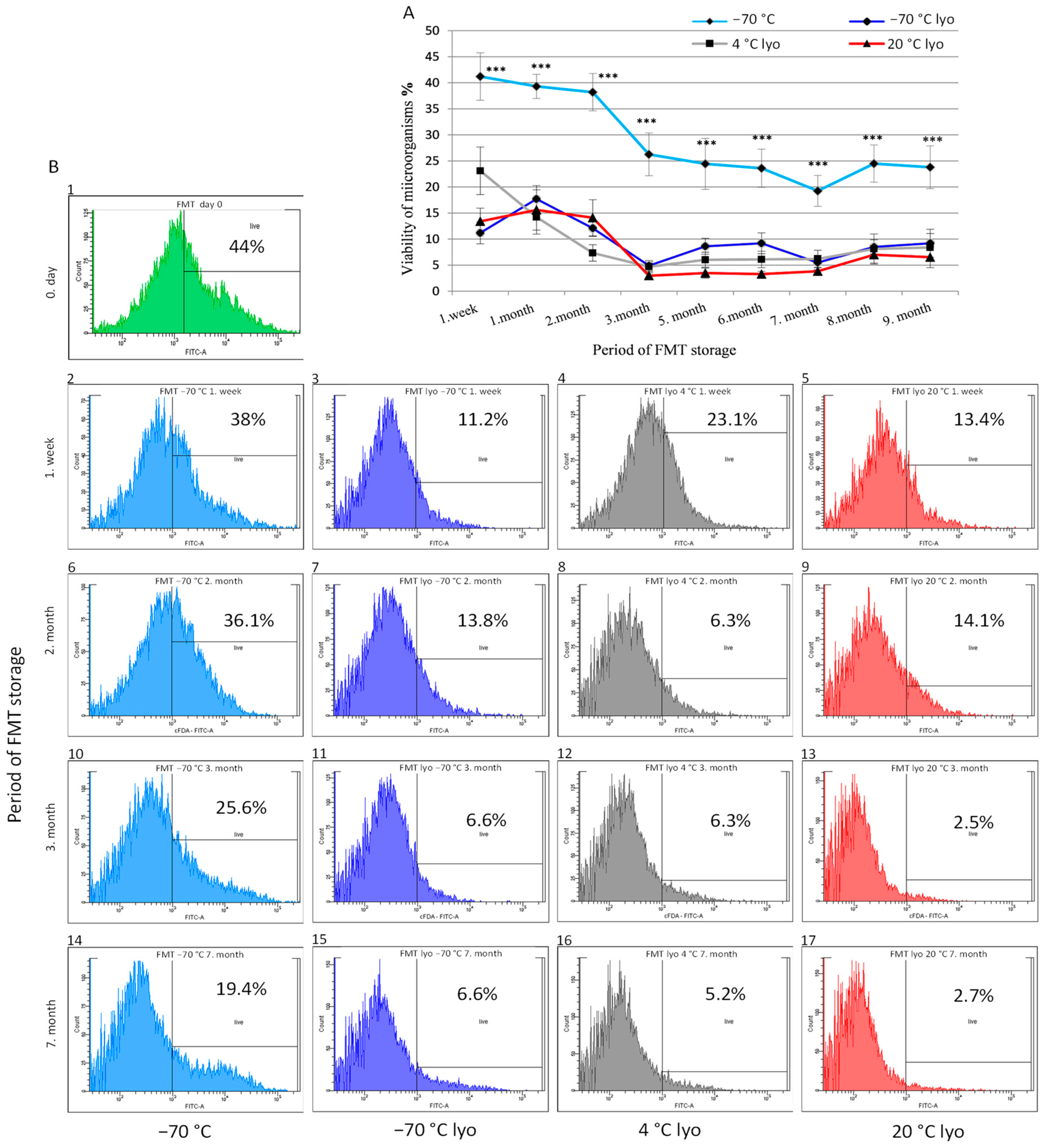
3.2. Monitoring the Metabolic Activity (Viability) Dynamics of Frozen and Lyophilised Forms of FMT across Prolonged Storage Periods

In the second phase of the experiments, we focused on assessing the viability of microbiota in frozen and lyophilised forms of FMT from a healthy donor under various storage conditions. Over a period of 9 months, we monitored changes in the frozen faecal material stored at −70 °C and in lyophilised faecal material stored at −70 °C, 4 °C, and 20 °C. The results are presented as the mean viability (%) of microorganisms (Figure 2A), while the individual representative measurements of microorganism viability are depicted in histograms (Figure 2B (1–17)).
Due to the high time demands of laboratory preparation and processing of the faecal microbiota transplant, and the impossibility of technically ensuring the preparation of FMT under anaerobic conditions, the viability of microorganisms detected by flow cytometry on day 0 reached only 44% (Figure 2B (1)). However, from the first week (Figure 2A,B (2)) up to 2nd month of storage of FMT (Figure 2B (6)), the metabolic activity of microorganisms in the frozen form of FMT remained consistently within the range of approximately 38% to 42%, comparable to pre-storage levels and significantly higher (p < 0.001) than all lyophilised forms. From month 3 of storage, the counts of viable microorganisms in this form of FMT consistently decreased over time, their mean viability ranging between 20 and 25% (Figure 2A,B (10,14)), which was a significantly higher level (p < 0.001) compared to the lyophilised forms (−70 °C, 4 °C and 20 °C) throughout the observation period.
Figure 2. The influence of storage conditions and the administration form of human FMT on viability of faecal microbiota (A). Period of FMT storage: −70 °C (n = 4)—viability of bacteria stored in frozen form of FMT at −70 °C, −70 °C lyo (n = 4)—viability of bacteria stored in lyophilised form of FMT at −70 °C, 4 °C lyo (n = 4)—viability of bacteria stored in lyophilised form of FMT at 4 °C, 20 °C lyo (n = 4)—viability of bacteria stored in lyophilised form of FMT at 20 °C. Results are presented as means ± SD. *** p < 0.001—statistical differences between the frozen form of FMT (−70 °C) and lyophilised forms of FMT (−70 °C, 4 °C, 20 °C). The statistical analysis of the results was conducted using GraphPad 5.0 software for Windows using the following methods: calculation of standard deviation (±), Student’s t-test, one-way analysis of variance (ANOVA) and Tukey’s multiple comparison test. Representative histograms of the influence of storage conditions and administration form on the viability of FMT bacteria (B); period and way of storage: (1) FMT after its processing (0 day); (2) (6) (10) (14) frozen FMT at −70 °C (1st week, 2nd, 3rd and 7th month); (3) (7) (11) (15) lyophilised frozen FMT stored at −70 °C (1st week, 2nd, 3rd and 7th month); (4) (8) (12) (16) lyophilised FMT stored at 4 °C (1st week, 2nd, 3rd and 7th month); (5) (9) (13) (17) lyophilised FMT stored at 20 °C (1st week, 2nd, 3rd and 7th month).
Applsci 14 04856 g002
The decrease in the metabolic activity of faecal microbiota observed in lyophilised forms of FMT storage (Figure 2A) resembled that in the frozen form of FMT (−70 °C); however, the activity in the lyophilised forms was lower by about 20% from the beginning. Up to the 2nd month of storage, all three lyophilised forms of FMT (−70 °C, 4 °C and 20 °C) showed a similar trend of viability of faecal microbiota (Figure 2A), with the exception of the form stored at 4 °C (decrease to 8% in month 2), ranging from 11 to 23% (Figure 2B (8)). While the viability of frozen, non-lyophilised FMT persisted in the range of 20–25% up to the end of the observation period, the metabolic activity of microorganisms in all lyophilised forms decreased between months 3 and 7, to below the acceptable level of 10% (Figure 2A,B (11–13,15–17)). In the form stored at 20 °C, the viability even failed to reach 5% (Figure 2A,B (13,17)).

4. Discussion

The concept of amendment of bowel microbiota in the pathology of ulcerative colitis (UC) has been generally accepted owing to the confirmation of significant differences in bowel microbiota between healthy individuals and UC patients [27,28]. However, it remains unclear whether the altered bowel microbiota is a cause or a consequence of intestinal inflammation [29]. However, the significant role of microbiota has been confirmed. Dysbiosis is frequently defined as an imbalance within the complex commensal communities associated with a particular disease. The whole-genome studies substantiated significant differences between bowel microbiota in healthy individuals and patients with UC with respect to biodiversity and species richness, which particularly diminish in terms of Firmicutes and Bacteroidetes and show a relative increase in the bacterial species of the Enterobacteriaceae family [27]. In patients suffering from UC, specific signs of bowel microbiota were observed, characterised by an increase in species Ruminococcus gnavus and a decrease in Bifidobacterium adolescentis, Dialister invisus, Faecalibacterium prausnitzi, and the species Clostridium cluster XIV [30], and a decrease in genera Blautia and Roseburia [31].
In the FMT from the donor used in our study, we detected an abundance of species Bifidobacterium adolescentis (22.35%) and Faecalibacterium prausnitzi (4%) and participation of representatives of genera Roseburia and Blautia. Faecalibacterium prausnitzii, the well-studied important producer of butyrate, is one of the most abundant bacteria in healthy human bowel microbiota, where it amounts to approximately 5% of the total faecal microbiota [32]. Butyrate plays a critical role in preventing the development of UC by promoting the regeneration of colonocytes and maintaining intestinal integrity [33]. In addition, butyrate also induces development of regulatory T cells (Treg) supporting immune tolerance of intestinal mucosa and maintains the balance between Th17 and Treg cells [34]. It was confirmed that the proportion of F. prausnitzii was extremely low in intestinal samples from patients with IBD (inflammatory bowel disease), as well as in those suffering from CD (Crohn’s disease) and UC [32]. Based on the ample evidence suggesting its significant replenishment in patients in remission, F. prausnitzii has been proposed as an important diagnostic marker of IBD [35]. In the FMT from the donor used in our study, we also detected the presence of other important producers of butyrate, such as representatives of the species Coprococcus catus and Eubacterium rectale, which also play an important role in the maintenance of health of human intestines.
One of the consequences of microbial dysbiosis in patients with UC is the increase in virulent intestinal bacteria of the family Enterobacteriaceae. Bacterial species included in this family comprise a highly endotoxic lipopolysaccharide, the major surface membrane component present in almost all Gram-negative bacteria [36]. Colonisation with Enterobacteriaceae may not itself induce the disease and more likely increases predisposition to the development of intestinal inflammation in IBD [37]. Numerous studies have confirmed a significant increase in enterobacteria, particularly Escherichia coli species, in patients with Crohn’s disease (CD) and UC. NGS analysis of FMT from our donor revealed an absence of a major proportion of bacteria of the Enterobacteriaceae family, namely genera Escherichia and Shigella. The studies mentioned above substantiate the key role of bowel microbiota in the development of intestinal inflammation, although it still remains unclear whether specific species or bacterial groups may be considered causative agents of UC and CD, or if they only contribute to the aggravation of pathogenesis.
The understanding of complex interactions and dynamics within the relevant microbial community could result in the development of a personalised plan of FMT treatment. Such a personalised approach takes into account specific patterns of dysbiosis associated with individual patients and their unique disease characteristics and focuses on targeted and more effective therapeutic interventions. Many studies [38,39] analysed the microbial profile of donors and made an effort to relate it to the clinical and laboratory results of patients with UC. The obtained clinical results and immunological changes following the FMT therapy in patients with UC were significantly related to variations in several specific strains in faecal microbiota recipients [38,39]. For example, the study that included patients with refractory UC who were subjected to preliminary antibiotic treatment followed by repeated FMT treatment with a transplant exhibiting high bacterial abundance and high relative proportion of species Akkermansia muciniphila, non-classified Ruminococcaceae, and Ruminococcus spp., revealed a higher probability of induction of remission in comparison with therapy by antibiotics alone [40,41]. Paramsothy et al. [42] conducted metagenomics and metabolomics analyses of patients and found out that the patients in remission after FMT showed increased counts of Eubacterium hallii and Roseburia inulivorans and increased levels of biosynthesis of short-chain fatty acids (SCFAs) and secondary biliary acids. These results indicated the importance of the diversity of donor′s microbiota for the effectiveness of FMT treatment. Presumably, just as described in the studies by some authors [41,43], the selection of donors based on taxonomic composition of their intestinal microbiota, particularly low or high abundance of specific strains, could become a subject of future studies involving treatment of UC or CD. These results indicate the importance of thorough inspection of each transplant of donor’s faecal microbiota by complex analysis including NGS analysis and various laboratory tests. Such analyses effectively define bacterial composition, identify harmful bacteria, and accurately determine genes resistant to medicaments. These strict analyses should be consistently carried out within each FMT procedure [44].
When looking for the rational approach to FMT preparation, the common assumption is to obtain an FMT product that captures the maximum amount of microbiota from the fresh stool of a healthy donor. Consequently, the majority of studies aimed at developing FMT production methods focus on processes that minimise the reduction in bacterial viability or diversity [45].
In the process of confirmation of standard quality of the prepared FMT it is important to select correct method for determination of viability of faecal microbiota. Characterisation of the composition of microbiome in the processed material obtained from FMT donor is not always performed, or the investigations are usually based on NGS analyses [46]. These analyses are based on detection of DNA derived from viable and non-viable organisms, and thus they cannot accurately indicate which bacteria in the recipient are viable and capable of replication. A number of studies investigated the viability of bacteria present in fresh forms of FMT subjected to various processes [47,48,49,50], but only a few of them referred to their stability in storage [47,51].
In the second stage of our experiments, we investigated the viability of microbiota in the frozen and lyophilised administration form of FMT from a healthy donor stored under various conditions. During the period of 9 months, we investigated frozen faecal material at −70 °C, and lyophilised faecal material stored at −70 °C, 4 °C, and 20 °C. An important factor affecting the viability of bacteria is the methodical procedure used for processing of FMT. Many bacteria found in the large intestine, that seem to be associated with positive results at CD and UC therapy, such as Faecalibacterium prausnitzii, are strict anaerobes that may be reduced by aerobic processing of FMT [52]. Because of that, it was suggested that the increase in clinical effectiveness of IBD therapy may be achieved by processing the faecal microbiota transplants by anaerobic technique introduced by Costello et al. [53]. However, controlled research evaluating the anaerobic versus the conventional processing of FMT is needed in order to decide whether the use of such a technique can bring improved results. In the study conducted by Papanicolas et al. [50], the authors compared anaerobic processing with processing carried out in the presence of oxygen. The viability of bacteria was affected the most by homogenisation of the transplant in mixers. High-speed mixing in comparison with manual homogenisation results in increased exposure to oxygen and higher damage to species susceptible to oxygen. On average, only about half of the bacteria present in faecal transplants retained their viability even after their immediate processing under strictly anaerobic conditions [50]. This correlates with the results obtained in our study that revealed 44% viability of microorganisms in FMT after its processing under aerobic conditions. With increasing time of storage, the number of viable bacteria showed a uniform decrease, and in the frozen form the number declined to 30% after 9 months of storage.
Discussions involved the following question: which form of processed FMT provides better results with respect to IBD therapy, fresh or frozen? Several studies compared the rates of clinical remission after applying FMT when using fresh or frozen form of faecal microbiota for CDI treatment. Results unambiguously showed that neither anaerobic or aerobic processing nor fresh or frozen stool significantly affected the effectiveness of FMT for rCDI treatment [54]. Several other randomised studies and meta-analyses showed that both the frozen form of FMT as well as fresh FMT showed the same effectiveness with respect to clinical improvement in recurrent and refractory CDI [55,56,57]. These results raised the following question: to what degree are the anaerobic species, and their metabolites in particular, and also the additional important soluble and insoluble bioactive components occurring in FMT, preserved during aerobic processing? One of the factors that may play an important role in equal successfulness regarding the clinical remission rates at CDI treatment when using both ways of FMT processing (anaerobic, aerobic) may presumably involve, besides successful colonisation and the effect of faecal microbiota of the donor, also the participation of metabolites of important taxonomic bacterial representatives the “healthy bowel markers” in the final product. Some researchers implied that viable bacteria are needed for recolonisation of the intestine, but other studies suggested that no anaerobic bacteria are necessary for effectiveness of rCDI treatment [50,58,59]. However, it was discovered that an adequate proportion of their metabolites is important [60,61,62]. It is surprising that in the study by Mahdi et al. [63], the authors observed equivalent or higher concentrations of all measured SCFAs in the stored lyophilised forms of stool kept at 4 °C for 12, 33, 60, and 130 days in comparison with the fresh stool. Samples of stool were processed and conserved using a procedure similar to the standard methodical procedure of FMT processing. Although Reygner et al. [48] and Ueyama et al. [64] arrived at the same conclusion, Zheng et al. [65] contrariwise observed lower concentrations of SCFAs in the lyophilised forms of stool. SCFAs are the main metabolite produced by intestinal microbiota during fermentation of fibres and their proportion in the digestive tract reflects the level of intestinal fermentation. However, the respective molecular ratio of SCFAs, 60:20:20 (acetate/propionate/butyrate), is constant in relatively healthy intestine, as in the large intestine section as in human stool [66]. The variations seen in SCFAs’ levels in patients in comparison with healthy controls led to an increased interest in their use as potential biomarkers in a wide spectrum of clinical settings [63]. The remarkable results of lyophilised forms of FMT turned them into an interesting tool for quantification of SCFAs as a supportive method for better evaluation of quality of processed FMT.
Despite the availability of several standardised and tested formulations of products based on FMT, there are still patients that may benefit from drinkable, fluid FMT formulation or a lyophilised, non-capsulated form. This includes adult and paediatric patients who may have problems with swallowing the capsules, particularly when the therapeutic procedure requires multiple repeating dosages. The proof of the clinical effectiveness of the lyophilised FMT for recurrent CDI was provided by two randomised controlled studies [67,68]. In the study [67], the authors reported no significant differences in the speed of recovery between groups of patients treated with lyophilised versus frozen FMT by means of colonoscopy. In spite of this, there was an insignificantly lower speed of recovery observed when using lyophilised product. No significant difference was observed between patients treated per os by lyophilised FMT and by enema administration of frozen FMT, with respect to safety or the results of 2-month cure rate [68]. From the beginning of observation, the viability of microorganisms in lyophilised forms decreased at a higher rate in comparison with the frozen form of FMT stored at −70 °C. All three lyophilised forms of FMT (−70 °C, 4 °C and 20 °C) showed a similar trend of viability of faecal microbiota that decreased to the range of 11–23% by the 2nd month of storage. Between months 3 and 7 of storage, the metabolic activity of microorganisms in all lyophilised forms continued to decrease to below the unacceptable level of 10%. It is important to note that the number of viable microorganisms throughout the storage period was significantly higher (p < 0.001) in the frozen FMT form (−70 °C) in comparison with all three lyophilised forms (−70 °C, 4 °C, and 20 °C). With its viability at the level of 20–30%, thus the highest observed metabolic activity of FMT, this form appeared most suitable for the purpose of transplantation.
After determination of the influence of methods of FMT processing on the viability of bacteria during storage, future investigations could focus on observation of the level of fermentation activity in long-term stored lyophilised forms of FMT. This could contribute to better understanding of the relationship between the viability of microorganisms and the level of their fermentation activity during aerobic processing of FMT and conservation by lyophilisation. Additional studies are needed to connect the viability of bacteria with the effectiveness of treatment with FMT, and the use of compact new formulations could facilitate such efforts.

5. Conclusions

The conducted NGS analyses that effectively defined the bacterial composition of faecal microbiota and identified harmful bacteria confirmed that the FMT donor used in our study was a suitable candidate for the target group of patients diagnosed with ulcerative colitis. In terms of optimising the utilisation of faecal material at its highest metabolic activity, or at least activity sufficient for the rapid revitalisation of causative organisms of faecal microbiota for transplantation purposes, our study indicated that either the frozen form of FMT (−70 °C) or all lyophilised FMT forms (−70 °C, 4 °C, and 20 °C) should be stored for a maximum of 2 months.

Author Contributions

Conceptualisation, S.G. and S.L.; methodology, S.G., S.L., V.H., V.D., M.J., M.R., P.A. and D.M.; validation, S.G., V.D., M.J. and S.L.; formal analysis, S.L., V.H., I.P., M.J., L.A., V.D., M.R., P.A., D.M., M.F. and D.N.; investigation, S.G., M.J., I.B. and L.A.; writing—original draft preparation, S.G., S.L. and I.P.; writing—review and editing, S.G. and V.D.; visualisation, S.L. and D.N.; supervision, V.D.; project administration, S.G. and I.B.; funding acquisition, S.G., M.F. and I.B. All authors have read and agreed to the published version of the manuscript.

Funding

This research was funded by the Slovak Research and Development Agency under the contract No. APVV-16-0176, the project VEGA No. 1/0015/21 and VEGA No. 1/0231/22.

Institutional Review Board Statement

The biological material that was collected from the selected donor of faecal microbiota transplant was obtained within the research project No. 14/2018/OVaV approved by the Ethical Committee of the University Hospital of L. Pasteur in Košice, Slovak Republic.

Informed Consent Statement

Health donor FMT signed informed consent for data collection and analysis for research purposes before inclusion.

Data Availability Statement

The original contributions presented in the study are included in the article, further inquiries can be directed to the corresponding author.

Conflicts of Interest

The authors declare no conflicts of interest.

References

  1. Allegretti, J.R.; Mullish, B.H.; Kelly, C.; Fischer, M. The evolution of the use of faecal microbiota transplantation and emerging therapeutic indications. Lancet 2019, 394, 420–431. [Google Scholar] [CrossRef] [PubMed]
  2. Gupta, A.; Saha, S.; Khanna, S. Therapies to modulate gut microbiota: Past, present and future. World J. Gastroenterol. 2020, 26, 777–788. [Google Scholar] [CrossRef] [PubMed]
  3. Antushevich, H. Fecal microbiota transplantation in disease therapy. Clin. Chim. Acta 2020, 503, 90–98. [Google Scholar] [CrossRef] [PubMed]
  4. Blanchaert, C.; Strubbe, B.; Peeters, H. Fecal microbiota transplantation in ulcerative colitis. Acta Gastro-Enterol. Belg. 2019, 82, 519–528. [Google Scholar]
  5. Rossen, N.; Fuentes, S.; Van Der Spek, M.J.; Tijssen, J.G.P.; Hartman, J.; Duflou, A.; Löwenberg, M.; Van Den Brink, G.R.; Mathus-Vliegen, E.M.H.; De Vos, W.M.; et al. Findings from a randomized controlled trial of fecal transplantation for patients with ulcerative colitis. Gastroenterology 2015, 149, 110–118.e4. [Google Scholar] [CrossRef] [PubMed]
  6. Zhang, F.; Cui, B.; He, X.; Nie, Y.; Wu, K.; Fan, D.; Feng, B.; Chen, D.; Ren, J.; Deng, M.; et al. Microbiota transplantation: Concept, methodology and strategy for its modernization. Protein Cell 2018, 9, 462–473. [Google Scholar] [CrossRef] [PubMed]
  7. Tian, H.; Ding, C.; Gong, J.; Ge, X.; McFarland, L.V.; Gu, L.; Yao, W.; Chen, Q.; Zhu, W.; Li, J.; et al. Treatment of slow transit constipation with fecal microbiota transplantation. J. Clin. Gastroenterol. 2016, 50, 865–870. [Google Scholar] [CrossRef] [PubMed]
  8. Johnsen, P.H.; Hilpüsch, F.; Cavanagh, J.P.; Leikanger, I.S.; Kolstad, C.; Valle, P.C.; Goll, R. Faecal microbiota transplantation versus placebo for moderate-to-severe irritable bowel syndrome: A double-blind, randomised, placebo-controlled, parallel-group, single-centre trial. Lancet Gastroenterol. Hepatol. 2018, 3, 17–24. [Google Scholar] [CrossRef] [PubMed]
  9. Bajaj, J.S.; Kassam, Z.; Fagan, A.; Gavis, E.; Liu, E.; Cox, I.J.; Kheradman, R.; Heuman, D.M.; Wang, J.; Gurry, T.; et al. Fecal microbiota transplant from a rational stool donor improves hepatic encephalopathy: A randomized clinical trial. Hepatology 2017, 66, 1727–1738. [Google Scholar] [CrossRef] [PubMed]
  10. Spindelboeck, W.; Schulz, E.; Uhl, B.; Kashofer, K.; Aigelsreiter, A.; Zinke-Cerwenka, W.; Mulabecirovic, A.; Kump, P.; Halwachs, B.; Gorkiewicz, G.; et al. Repeated fecal microbiota transplantations attenuate diarrhea and lead to sustained changes in the fecal microbiota in acute, refractory gastrointestinal graft- versus -host-disease. Haematologica 2017, 102, e210–e213. [Google Scholar] [CrossRef]
  11. Zhao, H.; Gao, X.; Luo, X.; Shi, Y.; Peng, L.; Wang, C.; Zou, L.; Yang, Y. Mo1667 Fecal microbiota transplantation for children with autism spectrum disorder. Gastrointest. Endosc. 2019, 89, AB512–AB513. [Google Scholar] [CrossRef]
  12. Lü, C.; Rong, J.; Fu, C.; Wang, W.; Xu, J.; Ju, X. Overall rebalancing of gut microbiota is key to autism intervention. Front. Psychol. 2022, 13, 862719. [Google Scholar] [CrossRef]
  13. He, Z.; Cui, B.; Zhang, T.; Li, P.; Long, C.; Ji, G.; Zhang, F. Fecal microbiota transplantation cured epilepsy in a case with Crohn’s disease: The first report. World J. Gastroenterol. 2017, 23, 3565–3568. [Google Scholar] [CrossRef] [PubMed]
  14. Vrieze, A.; Van Nood, E.; Holleman, F.; Salojärvi, J.; Kootte, R.S.; Bartelsman, J.F.W.M.; Dallinga-Thie, G.M.; Ackermans, M.T.; Serlie, M.J.; Oozeer, R.; et al. Transfer of intestinal microbiota from lean donors increases insulin sensitivity in individuals with metabolic syndrome. Gastroenterology 2012, 143, 913–916. [Google Scholar] [CrossRef] [PubMed]
  15. Wu, H.; Esteve, E.; Tremaroli, V.; Khan, M.I.; Caesar, R.; Mannerås-Holm, L.; Ståhlman, M.; Olsson, L.; Sérino, M.; Planas-Fèlix, M.; et al. Metformin alters the gut microbiome of individuals with treatment-naive type 2 diabetes, contributing to the therapeutic effects of the drug. Nat. Med. 2017, 23, 850–858. [Google Scholar] [CrossRef] [PubMed]
  16. Sampson, T.R.; Debelius, J.W.; Thron, T.; Janssen, S.; Shastri, G.G.; Ilhan, Z.E.; Challis, C.; Schretter, C.E.; Rocha, S.; Gradinaru, V.; et al. Gut microbiota regulate motor deficits and neuroinflammation in a model of Parkinson’s disease. Cell 2016, 167, 1469–1480.e12. [Google Scholar] [CrossRef]
  17. Borody, T.J.; Campbell, J. Fecal microbiota transplantation: Current status and future directions. Expert Rev. Gastroenterol. Hepatol. 2011, 5, 653–655. [Google Scholar] [CrossRef]
  18. Harach, T.; Marungruang, N.; Duthilleul, N.; Cheatham, V.; Coy, K.D.M.; Frisoni, G.B.; Neher, J.J.; Fåk, F.; Jucker, M.; Lasser, T.; et al. Reduction of Abeta amyloid pathology in APPPS1 transgenic mice in the absence of gut microbiota. Sci. Rep. 2017, 7, 41802. [Google Scholar] [CrossRef]
  19. Lauko, S.; Gancarčíková, S.; Hrčková, G.; Hajdučková, V.; Andrejčáková, Z.; Fecskeová, L.K.; Bertková, I.; Hijová, E.; Kamlárová, A.; Janíčko, M.; et al. Beneficial effect of faecal microbiota transplantation on mild, moderate and severe dextran sodium sulphate-induced ulcerative colitis in a pseudo germ-free animal model. Biomedicines 2023, 12, 43. [Google Scholar] [CrossRef]
  20. Magoč, T.; Salzberg, S.L. FLASH: Fast length adjustment of short reads to improve genome assemblies. Bioinformatics 2011, 27, 2957–2963. [Google Scholar] [CrossRef]
  21. Caporaso, J.G.; Kuczynski, J.; Stombaugh, J.; Bittinger, K.; Bushman, F.D.; Costello, E.K.; Fierer, N.; Peña, A.G.; Goodrich, J.K.; Gordon, J.; et al. QIIME allows analysis of high-throughput community sequencing data. Nat. Methods 2010, 7, 335–336. [Google Scholar] [CrossRef]
  22. Quast, C.; Pruesse, E.; Yilmaz, P.; Gerken, J.; Schweer, T.; Yarza, P.; Peplies, J.; Glöckner, F.O. The SILVA ribosomal RNA gene database project: Improved data processing and web-based tools. Nucleic Acids Res. 2013, 41, D590–D596. [Google Scholar] [CrossRef]
  23. Edgar, R.; Haas, B.J.; Clemente, J.C.; Quince, C.; Knight, R. UCHIME improves sensitivity and speed of chimera detection. Bioinformatics 2011, 27, 2194–2200. [Google Scholar] [CrossRef] [PubMed]
  24. Edgar, R.C. UPARSE: Highly accurate OTU sequences from microbial amplicon reads. Nat. Methods 2013, 10, 996–998. [Google Scholar] [CrossRef]
  25. Schloss, P.D.; Westcott, S.L.; Ryabin, T.; Hall, J.R.; Hartmann, M.; Hollister, E.B.; Lesniewski, R.A.; Oakley, B.B.; Parks, D.H.; Robinson, C.J.; et al. Introducing mothur: Open-source, platform-independent, community-supported software for describing and comparing microbial communities. Appl. Environ. Microbiol. 2009, 75, 7537–7541. [Google Scholar] [CrossRef]
  26. Edgar, R.C. MUSCLE: Multiple sequence alignment with high accuracy and high throughput. Nucleic Acids Res. 2004, 32, 1792–1797. [Google Scholar] [CrossRef] [PubMed]
  27. Khan, I.; Ullah, N.; Zha, L.; Bai, Y.; Khan, A.; Zhao, T.; Che, T.; Zhang, C. Alteration of gut microbiota in inflammatory bowel disease (IBD): Cause or consequence? IBD treatment targeting the gut microbiome. Pathogens 2019, 8, 126. [Google Scholar] [CrossRef] [PubMed]
  28. Ayobami, O.I.; Sunbare-Funto, O.J.; Mbah, C.E.; Ajibade, O.A.; Oyawoye, O.M.; Aborode, A.T.; Ogunleye, S.C.; Jamiu, A.T.; Bolarinwa, B.; Abanikannda, M.F.; et al. Faecal microbial transplant. Adv. Biomark. Sci. Technol. 2024, 6, 20–34. [Google Scholar]
  29. Rosen, C.E.; Palm, N.W. Navigating the microbiota seas: Triangulation finds a way forward. Cell Host Microbe 2018, 23, 1–3. [Google Scholar] [CrossRef]
  30. Nishida, A.; Inoüe, R.; Inatomi, O.; Bamba, S.; Naito, Y.; Andoh, A. Gut microbiota in the pathogenesis of inflammatory bowel disease. Clin. J. Gastroenterol. 2017, 11, 1–10. [Google Scholar] [CrossRef]
  31. Takahashi, K.; Nishida, A.; Fujimoto, T.; Fujii, M.; Shioya, M.; Imaeda, H.; Inatomi, O.; Bamba, S.; Sugimoto, M.; Andoh, A. Reduced abundance of butyrate-producing bacteria species in the fecal microbial community in Crohn’s disease. Digestion 2016, 93, 59–65. [Google Scholar] [CrossRef] [PubMed]
  32. Dong, L.; Wang, M.; Guo, J.; Wang, J. Role of intestinal microbiota and metabolites in inflammatory bowel disease. Chin. Med. J. 2019, 132, 1610–1614. [Google Scholar] [CrossRef] [PubMed]
  33. Cao, Y.; Shen, J.; Ran, Z.H. Association between Faecalibacterium prausnitzii reduction and inflammatory bowel disease: A meta-analysis and systematic review of the literature. Gastroenterol. Res. Pract. 2014, 2014, 872725. [Google Scholar] [CrossRef] [PubMed]
  34. Zhou, L.; Zhang, M.; Wang, Y.; Dorfman, R.; Liu, H.; Yu, T.; Chen, X.; Tang, D.; Xu, L.; Yin, Y.; et al. Faecalibacterium prausnitzii produces butyrate to maintain th17/treg balance and to ameliorate colorectal colitis by inhibiting histone deacetylase 1. Inflamm. Bowel Dis. 2018, 24, 1926–1940. [Google Scholar] [CrossRef] [PubMed]
  35. Ferreira-Halder, C.V.; De Sousa Faria, A.V.; Andrade, S.S. Action and function of Faecalibacterium prausnitzii in health and disease. Best Pract. Res. Clin. Gastroenterol. 2017, 31, 643–648. [Google Scholar] [CrossRef] [PubMed]
  36. Zuo, T.; Ng, S.C. The gut microbiota in the pathogenesis and therapeutics of inflammatory bowel disease. Front. Microbiol. 2018, 9, 2247. [Google Scholar] [CrossRef] [PubMed]
  37. Zeng, M.Y.; Inohara, N.; Núñez, G. Mechanisms of inflammation-driven bacterial dysbiosis in the gut. Mucosal Immunol. 2017, 10, 18–26. [Google Scholar] [CrossRef] [PubMed]
  38. Haifer, C.; Leong, R.W.; Paramsothy, S. The role of faecal microbiota transplantation in the treatment of inflammatory bowel disease. Curr. Opin. Pharmacol. 2020, 55, 8–16. [Google Scholar] [CrossRef] [PubMed]
  39. Zou, M.; Jie, Z.; Cui, B.; Wang, H.; Feng, Q.; Zou, Y.; Zhang, X.; Yang, H.; Wang, J.; Zhang, F.; et al. Fecal microbiota transplantation results in bacterial strain displacement in patients with inflammatory bowel diseases. FEBS Open Bio 2019, 10, 41–55. [Google Scholar] [CrossRef]
  40. Kump, P.; Wurm, P.; Gröchenig, H.; Wenzl, H.; Petritsch, W.; Halwachs, B.; Wagner, M.A.; Stadlbauer, V.; Eherer, A.; Hoffmann, K.M.; et al. The taxonomic composition of the donor intestinal microbiota is a major factor influencing the efficacy of faecal microbiota transplantation in therapy refractory ulcerative colitis. Aliment. Pharmacol. Ther. 2017, 47, 67–77. [Google Scholar] [CrossRef]
  41. Bibbò, S.; Settanni, C.R.; Porcari, S.; Bocchino, E.; Ianiro, G.; Cammarota, G.; Gasbarrini, A. Fecal microbiota transplantation: Screening and selection to choose the optimal donor. J. Clin. Med. 2020, 9, 1757. [Google Scholar] [CrossRef] [PubMed]
  42. Paramsothy, S.; Nielsen, S.; Kamm, M.A.; Deshpande, N.; Faith, J.J.; Clemente, J.C.; Paramsothy, R.; Walsh, A.; Van Den Bogaerde, J.; Samuel, D.B.; et al. Specific bacteria and metabolites associated with response to fecal microbiota transplantation in patients with ulcerative colitis. Gastroenterology 2019, 156, 1440–1454.e2. [Google Scholar] [CrossRef] [PubMed]
  43. Tian, H.; Zhang, S.; Qin, H.; Li, N.; Chen, Q. Long-term safety of faecal microbiota transplantation for gastrointestinal diseases in China. Lancet Gastroenterol. Hepatol. 2022, 7, 702–703. [Google Scholar] [CrossRef] [PubMed]
  44. Haindl, R.; Totzauer, L.; Kulozik, U. Preservation by lyophilization of a human intestinal microbiota: Influence of the cultivation pH on the drying outcome and re-establishment ability. Microb. Biotechnol. 2022, 15, 886–900. [Google Scholar] [CrossRef] [PubMed]
  45. Tian, H.; Wang, X.; Fang, Z.; Li, L.; Wu, C.; Bi, D.; Li, N.; Chen, Q.; Qin, H. Fecal microbiota transplantation in clinical practice: Present controversies and future prospects. hLife 2024, in press. [Google Scholar] [CrossRef]
  46. Fouhy, F.; Deane, J.; Rea, M.C.; O’Sullivan, Ó.; Ross, R.P.; O’Callaghan, G.; Plant, B.J.; Stanton, C. The effects of freezing on faecal microbiota as determined using MiSeq sequencing and culture-based investigations. PLoS ONE 2015, 10, e0119355. [Google Scholar] [CrossRef] [PubMed]
  47. Burz, S.D.; Abraham, A.L.; Fonseca, F.; David, O.; Chapron, A.; Béguet-Crespel, F.; Cénard, S.; Le Roux, K.; Patrascu, O.; Levenez, F.; et al. A guide for ex vivo handling and storage of stool samples intended for fecal microbiota transplantation. Sci. Rep. 2019, 9, 8897. [Google Scholar] [CrossRef]
  48. Reygner, J.; Charrueau, C.; Delannoy, J.; Mayeur, C.; Robert, V.; Cuinat, C.; Meylheuc, T.; Mauras, A.; Augustin, J.; Nicolis, I.; et al. Freeze-dried fecal samples are biologically active after long-lasting storage and suited to fecal microbiota transplantation in a preclinical murine model of Clostridioides difficile infection. Gut Microbes 2020, 11, 1405–1422. [Google Scholar] [CrossRef] [PubMed]
  49. Papanicolas, L.E.; Wang, Y.; Choo, J.M.; Gordon, D.L.; Wesselingh, S.L.; Rogers, G.B. Optimisation of a propidium monoazide based method to determine the viability of microbes in faecal slurries for transplantation. J. Microbiol. Methods 2019, 156, 40–45. [Google Scholar] [CrossRef]
  50. Papanicolas, L.E.; Choo, J.M.; Wang, Y.; Leong, L.E.X.; Costello, S.P.; Gordon, D.L.; Wesselingh, S.L.; Rogers, G.B. Bacterial viability in faecal transplants: Which bacteria survive. eBioMedicine 2019, 41, 509–516. [Google Scholar] [CrossRef]
  51. Jiang, Z.D.; Alexander, A.; Ke, S.; Valilis, E.M.; Hu, S.; Li, B.; DuPont, H.L. Stability and efficacy of frozen and lyophilized fecal microbiota transplant (FMT) product in a mouse model of Clostridium difficile infection (CDI). Anaerobe 2017, 48, 110–114. [Google Scholar] [CrossRef] [PubMed]
  52. Chu, N.D.; Smith, M.B.; Perrotta, A.R.; Kassam, Z.; Alm, E.J. Profiling living bacteria informs preparation of fecal microbiota transplantations. PLoS ONE 2017, 12, e0170922. [Google Scholar] [CrossRef] [PubMed]
  53. Costello, S.P.; Hughes, P.A.; Waters, O.; Bryant, R.V.; Vincent, A.D.; Blatchford, P.; Katsikeros, R.; Makanyanga, J.; Campaniello, M.A.; Mavrangelos, C.; et al. Effect of fecal microbiota transplantation on 8-week remission in patients with ulcerative colitis: A randomized clinical trial. JAMA 2019, 321, 156–164. [Google Scholar] [CrossRef] [PubMed]
  54. Hamilton, M.J.; Weingarden, A.R.; Sadowsky, M.J.; Khoruts, A. Standardized frozen preparation for transplantation of fecal microbiota for recurrent Clostridium difficile infection. Am. J. Gastroenterol. 2012, 107, 761–767. [Google Scholar] [CrossRef] [PubMed]
  55. Wise, J. Frozen faecal matter works as well as fresh for transplantation in C. difficile patients. BMJ 2016, 352, i138. [Google Scholar] [CrossRef] [PubMed]
  56. Tang, G.; Yin, W.; Liu, W. Is frozen fecal microbiota transplantation as effective as fresh fecal microbiota transplantation in patients with recurrent or refractory Clostridium difficile infection: A meta-analysis? Diagn. Microbiol. Infect. Dis. 2017, 88, 322–329. [Google Scholar] [CrossRef] [PubMed]
  57. Fang, H.; Fu, L.; Wang, J. Protocol for fecal microbiota transplantation in inflammatory bowel disease: A systematic review and meta-analysis. Biomed. Res. Int. 2018, 2018, 8941340. [Google Scholar] [CrossRef] [PubMed]
  58. Hirotaka, S.; Katsuhiro, A.; Ichiro, T.; Takuya, T.; Takashi, A.; Hirokazu, T.; Satoshi, M.; Yuichiro, Y. Anaerobic stool preparation method for fecal microbiota transplantation is not superior to conventional aerobic method in preserving anaerobic bacteria: P-125. Off. J. Am. College Gastroenterol. 2018, 113, 29. [Google Scholar] [CrossRef]
  59. Mendolia, G.; Kassam, Z.; McClure, E.L.; Bi, H.S. Mo1954 Anaerobic fecal microbiota transplantation preparations are not necessary for treatment successful engraftment microbial in recurrent C. difficile infection. Gastroenterology 2020, 158, 991–992. [Google Scholar] [CrossRef]
  60. Brunse, A.; Deng, L.; Pan, X.; Hui, Y.; Castro-Mejía, J.L.; Kot, W.; Nguyen, D.N.; Secher, J.B.; Nielsen, D.S.; Thymann, T. Fecal filtrate transplantation protects against necrotizing enterocolitis. ISME J. 2021, 16, 686–694. [Google Scholar] [CrossRef]
  61. Feuerstadt, P.; Louie, T.J.; Lashner, B.; Wang, E.E.L.; Diao, L.; Bryant, J.A.; Sims, M.; Kraft, C.S.; Cohen, S.H.; Berenson, C.S.; et al. SER109, an oral microbiome therapy for recurrent Clostridioides difficile infection. N. Engl. J. Med. 2022, 386, 220–229. [Google Scholar] [CrossRef] [PubMed]
  62. Ott, S.J.; Waetzig, G.H.; Rehman, A.; Moltzau-Anderson, J.; Bharti, R.; Grasis, J.A.; Cassidy, L.; Tholey, A.; Fickenscher, H.; Seegert, D.; et al. Efficacy of sterile fecal filtrate transfer for treating patients with Clostridium difficile infection. Gastroenterology 2017, 152, 799–811.e797. [Google Scholar] [CrossRef] [PubMed]
  63. Mahdi, T.; Desmons, A.; Krasniqi, P.; Lacorte, J.-M.; Kapel, N.; Lamazière, A.; Fourati, S.; Eguether, T. Effect of stool sampling on a routine clinical method for the quantification of six short chain fatty acids in stool using gas chromatography–mass spectrometry. Microorganisms 2024, 12, 828. [Google Scholar] [CrossRef] [PubMed]
  64. Ueyama, J.; Oda, M.; Hirayama, M.; Sugitate, K.; Sakui, N.; Hamada, R.; Ito, M.; Saito, I.; Ohno, K. Freeze-drying enables homogeneous and stable sample preparation for determination of fecal short-chain fatty acids. Anal. Biochem. 2020, 589, 113508. [Google Scholar] [CrossRef] [PubMed]
  65. Zheng, X.; Qiu, Y.; Zhong, W.; Baxter, S.; Su, M.; Li, Q.; Xie, G.; Ore, B.M.; Qiao, S.; Spencer, M.D.; et al. A targeted metabolomic protocol for short-chain fatty acids and branched-chain amino acids. Metabolomics 2013, 9, 818–827. [Google Scholar] [CrossRef] [PubMed]
  66. Den Besten, G.; van Eunen, K.; Groen, A.K.; Venema, K.; Reijngoud, D.-J.; Bakker, B.M. The role of short-chain fatty acids in the interplay between diet, gut microbiota, and host energy metabolism. J. Lipid Res. 2013, 54, 2325–2340. [Google Scholar] [CrossRef] [PubMed]
  67. Jiang, Z.D.; Ajami, N.J.; Petrosino, J.F.; Jun, G.; Hanis, C.L.; Shah, M.; Hochman, L.; Ankoma-Sey, V.; DuPont, A.W.; Wong, M.C.; et al. Randomised clinical trial: Faecal microbiota transplantation for recurrent clostridum difficile infection—Fresh, or frozen, or lyophilised microbiota from a small pool of healthy donors delivered by colonoscopy. Aliment. Pharmacol. Ther. 2017, 45, 899–908. [Google Scholar] [CrossRef]
  68. Jiang, Z.D.; Jenq, R.R.; Ajami, N.J.; Petrosino, J.F.; Alexander, A.A.; Ke, S.; Iqbal, T.; DuPont, A.W.; Muldrew, K.; Shi, Y.; et al. Safety and preliminary efficacy of orally administered lyophilized fecal microbiota product compared with frozen product given by enema for recurrent Clostridium difficile infection: A randomized clinical trial. PLoS ONE 2018, 13, e0205064. [Google Scholar] [CrossRef]
Figure 1. Bacterial composition of faecal microbiota transplant from a healthy FMT donor (A). The Krona diagram depicts the result of taxonomic analysis of the FMT donor at the strain level. A heatmap of species abundance clustering (B). The heatmap shows the hierarchical clustering of samples based on the relative abundance. The relative values illustrated in the heatmap by colours: minimum relative abundance—dark blue (−6), maximum relative abundance—dark red (6), provide picture of abundance of clusters among two replicates same sample of FMT (FMT, n = 2) at the level of (1) family, (2) genus, and (3) species. Taxonomy tree of FMT donors, expressed as a percentage of the relative abundance of species in the selected taxon (C). The first number below the taxonomic name represents the percentage in the entire taxon, while the second number represents the percentage in the selected taxon.
Applsci 14 04856 g001
Table 1. Criteria disqualifying the potential FMT donor.
Anamnesis
-
Must not have had past gastrointestinal disorders, recent diarrhoeic diseases, irritable bowel syndrome, coeliac disease, Inflammatory bowel disease, colitis, and Helicobacter pylori infection.
-
No ingestion of antibiotics and immunosuppressive preparations in the past 3 months.
-
No travel to developing countries in the past 6 months.
Complex laboratory examination
Bacterial examination
-
For the presence of pathogens Salmonella, Shigella, and Clostridioides difficile (GDH and toxins A, B).
Virological examination
-
For cytomegalovirus (CMV), rotavirus, norovirus I and II, and adenovirus 40 and 41.
Serological examination
-
For the presence of HIV, hepatitis A, B, C and E, Treponema pallidum, Escherichia coli (enterotoxigenic, enteroinvasive, enteropathogenic, enteroaggregative, and Shiga-like toxins producing strains), species Vibrio cholerae, Campylobacter spp.
Parasitic examination
-
For the presence of parasites such as Strongyloides stercoralis, Giardia intestinalis, Cryptosporidium spp., and Entamoeba histolytica.
Immunological examination
-
Determination of inflammatory marker calprotectin.
Disclaimer/Publisher’s Note: The statements, opinions and data contained in all publications are solely those of the individual author(s) and contributor(s) and not of MDPI and/or the editor(s). MDPI and/or the editor(s) disclaim responsibility for any injury to people or property resulting from any ideas, methods, instructions or products referred to in the content.

