Article

Investigation of the Photothermal Performance of the Composite Scaffold Containing Light-Heat-Sensitive Nanomaterial SiO2@Fe3O4

Xinjiang Key Laboratory of Additive Remanufacturing, School of Modern Industry for Smart Manufacturing, Xinjiang University, Urumchi 830017, China
*
Author to whom correspondence should be addressed.
Appl. Sci. 2024, 14(11), 4911; https://doi.org/10.3390/app14114911
Submission received: 19 April 2024 / Revised: 16 May 2024 / Accepted: 30 May 2024 / Published: 5 June 2024

Abstract

:
The objective of this investigation was to fabricate a photothermally responsive composite bone scaffold aimed at facilitating bone tissue regeneration and remedying bone defects via mild thermal stimulation. The photothermal-sensitive nanomaterial SiO2 coated Fe3O4 (SiO2@Fe3O4), synthesized through the hydrolysis–condensation process of tetraethyl orthosilicate (TEOS), displayed a uniform distribution of SiO2 coating, effectively preventing the aggregation of Fe3O4 particles within the scaffold matrix. The composite scaffold containing 5% mass fraction of photothermal-sensitive nanoparticles exhibited evenly dispersed microstructural porosity, a compressive strength of 5.722 MPa, and a water contact angle of 58.3°, satisfying the mechanical property requisites of cancellous bone while demonstrating notable hydrophilic characteristics. Upon exposure to near-infrared light at ambient temperature, the 5% composite scaffold underwent a temperature elevation of 3–6 °C within 40–45 s, attaining a temperature range (40–43 °C) conducive to fostering osteogenic differentiation. Experimental findings validated that the SiO2@Fe3O4/polyvinyl alcohol (PVA)/hydroxyapatite (HA)/polycaprolactone (PCL)/β-tricalcium phosphate (β-TCP) bone scaffold showcased outstanding mechanical and photothermal attributes, thereby presenting a pioneering avenue for advancing bone tissue cell proliferation and addressing bone defect rehabilitation.

1. Introduction

The repair of bone defects has long been a formidable challenge in the field of bone tissue engineering [1,2]. Bone defects resulting from surgical procedures or traumatic injuries exceeding the critical dimension of 10 mm manifest an inherent incapacity for structural bone regeneration [3,4]. To enhance the capability of bone scaffolds in inducing bone gene expression and facilitating bone cell regeneration, a strategy involving the combination of two or more types of biomaterials is employed to leverage the respective advantages of different biomaterials, thereby achieving the requisite criteria of native bone biocompatibility, porosity, and mechanical properties [5]. The bio-ceramic material hydroxyapatite (HA) is distinguished by its exceptional biocompatibility and stability and can be co-extruded with a polyvinyl alcohol (PVA) solution for the fabrication of bone scaffolds [6,7,8]. Its unique property of bonding directly to bone without forming any surrounding connective tissue has found widespread applications in various fields, such as bone scaffolds and coatings for dental implants [9,10]. Bio-ceramic materials, like beta-tricalcium phosphate (β-TCP) and HA, exhibit a high degree of resemblance to natural bone tissue, and a composite scaffold combining β-TCP and HA can address the degradation limitations of HA scaffolds made from a single material. Furthermore, the incorporation of the polymer biomaterial polycaprolactone (PCL) is beneficial for enhancing the mechanical performance of composite bone scaffolds owing to its superior toughness [11]. Peroglio and colleagues have validated that the mechanical properties of HA/β-TCP composite bone scaffolds are notably improved via the infiltrative effect of PCL. Additionally, they assessed the cell compatibility of the scaffolds by culturing human bone marrow mesenchymal stromal cells in vitro, demonstrating their favorable cell compatibility, enhanced cell proliferation, and non-cytotoxicity. This underscores the potential of these biomaterials in bone tissue engineering applications [12].
Localized hyperthermia is a widely utilized adjunctive therapeutic modality in clinical practice, known for its ability to expedite cellular metabolism, stimulate cell proliferation, and induce the expression of pertinent genes to facilitate the regeneration of bone tissue defects [13]. Traditional thermal therapy methods utilizing near-infrared light devices have limitations in effectively elevating the temperature of internal scaffolds, potentially resulting in adverse effects, such as skin burns, with prolonged exposure. Scaffolds incorporating thermosensitive compounds can be swiftly heated within the body through modulation by near-infrared light devices [14,15]. Nevertheless, challenges exist with organic photothermal agents due to their suboptimal photothermal stability and susceptibility to photobleaching effects, while the intricate fabrication processes associated with carbon nanomaterials and the heightened toxicity risks of noble metal nanomaterials present additional drawbacks. In contrast, photothermal-responsive Fe3O4 nanoparticles offer distinct advantages, including high stability, facile synthesis, low toxicity, and superior photothermal properties [16]. However, the propensity of Fe3O4 particles for aggregation necessitates modification for effective integration with scaffold materials to explore their full potential in harnessing photothermal properties.
This study aimed to promote the proliferation of bone tissue cells and induce the expression of bone-related genes by utilizing the hydrolysis–condensation method to synthesize photothermal-sensitive SiO2@Fe3O4 nanoparticles, thereby endowing them with excellent biocompatibility, stability, and negligible cytotoxicity, while preventing Fe₃O₄ particle aggregation [17]. Subsequently, a photothermal composite scaffold was fabricated using a state-of-the-art 3D bioprinting technique. The microstructural characteristics, mechanical properties, hydrophilicity/hydrophobicity, and photothermal behavior of the photothermal composite bone scaffold were systematically investigated through a combination of transmission electron microscopy (TEM), scanning electron microscopy (SEM), X-ray diffraction (XRD), compression testing, contact angle measurements, and near-infrared light stimulation. These comprehensive analyses elucidated the interplay of these factors, offering a novel strategy for advancing bone tissue regeneration and addressing bone defects.

2. Materials and Methods

2.1. Chemicals

Tri-ethoxy-silane (TEOS), iron(II) sulfate heptahydrate (FeSO4·7H2O), sodium dodecylbenzene-sulfonate (SDBS), and PVA were purchased from China National Pharmaceutical Group Corporation. Iron(III) chloride hexahydrate (FeCl3·6H2O) and ammonium hydroxide solution (NH4OH) were obtained from Merck. HA was purchased from Shanghai Yunwo Technology Co., Ltd. (Shanghai, China). β-TCP was acquired from Beijing Deke Island Technology Co., Ltd. (Beijing, China). PCL was sourced from US company SWI. Anhydrous ethanol was purchased from Tianjin Zhiyuan Chemical Reagent Co., Ltd. (Tianjin, China). Ultra-pure water was self-prepared.

2.2. Experimental Equipment

The experimental equipment utilized in this study comprises a multi-material high-precision biological 3D printer (CELLINK BIO X6, Gothenburg, Sweden), a magnetic stirrer (D1J1C-120S, Lichen Instruments, D1J1C-120S, Shaoxing, Zhejiang, China), a high-resolution transmission electron microscope (Tecnai G2 F30, Hillsboro, OR, USA), a scanning electron microscope (Regulus 8220, Hitachi, Tokyo, Japan), an X-ray powder diffractometer (D8 Advance, Bruker, Saarbrücken, Germany), a water contact angle tester (JY-82B Kruss DSA model, Kruss DSA, model, Hamburg, Germany), a computer-controlled universal mechanical testing machine (3005t, Shenzhen Rigel Instrument Co., Ltd., Shenzhen, China), a freeze dryer (Lichen Technology Co., Ltd., Shaoxing, Zhejiang, China), a high-precision single-channel digital display thermometer (YET-610 KJT, Shenzhen Yuwen Measurement Technology Co., Ltd., Shenzhen, China), and an 808 nm near-infrared light module (FU808MLKH-L2H, Shenzhen Fueray Technology Co., Ltd., Shenzhen, China).

2.3. Selection of Material Proportions for Bone Scaffolds

To meet the mechanical demands of bone scaffolds, the optimal material composition was determined using compressive strength as the benchmark. An orthogonal experimental design was employed, considering factors such as PVA solution concentration, the mass ratio of HA to β-TCP, and the mass ratio of HA to PCL, informed by literature review and preliminary experimentation (refer to Table 1) [18,19,20]. Additional blank columns were introduced to discern data fluctuations arising from alterations in experimental conditions from those attributable to experimental error. The proportion of PVA solution to other scaffold materials was maintained at 1.5 mL:1 g. Each group fabricated 8 specimens, subjected to a freezing period at −40 °C for 24 h followed by a 24-h vacuum freeze-drying process. The resultant samples, with a diameter of 10 mm and a height of 12 mm, underwent compression testing, with 5 specimens exhibiting superior morphology selected from each group.

2.4. Synthesis of Photothermal-Sensitive Nanoparticles

In a 250 mL beaker, 30 mL of ultra-pure water was heated in a water bath to 75 °C. Subsequently, 0.81 g of FeCl3·6H2O and 0.556 g of FeSO4·7H2O were added and dissolved. Upon complete dissolution, 3 mL of NH4OH was swiftly introduced into the solution. The reaction proceeded at 75 °C for 25 min before adding 0.12 g of SDBS. Stirring continued for 50 min to minimize the aggregation of Fe3O4 particles. The process was divided into two groups: the first group underwent triple washing with ultra-pure water to obtain Fe3O4 particles for the control group. For the second group, 100 mL of ultra-pure water was added, followed by 30 min of ultrasonic dispersion at room temperature. The solution was then transferred to a 1000 mL beaker and mixed with 450 mL of anhydrous ethanol. Magnetic stirring at 500 r/min was conducted at room temperature while slowly adding 15 mL of NH4OH. Subsequently, 2 mL, 5 mL, and 8 mL of TEOS were separately added, and the reaction was allowed to proceed for 8 h to yield the photo-thermosensitive nanocomposite SiO2@Fe3O4.

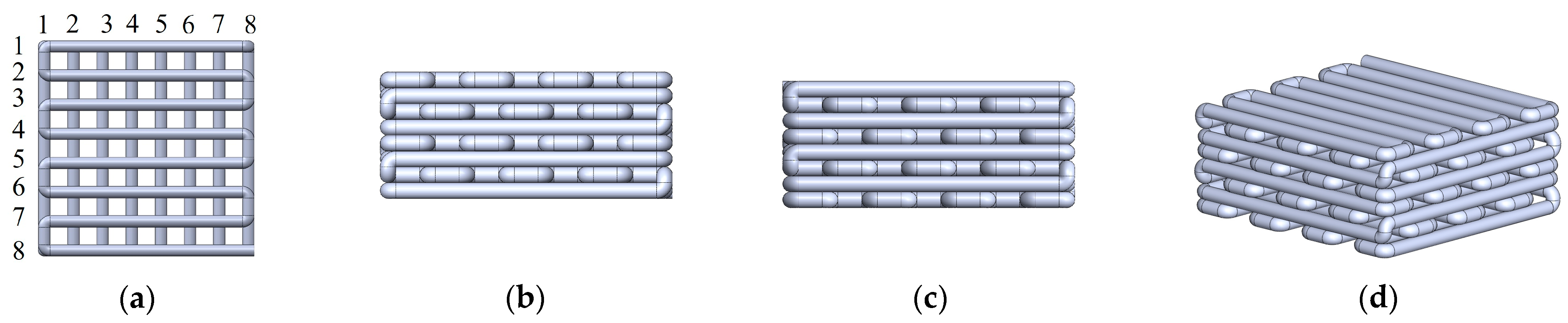
2.5. Preparation of Photothermal Composite Bone Scaffold

Composite bone scaffolds are fabricated using a multi-material high-precision biological 3D printer via direct ink writing technology. The printer platform is configured with various parameters, including a first-layer height set at 90%, an extrusion pressure ranging from 270 to 330 KPa, a nozzle movement speed of 2 mm/s, a nozzle temperature maintained between 45 and 55 °C, and an ambient platform temperature set at 10 °C. The design of bone scaffold models typically incorporates a large adhesive surface area for bone cells and pore shapes that enhance the circulation of oxygen, nutrients, and metabolic substances, thereby accelerating the growth rate of bone tissue cells within the porous bone scaffold [21]. As illustrated in Figure 1, the extrusion nozzle has a diameter of 0.6 mm, the pore spacing is 0.8 mm, and the extruded bone scaffold dimensions are 4.8 mm × 10.4 mm × 10.4 mm.
The material ratio used in the PVA/HA/β-TCP/PCL composite bone scaffold and the concentration of PVA solution are as follows: the mass ratio of HA to PCL is 4:1, the mass ratio of HA to β-TCP is 9:1, and the concentration of PVA solution is 13%. Among them, the mass ratio of the usage amount of PVA solution to other scaffold materials is 1.5 mL:1 g.
In the photothermal composite bone scaffold, the composition of HA, β-TCP, and PCL materials remained the same, with the addition of photo-thermosensitive nanoparticles SiO2@Fe3O4 at 3%, 5%, and 7% of the mass of the scaffold materials, respectively. The ratio of PVA solution used to the total mass of other scaffold materials and photo-thermosensitive nanoparticles was 1.5 mL to 1 g.
The prepared composite bone scaffolds underwent freezing at −40 °C for 4 h, followed by vacuum freeze-drying for 24 h to obtain PVA/HA/β-TCP/PCL bone scaffolds and photothermal composite bone scaffolds.

2.6. XRD Analysis

The nanoparticles of Fe3O4 and SiO2@Fe3O4 were subjected to X-ray diffraction (XRD) analysis using a scanning speed of 2°/min over a rotational range from 2θ = 10° to 80°.

2.7. Microstructural Analysis

The overall morphology, size, and elemental composition of SiO2@Fe3O4 particles were investigated using transmission electron microscopy and an energy dispersive spectrometer to analyze different regions. Additionally, scanning electron microscopy was utilized to assess the micro-porosity of PVA/HA/β-TCP/PCL composite bone scaffolds and 3%, 5%, and 7% photothermal composite bone scaffolds (where 3% denotes a mass fraction of 3% of photothermal-sensitive nanoparticles), with gold coating applied to the sample surfaces prior to testing.

2.8. Measurement of Water Contact Angle

The specimens of PVA/HA/β-TCP/PCL composite bone scaffolds and 3%, 5%, and 7% photothermal composite bone scaffolds were affixed onto glass slides. Each specimen exhibited a planar surface measuring 5 mm × 5 mm × 3 mm. Subsequently, a 2 µL water droplet was dispensed onto the surface of each specimen for contact angle evaluation using a water contact angle goniometer. The contact angle readings were averaged from three replicates of the same specimen, with a standardized reading time of 5 s.

2.9. Mechanical Properties Testing

The mechanical performance of PVA/HA/β-TCP/PCL composite bone scaffolds and 3%, 5%, and 7% photothermal composite bone scaffolds was evaluated using a microcomputer-controlled universal mechanical testing machine. The compression rate was maintained at 1 mm/min, and the results represent the averages of five consecutive measurements.

2.10. Photothermal Characterization

At room temperature, the 808 nm near-infrared light (2 W/cm2) was applied to the 3%, 5%, and 7% photothermal composite bone scaffolds, as well as the PVA/HA/β-TCP/PCL composite bone scaffold. Simultaneously, a high-precision single-channel digital thermometer was utilized to monitor the temperature variations of the scaffolds, aiming to explore their photothermal characteristics.

3. Results and Discussion

3.1. Orthogonal Experimental Analysis of Scaffold Materials

Optimizing the composition of biomaterials stands as a pivotal strategy for attaining superior mechanical performance. HA and β-TCP exhibit remarkable resemblance to the inorganic constituents of bone, offering a synergistic approach to circumvent the limitations associated with individual biomaterials in terms of their biological response and degradation kinetics. Furthermore, the incorporation of PCL and PVA solution serves to augment the mechanical robustness of composite bone scaffolds, ensuring sustained mechanical integrity within the physiological milieu and fostering an optimal environment for osteogenic cells. The outcomes of the orthogonal experimental analysis, delineated in Table 2, have validated the optimal formulation of composite scaffold materials through meticulous assessment of compressive strength. The minimal range (R) values observed in the empty columns signify negligible cross-factor interactions, underscoring the thorough consideration of influential parameters pivotal to experimental outcomes. Deconstruction of the R values has unveiled the primary and ancillary determinants governing the mechanical efficacy of composite scaffold materials: PVA solution concentration, HA to PCL mass ratio, and HA to β-TCP mass ratio. The identified optimal material parameters encompass a 13 wt% PVA solution concentration, a 9:1 ratio of HA to β-TCP, and a 4:1 ratio of HA to PCL.

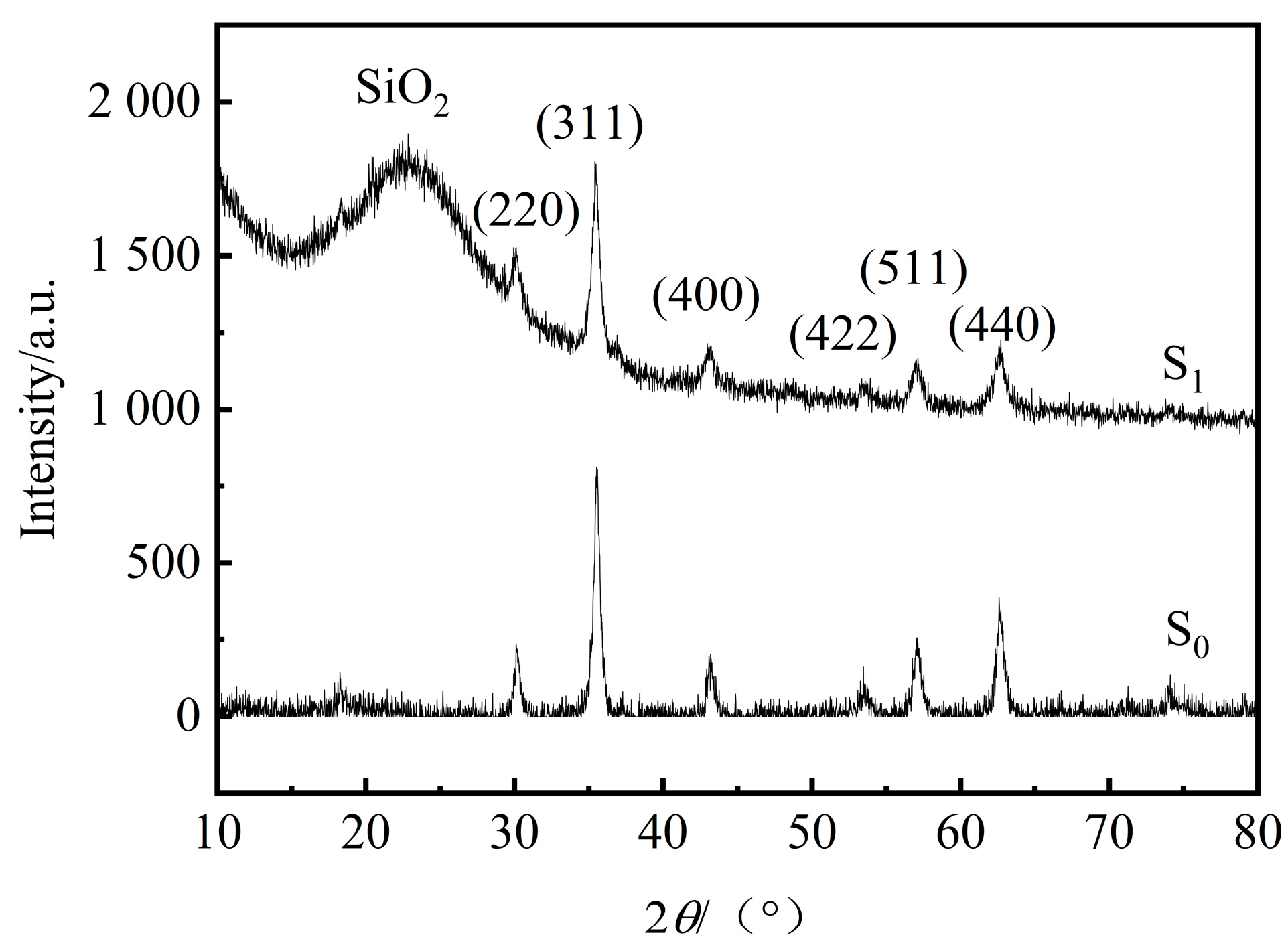
3.2. XRD Analysis of Photothermal Sensitive Nanoparticles

Fe3O4 nanoparticles, known for their exceptional photothermal properties, often encounter issues of local aggregation when used in conjunction with bone tissue engineering scaffolds, leading to hindered cellular growth, differentiation, and even apoptosis. To address this, SiO2@Fe3O4 nanoparticles were synthesized via TEOS hydrolysis–condensation, with the SiO2 coating offering superior biocompatibility and stability, effectively mitigating Fe3O4 particle aggregation. XRD analysis of both Fe3O4 particles (S0) and SiO2@Fe3O4 (S1) (as depicted in Figure 2) revealed diffraction peaks at 2θ = 29.8°, 35.1°, 42.7°, 52.9°, 56.4°, and 61.9°, corresponding to (220), (311), (400), (422), (511), and (440), consistent with the standard data for Fe3O4 powder diffraction. Additionally, a broad peak observed at 2θ = 15°–25° in the S1 curve may be attributed to amorphous SiO2 particles, although further testing is necessary to validate the encapsulation of Fe3O4 within SiO2. Nevertheless, the relative intensity of the Fe3O4 particle diffraction peaks in S1 has decreased, suggesting successful encapsulation by SiO2 and the formation of photothermal-sensitive nanoparticles SiO2@Fe3O4.

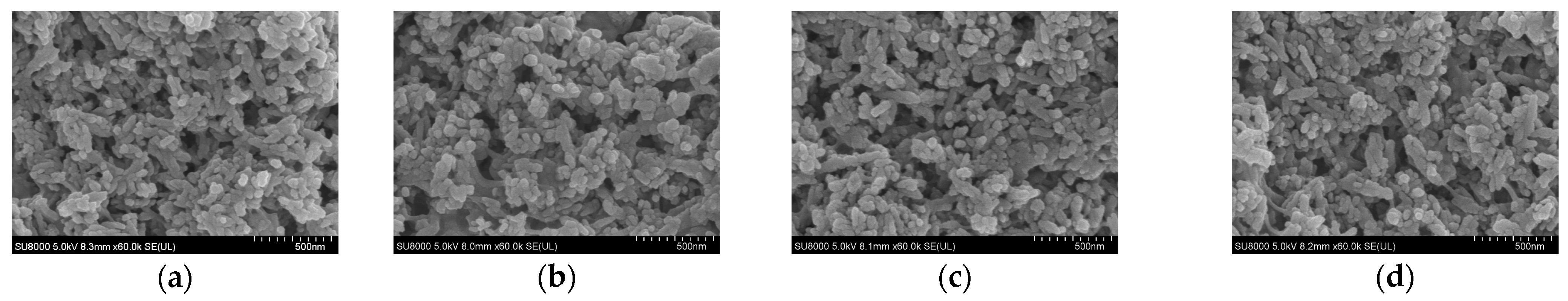
3.3. Microstructural Analysis

As illustrated in Figure 3, transmission electron microscopy images depict SiO2@Fe3O4 nanoparticles synthesized via hydrolysis of varying quantities of TEOS. Clearly discernible from the images is the encapsulation of Fe3O4 within SiO2. Notably, the sample prepared with 2 mL of TEOS exhibited suboptimal encapsulation and SiO2 coating, displaying a degree of disorder. Conversely, the utilization of 8 mL of TEOS resulted in an excessively thick SiO2 coating attributed to an overabundance of TEOS, leading to the gradual formation of Fe-O-Si bonds through interactions with solution-derived silicon species (Si4+). This phenomenon compromised the quality of Fe3O4@SiO2 nanoparticles for photothermal applications, rendering them unsuitable for biological contexts. In contrast, the introduction of 5 mL of TEOS yielded a homogeneously distributed sample devoid of irregularities, ensuring effective encapsulation of Fe3O4 within SiO2 to form approximately 18 nm coatings. Elemental mapping analysis, as depicted in Figure 4 for SiO2@Fe3O4, confirmed the encapsulation success by revealing a confined spatial distribution of Fe elements enveloped by Si and O species. Further scrutiny via scanning electron microscopy images in Figure 5 showcased the successful integration of Fe3O4@SiO2 with scaffold materials for bone scaffold fabrication. Both the PVA/HA/β-TCP/PCL composite bone scaffold and the 3%, 5%, and 7% photothermal composite scaffolds exhibited uniform micro-pore structures without localized aggregation, underscoring the proficient preparation of SiO2@Fe3O4 and its efficacious implementation in bone tissue engineering scaffolds, thereby mitigating the detrimental impact of Fe3O4 aggregation on cellular behavior.

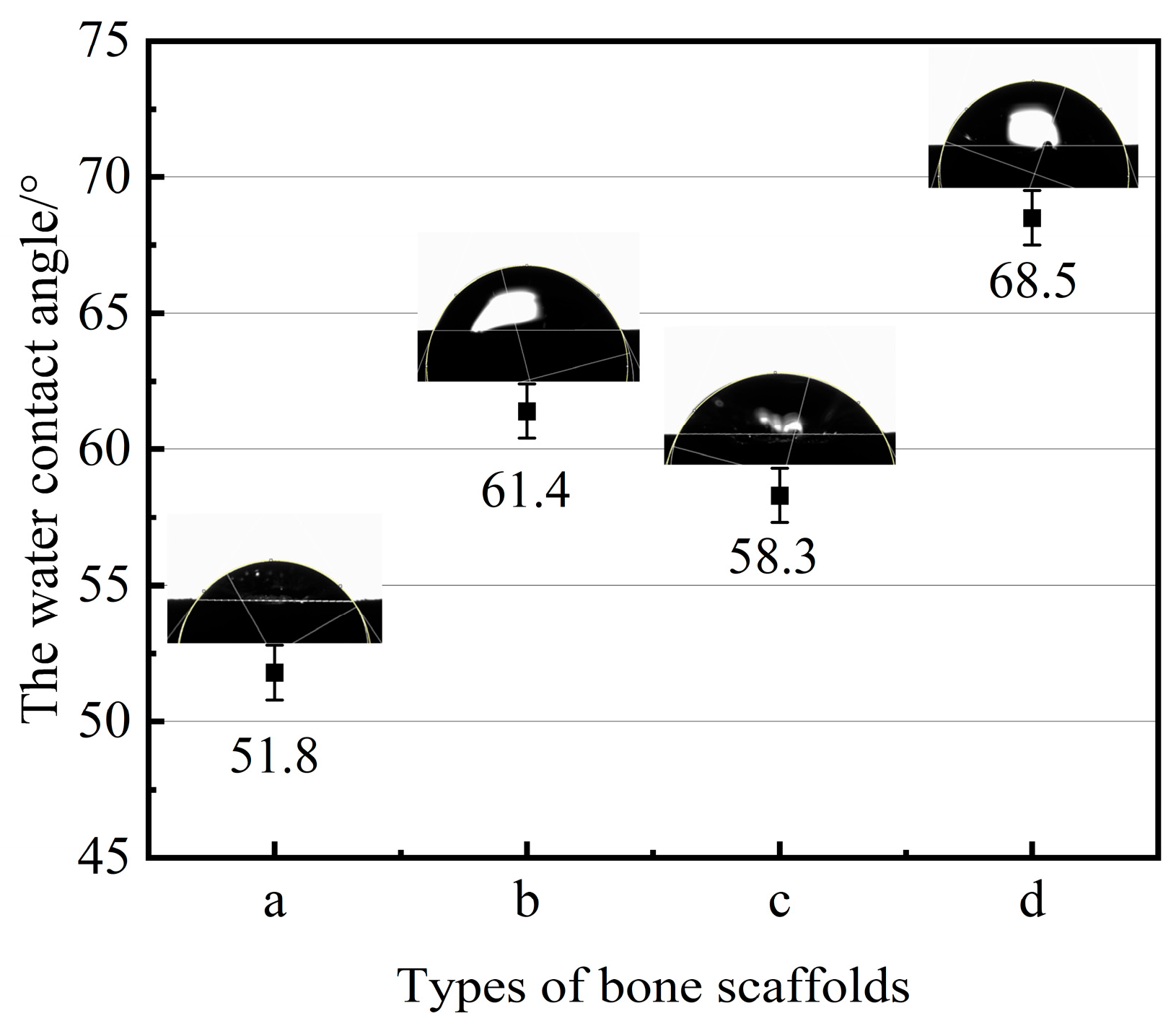
3.4. Measurement of Contact Angle and Mechanical Properties of Bone Scaffolds

In the field of bone tissue engineering applications, the size of the water contact angle can impact the interaction with bone tissue cells or biological fluids. A contact angle below 90° indicates that the material exhibits good wettability and hydrophilicity. As depicted in Figure 6, the contact angles of the PVA/HA/β-TCP/PCL composite bone scaffold and the 3%, 5%, and 7% photothermal composite scaffolds (a, b, c, d) are measured at 51.8°, 61.4°, 58.3°, and 68.5°, respectively, all conducive to cell adhesion and the spread of biological fluids. The compressive strengths of cancellous bone in the spine, tibia, and femur are (2.4 ± 1.6), (5.3 ± 2.9), and (6.8 ± 4.8) MPa, respectively [22,23]. As illustrated in Figure 7, which depicts the compressive strengths of various bone scaffolds, the PVA/HA/PCL/β-TCP composite bone scaffold and the 3%, 5%, and 7% photothermal composite scaffolds exhibit compressive strengths of 5.750, 5.700, 5.722, and 5.719 MPa, respectively. All of these scaffolds meet the mechanical strength requirements of cancellous bone in the spine, tibia, and femur. A comparison between the PVA/HA/PCL/β-TCP composite bone scaffold and the 3%, 5%, and 7% photothermal composite bone scaffolds reveals that the incorporation of photothermally sensitive nanoparticles SiO2@Fe3O4 does not significantly affect the compressive strength of the bone scaffolds, ensuring they can provide adequate mechanical support for bone tissue cells.

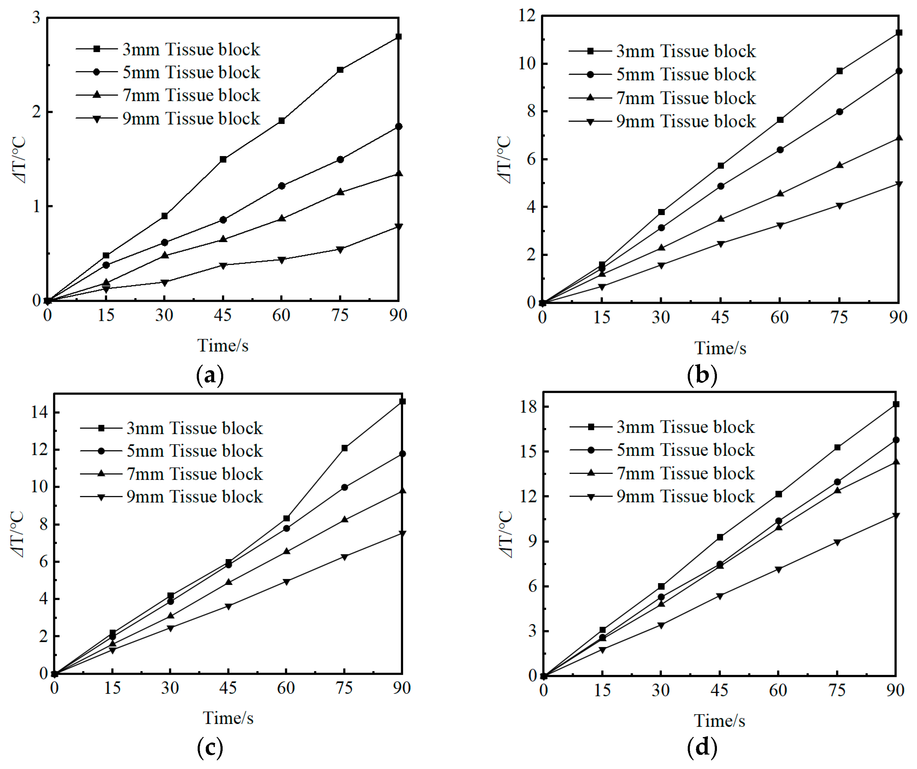
3.5. Evaluation of the Photothermal Properties of Bone Scaffolds

To assess the photothermal properties of the photothermal-sensitive nanocomposite SiO2@Fe3O4, the PVA/HA/β-TCP/PCL composite bone scaffold (control group) and varying mass fractions of the photothermal composite bone scaffolds were subjected to 808 nm (2 W/cm2) irradiation for 60 s at room temperature. The control group and the 3%, 5%, 7% photothermal composite bone scaffolds demonstrated temperature increases of 2.92 °C, 27.26 °C, 38.48 °C, and 52.88 °C, respectively. Relative to the control group, all photothermal composite bone scaffolds exhibited notably elevated temperatures in a short duration, affirming the superior photothermal properties of the photothermal-sensitive nanocomposite SiO2@Fe3O4.
In order to simulate the functional characteristics of photothermal composite bone scaffolds within different patient environments, control groups and photothermal composite scaffolds were placed within culture dishes. Specifically, tissue blocks of varying thicknesses (3, 5, 7, 9 mm; porcine tissue blocks) were positioned atop the scaffolds, while 2 mm tissue blocks were situated at the scaffold base to establish a sealed environment, thereby insulating the scaffold from external influences on temperature. It is noted that the optimal temperature range for promoting osteoblast differentiation falls between 40 and 43 °C [24]; temperatures exceeding this range may compromise the structural integrity of bone tissue cells, thus impeding their normal functionality and differentiation capacity. Conversely, lower temperatures are insufficient to induce osteogenic differentiation. However, SiO2@Fe3O4 demonstrates remarkable photothermal conversion capabilities, rendering it an optimal material for photothermal composite bone scaffolds. Upon irradiation by a near-infrared device within a tissue block, the absorbed light energy elicits the excitation of electrons into a highly energetic state. Subsequently, these excited electrons undergo a non-radiative relaxation process, efficiently converting the absorbed light energy into lattice vibrational energy or thermal kinetic energy of the electrons. This generates a thermal effect that attains the desired temperature for enhancing osteogenic differentiation. Illustratively, as depicted in Figure 8, a substantial temperature differential is observed between the control group and the photothermal composite scaffold, underscoring the superior photothermal properties inherent to the latter. Following a period of 90 s of near-infrared light exposure, an increase in internal temperature is registered within the PVA/HA/β-TCP/PCL composite bone scaffold. However, this temperature escalation does not attain the requisite threshold for fostering osteoblast differentiation. Prolonged irradiation poses the risk of inflicting damage upon superficial skin tissues. Notably, while the 3% photothermal composite scaffold exhibits a degree of photothermal efficacy, the disparate thickness of patient tissues precludes the standardization of irradiation durations necessary to elevate the scaffold temperature by 3–6 °C for the promotion of osteogenic differentiation. Conversely, the 7% photothermal composite scaffold, when subjected to a 30-s irradiation, elevates the temperature of sealed scaffolds with varying tissue thicknesses by 3–6 °C. However, owing to the robust photothermal attributes of the 7% composite scaffold, precise modulation of the near-infrared light exposure duration is imperative to preclude deleterious thermal effects on bone tissue cells. Conversely, the 5% photothermal composite scaffold, upon 40–45 s of irradiation, facilitates a 3–6 °C temperature increase across varying tissue thicknesses, thereby reaching the optimal temperature range of 40–43 °C for promoting osteogenic differentiation. This accomplishment signifies the realization of leveraging mild thermal stimuli to expedite the repair of bone tissue injuries.

4. Conclusions

The research conducted involved the design and synthesis of a composite bone scaffold comprising SiO2@Fe3O4/PVA/HA/β-TCP/PCL with notable photothermal characteristics. Conclusions were deduced from experimental investigations and analyses as outlined below:
(1)
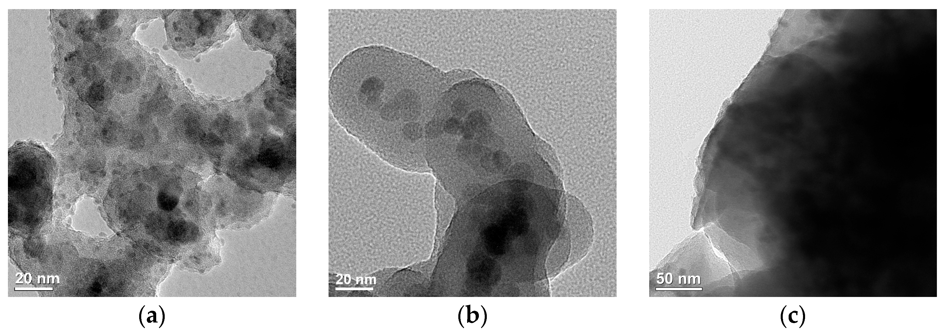
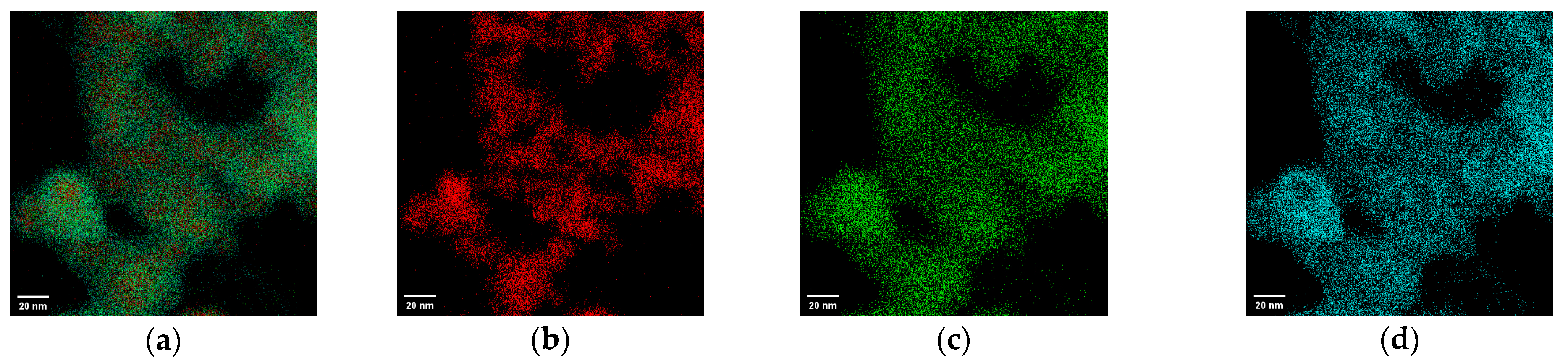
The photothermal-sensitive nanoparticles SiO2@Fe3O4, synthesized via hydrolysis and condensation using 5 mL TEOS, exhibited approximately 18 nm SiO2 coatings. These coatings were uniformly distributed, effectively preventing the aggregation of the photothermal-sensitive nanoparticles Fe3O4. Additionally, the micro-pore structure of the fabricated photothermal composite bone scaffold showed uniform distribution, devoid of localized aggregation phenomena. This ensured the avoidance of adverse effects on the properties and applications of the scaffold material.
(2)
The photothermal composite scaffold with a mass fraction of 5% for photothermal-sensitive nanoparticles exhibited a compressive strength of 5.722 MPa and a contact angle of 58.3°, demonstrating favorable mechanical support and hydrophilicity. This scaffold provides a stable environment for the adhesion and growth of bone tissue cells. Upon exposure to near-infrared light for 40–45 s, the 5% photothermal composite scaffold induced a temperature increase of 3–6 °C in different tissue depths, achieving controlled internal temperature variations through mild thermal stimulation. This approach aims to promote osteogenic differentiation and facilitate the repair of bone defects.

Author Contributions

Conceptualization, C.S., Y.X. and S.L.; methodology, C.S., Y.X. and S.L.; validation, C.S., Y.X. and S.L.; formal analysis, C.S., Y.X. and S.L.; investigation, C.S., Y.X. and S.L.; resources, Y.X.; data curation, C.S.; writing—original draft preparation, C.S.; writing—review and editing, Y.X. and S.L.; visualization, C.S., Y.X. and S.L.; supervision, Y.X. and S.L.; funding acquisition, Y.X. All authors have read and agreed to the published version of the manuscript.

Funding

This work was supported by the National Natural Science Foundation of China (52365053), Natural Science Foundation of Xinjiang Uyghur Autonomous Region (2022D01C34).

Institutional Review Board Statement

Not applicable.

Informed Consent Statement

Not applicable.

Data Availability Statement

The raw data supporting the conclusions of this article will be made available by the authors on request.

Conflicts of Interest

The authors declare that they have no known competing financial interests or personal relationships that could have appeared to influence the work reported in this paper.

References

  1. Konda, S.R.; Littlefield, C.P.; Carlock, K.D.; Ganta, A.; Leucht, P.; Egol, K.A. Autogenous iliac crest bone grafting for tibial nonunions revisited: Does approach matter? Arch. Orthop. Trauma Surg. 2021, 142, 961–968. [Google Scholar] [CrossRef] [PubMed]
  2. Xie, H.Y.; Ruan, S.J.; Zhao, M.L.; Long, J.; Ma, X.; Guo, J.; Lin, X. Preparation and characterization of 3D hydroxyapatite/collagen scaffolds and its application in bone regeneration with bone morphogenetic protein-2. RSC Adv. 2023, 13, 23010–23020. [Google Scholar] [CrossRef] [PubMed]
  3. Motamedian, S.R.; Khojaste, M.; Khojasteh, A. Success rate of implants placed in autogenous bone blocks versus allogenic bone blocks: A systematic literature review. Ann. Maxillofac. Surg. 2016, 6, 78–90. [Google Scholar] [PubMed]
  4. Sharmin, F.; O’Sullivan, M.; Malinowski, S.; Lieberman, J.R.; Khan, Y. Large scale segmental bone defect healing through the combined delivery of VEGF and BMP-2 from biofunctionalized cortical allografts. J. Biomed. Mater. Res. Part B Appl. Biomater. 2019, 107, 1002–1010. [Google Scholar] [CrossRef] [PubMed]
  5. Das, G.; Shin, H.S.; Campos, E.V.R.; Fraceto, L.F.; del Pilar Rodriguez-Torres, M.; Mariano, K.C.F.; de Araujo, D.R.; Fernández-Luqueño, F.; Grillo, R.; Patra, J.K. Sericin based nanoformulations: A comprehensive review on molecular mechanisms of interaction with organisms to biological applications. J. Nanobiotechnol. 2021, 19, 30. [Google Scholar] [CrossRef] [PubMed]
  6. Sangeetha, K.; Ashok, M.; Girija, E.K. Development of multifunctional cobalt ferrite/hydroxyapatite nanocomposites by microwave assisted wet precipitation method: A promising platform for synergistic chemo-hyperthermia therapy. Ceram. Int. 2019, 45, 12860–12869. [Google Scholar] [CrossRef]
  7. Menazea, A.A.; Mostafa, M.S.; Awwad, N.S.; Ali, H.E.; Moustapha, M.E.; Bajaber, M.A. Improvement of medical applicability of hydroxyapatite/antimonous oxide/graphene oxide mixed systems for biomedical application. J. Inorg. Organomet. Polym. Mater. 2022, 32, 3220–3234. [Google Scholar] [CrossRef]
  8. Kurtuldu, F.; Mutlu, N.; Boccaccini, A.R.; Galusek, D. Gallium containing bioactive materials: A review of anticancer, antibacterial, and osteogenic properties. Bioact. Mater. 2022, 17, 125–146. [Google Scholar] [CrossRef] [PubMed]
  9. Pokhrel, S. Hydroxyapatite: Preparation, properties and its biomedical applications. Adv. Chem. Eng. Sci. 2018, 8, 225. [Google Scholar] [CrossRef]
  10. Suhardi, J.V.; Morgan, D.F.A.; Muratoglu, O.K.; Oral, E. Radioprotection and cross-linking of allograft bone in the presence of vitamin E. J. Biomed. Mater. Res. Part B Appl. Biomater. 2020, 108, 2354–2367. [Google Scholar] [CrossRef]
  11. Park, H.; Choi, J.W.; Jeong, W.S. Clinical application of three-dimensional printing of polycaprolactone/beta-tricalcium phosphate implants for cranial reconstruction. J. Craniofacial Surg. 2022, 33, 1394–1399. [Google Scholar] [CrossRef] [PubMed]
  12. Peroglio, M.; Gremillard, L.; Gauthier, C.; Chazeau, L.; Verrier, S.; Alini, M.; Chevalier, J. Mechanical properties and cytocompatibility of poly (ε-caprolactone)-infiltrated biphasic calcium phosphate scaffolds with bimodal pore distribution. Acta Biomater. 2010, 6, 4369–4379. [Google Scholar] [CrossRef] [PubMed]
  13. Hang, K.; Ye, C.Y.; Chen, E.M.; Zhang, W.; Xue, D.; Pan, Z. Role of the heat shock protein family in bone metabolism. Cell Stress Chaperones 2018, 23, 1153–1164. [Google Scholar] [CrossRef] [PubMed]
  14. Han, L.; Guo, Y.; Jia, L.; Zhang, Q.; Sun, L.; Yang, Z.; Dai, Y.; Lou, Z.; Xia, Y. 3D magnetic nanocomposite scaffolds enhanced the osteogenic capacities of rat bone mesenchymal stem cells in vitro and in a rat calvarial bone defect model by promoting cell adhesion. J. Biomed. Mater. Res. Part A 2021, 109, 1670–1680. [Google Scholar] [CrossRef] [PubMed]
  15. Yun, H.M.; Ahn, S.J.; Park, K.R.; Kim, M.J.; Kim, J.J.; Jin, G.Z.; Kim, H.W.; Kim, E.C. Magnetic nanocomposite scaffolds combined with static magnetic field in the stimulation of osteoblastic differentiation and bone formation. Biomaterials 2016, 85, 88–98. [Google Scholar] [CrossRef] [PubMed]
  16. Beitollahi, H.; Ebadinejad, F.; Shojaie, F.; Torkzadeh-Mahani, M. A magnetic core–shell Fe3O4@SiO2/MWCNT nanocomposite modified carbon paste electrode for amplified electrochemical sensing of amlodipine and hydrochlorothiazide. Anal. Methods 2016, 8, 6185–6193. [Google Scholar] [CrossRef]
  17. Singh, R.K.; Kim, T.H.; Patel, K.D.; Knowles, J.C.; Kim, H.W. Biocompatible magnetite nanoparticles with varying silica-coating layer for use in biomedicine: Physicochemical and magnetic properties, and cellular compatibility. J. Biomed. Mater. Res. Part A 2012, 100, 1734–1742. [Google Scholar] [CrossRef] [PubMed]
  18. Duta, L.; Dorcioman, G.; Grumezescu, V. A review on biphasic calcium phosphate materials derived from fish discards. Nanomaterials 2021, 11, 2856. [Google Scholar] [CrossRef] [PubMed]
  19. Wang, H.; Hu, B.; Li, H.; Feng, G.; Pan, S.; Chen, Z.; Li, B.; Song, J. Biomimetic mineralized hydroxyapatite nanofiber-incorporated methacrylated gelatin hydrogel with improved mechanical and osteoinductive performances for bone regeneration. Int. J. Nanomed. 2022, 17, 1511–1529. [Google Scholar] [CrossRef]
  20. Arif, Z.U.; Khalid, M.Y.; Noroozi, R.; Sadeghianmaryan, A.; Jalalvand, M.; Hossain, M. Recent advances in 3D-printed polylactide and polycaprolactone-based biomaterials for tissue engineering applications. Int. J. Biol. Macromol. 2022, 218, 930–968. [Google Scholar] [CrossRef]
  21. Entezari, A.; Roohani, I.; Li, G.; Dunstan, C.R.; Rognon, P.; Li, Q.; Jiang, X.; Zreiqat, H. Architectural design of 3D printed scaffolds controls the volume and functionality of newly formed bone. Adv. Healthc. Mater. 2019, 8, 1801353. [Google Scholar] [CrossRef] [PubMed]
  22. Sola, A.; Bellucci, D.; Cannillo, V. Functionally graded materials for orthopedic applications–an update on design and manufacturing. Biotechnol. Adv. 2016, 34, 504–531. [Google Scholar] [CrossRef] [PubMed]
  23. Wang, X.J.; Xu, S.Q.; Zhou, S.W.; Xu, W.; Leary, M.; Choong, P.; Qian, M.; Brandt, M.; Xie, Y.M. Topological design and additive manufacturing of porous metals for bone scaffolds and orthopaedic implants: A review. Biomaterials 2016, 83, 127–141. [Google Scholar] [CrossRef] [PubMed]
  24. Li, W.H.; Li, S.T.; Zhang, J.W.; Zhong, H.; Liang, J.; Huang, S.; Liao, G.; Zhang, B.; Liu, C. Fabrication and evaluation of bone morphogenetic protein-2 microspheres coated black phosphorus nanosheets@ polylactic-glycolic acid copolymers scaffold: A multifunctional antibacterial photothermal scaffold for bone regeneration. Int. J. Biol. Macromol. 2022, 210, 350–364. [Google Scholar] [CrossRef]
Figure 1. Composite bone scaffold models. (a) Top view of the bone scaffold model; (b) Front view of the bone scaffold model; (c) Left side view of the bone scaffold model; (d) Three-dimensional view of the bone scaffold model.
Applsci 14 04911 g001
Figure 2. XRD patterns of Fe3O4 and SiO2@Fe3O4.
Applsci 14 04911 g002
Figure 3. Transmission electron microscopy image of SiO2@Fe3O4. (a) SiO2@Fe3O4 prepared with 2 mL TEOS; (b) SiO2@Fe3O4 prepared with 5 mL TEOS; (c) SiO2@Fe3O4 prepared with 8 mL TEOS.
Applsci 14 04911 g003
Figure 4. SiO2@Fe3O4 element distribution map. (a) Element distribution map; (b) Spatial distribution of Fe elements; (c) Spatial distribution of Si elements; (d) Spatial distribution of O elements.
Applsci 14 04911 g004
Figure 5. SEM image of composite bracket. (a) PVA/HA/β-TCP/PCL composite bone scaffold; (b) 3% photothermal composite scaffold; (c) 5% photothermal composite scaffold; (d) 7% photothermal composite scaffold.
Applsci 14 04911 g005
Figure 6. Water contact angle of composite scaffold.
Applsci 14 04911 g006
Figure 7. Compressive strength of the composite scaffold.
Applsci 14 04911 g007
Figure 8. Temperature variation of composite support. (a) PVA/HA/β-TCP/PCL composite bone scaffold; (b) 3% photothermal composite scaffold; (c) 5% photothermal composite scaffold; (d) 7% photothermal composite scaffold.
Applsci 14 04911 g008
Table 1. Orthogonal parameters of materials.
LevelsFactors
PVA Conc. (%)HA: β-TCP (g:g)HA: PCL (g:g)Blank Column
189:14:11
210.54:17:32
3137:33:23
Table 2. Orthogonal test results table.
NumberPVA Conc. (%)HA: β-TCP (g:g)HA: PCL (g:g)Blank ColumnCompressive Strength (MPa)
189:14:1110.823
284:17:327.661
387:33:237.881
410.59:17:3316.713
510.54:13:2116.239
610.57:34:1215.557
7139:13:2228.546
8134:14:1331.173
9137:37:3123.365
k18.78818.69419.18416.809
k216.17018.35815.91317.255
k327.69515.60117.55518.589
R18.9073.0933.2711.78
Order1324
Optimal levelA3B1C1
Optimal combinationA3B1C1
Disclaimer/Publisher’s Note: The statements, opinions and data contained in all publications are solely those of the individual author(s) and contributor(s) and not of MDPI and/or the editor(s). MDPI and/or the editor(s) disclaim responsibility for any injury to people or property resulting from any ideas, methods, instructions or products referred to in the content.

