Article

Exploratory Anterior Cruciate Ligament Graft Stress during Medial Knee Rotation with and without Iliotibial Band–Intermuscular Septum Lateral Extra-Articular Tenodesis for Transtibial and Anteromedial Femoral Tunnels

1
Unidad de Biomecanica, Centro de Innovación, Clínica MEDS, Santiago 7691236, Chile
2
Traumatología, Clínica MEDS, Santiago 7691236, Chile
3
Carrera de Kinesiología, Departamento de Cs. de la Salud, Facultad de Medicina, Pontificia Universidad Católica de Chile, Santiago 7820436, Chile
4
Escuela de Kinesiología, Facultad de Medicina y Ciencias de la Salud, Universidad Mayor, Santiago 8580745, Chile
5
Facultad de Ingeniería, Universidad de Chile, Santiago 7800003, Chile
6
Exercise and Rehabilitation Sciences Institute, Postgraduate, Faculty of Rehabilitation Sciences, Universidad Andres Bello, Santiago 7691236, Chile
*
Author to whom correspondence should be addressed.
Appl. Sci. 2024, 14(12), 5160; https://doi.org/10.3390/app14125160
Submission received: 23 March 2024 / Revised: 10 April 2024 / Accepted: 16 April 2024 / Published: 13 June 2024
(This article belongs to the Special Issue Recent Advances in Applied Biomechanics and Sports Sciences)

Abstract

:
Traditional lateral extra-articular tenodesis (LET) using fixation elements constrains medial knee rotation laxity after anterior cruciate ligament reconstruction (ACLr). However, the mechanical behavior of an LET made with an iliotibial band–intermuscular septum is unknown using different anterior cruciate ligament (ACL) reconstruction drillings and would be crucial for constraining the rotatory components of direction change movements. Thus, this study aimed to explore the maximum principal stresses and their distribution in grafts during medial knee rotation with and without iliotibial band–intermuscular septum lateral extra-articular tenodesis for the transtibial technique (TT), hybrid transtibial technique (HTT), and anteromedial portal technique (AM) in single-bundle ACLr. The maximum von Mises principal stresses and their distribution under medial knee rotation were described using a finite element model generated from a healthy knee. LET with HTT, TT, and AM decreases stress by 97%, 93%, and 86% during medial rotation compared to each technique without LET, respectively. The stress concentration for the AM portal and TT techniques was located at the femoral tunnel, and for HTT with LET, it was located across the distal thirds of the anterior aspect of the graft. In conclusion, the HTT with LET diminishes graft stress more than the HTT, TT, and AM without LET, and the TT and AM with LET during medial knee rotation. The AM portal, HTT, and TT techniques without LET show higher stress concentration patterns at the femoral tunnel, establishing a biomechanical risk of femoral tunnel enlargement when LET is not performed.

1. Introduction

Medial rotation laxity after anterior cruciate ligament reconstruction (ACLr) has a profound impact on the quality of life [1,2] and is a key clinical challenge for single-bundle reconstruction in different femoral drillings [3,4,5,6]. Thus, lateral extra-articular tenodesis (LET) has developed because all anterolateral knee structures contribute to minimizing anterolateral rotational laxity, and LET accounts for an increased rotatory knee arm [7,8,9], complementing single-bundle ACLr. Unfortunately, double-bundle ACLr promotes enlarged femoral tunnels, being the highest obstacle for ACLr revision surgeries due to a lack of bone stock [10]. Because of that, there is clinical interest in exploring how LET and femoral tunnel drillings for single-bundle ACLr may diminish the maximal stress of grafts during knee medial rotation and how this can change the graft stress distribution to avoid early graft failure or femoral tunnel windowing or enlargement [5,6,11]. It is expected that a more deflected graft as part of the anteromedial (AM) portal technique would better constrain the knee rotation compared to the traditional transtibial technique (TT) and the hybrid transtibial technique (HTT) [3,12,13,14,15], but whether LET combined with the HTT or TT would diminish the stress graft during medial knee rotation is unknown.
The LET is an augmentation technique made for residual rotatory knee movements [6,16,17] and is able to reduce up to 43% of graft stress [18]. This stress reduction is relevant to prevent graft failure and stress concentration in single-bundle ACLr [6]. LET biomechanics is generally similar to anterolateral ligament biomechanics because they have similar orientations, and both constrain medial knee rotation [19,20]. Lemaire, Macintosh, Losee, Arnold-Coker, Ellison, and others are different LET choices [6,17,18]. However, these techniques demand a higher surgical time and are expensive due to the need for fixation elements and tunneling, procedures that can increase the risk of persistent knee pain [21,22]. As an alternative, one of the LET techniques that involves only soft tissue is the iliotibial band, which passes under the fibular or lateral collateral ligament (LCL) and goes through the femoral intermuscular septum [16]. The final fixation is distally made with five independent high-resistance stitches [23]. This technique is based on the Macintosh and Arnold–Coker procedure, discarding the osteoperiosteal flap [24]. It is reproducible, easy to replicate, inexpensive, and without risk of tunnel coalition [24]. In addition, according to the LET state of the art, a proximal attachment to the lateral femoral epicondyle and beneath the LCL will prevent excessive tightening or slackening during knee movement [18].
On the other hand, finite element (FE) is a numerical and computational method that allows the division of complex structures into smaller ones to simplify the analysis of the entire structure [25]. It has emerged as a potent method in orthopedic surgery, providing surgeons, physical therapists, and biomechanists with enhanced insights into biomechanical factors associated with surgical techniques [25]. Although the FE method could give interesting outcomes and allow an understanding of the mechanical factor roles, there are several limitations associated with geometric complexity or material properties that can complicate the accuracy of the models [25,26], as well as other non-modeling elements that can occur during clinical settings with patients that limit its complete extrapolation to real-life applications. Regarding FE advantages, this technique would allow an understanding of the maximum principal stresses and the stress distribution on grafts following the addition of an iliotibial band–intermuscular septum LET for TT, HTT, and AM femoral tunnels in ACLr under medial knee rotation.
Therefore, this study aimed to explore the maximum principal stresses and their distribution on grafts during medial knee rotation with and without iliotibial band–intermuscular septum lateral extra-articular tenodesis for the TT, HTT, and AM techniques in single-bundle ACLr. This aim covers the relevant problem of movement involved in ACL rupture and rotatory laxity, which promotes knee osteoarthritis, and highlights the mechanical aspects (stress concentration) that typically cannot be determined in vitro or in vivo. In this manner, this study will contribute to the mechanical understanding of ACLr with and without LET.

2. Material and Methods

2.1. Study Design

In this exploratory study, we describe the graft stress during medial knee rotation with and without iliotibial band–intermuscular septum lateral extra-articular tenodesis for the TT, HTT, and AM techniques in single-bundle ACLr femoral tunnels modeled with the FE technique. The IRB #202103 (MEDS clinic, Santiago, Chile) authorized the retrospective and anonymous use of X-ray computed tomography (CT) images for FE modeling.

2.2. Geometry and Mesh Generation

The anatomical FE model was obtained from a healthy knee of a 26-year-old without injured ACL records. DICOM data were collected in a CT (Brivo CT 385 series, GE Healthcare, Chicago, IL, USA) using 16 slices with a 0.62 mm resolution and pixel spacing of 0.35 mm × 0.35 mm. The tibia, femur, and patella were segmented using a density filter in the 3DSlicer software, version 5.2.1 [27]. Then, a mesh model was created, and artifacts (internal residuals) were eliminated manually. The whole structures were smoothed, and a solid structure was created from the bone surfaces using Autodesk Meshmixer 3.5 (Autodesk, Inc., San Rafael, CA, USA).

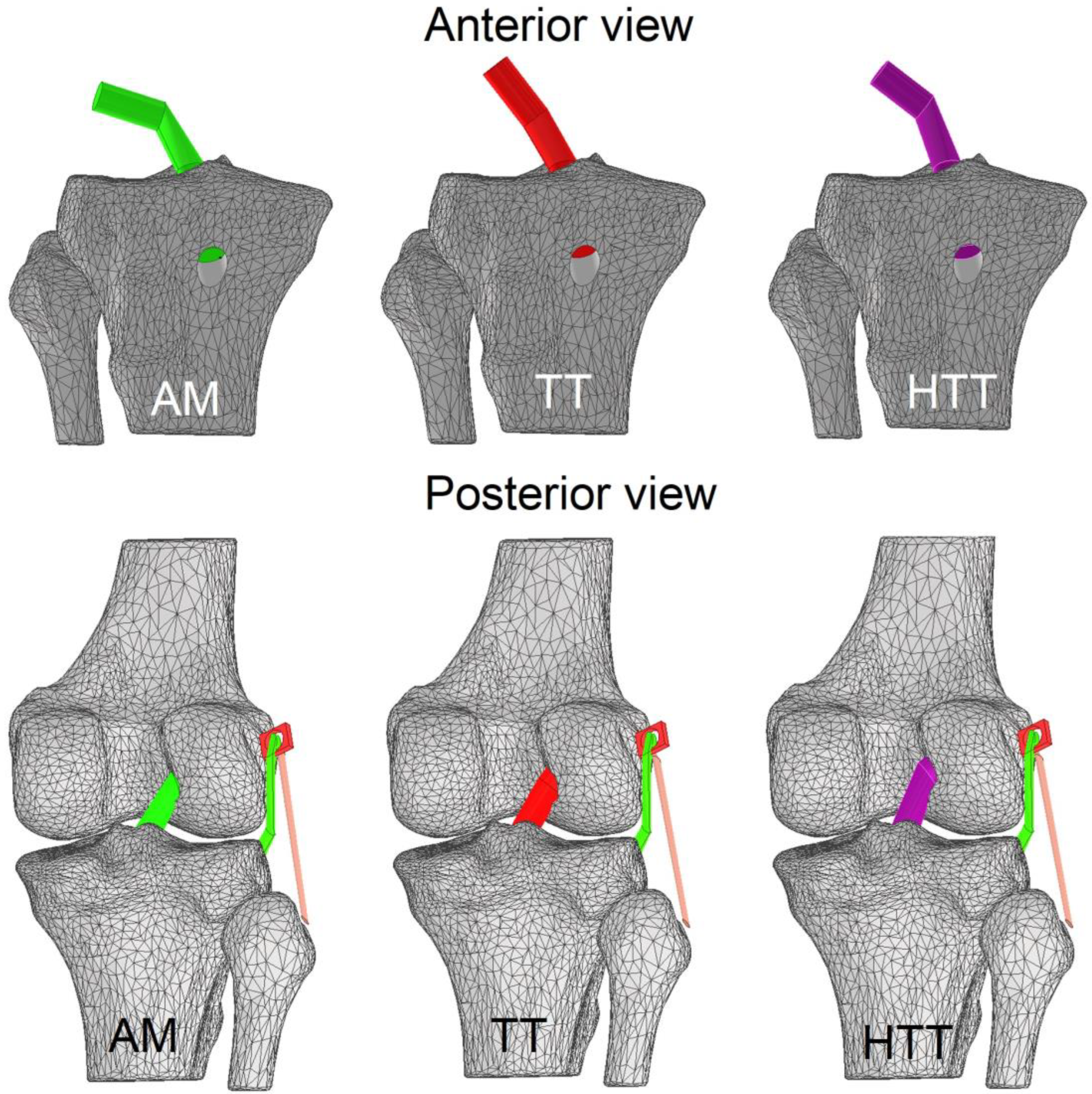
2.3. Knee Tunnels and ACLr Graft

The tibial and femoral drilling procedures followed the method outlined by Troffa et al. [28], involving the creation of a circular section of 9 mm in diameter. The tibia underwent drilling, followed by subsequent drilling in the femur. The drillings were made via Boolean operations [26]. The sagittal orientation angles for the AM portal, TT, and HTT were 146.3°, 155.4°, and 158.8°, respectively, while the coronal orientation angles were 100.5°, 129.4°, and 111.0° for the AM portal, TT, and HTT, respectively [28]. Subsequently, an ACLr graft with a diameter of 9 mm and a width of 25 mm was affixed based on Achilles graft preparation [29]. In this research, the TT, AM portal, and HTT drillings were performed using the same ACLr graft. The node and total element counts were 8812/42,454, 11,482/53,070, and 9776/48,076 units for the AM portal, TT, and HTT, respectively. The entire surgical model in the FE was created using Inventor software version 25.0.18391.0 (Autodesk, Inc., San Francisco, CA, USA) (Figure 1). The femoral and tibial attachments were set as rigid contacts.

2.4. Iliotibial Band–Intermuscular Septum LET

The septum, the iliotibial band LET, and the LCL were created using Autodesk Inventor software version 25.0.18391.0 (Autodesk, Inc., San Francisco, CA, USA). The geometry of the iliotibial band originates from the tubercle of Geardy (a natural protuberance of the lateral tibial condyle) and crosses a dense, rigid, and structural tissue [30] known as the septum, according to Abusleme et al. [23], which is located at the lateral side of the femoral bone [30]. The cross-sectional diameter of the modeled iliotibial band LET was 5 mm at its narrowest points and 7 mm at its thickest points [23]. This tenodesis technique takes into account the support of the LCL. Hence, this ligament was created with a circular section of 7 mm in diameter (Figure 2). Finally, the contacts between the LCL and fibula, the LCL and femur, the septum and iliotibial band LET, and the tibia and iliotibial band were considered rigid contacts.

2.5. Materials Properties

Material properties were based on Cheng et al. [31]. The tibial and femoral properties were a rigid body material (Table 1) [31]. The reconstructed ACLr graft was modeled as isotropic hyperelastic (no linear) material with Veronda–Westmann coefficients (Table 1). The mechanical properties of the LCL and iliotibial band LET were modeled as hyperelastic isotropic materials of Mooney–Rivlin (Table 1). The septum was considered a rigid material as a proximal attachment based on the previous anatomical description [30] and on the experience of the knee orthopedic senior surgeon (RY).

2.6. Boundary Conditions

A knee model underwent medial rotation, allowing for the determination of maximum von Mises principal stresses and the corresponding stress locations. The boundary conditions were 1 degree of freedom (DoF) in the craniocaudal axis of the femur and septum, and 6 DoFs for ACLr, iliotibial band LET, and LCL. A force of 16 N in the posterior direction was applied to the most lateral aspect of the lateral femoral condyle, causing a medial rotation moment of 0.8 Nm. This magnitude was used to test the model both with and without LET. Empirically, the 0.8 Nm magnitude was sufficient to cause controlled medial knee rotation movement into the physiological range in the absence of different constrained knee elements. Other models presented higher moments to cause the same rotation because they added a greater number of anatomical constraints. The magnitude of the differences in the rotation moment compared to other studies is related to the lower number of constraints used in this study [26]. All biomechanical simulations were performed using FEBio software version 3.2 (University of Utah, Salt Lake City, UT, USA, and Columbia University, New York, NY, USA).

2.7. Validation of the FE Model

The outcomes of our model were validated regarding the ACLr graft model of Cheng et al. [31]. The fifth case of the simulation was used to observe the similarity of outcomes between the models. Here, we observed the von Mises stress and strain distribution for the reconstructed ACLr. The graft was set for AM portal drilling, 45° in the coronal plane and 60° in the sagittal plane [31]. Our model used the same properties of tissues described in Table 1, and a load of 104 N (15% of body weight) was applied from the anterior aspect of the femur towards the posterior direction, according to Cheng et al. [31]. The outcomes showed a maximal stress location at the lateral aspect of the femoral tunnel graft attachment, and the strain distribution of our model occurred mainly at the femoral tunnel graft attachment, as expected [31]. All biomechanical simulations were performed using FEBio software version 3.2 (University of Utah, Salt Lake City, UT, USA, and Columbia University, New York, NY, USA).

2.8. Data Analysis

The maximum principal stresses and the stress distribution (body graft, femoral, and tibial graft attachment) on the graft reconstructed by FE during the medial knee rotation models for the TT, AM portal, and HTT techniques with and without LET were statistically described. Hot color scale maps were scaled to the maximum principal stress for each simulation.

3. Results

The iliotibial band–intermuscular septum LET during medial rotation with the HTT, TT, and AM techniques compared to without LET decreases the stress by 97%, 93%, and 86%, respectively. The maximal stress using LET was 0.6 MPa, 1.4 MPa, and 2.6 MPa for the HTT, TT, and AM techniques under a medial rotation moment of 0.8 Nm (Figure 3). The maximal stress without LET was 21.6 MPa, 20.3 MPa, and 18.4 MPa for the HTT, TT, and AM techniques under a medial rotation moment of 0.8 Nm (Figure 3). The stress concentration for the AM portal and TT techniques was located at the anterior aspect of the femoral tunnel, and that of the HTT technique was located across the distal thirds of the anterior aspect of the graft (Figure 4).

4. Discussion

In this exploratory study, the main findings were that (1) the HTT with LET diminishes ACLr graft stress more than HTT, TT, and AM without LET, and TT and AM with LET under medial rotation moment, and (2) for the AM portal, HTT, and TT techniques without LET show higher stress concentration patterns in the femoral tunnel. Thus, the HTT with LET FE model is suggested to be a surgical alternative to constrain isolated medial rotation moments during ACLr in agreement with Lemaire’s principles for adding an LET technique [20,32]. The LET technique based on soft tissue modeled here reduced the ACLr stress in three surgical choices (TT, HTT, and AM techniques) and shifted the stress concentration from the femoral tunnel for HTT with LET. The novelty of lower stress requirement and better stress distribution for HTT with LET found here would impact surgery costs, time of surgery and planning [17], complications associated with fixation elements during rehabilitation [21,22], and the risk of tunnel coalition [24]. Thus, it might be a better surgical design for single-bundle ACLr than TT and AM. Our findings gave insight into differentiating the ACLr stress distribution and concentration between HTT, TT, and AM with and without LET to constrain excessive medial knee rotation in the same direction as previous clinical and preclinical findings using LET with fixation elements found [32,33]. The simulated LET technique here would be an alternative for sport and exercise movements involving directional changes and pivoting [34], as it necessitates medial knee rotation. The absence of LET may lead to increased stress concentration at the femoral tunnel, tunnel enlargement, and, consequently, altered tibial stress displacement, potentially contributing to long-term tibial osteoarthritis.
In this study, it was found that the HTT with LET diminishes the maximal stress of ACLr grafts (∆ = 97%) more than TT (∆ = 93%) and AM (∆ = 86%) with LET under a medial rotation moment. Other LET techniques (non-soft tissue LET [16]) reported a 43% graft stress reduction [18]. The stress reduction in our study establishes that when the ACLr femoral tunnel varied from the AM to TT technique combined with LET under medial knee rotation, the ACLr graft stress changed from higher values due to a more deflected graft in the coronal and sagittal plane to lower values due to a more verticalized ACLr graft, in agreement with past analysis [13,32,33]. The HTT is a femoral tunnel choice within the spectrum of all possible tunnels between the AM and TT techniques, with the HTT being a surgical alternative to the TT technique. Our study showed how the femoral condyle is constrained under medial knee rotation internally at the ACLr footprint and externally at the lateral condyle septum by the ACLr graft and LET [35]. Hence, a more deflected graft derived from an AM tunnel tightens the ACLr graft more than the TT technique, suggesting that the HTT femoral tunnel and LET diminish the ACLr graft stress. Additionally, the biomechanical superiority of adding LET to constrain knee movements following an ACLr is in agreement with past reports [32,33]. However, it is important to indicate that an unbalanced ACLr and LET would change our findings i.e., a shortened ACLr graft and lengthened LET. Thus, the tightened balance for the ACLr graft and LET ought to be considered during the surgery plan. In the case of the iliotibial band–intermuscular septum LET technique, a key point for surgeons is not to overconstrain LET, and it is recommended not to apply too much tension and always check that the full range of motion is permitted before closure distally [23]. Real-time fluoroscopy of the medial knee rotation might provide good feedback on how the lateral condyle displaces over the tibia to adjust the LET tension.
In our model, we have found that the femoral tunnel (femoral footprint) in the AM portal, HTT, and TT techniques without LET is the place of the stress concentration, but the stress concentration shifts to the body graft for the HTT when LET is performed. Thus, the HTT combined with LET is suggested to be an acceptable surgical alternative both for the expected lower loads at the femoral tunnel contact and for hypothetical fatiguing loads because the distribution of the stress is shifted across the graft and not concentrated at the femoral tunnel contact, avoiding femoral tunnel windowing [10,11]. Considering that the physiological aim of an ACLr is to restore the anatomical and physiological mechanical environment, our stress pattern for the HTT with LET is in accordance with recent findings of healthy ACL modeling with higher femoral footprint stress concentrations during 0 to 30° of knee flexion [36] and combined loads (anterior translation, medial rotation, and valgus moment) with a joint flexed at 30° [11]. Unfortunately, the femoral footprint stress concentration is relevant to causing tunnel enlargement and graft wear following ACLr, causing graft lengthening and altered tibial stress contacts [1,11]. Because of that, early interventions during the ligamentation stage for the AM and TT techniques with and without LET might predispose patients to tunnel enlargement and graft wear rather than HTT with LET. Undertaking the problem of biological graft healing with osteoinductive, angiogenic, or anti-inflammatory strategies has been recommended to promote tunnel mineralization and mid-substance graft ligamentation to mitigate higher and relatively low strains under repetitive loads concentrated at the femoral tunnel [37].
We acknowledge that our research is not without limitations. The primary limitation we recognize pertains to the inherent geometry of our ACLr graft model, with no higher knee constraint elements and parameters limited only to the validation model. Nonetheless, it is noteworthy that we have employed a comparable graft across three distinct techniques. On the other hand, the biological variability was controlled by simplifying the modeling to a healthy participant under a validated model. Also, our sample size is typical of finite element simulations [15]. Future directions should explore different anatomy graft and LET effects, different tissue parameters, and a tightened balance between the ACLr and LET to compare different kinds of LET on graft stress, pivoting movements [34], and in vivo knee-rotating movements following ACLr and LET to obtain strong clinical evidence.

5. Conclusions

In conclusion, the HTT technique with LET diminishes graft stress more than the HTT, TT, and AM techniques without LET, and the TT and AM techniques with LET during medial knee rotation. In addition, the AM portal, HTT, and TT techniques without LET show higher stress concentration patterns at the femoral tunnel, establishing a biomechanical risk of tunnel enlargement. Tunnel enlargement is the most significant mechanical factor that can alter the tibial stress displacement by graft enlargement and potentially contribute to long-term tibial osteoarthritis being accelerated under repetitive and/or intense changes in directional movement.

Author Contributions

Conceptualization, R.Y., R.S., M.R., A.N., S.M. and C.D.l.F.; methodology, R.S., S.M. and C.D.l.F.; software, S.M. and C.D.l.F.; validation, R.Y., R.S., M.R., A.N., S.M. and C.D.l.F.; formal analysis, S.M. and C.D.l.F.; investigation, R.S., S.M. and C.D.l.F.; resources, S.M. and C.D.l.F.; data curation, S.M. and C.D.l.F.; writing—original draft preparation, R.Y., R.S., M.R., A.N., S.M. and C.D.l.F.; writing—review and editing, R.Y., R.S., M.R., A.N., S.M. and C.D.l.F.; visualization, R.Y., R.S., M.R., A.N., S.M. and C.D.l.F.; supervision, R.Y., R.S., M.R., A.N., S.M. and C.D.l.F.; project administration, R.S., S.M. and C.D.l.F.; funding acquisition, A.N. and C.D.l.F. All authors have read and agreed to the published version of the manuscript.

Funding

The APC was funded by Universidad Mayor de Chile.

Institutional Review Board Statement

The study was conducted in accordance with the Declaration of Helsinki, and approved by the Institutional Review Board of MEDS Clinic #202103 (March 2021).

Informed Consent Statement

Informed consent was obtained from all subjects involved in the study.

Data Availability Statement

Publicly available datasets were analyzed in this study. This data can be found here: https://www.researchgate.net/profile/Carlos-De-La-Fuente-3.

Conflicts of Interest

The authors declare no conflict of interest.

References

  1. Grassi, A.; Pizza, N.; Al-Zu’bi, B.B.H.; Fabbro, G.D.; Lucidi, G.A.; Zaffagnini, S. Clinical Outcomes and Osteoarthritis at Very Long-Term Follow-up After ACL Reconstruction: A Systematic Review and Meta-Analysis. Orthop. J. Sports Med. 2022, 10, 23259671211062238. [Google Scholar] [CrossRef] [PubMed]
  2. Michel, E.; Jordan, E.; Canovas, F.; Bouchdoug, K.; Dagneaux, L.; Gaillard, F. Influence of Residual Anterior Laxity on Functional Outcomes after Anterior Cruciate Ligament Reconstruction. Orthop. Traumatol. Surg. Res. 2022, 108, 103264. [Google Scholar] [CrossRef] [PubMed]
  3. Tudisco, C.; Bisicchia, S.; Cosentino, A.; Chiozzi, F.; Piva, M. Knee Stability, Athletic Performance and Sport-Specific Tasks in Non-Professional Soccer Players after ACL Reconstruction: Comparing Trans-Tibial and Antero-Medial Portal Techniques. Muscles Ligaments Tendons J. 2015, 5, 175–180. [Google Scholar] [CrossRef] [PubMed]
  4. Kulczycka, P.; Larbi, A.; Malghem, J.; Thienpont, E.; Vande Berg, B.; Lecouvet, F. Imaging ACL Reconstructions and Their Complications. Diagn. Interv. Imaging 2015, 96, 11–19. [Google Scholar] [CrossRef] [PubMed]
  5. Moon, H.-S.; Song, S.Y.; Oh, J.U.; Seo, Y.-J. Effects of Modified Trans-Tibial versus Trans-Portal Technique on Stress Patterns around the Femoral Tunnel in Anatomical Single-Bundle ACL Reconstruction with Different Knee Flexion Angles Using Finite Element Analysis. BMC Musculoskelet. Disord. 2022, 23, 759. [Google Scholar] [CrossRef] [PubMed]
  6. Na, B.-R.; Kwak, W.-K.; Seo, H.-Y.; Seon, J.-K. Clinical Outcomes of Anterolateral Ligament Reconstruction or Lateral Extra-Articular Tenodesis Combined with Primary ACL Reconstruction: A Systematic Review With Meta-Analysis. Orthop. J. Sports Med. 2021, 9, 23259671211023099. [Google Scholar] [CrossRef] [PubMed]
  7. Weber, A.E.; Zuke, W.; Mayer, E.N.; Forsythe, B.; Getgood, A.; Verma, N.N.; Bach, B.R.; Bedi, A.; Cole, B.J. Lateral Augmentation Procedures in Anterior Cruciate Ligament Reconstruction: Anatomic, Biomechanical, Imaging, and Clinical Evidence. Am. J. Sports Med. 2019, 47, 740–752. [Google Scholar] [CrossRef]
  8. Inderhaug, E.; Stephen, J.M.; Williams, A.; Amis, A.A. Biomechanical Comparison of Anterolateral Procedures Combined With Anterior Cruciate Ligament Reconstruction. Am. J. Sports Med. 2017, 45, 347–354. [Google Scholar] [CrossRef]
  9. Devitt, B.M.; Bouguennec, N.; Barfod, K.W.; Porter, T.; Webster, K.E.; Feller, J.A. Combined Anterior Cruciate Ligament Reconstruction and Lateral Extra-Articular Tenodesis Does Not Result in an Increased Rate of Osteoarthritis: A Systematic Review and Best Evidence Synthesis. Knee Surg. Sports Traumatol. Arthrosc. 2017, 25, 1149–1160. [Google Scholar] [CrossRef]
  10. Taketomi, S. Editorial Commentary: Tunnel Widening After Anterior Cruciate Ligament Reconstruction May Increase Laxity and Complicate Revision. Arthroscopy 2021, 37, 2564–2566. [Google Scholar] [CrossRef]
  11. Wang, H.; Zhang, B.; Cheng, C.-K. Stiffness and Shape of the ACL Graft Affects Tunnel Enlargement and Graft Wear. Knee Surg. Sports Traumatol. Arthrosc. 2020, 28, 2184–2193. [Google Scholar] [CrossRef] [PubMed]
  12. Li, G.; Papannagari, R.; DeFrate, L.E.; Yoo, J.D.; Park, S.E.; Gill, T.J. Comparison of the ACL and ACL Graft Forces before and after ACL Reconstruction: An in-Vitro Robotic Investigation. Acta Orthop. 2006, 77, 267–274. [Google Scholar] [CrossRef] [PubMed]
  13. Jennings, J.K.; Leas, D.P.; Fleischli, J.E.; D’Alessandro, D.F.; Peindl, R.D.; Piasecki, D.P. Transtibial Versus Anteromedial Portal ACL Reconstruction: Is a Hybrid Approach the Best? Orthop. J. Sports Med. 2017, 5, 2325967117719857. [Google Scholar] [CrossRef] [PubMed]
  14. Dargel, J.; Schmidt-Wiethoff, R.; Fischer, S.; Mader, K.; Koebke, J.; Schneider, T. Femoral Bone Tunnel Placement Using the Transtibial Tunnel or the Anteromedial Portal in ACL Reconstruction: A Radiographic Evaluation. Knee Surg. Sports Traumatol. Arthrosc. 2009, 17, 220–227. [Google Scholar] [CrossRef] [PubMed]
  15. Bae, J.Y.; Kim, G.-H.; Seon, J.K.; Jeon, I. Finite Element Study on the Anatomic Transtibial Technique for Single-Bundle Anterior Cruciate Ligament Reconstruction. Med. Biol. Eng. Comput. 2016, 54, 811–820. [Google Scholar] [CrossRef] [PubMed]
  16. Vaudreuil, N.J.; Rothrauff, B.B.; de Sa, D.; Musahl, V. The Pivot Shift: Current Experimental Methodology and Clinical Utility for Anterior Cruciate Ligament Rupture and Associated Injury. Curr. Rev. Musculoskelet. Med. 2019, 12, 41–49. [Google Scholar] [CrossRef] [PubMed]
  17. Guarino, A.; Farinelli, L.; Iacono, V.; Screpis, D.; Piovan, G.; Rizzo, M.; Mariconda, M.; Zorzi, C. Lateral Extra-Articular Tenodesis and Anterior Cruciate Ligament Reconstruction in Young Patients: Clinical Results and Return to Sport. Orthop. Rev. (Pavia) 2022, 14, 33696. [Google Scholar] [CrossRef] [PubMed]
  18. Wascher, D.C.; Lording, T.D.; Neyret, P. Extra-Articular Procedures for the ACL-Deficient Knee: A State of the Art Review. J. ISAKOS 2016, 1, 174–182. [Google Scholar] [CrossRef]
  19. Moreno Mateo, F.; Perea, S.H.; Green, D.W. Lateral-Extra Articular Tenodesis vs. Anterolateral Ligament Reconstruction in Skeletally Immature Patients Undergoing Anterior Cruciate Ligament Reconstruction. Curr. Opin. Pediatr. 2022, 34, 71–75. [Google Scholar] [CrossRef]
  20. Nazzal, E.M.; Keeling, L.E.; Ryan, P.M.; Herman, Z.J.; Hughes, J.D. The Role of Lateral Extra-Articular Tenodesis in Anterior Cruciate Ligament Reconstruction and Treatment of Rotatory Knee Instability: A Scoping Review. Curr. Rev. Musculoskelet. Med. 2023, 16, 235–245. [Google Scholar] [CrossRef]
  21. Kurzweil, P.R.; Frogameni, A.D.; Jackson, D.W. Tibial Interference Screw Removal Following Anterior Cruciate Ligament Reconstruction. Arthroscopy 1995, 11, 289–291. [Google Scholar] [CrossRef] [PubMed]
  22. Hirschmann, M.T.; Adler, T.; Rasch, H.; Hügli, R.W.; Friederich, N.F.; Arnold, M.P. Painful Knee Joint after ACL Reconstruction Using Biodegradable Interference Screws- SPECT/CT a Valuable Diagnostic Tool? A Case Report. BMC Sports Sci. Med. Rehabil. 2010, 2, 24. [Google Scholar] [CrossRef] [PubMed]
  23. Abusleme, S.; Strömbäck, L.; Caracciolo, G.; Zamorano, H.; Cheyre, J.; Vergara, F.; Yañez, R. Lateral Extra-Articular Tenodesis: A Technique With an Iliotibial Band Strand Without Implants. Arthrosc. Tech. 2021, 10, e85–e89. [Google Scholar] [CrossRef] [PubMed]
  24. Slette, E.L.; Mikula, J.D.; Schon, J.M.; Marchetti, D.C.; Kheir, M.M.; Turnbull, T.L.; LaPrade, R.F. Biomechanical Results of Lateral Extra-Articular Tenodesis Procedures of the Knee: A Systematic Review. Arthroscopy 2016, 32, 2592–2611. [Google Scholar] [CrossRef] [PubMed]
  25. Herrera, A.; Ibarz, E.; Cegoñino, J.; Lobo-Escolar, A.; Puértolas, S.; López, E.; Mateo, J.; Gracia, L. Applications of Finite Element Simulation in Orthopedic and Trauma Surgery. World J. Orthop. 2012, 3, 25–41. [Google Scholar] [CrossRef] [PubMed]
  26. Benos, L.; Stanev, D.; Spyrou, L.; Moustakas, K.; Tsaopoulos, D.E. A Review on Finite Element Modeling and Simulation of the Anterior Cruciate Ligament Reconstruction. Front. Bioeng. Biotechnol. 2020, 8, 967. [Google Scholar] [CrossRef] [PubMed]
  27. Fedorov, A.; Beichel, R.; Kalpathy-Cramer, J.; Finet, J.; Fillion-Robin, J.-C.; Pujol, S.; Bauer, C.; Jennings, D.; Fennessy, F.; Sonka, M.; et al. 3D Slicer as an Image Computing Platform for the Quantitative Imaging Network. Magn. Reson. Imaging 2012, 30, 1323–1341. [Google Scholar] [CrossRef]
  28. Trofa, D.P.; Saltzman, B.M.; Corpus, K.T.; Connor, P.M.; Fleischli, J.E.; Piasecki, D.P. A Hybrid Transtibial Technique Combines the Advantages of Anteromedial Portal and Transtibial Approaches: A Prospective Randomized Controlled Trial. Am. J. Sports Med. 2020, 48, 3200–3207. [Google Scholar] [CrossRef]
  29. Zamorano, H.; Yáñez-Diaz, R.; Vergara, F.; Caracciolo, G.; Valdés, S.; Yáñez-Rojo, R.; De la Fuente, C. Achilles Tendon Allograft Preparation Technique for Anterior Cruciate Ligament Reconstruction: A Technical Note. Arthrosc. Tech. 2021, 10, e2143–e2150. [Google Scholar] [CrossRef]
  30. Hayashi, A.; Maruyama, Y. Lateral Intermuscular Septum of the Thigh and Short Head of the Biceps Femoris Muscle: An Anatomic Investigation with New Clinical Applications. Plast. Reconstr. Surg. 2001, 108, 1646–1654. [Google Scholar] [CrossRef]
  31. Cheng, R.; Wang, H.; Jiang, Z.; Dimitriou, D.; Cheng, C.-K.; Tsai, T.-Y. The Femoral Tunnel Drilling Angle at 45° Coronal and 45° Sagittal Provided the Lowest Peak Stress and Strain on the Bone Tunnels and Anterior Cruciate Ligament Graft. Front. Bioeng. Biotechnol. 2021, 9, 797389. [Google Scholar] [CrossRef] [PubMed]
  32. Guenther, D.; Irarrázaval, S.; Bell, K.M.; Rahnemai-Azar, A.A.; Fu, F.H.; Debski, R.E.; Musahl, V. The Role of Extra-Articular Tenodesis in Combined ACL and Anterolateral Capsular Injury. J. Bone Jt. Surg. Am. 2017, 99, 1654–1660. [Google Scholar] [CrossRef] [PubMed]
  33. Risvas, K.; Stanev, D.; Moustakas, K. Can Lateral Tenodesis Improve the Rotational Stability of the ACL Reconstruction? A Finite Element Analysis. PLoS ONE 2024, 19, e0293161. [Google Scholar] [CrossRef] [PubMed]
  34. Neira, A.; Silvestre, R.; Debandi, A.; Darras, D.; Cristi-Sánchez, I.; Barra, I.; Peñailillo, L.; De La Fuente, C. Is the Rotatory Knee Stability Immediately Decreased Following a Competitive Soccer Match? Front. Bioeng. Biotechnol. 2022, 10, 903131. [Google Scholar] [CrossRef] [PubMed]
  35. Zaffagnini, S.; Lucidi, G.A.; Macchiarola, L.; Agostinone, P.; Neri, M.P.; Marcacci, M.; Grassi, A. The 25-Year Experience of over-the-Top ACL Reconstruction plus Extra-Articular Lateral Tenodesis with Hamstring Tendon Grafts: The Story so Far. J. Exp. Orthop. 2023, 10, 36. [Google Scholar] [CrossRef] [PubMed]
  36. Yang, S.; Liu, Y.; Ma, S.; Ding, C.; Kong, Z.; Li, H.; Huang, F.; Chen, H.; Zhong, H. Stress and Strain Changes of the Anterior Cruciate Ligament at Different Knee Flexion Angles: A Three-Dimensional Finite Element Study. J. Orthop. Sci. 2023; in press. [Google Scholar] [CrossRef]
  37. Yao, S.; Yung, P.S.H.; Lui, P.P.Y. Tackling the Challenges of Graft Healing After Anterior Cruciate Ligament Reconstruction-Thinking From the Endpoint. Front. Bioeng. Biotechnol. 2021, 9, 756930. [Google Scholar] [CrossRef]
Figure 1. Iliotibial band–intermuscular septum lateral extra-articular tenodesis modeling. (A) The collateral lateral ligament. (B) The iliotibial band. (C) Intermuscular septum. (D) The iliotibial band–intermuscular lateral extra-articular tenodesis, according to Abusleme et al. [23].
Applsci 14 05160 g001
Figure 2. Anteromedial (AM) portal, transtibial (TT), and hybrid transtibial (HTT) tunnel and iliotibial bandintermuscular septum lateral extra-articular tenodesis.
Applsci 14 05160 g002
Figure 3. Maximum von Mises stress under medial rotation moment (0.8 Nm). AM = anteromedial portal technique. HTT = hybrid transtibial technique. TT = transtibial technique. LET = lateral extra-articular tenodesis.
Applsci 14 05160 g003
Figure 4. Stress concentration with and without iliotibial band–intermuscular septum lateral extra-articular tenodesis (LET). The magnitude of stress is not normalized between the surgical techniques to see each stress concentration in each surgical combination independently of its magnitude. AM = anteromedial portal technique. HTT = hybrid transtibial technique. TT = transtibial technique. White and black arrows show the stress concentration in the stress field on the anterior cruciate ligament graft.
Applsci 14 05160 g004
Table 1. Material properties of the study.
Knee ElementMaterialParameters
BonesRigidYoung modulus = 0.4 GPa
Poisson coefficient = 0.33
ACLr graftIsotropic hyperelastic
(no linear)
Veronda–Westmann coefficients of
α = 0.3 MPa
β = 12.20
Lateral collateral ligamentHyperelastic isotropicMooney–Rivlin with
c1 = 30.1 MPa
c2 = −27.1 MPa
Iliotibial band LETHyperelastic isotropicMooney–Rivlin with
c1 = 30.1 MPa
c2 = −27.1 MPa
SeptumRigidYoung modulus = 0.4 GPa
Poisson coefficient = 0.33
Disclaimer/Publisher’s Note: The statements, opinions and data contained in all publications are solely those of the individual author(s) and contributor(s) and not of MDPI and/or the editor(s). MDPI and/or the editor(s) disclaim responsibility for any injury to people or property resulting from any ideas, methods, instructions or products referred to in the content.

